Calcium-sensing receptors promoted Homer1 expression and osteogenic differentiation in bone marrow mesenchymal stem cells
-
Kainan Liu


Abstract
Homer1 interacts with calcium-sensing receptors (CaSRs) in osteoblasts (OBs), with both CaSR and Homer1 playing essential roles in AKT phosphorylation. This study investigated the impact of CaSR on Homer1 expression during the differentiation of rat bone marrow mesenchymal stem cells (BMSCs) at morphological, imaging, and molecular levels, both in vivo and in vitro. A post-oophorectomy osteoporosis model was established in Sprague-Dawley rats, validated through micro-computed tomography, hematoxylin-eosin staining, and biomechanical testing to assess in vivo changes in CaSR expression. BMSCs were isolated from 3 week-old SD rats for in vitro OB differentiation studies, wherein osteogenic differentiation was induced alongside changes in CaSR expression. Morphological alterations were examined using transmission electron microscopy and immunofluorescence staining. Furthermore, the protein and mRNA levels of OB-specific genes were quantified by Western blot and reverse transcription quantitative real-time polymerase chain reaction, with Homer1-related proteins also assessed. Results showed a reduction in CaSR and Homer1 expression in the ovariectomized group. In cellular studies, CaSR activation upregulated AKT, Homer1, and osteogenic markers, promoting cell differentiation. In conclusion, CaSR enhances OB differentiation, likely via Homer1-mediated regulation of AKT signaling, suggesting CaSR as a potential therapeutic target for osteoporosis.
1 Introduction
Osteoporosis (OP) is a systemic bone disorder characterized by reduced bone mass, impaired bone microstructure, increased bone fragility, and an elevated risk of fractures [1]. It arises from an imbalance between bone resorption by osteoclasts (OCs) and bone formation by osteoblasts (OBs) [2]. Bone marrow-derived mesenchymal stem cells (BMSCs) have the potential to differentiate into multiple cell types, maintaining a critical balance essential for normal bone metabolism. This balance is disrupted in the OP state [3]. A central mechanism underlying OP is the impaired differentiation of BMSCs into OBs [4]. Research has demonstrated that both the number of BMSCs and their osteogenic differentiation capacity are altered in patients with OP, resulting in impaired bone formation [5]. Consequently, exploring the molecular mechanisms regulating BMSC differentiation into OBs is essential for understanding OP pathogenesis and developing effective therapeutic strategies for its prevention and treatment.
The calcium-sensing receptor (CaSR), a G-protein-coupled receptor (GPCR), plays a pivotal role in calcium homeostasis [6]. It regulates Ca2+ and other metal ions while promoting cell proliferation and differentiation [7]. Activation of CaSR can modulate the release of transforming growth factor-β (TGF-β), which in turn stimulates OB proliferation, differentiation, and mineralization via the TGF-β type I/II receptor (TβRI/II)-Smad 2/3 signaling pathway. Studies have shown that CaSR activation triggers various signaling cascades in osteocytes, promoting OB proliferation and differentiation, inhibiting OC activity, and thereby exerting therapeutic effects in OP treatment [8,9]. Moreover, the ERK1/2-MAPK, Wnt/β-catenin, and Akt signaling pathways rely on CaSR to regulate OB function [9].
Homer proteins, initially discovered in the nervous system [10,11], function as scaffold proteins. The primary isoforms include Homer1, Homer2, and Homer3. Homer1 is known to regulate calcium channels, act as a scaffold protein, and modulate the endoplasmic reticulum calcium release channel [12]. Additionally, Homer1 plays a role in mediating AKT phosphorylation. Activation of CaSR induces AKT Ser 473 phosphorylation and nuclear β-catenin translocation in OBs, with Homer proteins promoting AKT phosphorylation at the cell membrane [13]. The involvement of CaSR and Homer1 in AKT Ser 473 and GSK 3β-S9 phosphorylation has been demonstrated in MG 63 osteosarcoma cells. However, the exact role of Homer1 in osteogenic differentiation and OP remains unclear.
This study hypothesizes that Homer1 plays a role in OP pathogenesis, with CaSR promoting OB proliferation and differentiation by modulating Homer1, thereby exerting a therapeutic effect on OP. To test this hypothesis, a bilateral ovariectomized rat model of OP was established, as ovarian ligation-induced models are commonly used to study OP pathology and explore potential therapeutic strategies [14]. Additionally, rat BMSCs were used to investigate the mechanism by which CaSR influences osteogenic differentiation. Specifically, this study aimed to determine whether Homer1 expression was altered in the rat model of postmenopausal OP and whether Homer1 participates in osteogenic differentiation through CaSR signaling. These findings may provide insights for the development and optimization of novel diagnostic and therapeutic approaches for OP.
2 Materials & methods
2.1 Animals
All animal procedures were performed in compliance with the Guidelines for the Care and Use of Laboratory Animals set by the National Institutes of Health. The animals were provided by the Animal Center of North China University of Science and Technology (License No.: SYXK(Ji)2020-007). Eight week-old female rats (180–200 g) and 3 week-old male SD rats were selected for the study. The animals were housed under standard laboratory conditions.
-
Ethical approval: The research related to animal use has been complied with all the relevant national regulations and institutional policies for the care and use of animals, and has been approved by the Experimental Animal Ethics Committee of North China University of Science and Technology (Approval No.: 2023-SY-015).
2.2 Rat model of ovariectomized osteoporosis (OVX)
Female SD rats were placed in a barrier environment. After a 1 week acclimation period, the rats were randomly assigned to two groups (n = 5): the sham group and the ovariectomized (OVX) group. Prior to surgery, the rats were fasted for 12 h and anesthetized with 60 mg/kg sodium pentobarbital. The rats were fixed in a lateral position, and a 1–2 cm incision was made on the lateral abdomen. The skin was incised with scissors, and the fascia and muscle were separated bluntly. White adipose tissue was exposed, and the ovaries, which appeared as pink, cauliflower-shaped structures, were visible upon removal of the fat folds. The ovariectomy model was established by ligating the fallopian tubes and removing the ovarian tissue (Specific operations shown schematically in Figure 1. Created with BioRender.com.) [15]. In the Sham group, adipose tissue of equivalent weight to the ovaries was used. The adipose tissue was then returned to the abdominal cavity, and an appropriate amount of penicillin was applied to prevent infection. The surgical sites were closed in sequence: muscle, fascia, and skin. Postoperatively, the rats were monitored, and after recovery, they were fed routinely with ad libitum access to food and water.
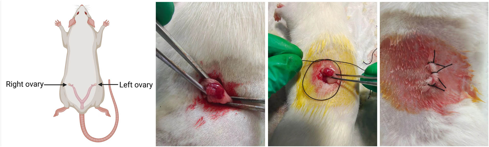
Diagram of the OVX rat model. Created with BioRender.com.
Eight weeks post-surgery, all female rats were anesthetized and sacrificed via cervical dislocation. Tibias and femurs were carefully isolated, fixed in 4% paraformaldehyde, or frozen for subsequent experiments. The fixed tibias were decalcified in 4% ethylenediaminetetraacetic acid disodium salt (EDTA disodium) (Coolaber, China, CE31192504) for 3 months. After decalcification, the samples were dehydrated in a graded series of ethanol. Following clearing in xylene, the samples were embedded in paraffin and sectioned into 6 μm thick slices.
2.3 Micro-computed tomography (Micro-CT) determination
Micro-CT (PINGSENG Healthcare Inc., China) was used to scan the proximal femur in both sagittal and horizontal planes.
The scanned data were analyzed to calculate key bone parameters, including bone mineral density (BMD, mgHA/cm3), which correlates positively with OP and bone strength, and bone volume/tissue volume (BV/TV, %), which correlates positively with load-bearing capacity. Additional parameters assessed included the structural model index (SMI), which is negatively correlated with trabecular resistance to compression; trabecular number (Tb.N, mm−1); trabecular thickness (Tb.Th, %), negatively correlated with OP, fractures, and degeneration; and trabecular separation (Tb.Sp, μm), which is inversely related to bone quality.
2.4 Biomechanical properties
A three-point bending test was performed on the tibia using a universal electronic testing machine (Shimadzu, Japan) to evaluate the biomechanical properties of the rats’ tibiae. The tibia samples were carefully positioned on a low support, with an 8 mm span between the two fixed loading points. A force of 2 N was applied to the tibia at a constant displacement rate of 2 mm/min until fracture occurred. The primary parameters measured during the test included maximum load, flexural stiffness, and elastic modulus.
2.5 Hematoxylin and eosin (H&E) staining
H&E staining was performed according to standard protocols [16]. Briefly, paraffin sections were deparaffinized in xylene and rehydrated through a graded ethanol series. The sections were stained with hematoxylin for 10 minutes, followed by thorough washing with distilled water. Differentiation was carried out using 1% hydrochloric acid alcohol for 3 s, followed by rinsing. The sections were then stained with eosin for 1 min and rinsed again. Finally, the sections were sealed and observed under a microscope. Digital images were captured using a scanning system (Olympus, Japan, CX31).
2.6 Immunohistochemistry (IHC)
For immunohistochemical analysis, the paraffin sections were deparaffinized using xylene and rehydrated through graded ethanol. Antigen retrieval was performed using 0.1% pancreatic enzyme at room temperature for 15 min. The sections were washed with phosphate-buffered saline (PBS) and treated with an endogenous peroxidase blocker (Zhongshan Jinqiao, Beijing, China, PV-6001). Following this, the sections were incubated overnight at 4°C with a primary antibody diluted 1:200. Then, the sections were incubated with an enzyme-conjugated goat anti-rabbit IgG polymer at room temperature for 1 h. Color development was achieved using 3,3′-diaminobenzidine (DAB), and the sections were counterstained, sealed, and observed under a microscope. Digital images were captured using a microscope scanning system. The primary antibodies used included CaSR (Abclonal, China, A1426), collagen type I (Col I; Abclonal, China, A1352), and Homer1 (Affinity, USA, DF12280).
2.7 Cell culture and identification
Primary BMSCs were isolated from male SD rats through standard procedures [17]. Briefly, rats were anesthetized and euthanized by cervical dislocation. The tibia and fibula were harvested under sterile conditions, with surrounding subcutaneous tissue and muscle removed. Both ends of the bones were excised, and the bone marrow cavity was flushed with α-modified Eagle’s medium (α-MEM, Eallbio, China, 03.18001A) to collect the bone marrow. The isolated cells were dissociated by pipetting and transferred to culture flasks, and a single-cell suspension was prepared. Cells were cultured in α-MEM supplemented with 10% fetal bovine serum (FBS) and 1% penicillin–streptomycin (Eallbio, China) and maintained in a humidified incubator at 37°C with 5% CO₂. Upon reaching 80% confluence, cells were detached using 0.25% trypsin and subcultured. Passage 3 (P3) cells were utilized for subsequent experiments. Characterization of third-generation BMSCs was performed via flow cytometry using antibodies against CD90 (positive marker) and CD45 (negative marker) (BD Pharmingen, China).
2.8 Cell viability assay
For cell proliferation assays, BMSCs were seeded at 2 × 10³ cells/well in 96-well plates and cultured in basal medium for 24 h. The cells were then incubated with varying concentrations of GdCl3 (Adams, China, P1616025) (200, 250, 300, 350, and 400 μg/mL) or NPS2390 (Cayman, USA, 11989) (5, 10, 15, and 20 μg/mL) for 5 days. Every one day, 10 μL of CCK-8 solution was added to each well and incubated for 1 h. Absorbance at 450 nm was measured using a microplate reader (Thermo Scientific, China, 117123001).
2.9 Cell treatment
For osteogenic differentiation, the third-generation BMSCs were induced with GdCl3 to activate CaSR and NPS2390 to inhibit CaSR expression. The cells were grouped into four conditions: undifferentiated (Undiff), differentiated (Diff), GdCl3, and NPS. In the Undiff group, BMSCs were cultured in a complete medium (α-MEM + 10% FBS). The Diff group received osteogenic induction medium (complete medium + 10 mmol/L β-sodium glycerophosphate (Alfa Aesar, USA, 41099500), 50 mg/L ascorbic acid (TCI, China, QGNMA-SG), 10−8 mol/L dexamethasone (TCI, China, AAVYJ-BC)). The GdCl3 group was treated with 300 μmol/L GdCl3 in the osteogenic induction medium, while the NPS group was treated with 10 μmol/L NPS2390 in the osteogenic induction medium.
2.10 Transmission electron microscopy (TEM)
TEM (Hitachi High-Technologies Corporation, Japan, HITACHI HT7800) was employed to visualize autophagy. On day 14, the cells were scraped, aggregated, and centrifuged to form pellets. The pellets were fixed in an electron microscopy fixation solution (Servicebio, China, G1102) at 4°C, rinsed, and then treated with 1% osmium tetroxide until blackened. Following fixation, the samples underwent dehydration, infiltration, and embedding, after which resin blocks were sectioned into 70 nm ultrathin slices using an ultramicrotome. The sections were stained with 2% uranyl acetate in ethanol for 8 min in the dark, followed by 2.6% lead citrate staining for 8 min, also in the dark, to prevent CO₂ contamination. Finally, the sections were analyzed using a HITACHI HT7800 transmission electron microscope.
2.11 Cell immunofluorescence staining
Cell suspensions were plated in confocal culture dishes at a density of 1 × 10⁴ cells/mL. After 24 h of stable growth, the medium was replaced according to the conditions of the experimental groups. Calcium ions were labeled using the Fluo-4/AM (Oregon, USA, F14217) calcium fluorescent indicator on days 7, 14, and 21 of culture. Fluorescence images were captured with a laser confocal microscope (Olympus, Japan, CX31), and fluorescence intensity was quantified using ImageJ software.
2.12 Western blot (WB)
For protein analysis, cell suspensions were seeded into culture dishes at a density of 1 × 10⁴ cells/mL. After 24 h, the medium was replaced based on the experimental group conditions. Protein expression at days 7, 14, and 21 post-culture was assessed via WB. Total protein was extracted using RIPA lysis buffer containing protease inhibitors (Beijing Zoman Biotechnology Co., Ltd., ZS306-1), and protein concentration was measured using a BCA kit (Beijing Zoman Biotechnology Co., Ltd., ZS301-2). Equal amounts of protein (20 µg) were separated by 10% sodium dodecyl sulfate polyacrylamide gel electrophoresis (Biotides, Beijing, China) and transferred onto polyvinylidene fluoride membranes (Immobilon, USA, 3098530). Following blocking with 5% skimmed milk, the membranes were cut according to the molecular weight of the prestained marker protein and incubated overnight at 4°C with primary antibodies. The membranes were subsequently incubated with horseradish peroxidase-conjugated anti-rabbit or anti-mouse IgG secondary antibodies (1:5,000; Yeasen, China, 34854ES60). Protein bands were detected using an ECL reagent (Zoman, China, ZD310) and visualized with a 6100 EXP system (Clinx Science Instruments, Shanghai, China). Band intensities (gray values) were quantified with ImageJ software for comparative analysis. Primary antibodies included anti-Col I, anti-Homer1, anti-Runx2 (1:1000; Affinity, USA, AF5186), anti-AKT (1:5,000; Huabio, China, WL0003b), and anti-ACTB (β-actin; 1:50,000; Abclonal, China; AC026).
2.13 RNA extraction and quantitative real-time PCR
Total RNA was extracted from tissues and cells using the EaStep® Super Total RNA extraction kit (Mei5 Biotechnology Co., Ltd., Beijing, China) as previously described in Section 1.12. RNA concentration and quality were assessed using a spectrophotometer. Reverse transcription was performed according to the manufacturer’s protocol (Mei5 Biotechnology, Beijing, China, MF167-01) to synthesize cDNA. qRT-PCR was conducted using the 2× M5 HiPer SYBR Premix EsTaq (Mei5 Biotechnology Co., Ltd., Beijing, China, MF787-T) and a QuantStudio™ Real-time PCR Detection System (Applied Biosystems). The PCR program consisted of an initial denaturation at 95°C for 30 s, followed by 40 cycles of 5 s at 95°C and 32 s at 60°C. Relative gene expression levels were calculated using the 2‒ΔΔCt method, with GAPDH as the internal reference. Primer sequences for qRT-PCR are provided in Table 1.
Nucleotide sequences of real-time PCR primers
| Name | Primer sequence |
|---|---|
| CaSR | Forward: 5′-CTTTGTGCTCTGTATCTCGT-3′ |
| Reverse: 5′-TTGGCTTCAAATACCAGGAG-3′ | |
| Homer1 | Forward: 5′-TGAGTGTTTTCCATGTCCAA-3′ |
| Reverse: 5′-CTGACAAGCTGGGACTTTAT-3′ | |
| Runx2 | Forward: 5′-ACCAACCGAGTCATTTAAGG-3′ |
| Reverse: 5′-TCCCAAAAGAAGTTTTGCTG-3′ | |
| AKT | Forward: 5′-TCACAGATGCAGCTACCATGAAGAC-3′ |
| Reverse: 5′-AGGCAACCTCCCACACATCATTTC-3′ | |
| Col Ⅰ | Forward: 5′-CAAAGGTGCTACATCTCTGT-3′ |
| Reverse: 5′-CACACAAGTCCCTATCCATT-3′ | |
| GAPDH | Forward: 5′-AGTGCCAGCCTCGTCTCATA-3′ |
| Reverse: 5′-ATGAAGGGGTCGTTGATGGC-3′ |
2.14 Statistical analyses
Data are presented as mean ± standard deviation (SD) and analyzed using SPSS 20 software (SPSS Inc., Chicago, IL). Group comparisons were made using a t-test. For normally distributed data with homogeneity of variance, one-way ANOVA was used for multiple group comparisons, followed by the least significant difference test for pairwise comparisons. For non-normally distributed data, the rank-sum test was applied. For normally distributed data with unequal variances, Tamhane’s T2 test was used. A P-value <0.05 was considered statistically significant.
3 Results
3.1 Postmenopausal osteoporosis in rats leads to decreased expression of CaSR and Homer1
OP development in bilateral OVX rats was evaluated using micro-CT, three-point bending tests, and H&E staining.
Micro-CT analysis revealed significant differences between OVX and sham rats. Trabecular bone imaging indicated a marked reduction in bone mass in OVX rats compared to sham rats (P < 0.05, Figure 2a). Longitudinal changes in bone microarchitecture are summarized in Figure 2. OVX rats exhibited significant reductions in BMD, BV/TV, Tb.Th, and Tb.N, along with increases in SMI and Tb.Sp (P < 0.05, n = 3, Figure 2b). Biomechanical testing by three-point bending demonstrated significantly lower maximum load, bending stiffness, and elastic modulus in OVX rats relative to sham rats (P < 0.05, n = 3, Figure 2c). H&E staining of the proximal tibia revealed clear morphological differences between the groups. The growth plate in OVX rats appeared thinner, and the trabecular bone beneath it showed a markedly thinner reticular structure compared to the sham group (Figure 2d). These results confirm the development of OP in OVX rats, characterized by reduced bone mass, impaired biomechanical properties, and altered bone microarchitecture.

Expression of CaSR and Homer1 in OVX rats: (a) trabecular bone images of rats; (b) bone formation indices, including BMD, BV/TV, SMI, Tb.Th, Tb.N, and Tb.Sp as analyzed by micro-CT; (c) biomechanical parameters of rat tibia: maximum load, bending stiffness, and elastic modulus; (d) bone formation in rats assessed by H&E staining (40 ×, 100 ×); (e) CaSR mRNA expression; (f) Runx2 and Homer1 protein expression in femur tissues; and (g) IHC analysis of CaSR, Col I, and Homer1 expression in tibia. * P < 0.05; ** P < 0.01.
Additionally, WB and qPCR analyses showed significant reductions in both protein and mRNA expression levels in OVX rats compared to sham controls. The relative protein expression of Runx2 and Homer1 was significantly decreased in OVX rats (n = 3, P < 0.05, Figure 2e). Similarly, the relative mRNA expression of CaSR was notably lower in OVX rats compared to sham rats (n = 3, P < 0.05, Figure 2f).
Immunohistochemical analysis confirmed decreased expression and distribution of Col I, CaSR, and Homer1 proteins near the growth plate in OVX rats compared to sham rats (P < 0.05, Figure 2g).
These results suggest that Homer1 and CaSR are closely associated with the development of OP. Therefore, Homer1 and CaSR may modulate OP through the regulation of osteogenesis.
3.2 CaSR regulates the OB differentiation
To investigate the role of CaSR in OB differentiation, BMSCs at passage 3 were first identified. Flow cytometry analysis showed a high purity of the BMSCs, with 93.29% positive expression for CD90 and 0.91% negative expression for CD45 (Figure 3a). Cells without antibodies served as the control group. The concentrations of the CaSR agonist (GdCl3) and inhibitor (NPS2390) were then optimized by evaluating cell viability through CCK-8 assays. BMSCs were treated with various concentrations of GdCl3 (200, 250, 300, 350, and 400 μmol/L) and NPS2390 (5, 10, 15, and 20 μmol/L). The results indicated that 300 μmol/L GdCl3 and 10 μmol/L NPS2390 were optimal for promoting cell proliferation (P < 0.05, n = 6, Figure 3b). These concentrations were used in subsequent experiments.

CaSR regulates the OB differentiation of BMSCs: (a) flow cytometry analysis for BMSC identification; (b) determination of optimal concentrations for GdCl3 and/or NPS 2390 based on cell proliferation; (c) experimental design schematic illustrating the impact of CaSR on BMSC osteogenic differentiation; (d) effect of CaSR on calcium distribution in BMSCs. Green fluorescence represents Fluo-4/AM calcium indicator staining in the cytoplasm, with arrows highlighting calcium-positive areas (×400 magnification); and (e) ultrastructural observation of cells after 14 days of osteogenic induction. * P < 0.05; ** P < 0.01; *** P < 0.001.
To validate the experimental setup, group experiments were conducted (Figure 3c). The activation or inhibition of CaSR was confirmed by assessing intracellular calcium levels in different experimental groups after induction.
The fluorescence intensity in NPS2390-treated cells was significantly lower than that in the other three groups on day 7 of induction (P < 0.05). Over time, changes in calcium ion fluorescence were observed. By days 14 and 21, fluorescence intensity in the GdCl3 group was notably stronger than in the Diff group, while the NPS group exhibited weaker fluorescence. These results suggest that calcium ion pathways can be modulated by altering CaSR expression, confirming the successful activation and inhibition of CaSR in the experimental setup (P < 0.05, n = 3, Figure 3d).
Further examination of intracellular organelles using transmission electron microscopy revealed distinct alterations between experimental groups. In the Undiff group, BMSCs exhibited typical mesenchymal cell characteristics, including short microvilli, abundant rough endoplasmic reticulum, and intact mitochondria. After 14 days of induction, noticeable changes were observed. In the Diff group, BMSCs exhibited increased microvilli, irregular nuclear morphology, and significant glycogen accumulation in the cytoplasm. Prominent organelles such as ribosomes, rough endoplasmic reticulum, and Golgi apparatus suggested OB-like differentiation. In the GdCl3 group, BMSCs showed normal mitochondrial structure, with a significant increase in mitochondrial numbers compared to the Undiff group. In contrast, the NPS2390 group displayed a reduction in mitochondrial numbers and evident structural abnormalities, such as cristae breakage, dissolution, and vacuolation (Figure 3e). These results confirm that CaSR modulation influences osteogenic differentiation.
3.3 Effect of CaSR on osteogenic differentiation of BMSCs
To elucidate the role of CaSR in osteogenic differentiation, the effects of CaSR activation and inhibition on the expression of key osteogenic markers, including Homer1, AKT, Runx2, and Col I, were assessed at both protein and mRNA levels.
The results indicated that, compared to the Diff group, the relative protein expression and mRNA levels of these markers were significantly increased with the addition of the CaSR agonist (GdCl3) and significantly decreased with CaSR inhibition (NPS2390) (P < 0.05, n = 6, Figure 4a and b). These results suggest that CaSR activation enhances the osteogenic differentiation of BMSCs by promoting the expression of AKT, Homer1, Runx2, and Col I.

Effect of CaSR on osteogenic differentiation of BMSCs: (a) Reverse transcription quantitative real-time polymerase chain reaction analysis of mRNA expression for Col I, AKT, Homer1, and Runx2 in the four groups and (b) protein expression of Col I, AKT, Runx2, and Homer1 detected by WB. *P < 0.05; **P < 0.01; ***P < 0.001.
4 Discussion
This research indicates that the expression levels of CaSR and Homer1 were significantly lower in the OVX group compared to the Sham group, which may contribute to the impaired osteogenic differentiation capacity observed in the OVX rats. OP accelerates the aging of ovariectomized stem cells, thereby diminishing their osteogenic potential. Previous studies have suggested that BMSCs may be negatively affected in OP, which could influence the progression of the disease [18]. Consequently, understanding the mechanisms underlying the osteogenic differentiation of BMSCs is critical for advancing our knowledge of OP pathogenesis.
BMD is a widely accepted metric for assessing bone loss, while BV/TV serves as a reliable indicator of trabecular and cortical bone mass. Parameters such as Tb.Sp and Tb.N are primarily used to evaluate the spatial structure of trabeculae, and SMI and Tb.Sp are commonly employed to assess bone compressive strength [19]. Runx2 and Col I are the well-established markers of osteogenesis [20]. Osteoporotic animal models are invaluable for studying bone formation and remodeling processes [15]. In this study, an osteoporotic rat model (OVX model) was successfully established through ovariectomy. The OVX rats exhibited significant reductions in BMD, BV/TV, Tb.N, and Tb.Th, along with marked increases in SMI and Tb.Sp. The trabecular bone structure was sparse and defective, with evident microstructural damage, leading to a substantial reduction in bone strength. Additionally, the expression of Runx2 and Col I proteins in femoral tissue was significantly downregulated. These results align with previous studies on osteoporosis, confirming that the ovariectomy procedure effectively replicated a postmenopausal osteoporosis (PMOP) rat model [21].
Significant downregulation of CaSR expression was observed in the femoral tissue and growth plate region of OVX rats, suggesting that CaSR may play a key regulatory role in the physiological changes in these animals. As a GPCR, CaSR is critical for osteogenic differentiation and bone repair in BMSCs [22]. Gadolinium chloride (GdCl3), a potent activator of CaSR [23], and NPS2390, a highly selective antagonist of Group I metabolic glutamate receptors (mGluRs), share high homology with CaSR and mGluR1, making NPS2390 a widely used CaSR antagonist [7,24]. In this study, Ca2+ expression increased upon CaSR activation and decreased with its inhibition, confirming the successful modulation of CaSR activity. Ultrastructural observations via transmission electron microscopy revealed abundant mitochondria following CaSR activation, suggesting enhanced energy availability, which may directly support OB function. Col I, a key marker of early osteogenic differentiation, is essential for determining the mechanical and elastic properties of bone tissue [25]. Similarly, Runx2, known as a master regulator of bone development and OB differentiation, promotes the commitment of mesenchymal stem cells to the OB lineage [26]. Runx2 activation regulates Col I gene expression, facilitating successful OB differentiation [27].
CaSR activation significantly elevated serum levels of Runx2 and Col I in OVX rats, while the inhibition of CaSR reduced these levels. This suggests that CaSR activation regulates Runx2 gene expression, promoting Col I synthesis, thereby maintaining extracellular matrix homeostasis and promoting osteogenic differentiation of BMSCs. These observations align with previous studies, such as those by Xu et al. [28], which demonstrated that CaSR enhances BMSC proliferation, and An [29], who reported that CaSR activation induces both vasodilation and osteogenic differentiation.
Furthermore, CaSR has been shown to promote osteogenic differentiation of Wharton’s jelly mesenchymal stem cells by modulating the PI3K/AKT signaling pathway [30]. CaSR also influences AKT expression in various contexts, including osteosarcoma cell proliferation [31], human telomerase reverse transcriptase in gastric cancer [32], and neurite outgrowth in developing chicken embryos [33]. The PI3K/AKT pathway plays a pivotal role in numerous cellular processes, including metabolism, growth, and proliferation. Upon activation by protein kinases and GPCRs, PI3K converts PIP2 to PIP3, creating high-affinity binding sites for the PH domain of AKT, which promotes AKT translocation to the membrane. There, phosphorylation at Ser473 and Thr308 activates downstream transcription factors. The PI3K/AKT pathway is also a central regulator of OB differentiation, facilitating OB differentiation and mineralization by upregulating osteogenic transcription factors such as Runx2 and Osterix, as well as extracellular matrix proteins like Col I while enhancing ALP activity. Additionally, traditional Chinese medicine compounds, such as myricetin, have been shown to enhance osteogenic differentiation of immortalized bone marrow mesenchymal stem cells and reduce bone loss in OVX mice via the PI3K/AKT pathway. However, the specific involvement of AKT in the mechanisms observed in this study remains to be determined.
The HOMER1 gene, which encodes the HOMER1 protein, is a critical scaffold involved in signal transduction and various neurodevelopmental processes, including chronic pain and drug addiction [34,35]. In OBs, studies have established a link between CaSR, HOMER1, and AKT, where CaSR activation induces AKT phosphorylation at Ser473 and facilitates the nuclear translocation of β-catenin [36]. Additionally, membrane-bound HOMER1 proteins are known to promote AKT phosphorylation [37]. CaSR-mediated phosphorylation of AKT at Ser473, subsequent phosphorylation of GSK3β, and the nuclear translocation of β-catenin are closely associated with HOMER1. In MG63 osteosarcoma cells, CaSR and HOMER1 co-regulate AKT Ser473 and GSK3β-S9 phosphorylation. Similarly, studies in human OBs have identified a protein complex consisting of CaSR, HOMER1, and mTORC2 that governs the AKT phosphorylation pathway. This interaction significantly enhances OB proliferation and differentiation [13]. These findings emphasize the pivotal role of HOMER1 in integrating CaSR and AKT signaling during osteogenesis. By acting as a key mediator, HOMER1 facilitates bone remodeling and OB differentiation, positioning it as a potential therapeutic target for promoting bone formation and repair.
In this study, CaSR upregulation led to a significant increase in Homer1 and AKT protein levels, while CaSR inhibition resulted in a marked reduction in these proteins. This suggests that, during osteogenic differentiation, CaSR and Homer1 are co-regulated, with CaSR acting as a positive regulator that enhances Homer1 expression and promotes AKT activation.
In conclusion, an OVX rat model was established, revealing reduced CaSR and Homer1 expression in OVX rats. In vitro experiments further examined the roles and mechanisms of CaSR and Homer1 in osteogenic differentiation of BMSCs. However, this study has several limitations. First, while the activity of Homer1 and AKT was confirmed following CaSR modulation in vitro, the precise mechanisms underlying CaSR regulation remain incompletely understood. Second, the role of Homer1 in these regulatory pathways requires further validation. Finally, whether CaSR can mitigate OP by modulating Homer1 in vivo remains to be explored. Future research should address these limitations by investigating the underlying mechanisms of CaSR regulation and further validating the involvement of Homer1. These efforts will provide deeper insights into targeting CaSR and Homer1 as potential therapeutic strategies for OP.
5 Conclusions
In summary, this study demonstrates the expression of CaSR in the bone tissue of SD rats and highlights the potential involvement of CaSR and Homer1 pathways in OB differentiation. CaSR effectively promotes osteogenic cell differentiation, likely through Homer1-mediated regulation of AKT phosphorylation, which in turn facilitates BMSC differentiation. These findings suggest that modulating CaSR activity may offer a promising therapeutic strategy for bone diseases. Future research should focus on investigating various CaSR activators and inhibitors, as well as their underlying mechanisms in bone tissue under both physiological and pathological conditions, to enhance our understanding and inform potential therapeutic applications.
Acknowledgement
The authors would like to thank the reviewer for their insightful comments, which contributed to the improvement of this research.
-
Funding information: This work was supported by the 333 Talent Project (C20221117) and North China University of Science and Technology. The funders had no role in study design, data collection and analysis, decision to publish, or preparation of the manuscript.
-
Author contributions: KNL, QW, and TJX designed the experiment; TJX, JXF, and XLG conducted the experiment; LKN, JXF, and YYL analyzed the experimental data, while XLG and QW prepared and revised the manuscript; KNL and QW proofread the manuscript; HZ provided technical guidance for animal model preparation. All the authors read and approved the final submitted manuscript.
-
Conflict of interest: Authors state no conflict of interest.
-
Data availability statement: The datasets generated during and/or analyzed during the current study are available from the corresponding author on reasonable request.
References
[1] Ensrud KE, Crandall CJ. Osteoporosis. Ann Intern Med. 2024;177(1):ITC1–16.10.7326/AITC202401160Search in Google Scholar PubMed
[2] Luo B, Zhou X, Tang Q, Yin Y, Feng G, Li S, et al. Circadian rhythms affect bone reconstruction by regulating bone energy metabolism. J Transl Med. 2021;19(1):410.10.1186/s12967-021-03068-xSearch in Google Scholar PubMed PubMed Central
[3] Suo J, Zou S, Wang J, Han Y, Zhang L, Lv C, et al. The RNA-binding protein Musashi2 governs osteoblast-adipocyte lineage commitment by suppressing PPARγ signaling. Bone Res. 2022;10(1):31.10.1038/s41413-022-00202-3Search in Google Scholar PubMed PubMed Central
[4] Li H, Qu J, Zhu H, Wang J, He H, Xie X, et al. CGRP regulates the age-related switch between osteoblast and adipocyte differentiation. Front Cell Dev Biol. 2021;9:675503.10.3389/fcell.2021.675503Search in Google Scholar PubMed PubMed Central
[5] Wang H, Shan K, Li Y, Wu S, Zhou C, Tao S, et al. Therapeutic potential of Chinese medicinal herbs stimulating osteogenic differentiation of bone marrow-derived mesenchymal stem cells in osteoporosis. Front Pharmacol. 2024;15:1423555.10.3389/fphar.2024.1423555Search in Google Scholar PubMed PubMed Central
[6] Iamartino L, Brandi ML. The calcium-sensing receptor in inflammation: Recent updates. Front Physiol. 2022;13:1059369.10.3389/fphys.2022.1059369Search in Google Scholar PubMed PubMed Central
[7] Qi L, Zhang H, Guo Y, Zhang C, Xu Y. Novel calcium-binding peptide from bovine bone collagen hydrolysates and its potential pro-osteogenic activity via calcium-sensing receptor (CaSR). Mol Nutr Food Res. 2024;68(4):e2200726.10.1002/mnfr.202200726Search in Google Scholar PubMed
[8] Christensen TEK, Berglund Davidsen M, Van Malderen S, Garrevoet J, Offermanns V, Andersen OZ, et al. Local release of strontium from sputter-deposited coatings at implants increases the strontium-to-calcium ratio in peri-implant bone. ACS Biomater Sci Eng. 2022;8(2):620–5.10.1021/acsbiomaterials.1c01004Search in Google Scholar PubMed
[9] Walker MD, Shane E. Postmenopausal osteoporosis. N Engl J Med. 2023;389(21):1979–91.10.1056/NEJMcp2307353Search in Google Scholar PubMed
[10] Zhu J, Chen C, Liu X, He M, Fang Y, Wang L, et al. Cerebellar Purkinje cell firing promotes conscious recovery from anesthesia state through coordinating neuronal communications with motor cortex. Theranostics. 2024;14(2):480–95.10.7150/thno.89592Search in Google Scholar PubMed PubMed Central
[11] Bockaert J, Perroy J, Ango F. The complex formed by group I metabotropic glutamate receptor (mGluR) and homer1a plays a central role in metaplasticity and homeostatic synaptic scaling. J Neurosci. 2021;41(26):5567–78.10.1523/JNEUROSCI.0026-21.2021Search in Google Scholar PubMed PubMed Central
[12] Bartolomeis A, Barone A, Buonaguro EF, Tomasetti C, Vellucci L, Iasevoli F. The Homer1 family of proteins at the crossroad of dopamine-glutamate signaling: An emerging molecular “Lego” in the pathophysiology of psychiatric disorders. A systematic review and translational insight. Neurosci Biobehav Rev. 2022;136:104596.10.1016/j.neubiorev.2022.104596Search in Google Scholar PubMed
[13] Rybchyn MS, Brennan-Speranza TC, Mor D, Cheng Z, Chang W, Conigrave AD, et al. The mTORC2 regulator homer1 modulates protein levels and sub-cellular localization of the CaSR in osteoblast-lineage cells. Int J Mol Sci. 2021;22(12):6509.10.3390/ijms22126509Search in Google Scholar PubMed PubMed Central
[14] Komori T. Animal models for osteoporosis. Eur J Pharmacol. 2015;759:287–94.10.1016/j.ejphar.2015.03.028Search in Google Scholar PubMed
[15] Yousefzadeh N, Kashfi K, Jeddi S, Ghasemi A. Ovariectomized rat model of osteoporosis: a practical guide. Excli J. 2020;19:89–107.Search in Google Scholar
[16] Huang C, Wang Y. Downregulation of METTL14 improves postmenopausal osteoporosis via IGF2BP1 dependent posttranscriptional silencing of SMAD1. Cell Death Dis. 2022;13(11):919.10.1038/s41419-022-05362-ySearch in Google Scholar PubMed PubMed Central
[17] Zhang RF, Wang Q, Zhang AA, Xu JG, Zhai LD, Yang XM, et al. Low-level laser irradiation promotes the differentiation of bone marrow stromal cells into osteoblasts through the APN/Wnt/β-catenin pathway. Eur Rev Med Pharmacol Sci. 2018;22(9):2860–8.Search in Google Scholar
[18] Liu Z, Lee HL, Suh JS, Deng P, Lee CR, Bezouglaia O, et al. The ERα/KDM6B regulatory axis modulates osteogenic differentiation in human mesenchymal stem cells. Bone Res. 2022;10(1):3.10.1038/s41413-021-00171-zSearch in Google Scholar PubMed PubMed Central
[19] Oliviero S, Cheong VS, Roberts BC, Orozco Diaz CA, Griffiths W, Bellantuono I, et al. Reproducibility of densitometric and biomechanical assessment of the mouse tibia from in vivo micro-CT images. Front Endocrinol (Lausanne). 2022;13:915938.10.3389/fendo.2022.915938Search in Google Scholar PubMed PubMed Central
[20] Bai W, Cheng M, Jin J, Zhang D, Li L, Bai Y, et al. KAP1 modulates osteogenic differentiation via the ERK/Runx2 cascade in vascular smooth muscle cells. J Mol Biol Rep. 2023;50(7):6305.10.1007/s11033-023-08494-2Search in Google Scholar PubMed
[21] Tao ZS, Li TL, Xu HG, Yang M. Hydrogel contained valproic acid accelerates bone-defect repair via activating Notch signaling pathway in ovariectomized rats. J Mater Sci Mater Med. 2021;33(1):4.10.1007/s10856-021-06627-2Search in Google Scholar PubMed PubMed Central
[22] Ranieri M, Schepelmann M, Valenti G, Kallay E, Riccardi D. Editorial: The calcium-sensing receptor: From physiology to pharmacology. Front Physiol. 2023;14:1225074.10.3389/fphys.2023.1225074Search in Google Scholar PubMed PubMed Central
[23] Eisner D, Neher E, Taschenberger H, Smith G. Physiology of intracellular calcium buffering. [published correction appears in Physiol Rev]. 2024;104(1):327.Search in Google Scholar
[24] Li X, Chen S, Feng D, Fu Y, Wu H, Lu J, et al. Calcium-sensing receptor promotes calcium oxalate crystal adhesion and renal injury in Wistar rats by promoting ROS production and subsequent regulation of PS ectropion, OPN, KIM-1, and ERK expression. Ren Fail. 2021;43(1):465–76.10.1080/0886022X.2021.1881554Search in Google Scholar PubMed PubMed Central
[25] Romero-Castillo I, López-Ruiz E, Fernández-Sánchez JF, Marchal JA, Gómez-Morales J. Self-assembled type I collagen-apatite fibers with varying mineralization extent and luminescent terbium promote osteogenic differentiation of mesenchymal stem cells. Macromol Biosci. 2021;21(3):e2000319.10.1002/mabi.202170006Search in Google Scholar
[26] Yoshida G, Kawabata T, Takamatsu H, Saita S, Nakamura S, Nishikawa K, et al. Degradation of the NOTCH intracellular domain by elevated autophagy in osteoblasts promotes osteoblast differentiation and alleviates osteoporosis. Autophagy. 2022;18(10):2323–32.10.1080/15548627.2021.2017587Search in Google Scholar PubMed PubMed Central
[27] Komori T. Regulation of osteoblast differentiation by Runx2. Adv Exp Med Biol. 2010;658:43–9.10.1007/978-1-4419-1050-9_5Search in Google Scholar PubMed
[28] Xu Z, Yan L, Ge Y, Zhang Q, Yang N, Zhang M, et al. Effect of the calcium sensing receptor on rat bone marrow-derived mesenchymal stem cell proliferation through the ERK1/2 pathway. Mol Biol Rep. 2012;39(7):7271–9.10.1007/s11033-012-1557-4Search in Google Scholar PubMed
[29] An S. The emerging role of extracellular Ca2+ in osteo/odontogenic differentiation and the involvement of intracellular Ca2+ signaling: From osteoblastic cells to dental pulp cells and odontoblasts. J Cell Physiol. 2019;234(3):2169–93.10.1002/jcp.27068Search in Google Scholar PubMed
[30] Chiu YC, Lin YH, Chen YW, Kuo TY, Shie MY. Additive manufacturing of barium-doped calcium silicate/poly-ε-caprolactone scaffolds to activate CaSR and AKT signalling and osteogenic differentiation of mesenchymal stem cells. J Mater Chem B. 2023;11(21):4666–76.10.1039/D3TB00208JSearch in Google Scholar
[31] Yang T, Liu P, Qiu Z, Zhang Y, An S. Calcium-sensing receptor regulates the angiogenic differentiation of LPS-treated human dental pulp cells via the phosphoinositide 3-kinase/Akt pathway in vitro. Int Endod J. 2024;57(11):1655–68.10.1111/iej.14129Search in Google Scholar PubMed
[32] Orduña-Castillo LB, Del-Río-Robles JE, García-Jiménez I, Zavala-Barrera C, Beltrán-Navarro YM, Hidalgo-Moyle JJ, et al. Calcium sensing receptor stimulates breast cancer cell migration via the Gβγ-AKT-mTORC2 signaling pathway. J Cell Commun Signal. 2022;16(2):239–52.10.1007/s12079-021-00662-ySearch in Google Scholar PubMed PubMed Central
[33] Markworth R, Adolfs Y, Dambeck V, Steinbeck LM, Lizé M, Pasterkamp RJ, et al. Sensory axon growth requires spatiotemporal integration of CaSR and TrkB signaling. J Neurosci. 2019;39(30):5842–60.10.1523/JNEUROSCI.0027-19.2019Search in Google Scholar PubMed PubMed Central
[34] Lv W, Zhang Q, Li Y, Liu D, Wu X, He X, et al. Homer1 ameliorates ischemic stroke by inhibiting necroptosis-induced neuronal damage and neuroinflammation. Inflamm Res. 2024;73(1):131–44.10.1007/s00011-023-01824-xSearch in Google Scholar PubMed PubMed Central
[35] Ateaque S, Merkouris S, Wyatt S, Allen ND, Xie J, DiStefano PS, et al. Selective activation and down-regulation of Trk receptors by neurotrophins in human neurons co-expressing TrkB and TrkC. J Neurochem. 2022;161(6):463–77.10.1111/jnc.15617Search in Google Scholar PubMed PubMed Central
[36] Fan JB, Yuan K, Zhu XH, Cui SY, Yi H, Zhang W. Neuroligin-3 activates Akt-dependent Nrf2 cascade to protect osteoblasts from oxidative stress. Free Radic Biol Med. 2023;208:807–19.10.1016/j.freeradbiomed.2023.09.032Search in Google Scholar PubMed
[37] Mao L, Yang L, Tang Q, Samdani S, Zhang G, Wang JQ. The scaffold protein Homer1b/c links metabotropic glutamate receptor 5 to extracellular signal-regulated protein kinase cascades in neurons. J Neurosci. 2005;25(10):2741–52.10.1523/JNEUROSCI.4360-04.2005Search in Google Scholar PubMed PubMed Central
© 2025 the author(s), published by De Gruyter
This work is licensed under the Creative Commons Attribution 4.0 International License.
Articles in the same Issue
- Safety assessment and modulation of hepatic CYP3A4 and UGT enzymes by Glycyrrhiza glabra aqueous extract in female Sprague–Dawley rats
- Adult-onset Still’s disease with hemophagocytic lymphohistiocytosis and minimal change disease
- Role of DZ2002 in reducing corneal graft rejection in rats by influencing Th17 activation via inhibition of the PI3K/AKT pathway and downregulation of TRAF1
- Biomedical Sciences
- Mechanism of triptolide regulating proliferation and apoptosis of hepatoma cells by inhibiting JAK/STAT pathway
- Maslinic acid improves mitochondrial function and inhibits oxidative stress and autophagy in human gastric smooth muscle cells
- Comparative analysis of inflammatory biomarkers for the diagnosis of neonatal sepsis: IL-6, IL-8, SAA, CRP, and PCT
- Post-pandemic insights on COVID-19 and premature ovarian insufficiency
- Proteome differences of dental stem cells between permanent and deciduous teeth by data-independent acquisition proteomics
- Optimizing a modified cetyltrimethylammonium bromide protocol for fungal DNA extraction: Insights from multilocus gene amplification
- Preliminary analysis of the role of small hepatitis B surface proteins mutations in the pathogenesis of occult hepatitis B infection via the endoplasmic reticulum stress-induced UPR-ERAD pathway
- Efficacy of alginate-coated gold nanoparticles against antibiotics-resistant Staphylococcus and Streptococcus pathogens of acne origins
- Battling COVID-19 leveraging nanobiotechnology: Gold and silver nanoparticle–B-escin conjugates as SARS-CoV-2 inhibitors
- Neurodegenerative diseases and neuroinflammation-induced apoptosis
- Impact of fracture fixation surgery on cognitive function and the gut microbiota in mice with a history of stroke
- COLEC10: A potential tumor suppressor and prognostic biomarker in hepatocellular carcinoma through modulation of EMT and PI3K-AKT pathways
- High-temperature requirement serine protease A2 inhibitor UCF-101 ameliorates damaged neurons in traumatic brain-injured rats by the AMPK/NF-κB pathway
- SIK1 inhibits IL-1β-stimulated cartilage apoptosis and inflammation in vitro through the CRTC2/CREB1 signaling
- Rutin–chitooligosaccharide complex: Comprehensive evaluation of its anti-inflammatory and analgesic properties in vitro and in vivo
- Knockdown of Aurora kinase B alleviates high glucose-triggered trophoblast cells damage and inflammation during gestational diabetes
- Calcium-sensing receptors promoted Homer1 expression and osteogenic differentiation in bone marrow mesenchymal stem cells
- ABI3BP can inhibit the proliferation, invasion, and epithelial–mesenchymal transition of non-small-cell lung cancer cells
- Changes in blood glucose and metabolism in hyperuricemia mice
- Rapid detection of the GJB2 c.235delC mutation based on CRISPR-Cas13a combined with lateral flow dipstick
- IL-11 promotes Ang II-induced autophagy inhibition and mitochondrial dysfunction in atrial fibroblasts
- Short-chain fatty acid attenuates intestinal inflammation by regulation of gut microbial composition in antibiotic-associated diarrhea
- Application of metagenomic next-generation sequencing in the diagnosis of pathogens in patients with diabetes complicated by community-acquired pneumonia
- NAT10 promotes radiotherapy resistance in non-small cell lung cancer by regulating KPNB1-mediated PD-L1 nuclear translocation
- Phytol-mixed micelles alleviate dexamethasone-induced osteoporosis in zebrafish: Activation of the MMP3–OPN–MAPK pathway-mediating bone remodeling
- Association between TGF-β1 and β-catenin expression in the vaginal wall of patients with pelvic organ prolapse
- Primary pleomorphic liposarcoma involving bilateral ovaries: Case report and literature review
- Effects of de novo donor-specific Class I and II antibodies on graft outcomes after liver transplantation: A pilot cohort study
- Sleep architecture in Alzheimer’s disease continuum: The deep sleep question
- Ephedra fragilis plant extract: A groundbreaking corrosion inhibitor for mild steel in acidic environments – electrochemical, EDX, DFT, and Monte Carlo studies
- Langerhans cell histiocytosis in an adult patient with upper jaw and pulmonary involvement: A case report
- Inhibition of mast cell activation by Jaranol-targeted Pirin ameliorates allergic responses in mouse allergic rhinitis
- Aeromonas veronii-induced septic arthritis of the hip in a child with acute lymphoblastic leukemia
- Clusterin activates the heat shock response via the PI3K/Akt pathway to protect cardiomyocytes from high-temperature-induced apoptosis
- Research progress on fecal microbiota transplantation in tumor prevention and treatment
- Low-pressure exposure influences the development of HAPE
- Stigmasterol alleviates endplate chondrocyte degeneration through inducing mitophagy by enhancing PINK1 mRNA acetylation via the ESR1/NAT10 axis
- AKAP12, mediated by transcription factor 21, inhibits cell proliferation, metastasis, and glycolysis in lung squamous cell carcinoma
- Association between PAX9 or MSX1 gene polymorphism and tooth agenesis risk: A meta-analysis
- A case of bloodstream infection caused by Neisseria gonorrhoeae
- Case of nasopharyngeal tuberculosis complicated with cervical lymph node and pulmonary tuberculosis
- p-Cymene inhibits pro-fibrotic and inflammatory mediators to prevent hepatic dysfunction
- GFPT2 promotes paclitaxel resistance in epithelial ovarian cancer cells via activating NF-κB signaling pathway
- Transfer RNA-derived fragment tRF-36 modulates varicose vein progression via human vascular smooth muscle cell Notch signaling
- RTA-408 attenuates the hepatic ischemia reperfusion injury in mice possibly by activating the Nrf2/HO-1 signaling pathway
- Decreased serum TIMP4 levels in patients with rheumatoid arthritis
- Sirt1 protects lupus nephritis by inhibiting the NLRP3 signaling pathway in human glomerular mesangial cells
- Sodium butyrate aids brain injury repair in neonatal rats
- Interaction of MTHFR polymorphism with PAX1 methylation in cervical cancer
- Convallatoxin inhibits proliferation and angiogenesis of glioma cells via regulating JAK/STAT3 pathway
- The effect of the PKR inhibitor, 2-aminopurine, on the replication of influenza A virus, and segment 8 mRNA splicing
- Effects of Ire1 gene on virulence and pathogenicity of Candida albicans
- Small cell lung cancer with small intestinal metastasis: Case report and literature review
- GRB14: A prognostic biomarker driving tumor progression in gastric cancer through the PI3K/AKT signaling pathway by interacting with COBLL1
- 15-Lipoxygenase-2 deficiency induces foam cell formation that can be restored by salidroside through the inhibition of arachidonic acid effects
- FTO alleviated the diabetic nephropathy progression by regulating the N6-methyladenosine levels of DACT1
- Clinical relevance of inflammatory markers in the evaluation of severity of ulcerative colitis: A retrospective study
- Zinc valproic acid complex promotes osteoblast differentiation and exhibits anti-osteoporotic potential
- Primary pulmonary synovial sarcoma in the bronchial cavity: A case report
- Metagenomic next-generation sequencing of alveolar lavage fluid improves the detection of pulmonary infection
- Uterine tumor resembling ovarian sex cord tumor with extensive rhabdoid differentiation: A case report
- Genomic analysis of a novel ST11(PR34365) Clostridioides difficile strain isolated from the human fecal of a CDI patient in Guizhou, China
- Effects of tiered cardiac rehabilitation on CRP, TNF-α, and physical endurance in older adults with coronary heart disease
- Changes in T-lymphocyte subpopulations in patients with colorectal cancer before and after acupoint catgut embedding acupuncture observation
- Modulating the tumor microenvironment: The role of traditional Chinese medicine in improving lung cancer treatment
- Alterations of metabolites related to microbiota–gut–brain axis in plasma of colon cancer, esophageal cancer, stomach cancer, and lung cancer patients
- Research on individualized drug sensitivity detection technology based on bio-3D printing technology for precision treatment of gastrointestinal stromal tumors
- CEBPB promotes ulcerative colitis-associated colorectal cancer by stimulating tumor growth and activating the NF-κB/STAT3 signaling pathway
- Oncolytic bacteria: A revolutionary approach to cancer therapy
- A de novo meningioma with rapid growth: A possible malignancy imposter?
- Diagnosis of secondary tuberculosis infection in an asymptomatic elderly with cancer using next-generation sequencing: Case report
- Hesperidin and its zinc(ii) complex enhance osteoblast differentiation and bone formation: In vitro and in vivo evaluations
- Research progress on the regulation of autophagy in cardiovascular diseases by chemokines
- Anti-arthritic, immunomodulatory, and inflammatory regulation by the benzimidazole derivative BMZ-AD: Insights from an FCA-induced rat model
- Immunoassay for pyruvate kinase M1/2 as an Alzheimer’s biomarker in CSF
- The role of HDAC11 in age-related hearing loss: Mechanisms and therapeutic implications
- Evaluation and application analysis of animal models of PIPNP based on data mining
- Therapeutic approaches for liver fibrosis/cirrhosis by targeting pyroptosis
- Fabrication of zinc oxide nanoparticles using Ruellia tuberosa leaf extract induces apoptosis through P53 and STAT3 signalling pathways in prostate cancer cells
- Haplo-hematopoietic stem cell transplantation and immunoradiotherapy for severe aplastic anemia complicated with nasopharyngeal carcinoma: A case report
- Modulation of the KEAP1-NRF2 pathway by Erianin: A novel approach to reduce psoriasiform inflammation and inflammatory signaling
- The expression of epidermal growth factor receptor 2 and its relationship with tumor-infiltrating lymphocytes and clinical pathological features in breast cancer patients
- Innovations in MALDI-TOF Mass Spectrometry: Bridging modern diagnostics and historical insights
- BAP1 complexes with YY1 and RBBP7 and its downstream targets in ccRCC cells
- Hypereosinophilic syndrome with elevated IgG4 and T-cell clonality: A report of two cases
- Electroacupuncture alleviates sciatic nerve injury in sciatica rats by regulating BDNF and NGF levels, myelin sheath degradation, and autophagy
- Polydatin prevents cholesterol gallstone formation by regulating cholesterol metabolism via PPAR-γ signaling
- RNF144A and RNF144B: Important molecules for health
- Analysis of the detection rate and related factors of thyroid nodules in the healthy population
- Artesunate inhibits hepatocellular carcinoma cell migration and invasion through OGA-mediated O-GlcNAcylation of ZEB1
- Endovascular management of post-pancreatectomy hemorrhage caused by a hepatic artery pseudoaneurysm: Case report and review of the literature
- Efficacy and safety of anti-PD-1/PD-L1 antibodies in patients with relapsed refractory diffuse large B-cell lymphoma: A meta-analysis
- SATB2 promotes humeral fracture healing in rats by activating the PI3K/AKT pathway
- Overexpression of the ferroptosis-related gene, NFS1, corresponds to gastric cancer growth and tumor immune infiltration
- Understanding risk factors and prognosis in diabetic foot ulcers
- Atractylenolide I alleviates the experimental allergic response in mice by suppressing TLR4/NF-kB/NLRP3 signalling
- FBXO31 inhibits the stemness characteristics of CD147 (+) melanoma stem cells
- Immune molecule diagnostics in colorectal cancer: CCL2 and CXCL11
- Inhibiting CXCR6 promotes senescence of activated hepatic stellate cells with limited proinflammatory SASP to attenuate hepatic fibrosis
- Cadmium toxicity, health risk and its remediation using low-cost biochar adsorbents
- Pulmonary cryptococcosis with headache as the first presentation: A case report
- Solitary pulmonary metastasis with cystic airspaces in colon cancer: A rare case report
- RUNX1 promotes denervation-induced muscle atrophy by activating the JUNB/NF-κB pathway and driving M1 macrophage polarization
- Morphometric analysis and immunobiological investigation of Indigofera oblongifolia on the infected lung with Plasmodium chabaudi
- The NuA4/TIP60 histone-modifying complex and Hr78 modulate the Lobe2 mutant eye phenotype
- Experimental study on salmon demineralized bone matrix loaded with recombinant human bone morphogenetic protein-2: In vitro and in vivo study
- A case of IgA nephropathy treated with a combination of telitacicept and half-dose glucocorticoids
- Analgesic and toxicological evaluation of cannabidiol-rich Moroccan Cannabis sativa L. (Khardala variety) extract: Evidence from an in vivo and in silico study
- Wound healing and signaling pathways
- Combination of immunotherapy and whole-brain radiotherapy on prognosis of patients with multiple brain metastases: A retrospective cohort study
- To explore the relationship between endometrial hyperemia and polycystic ovary syndrome
- Research progress on the impact of curcumin on immune responses in breast cancer
- Biogenic Cu/Ni nanotherapeutics from Descurainia sophia (L.) Webb ex Prantl seeds for the treatment of lung cancer
- Dapagliflozin attenuates atrial fibrosis via the HMGB1/RAGE pathway in atrial fibrillation rats
- Glycitein alleviates inflammation and apoptosis in keratinocytes via ROS-associated PI3K–Akt signalling pathway
- ADH5 inhibits proliferation but promotes EMT in non-small cell lung cancer cell through activating Smad2/Smad3
- Apoptotic efficacies of AgNPs formulated by Syzygium aromaticum leaf extract on 32D-FLT3-ITD human leukemia cell line with PI3K/AKT/mTOR signaling pathway
- Novel cuproptosis-related genes C1QBP and PFKP identified as prognostic and therapeutic targets in lung adenocarcinoma
- Bee venom promotes exosome secretion and alters miRNA cargo in T cells
- Treatment of pure red cell aplasia in a chronic kidney disease patient with roxadustat: A case report
- Comparative bioinformatics analysis of the Wnt pathway in breast cancer: Selection of novel biomarker panels associated with ER status
- Kynurenine facilitates renal cell carcinoma progression by suppressing M2 macrophage pyroptosis through inhibition of CASP1 cleavage
- RFX5 promotes the growth, motility, and inhibits apoptosis of gastric adenocarcinoma cells through the SIRT1/AMPK axis
- ALKBH5 exacerbates early cardiac damage after radiotherapy for breast cancer via m6A demethylation of TLR4
- Phytochemicals of Roman chamomile: Antioxidant, anti-aging, and whitening activities of distillation residues
- Circadian gene Cry1 inhibits the tumorigenicity of hepatocellular carcinoma by the BAX/BCL2-mediated apoptosis pathway
- The TNFR-RIPK1/RIPK3 signalling pathway mediates the effect of lanthanum on necroptosis of nerve cells
- Longitudinal monitoring of autoantibody dynamics in patients with early-stage non-small-cell lung cancer undergoing surgery
- The potential role of rutin, a flavonoid, in the management of cancer through modulation of cell signaling pathways
- Construction of pectinase gene engineering microbe and its application in tobacco sheets
- Construction of a microbial abundance prognostic scoring model based on intratumoral microbial data for predicting the prognosis of lung squamous cell carcinoma
- Sepsis complicated by haemophagocytic lymphohistiocytosis triggered by methicillin-resistant Staphylococcus aureus and human herpesvirus 8 in an immunocompromised elderly patient: A case report
- Sarcopenia in liver transplantation: A comprehensive bibliometric study of current research trends and future directions
- Advances in cancer immunotherapy and future directions in personalized medicine
- Can coronavirus disease 2019 affect male fertility or cause spontaneous abortion? A two-sample Mendelian randomization analysis
- Heat stroke associated with novel leukaemia inhibitory factor receptor gene variant in a Chinese infant
- PSME2 exacerbates ulcerative colitis by disrupting intestinal barrier function and promoting autophagy-dependent inflammation
- Hyperosmolar hyperglycemic state with severe hypernatremia coexisting with central diabetes insipidus: A case report and literature review
- Efficacy and mechanism of escin in improving the tissue microenvironment of blood vessel walls via anti-inflammatory and anticoagulant effects: Implications for clinical practice
- Merkel cell carcinoma: Clinicopathological analysis of three patients and literature review
- Genetic variants in VWF exon 26 and their implications for type 1 Von Willebrand disease in a Saudi Arabian population
- Lipoxin A4 improves myocardial ischemia/reperfusion injury through the Notch1-Nrf2 signaling pathway
- High levels of EPHB2 expression predict a poor prognosis and promote tumor progression in endometrial cancer
- Knockdown of SHP-2 delays renal tubular epithelial cell injury in diabetic nephropathy by inhibiting NLRP3 inflammasome-mediated pyroptosis
- Exploring the toxicity mechanisms and detoxification methods of Rhizoma Paridis
- Concomitant gastric carcinoma and primary hepatic angiosarcoma in a patient: A case report
- YAP1 inhibition protects retinal vascular endothelial cells under high glucose by inhibiting autophagy
- Identification of secretory protein related biomarkers for primary biliary cholangitis based on machine learning and experimental validation
- Integrated genomic and clinical modeling for prognostic assessment of radiotherapy response in rectal neoplasms
- Stem cell-based approaches for glaucoma treatment: a mini review
- Bacteriophage titering by optical density means: KOTE assays
- Neutrophil-related signature characterizes immune landscape and predicts prognosis of esophageal squamous cell carcinoma
- Integrated bioinformatic analysis and machine learning strategies to identify new potential immune biomarkers for Alzheimer’s disease and their targeting prediction with geniposide
- TRIM21 accelerates ferroptosis in intervertebral disc degeneration by promoting SLC7A11 ubiquitination and degradation
- TRIM21 accelerates ferroptosis in intervertebral disc degeneration by promoting SLC7A11 ubiquitination and degradation
- Histone modification and non-coding RNAs in skin aging: emerging therapeutic avenues
- A multiplicative behavioral model of DNA replication initiation in cells
- Biogenic gold nanoparticles synthesized from Pergularia daemia leaves: a novel approach for nasopharyngeal carcinoma therapy
- Creutzfeldt-Jakob disease mimicking Hashimoto’s encephalopathy: steroid response followed by decline
- Impact of semaphorin, Sema3F, on the gene transcription and protein expression of CREB and its binding protein CREBBP in primary hippocampal neurons of rats
- Iron overloaded M0 macrophages regulate hematopoietic stem cell proliferation and senescence via the Nrf2/Keap1/HO-1 pathway
- Revisiting the link between NADPH oxidase p22phox C242T polymorphism and ischemic stroke risk: an updated meta-analysis
- Exercise training preferentially modulates α1D-adrenergic receptor expression in peripheral arteries of hypertensive rats
- Overexpression of HE4/WFDC2 gene in mice leads to keratitis and corneal opacity
- Tumoral calcinosis complicating CKD-MBD in hemodialysis: a case report
- Mechanism of KLF4 Inhibition of epithelial-mesenchymal transition in gastric cancer cells
- Dissecting the molecular mechanisms of T cell infiltration in psoriatic lesions via cell-cell communication and regulatory network analysis
- Circadian rhythm-based prognostic features predict immune infiltration and tumor microenvironment in molecular subtypes of hepatocellular carcinoma
- Ecology and Environmental Science
- Optimization and comparative study of Bacillus consortia for cellulolytic potential and cellulase enzyme activity
- The complete mitochondrial genome analysis of Haemaphysalis hystricis Supino, 1897 (Ixodida: Ixodidae) and its phylogenetic implications
- Epidemiological characteristics and risk factors analysis of multidrug-resistant tuberculosis among tuberculosis population in Huzhou City, Eastern China
- Indices of human impacts on landscapes: How do they reflect the proportions of natural habitats?
- Genetic analysis of the Siberian flying squirrel population in the northern Changbai Mountains, Northeast China: Insights into population status and conservation
- Diversity and environmental drivers of Suillus communities in Pinus sylvestris var. mongolica forests of Inner Mongolia
- Global assessment of the fate of nitrogen deposition in forest ecosystems: Insights from 15N tracer studies
- Fungal and bacterial pathogenic co-infections mainly lead to the assembly of microbial community in tobacco stems
- Influencing of coal industry related airborne particulate matter on ocular surface tear film injury and inflammatory factor expression in Sprague-Dawley rats
- Temperature-dependent development, predation, and life table of Sphaerophoria macrogaster (Thomson) (Diptera: Syrphidae) feeding on Myzus persicae (Sulzer) (Homoptera: Aphididae)
- Eleonora’s falcon trophic interactions with insects within its breeding range: A systematic review
- Agriculture
- Integrated analysis of transcriptome, sRNAome, and degradome involved in the drought-response of maize Zhengdan958
- Variation in flower frost tolerance among seven apple cultivars and transcriptome response patterns in two contrastingly frost-tolerant selected cultivars
- Heritability of durable resistance to stripe rust in bread wheat (Triticum aestivum L.)
- Molecular mechanism of follicular development in laying hens based on the regulation of water metabolism
- Molecular identification and control studies on Coridius sp. (Hemiptera: Dinidoridae) in Al-Khamra, south of Jeddah, Saudi Arabia
- 10.1515/biol-2025-1218
- Animal Science
- Effect of sex ratio on the life history traits of an important invasive species, Spodoptera frugiperda
- Plant Sciences
- Hairpin in a haystack: In silico identification and characterization of plant-conserved microRNA in Rafflesiaceae
- Widely targeted metabolomics of different tissues in Rubus corchorifolius
- The complete chloroplast genome of Gerbera piloselloides (L.) Cass., 1820 (Carduoideae, Asteraceae) and its phylogenetic analysis
- Field trial to correlate mineral solubilization activity of Pseudomonas aeruginosa and biochemical content of groundnut plants
- Correlation analysis between semen routine parameters and sperm DNA fragmentation index in patients with semen non-liquefaction: A retrospective study
- Plasticity of the anatomical traits of Rhododendron L. (Ericaceae) leaves and its implications in adaptation to the plateau environment
- Effects of Piriformospora indica and arbuscular mycorrhizal fungus on growth and physiology of Moringa oleifera under low-temperature stress
- Effects of different sources of potassium fertiliser on yield, fruit quality and nutrient absorption in “Harward” kiwifruit (Actinidia deliciosa)
- Comparative efficiency and residue levels of spraying programs against powdery mildew in grape varieties
- The DREB7 transcription factor enhances salt tolerance in soybean plants under salt stress
- Using plant electrical signals of water hyacinth (Eichhornia crassipes) for water pollution monitoring
- Response of hybrid grapes (Vitis spp.) to two biotic stress factors and their seedlessness status
- Metabolomic profiling reveals systemic metabolic reprogramming in Alternaria alternata under salt stress
- Effects of mixed salinity and alkali stress on photosynthetic characteristics and PEPC gene expression of vegetable soybean seedlings
- Food Science
- Phytochemical analysis of Stachys iva: Discovering the optimal extract conditions and its bioactive compounds
- Review on role of honey in disease prevention and treatment through modulation of biological activities
- Computational analysis of polymorphic residues in maltose and maltotriose transporters of a wild Saccharomyces cerevisiae strain
- Optimization of phenolic compound extraction from Tunisian squash by-products: A sustainable approach for antioxidant and antibacterial applications
- Liupao tea aqueous extract alleviates dextran sulfate sodium-induced ulcerative colitis in rats by modulating the gut microbiota
- Toxicological qualities and detoxification trends of fruit by-products for valorization: A review
- Polyphenolic spectrum of cornelian cherry fruits and their health-promoting effect
- Optimizing the encapsulation of the refined extract of squash peels for functional food applications: A sustainable approach to reduce food waste
- Advancements in curcuminoid formulations: An update on bioavailability enhancement strategies curcuminoid bioavailability and formulations
- Impact of saline sprouting on antioxidant properties and bioactive compounds in chia seeds
- The dilemma of food genetics and improvement
- Causal effects of trace elements on congenital foot deformities and their subtypes: a Mendelian randomization study with gut microbiota mediation
- Honey meets acidity: a novel biopreservative approach against foodborne pathogens
- Bioengineering and Biotechnology
- Impact of hyaluronic acid-modified hafnium metalorganic frameworks containing rhynchophylline on Alzheimer’s disease
- Emerging patterns in nanoparticle-based therapeutic approaches for rheumatoid arthritis: A comprehensive bibliometric and visual analysis spanning two decades
- Application of CRISPR/Cas gene editing for infectious disease control in poultry
- Preparation of hafnium nitride-coated titanium implants by magnetron sputtering technology and evaluation of their antibacterial properties and biocompatibility
- Preparation and characterization of lemongrass oil nanoemulsion: Antimicrobial, antibiofilm, antioxidant, and anticancer activities
- Fluorescent detection of sialic acid–binding lectins using functionalized quantum dots in ELISA format
- Smart tectorigenin-loaded ZnO hydrogel nanocomposites for targeted wound healing: synthesis, characterization, and biological evaluation
- Corrigendum
- Corrigendum to “Utilization of convolutional neural networks to analyze microscopic images for high-throughput screening of mesenchymal stem cells”
- Corrigendum to “Effects of Ire1 gene on virulence and pathogenicity of Candida albicans”
- Retraction
- Retraction of “Down-regulation of miR-539 indicates poor prognosis in patients with pancreatic cancer”