Preparation of hafnium nitride-coated titanium implants by magnetron sputtering technology and evaluation of their antibacterial properties and biocompatibility
-
 and
and

Abstract
Hafnium nitride (HfN) coatings with different thicknesses and various composition ratios were successfully fabricated on the surface of titanium alloys by magnetron sputtering, and their antibacterial properties and biocompatibility were evaluated. The structures of the coatings were characterized by X-ray diffraction (XRD; DX-2700BH diffractometer) in symmetric θ–2θ scanning mode. The surface morphologies of the coatings were characterized by scanning electron microscopy (SEM; Nova NanoSEM450). The surface element distribution of the film was characterized by the energy-dispersive spectrometer (EDAX ELECT PLUS). The antibacterial properties of different materials against Staphylococcus aureus (ATCC 25923) were evaluated by plate colony counting, crystal violet staining, SEM, and SYTO-9/PI live/dead bacterial staining. The proliferation of bone marrow mesenchymal stem cells was detected by CCK-8 assays, and the biocompatibilities of different materials were evaluated by calcein-acetoxymethyl/propidium iodide (PI) live/dead cell staining. The surface morphology and element analyses revealed that the HfN coatings and titanium substrates did not contain other substances, and their surfaces were relatively uniform. After 24 h incubated with Staphylococcus aureus, sample 2 (50 nm thick, N2 flow rate of 2.5 sccm) displayed the best antibacterial performance. CCK-8 cell proliferation and calcein-AM/PI live/dead cell staining assays indicated that sample 2 had the best biocompatibility. The modified titanium implants had good biocompatibility and antibacterial properties. The results presented here can potentially guide and inspire additional ideas to alter the surfaces of titanium implants.
Graphical abstract

1 Introduction
Orthopedic implants have been widely used for fracture trauma fixation, spinal fusion internal fixation, and artificial joint replacement because they can relieve the pain of patients; however, they also carry the risk of infection [1]. An important mechanism of orthopedic implant infection is the formation of a bacterial biofilm, which greatly extends the treatment cycle and increases the difficulty of treatment [2,3]. Therefore, preventing implant infection is particularly important, which has made anti-infection orthopedic implants a research hotspot.
Due to its superior biocompatibility, good machinability, strong osseointegration ability, density, and elasticity that match human bone, titanium (Ti) has attracted much attention in biomedicine, such as dentistry, orthopedics, and orthopedic implants [4,5]. However, a 2–5 nm thick surface oxide layer, mainly composed of TiO2, readily forms on the surface of pure titanium implants, which can affect the osteointegration and biocompatibility of the implant [6,7]. In addition, Ti and its alloys do not have antibacterial properties. After implantation into the human body, bacteria can easily accumulate and eventually form a biofilm, which resists antibacterial treatment and hinders osseointegration [8,9]. Ti and its alloys are the most commonly used materials for permanent implants that contact bone, so preventing infections on the surface of Ti implants is a major challenge for orthopedic surgeons.
Previous studies have shown that the surface parameters of titanium-based alloys, including the roughness, morphology, and elemental composition, affect the infectivity, cell proliferation, cell adhesion, and gene expression of the tissues surrounding Ti implants, thereby affecting the bone healing process [10,11]. The currently used antibacterial surface modification methods can be divided into two types: surface topography construction and the formation of an antibacterial coating on the surface [12,13,14,15,16].
Magnetron sputtering is a commonly used thin film preparation technique that utilizes the principles of ion bombardment and sputtering by applying high-frequency electric and static magnetic fields in a vacuum environment [17]. Compared with traditional sputtering technology, magnetron sputtering has a higher ionization efficiency and a more stable plasma environment, so it can achieve a more efficient and uniform coating process. We use this technology to form an antibacterial coating on the surface of titanium substrates.
The periodic table element, hafnium, falls into the same period, similar to standard titanium. Hafnium has been researched as an alloy combination or coating in various in vitro scenarios [18,19,20]. Hafnium is also similar to titanium in its behavior with osseous tissues [19,20]. This hafnium metal coating of commercially available titanium implants has proven to show some superior properties to titanium with in vitro studies and soft tissue [21]. Hafnium nitride (HfN) is a typical covalently bound material with free electrons, high corrosion resistance, and good mechanical properties. It can maintain its good surface characteristics after 10,000 friction and wear tests [22,23]. According to our previous research, HfN has a small corrosion current density (5.09 × 10⁻⁴ A/cm2, 4.64 × 10⁻⁴ A/cm2) in 0.5 mol/L H2SO4 and 3.5 wt% NaCl solution, indicating that HfN membrane has excellent acid and salt corrosion resistance [24]. The surface roughness of HfN film was increased from 0.92 to 2.93 nm. The static water contact angle only increased from 98.1° to 106.6°, indicating a hydrophobic state [24], and it can reduce the risk of osteolysis. HfN coatings have potential as an osteoinductive materials, and its advantages are mainly reflected in the excellent biocompatibility and chemical stability, surface modification to enhance osseointegration, mechanical properties adapted to bone tissue, immune regulation, and so on [25,26]. HfN has shown significant antibacterial potential by multiple mechanisms such as metal ion release, oxide layer catalysis, and nanostructure effect [21,27]. However, further research is needed to optimize its clinical application feasibility.
This study is the first attempt to replace traditional microfabrication technologies, such as ion etching, with magnetron sputtering, construct a novel HfN nanostructured antibacterial coating on the surface of an orthopedic Ti implant, and explore its antibacterial properties and biocompatibility.
2 Experimental details
2.1 Materials and preparation
A pure Ti (TA1 grade) sheet with a diameter of 10 mm × 10 mm × 1 mm was used as the matrix; continuously washed with acetone, ethanol, and deionized water under high-frequency ultrasonic; and then dried for later use. In the atmosphere of N2 and Ar, HfN films of different thicknesses and different composition ratios were deposited on the titanium substrate by sputtering a pure hafnium target (purity 99.99%). The thicknesses of coatings were 50 and 200 nm. The compositional ratio of HfN was achieved by controlling the flow ratio of Ar and N2; the thickness was controlled by the sputtering time in Table 1.
Process parameters by magnetron sputtering technology
| Number | Parameters |
|---|---|
| Control 1 | Ti |
| Sample 1 | HfN, 150 W, Ar 80 sccm, N2 3.0 sccm, 50 nm, 12 min 30 s |
| Sample 2 | HfN, 150 W, Ar 80 sccm, N2 2.5 sccm, 50 nm, 13 min 3 s |
| Sample 3 | HfN, 150 W, Ar 80 sccm, N2 2.0 sccm, 50 nm, 13 min 38 s |
| Sample 4 | HfN, 150 W, Ar 80 sccm, N2 3.0 sccm, 200 nm, 50 min |
| Sample 5 | HfN, 150 W, Ar 80 sccm, N2 2.5 sccm, 200 nm, 52 min 10 s |
| Sample 6 | HfN, 150 W, Ar 80 sccm, N2 2.0 sccm, 200 nm, 54 min 33 s |
The radio frequency power was 150 W; the Ar traffic was always 80 sccm; and the N2 traffic was 2, 2.5, and 3 sccm, respectively. In this way, the purpose of changing the coating composition ratio was achieved. The background vacuum was 4 × 10⁻⁴ Pa, the working pressure was 1.1 Pa, the stage speed was 5 rpm, and the bias voltage was −80 V. The samples were divided into seven groups according to the different coating thickness and composition ratio, as shown in Table 1. The prepared sheets were wiped with 75% alcohol on a sterile bench and sterilized by ultraviolet irradiation for 15 min.
The crystal structure of the coatings was characterized by an X-ray diffractometer (XRD, DX-2700BH diffractometer, Dandong, China) in symmetric θ–2θ scanning mode. The surface element distribution of the film was characterized by the energy-dispersive spectrometer (EDS, EDAX ELECT PLUS,USA).
2.2 Biofilm formation on implants
This study used Staphylococcus aureus (S. aureus) ATCC 25923, provided by the Department of Clinical Laboratory, Jiaxing First Hospital. The strain was grown on tryptic soy agar (TSA; Fushenbio, Shanghai, China) at 37°C, 5% CO2. Then, representative colonies were picked and suspended in tryptic soy broth (TSB; Fushenbio, Shanghai, China), growing at 37°C overnight with agitation (200 rpm). Bacteria were harvested and resuspended in TSB, adjusted to a turbidity equivalent to that of a 1 McFarland standard, and diluted 1:300, achieving the final cell concentration of approximately 1 × 10⁶ colony forming units (CFU)/mL.
The sterile sheets were put in the 24-well clear bottom microtiter plates (Corning Inc, Corning, NY). Subsequently, 1 mL of bacteria suspension was added to each well and incubated for 24 h at 37°C, 5% CO2.
2.3 Colony plate counting
Each sample was rinsed with sterile phosphate buffered saline (PBS) after being incubated for 24 h. After being cleaned, the sample was put in a 15 mL tube with 10 mL of PBS. About 10 mL of PBS was subjected to a 35 kHz sonication frequency for 15 min to remove the biofilm. There was an additional 10 s vortex period added in between each of the three sonication cycles. Ten serial dilutions were prepared and plated onto TSA, which was subsequently incubated at 37°C with 5% CO2 for a full day. Next, the number of colonies observed on the TSA was summed, and the CFU was calculated based on the dilution.
2.4 Crystal violet method for biofilm detection
After incubation with the bacterial inoculum for 24 h, the solution was aspirated, and 1 mL of 0.1% crystal violet staining solution (Beyotime, Shanghai, China) was added to each well. The plate was then incubated at room temperature for 15 min to stain the biofilm on the material surface. Following stain removal, the wells were rinsed three times with PBS and air-dried. Subsequently, 1 mL of 95% ethanol was added to each well and incubated for 15 min to dissolve the biofilm. A microplate reader (ELX800, Bio-Tek, USA) was used to measure absorbance at a wavelength of λ = 570 nm. To obtain stable and accurate data, six replicates were set for each data to take the average value, and three tests were performed.
2.5 Confocal laser scanning microscopy (CLSM)
After 24 h incubation of the material and bacterial suspension, the bacterial solution was aspirated and washed twice in sterile PBS. SYTO-9/PI live/dead bacteria fluorescent stain (Fushen Biotech, Shanghai, China) was used to fluoresce the aforementioned samples, according to the instructions of the staining kit, stained at room temperature for 15 min, pay attention to protect from light. The stain was aspirated and rinsed with sterile PBS buffer, and the adhesion of live dead bacteria on the surface of the material was observed using a laser confocal microscope (CLSM, LSM800, ZEISS, Germany).
2.6 Scanning electron microscopy (SEM)
After 24 h of incubation, the samples were washed three times with PBS, fixed overnight with 1 mL of 2.5% glutaraldehyde solution, washed three times with PBS, and dehydrated with 30, 50, 70, 80, 90, and 100% ethanol solution for 15 min. The samples were placed in a 1:1 solution of ethanol and tert-butanol for 15 min and placed in a freeze dryer for freeze drying. SEM (Nova NanoSEM450, FEI, USA) was used to detect the adhesion of S. aureus on the surface of the sample.
2.7 Isolation and culture of bone mesenchymal stem cells (BMSCs)
BMSCs were obtained from the femurs and tibias of a 6-week-old female Sprague-Dawley. All animal experiments and protocols were approved by the Laboratory Animal Ethics Committee of Jiaxing First Hospital (permit number: JXYY2024-004). The culture medium was used to flush the bone marrow cavities of isolated femurs and tibias until the diaphysis turned completely white. Then, the resulting cell suspension was centrifuged at 1,200 rpm for 5 min. The cells were then resuspended with erythrocyte lysis buffer and left to settle for 1 min, and the centrifugation process was repeated at 1,200 rpm for 5 min. Finally, the cells were resuspended in Dulbecco’s modified Eagle medium (DMEM; Gibco, USA), which contained 10% fetal bovine serum (FBS; Gibco, USA) and 1% penicillin and streptomycin (PS; Gibco, USA).
After three passages, BMSCs can be obtained for experimentation. The sterilized samples were placed into 24-well clear bottom microtiter plates with 2 × 10⁵ digested BMSCs per well, and BMSCs were grown in 1 mL DMEM and seeded on the surface of the samples for 48 h.
-
Ethical approval: The research related to animal use has been complied with all the relevant national regulations and institutional policies for the care and use of animals, and has been approved by the Laboratory Animal Ethics Committee of Jiaxing First Hospital (permit number: JXYY2024-004).
2.8 Cell counting kit-8 assay
After BMSCs were incubated on sterile material for 48 h, the Cell counting kit-8 assay (CCK8; MCE, China) was used to determine cell viability. Medium containing 10% CCK-8 solution was added to each well at the indicated time. BMSCs were placed in the dark and incubated for 2 h. Then, absorbance was measured by the absorbance microplate reader (Bio-Tek, Winooski, USA) at a wavelength of 450 nm.
2.9 Calcein-acetoxymethyl/propidium iodide (PI) live/dead cell staining
Sterile samples were prepared by wiping the samples by 75% ethanol and then by UV irradiation for 30 min. After 48 h of culture on sterile substrates, adherent BMSCs were harvested by trypsinization (0.25% trypsin–EDTA) and adjusted to a final concentration of 1 × 10⁵ cells/mL in complete medium. The Calcein-AM/PI Live/Dead Cell Staining Kit (Solarbio, China) was used to stain the cell suspension according to the instructions. About 2 μL Calcein-AM was added into 1 mL of cell suspension and incubated at 37°C for 25 min in the dark; 2 μL of PI was added for staining at room temperature for 5 min in the dark. Fluorescence microscopy detected live cells (yellow-green fluorescence) as well as dead cells (red fluorescence) at 490 ± 10 nm excitation filters.
2.10 Statistical analysis
GraphPad Prism 5 software (GraphPad Prism Software, Inc.; San Diego, CA, USA) was used for statistical analysis. A two-tailed, unpaired Student’s t-test with equal variance was used. A p-value <0.05 was considered to indicate statistical significance.
3 Results
3.1 Topography analysis of the coatings
XRD analysis provides crucial indirect evidence for evaluating coating uniformity. As demonstrated in Figure 1, the characteristic peaks of the Ti substrate show significant attenuation, indicating complete coverage by the HfN coating (X-ray penetration depth ∼1–5 μm). This observation is further corroborated by SEM characterization (Figure 2), which reveals a continuous coating morphology without exposed substrate areas. Our findings align with the methodology employed by Xu et al. [28]. In their study of nano-silver coatings on titanium substrates, combined XRD and SEM analysis was used to verify complete surface coverage. Based on this comprehensive characterization approach, we conclude that the HfN coating forms a uniform and continuous layer over the entire Ti substrate surface.
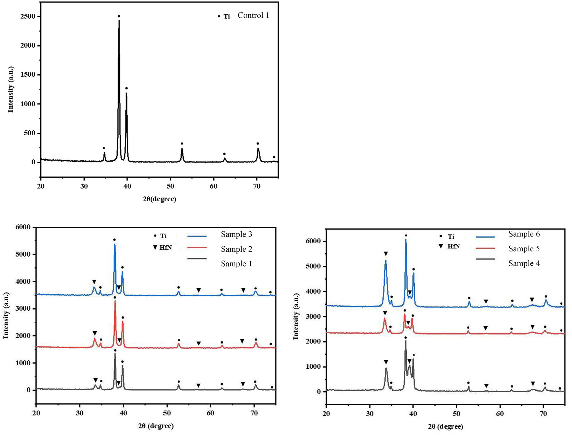
The crystal structures of the coatings were characterized by XRD in symmetric θ–2θ scanning mode.

Surface topography of the material coating observed by SEM; scale bar = 50 μm.
Figure 3 is obtained by EDS elemental mapping. EDS analyzed the weight percentage and atomic percentage of key elements on the coating surface. From Figure 6, it could be found that Ti and HfN were detected in all the samples, proved that 50 and 200 nm thicknesses of coatings magnetron sputtering could successfully attach different compositional ratio of HfN to the surfaces of the samples.

Surface element distribution of the film was characterized by EDS after magnetron sputtering with different parameters. wt%: weight percentage; at%: atomic percentage.
3.2 Evaluation of the antibacterial properties of the coatings
In Figure 4, crystal violet staining showed that biofilm formation on the surfaces of sample 2 (with a thickness of 50 nm and an N2 flow rate of 2.5 sccm) and sample 5 (with a thickness of 200 nm and an N2 flow rate of 2.5 sccm) was significantly inhibited. Among them, the surface of sample 2 had the least amount of biofilm. Notably, the antibacterial properties of sample 2 was better than those of samples 1 and 3, and the antibacterial properties of sample 5 was better than those of samples 4 and 6, suggesting that the antibacterial properties of N2 flow at 2.5 sccm may be better than those of N2 at 2 sccm or 3 sccm.

Quantitative results of crystal violet staining of the surfaces of biofilms on the implant material (*p < 0.05; **p < 0.01, ***p < 0.001).
In Figure 5, SYTO-9/PI live/dead bacterial fluorescence staining indicated that the surface of sample 2 had less green fluorescence than those of control 1 and other samples. It is illustrated that the surviving S. aureus in sample 2 was less than other samples.

Control 1 and samples 1–6 were stained with SYTO-9/PI. SYTO-9-stained live bacteria to produce green fluorescence, while PI stained dead bacteria to produce red fluorescence; scale bar = 100 μm.
SEM was used to compare bacterial adhesion to the coating surfaces in Figure 6. Notably, most S. aureus adhered to the surface of control 1, while the least bacteria were found on samples 2 and 5. In Figure 5, the bacteria adhered to the surface of sample 2 were relatively scattered and not stacked, while those on the surface of sample 5 were stacked and accumulated to form aggregates. Moreover, the SEM results showed that the morphology of S. aureus remained unchanged.

SEM was used to detect bacterial adhesion to the surface of the coating: a large scale bar = 10 μm and a small scale bar = 3 μm.
3.3 Biocompatibility of the coatings
After 2 days of coculture with the implant material, the CCK-8 method was used to analyze the proliferation of BMSCs, as shown in Figure 7. Compared to control 1, there was no decrease in cell viability on the coated surface of sample 2, while cell viability on the surface of the other samples decreased. In addition, the cell activity on the surface of sample 2 was higher than that of other samples.

Viability of BMSCs cultured on control 1 and samples 1–6 was determined by CCK-8 assays (***p < 0.001).
Moreover, the calcein-AM/PI live/dead cell staining results showed that sample 2 had the highest number of viable cells compared to control 1 and other samples in Figure 8. It was consistent with the CCK-8 assay and indicated that sample 2 had the best biocompatibility.

Calcein-AM/PI live/dead cell staining results. Calcein-AM fluoresces green in live cells, and PI fluoresces red in dead cells; scale bar = 400 μm.
4 Discussion
One of the most serious complications after the implantation of orthopedic materials is the development of an infection, which can cause clinical problems and patient suffering [28]. Thus, developing biomaterials with anti-infective properties is highly important for the prevention and treatment of related infections [29,30].
Graphene has been studied for its antimicrobial and coating applications [31], and the antibacterial, osteogenic, and angiogenic properties of the novel pH-responsive CaO2@ZIF-67-HA-ADH coating on titanium implants have also been investigated [32]. Ti and its alloys have been widely used in industry and the biomedical field, particularly for bone fusion, bone fixation, and joint replacement surgery [6]. Many researchers have modified the surface of titanium with antibiotics, antibacterial peptides, inorganic antibacterial metal elements, and antibacterial polymers to increase its antibacterial properties [33–35].
This is the first study applying magnetron sputtering technology to form an HfN coating on the surface of Ti. Grossman developed a classic method to quantify S. aureus biofilm formation by crystal violet staining and confocal microscopy, in an approach that has been standardized [36]. We used the same methodology to test the antibacterial properties of our coatings.
The SEM analysis showed that the HfN coating and Ti substrate formed relatively uniform nanoscale filaments, combined the XRD analysis shows the uniformity of the coating. The SEM analysis of bacteria adherence to the surface of the material revealed that the morphology of S. aureus was unchanged, indicating that the coating may have produced an antibacterial effect by inhibiting bacterial growth rather than killing the bacteria by changing their state, similarly inhibiting the growth of bacteria on material surfaces [37]. It is also possible that the dead S. aureus is not adherent therefore barely visible on the surface of the samples.
CCK-8 and live/dead cell staining assays have been used in many studies to evaluate biocompatibility [38]. Additionally, BMSCs are widely used in various orthopedic studies [39,40]. We also used these methods here to demonstrate that sample 2 did not exhibit significant toxicity to BMSCs. According to the calcein-AM/PI live/dead cell staining results, there were obvious live cells (green fluorescence), but not obvious dead cells (red fluorescence), indicating that the dead BMSCs may have been washed away without adhering.
Our study revealed that HfN coatings with a thickness of 50 nm and an N2 flow rate of 2.5 sccm had the greatest antibacterial effect on S. aureus compared to the other coatings studied in the present manuscript. In addition, SEM was used to detect bacterial adhesion to the surface of the materials, and the results were consistent. Notably, the antibacterial properties of sample 5 are second only to those of sample 2, indicating that the antibacterial performance of the N2 flow of 2.5 sccm may be better than that of the N2 flow of 2 sccm or 3 sccm. The EDS results showed that the composition of HfN element on the coating surface also changed accordingly. At 50 nm thickness, the Hf wt% of 2.5 sccm N2 flow was 95.9 and the Hf at% of 2.5 sccm N2 flow was 66.9, which was higher than 2 sccm or 3 sccm N2 flow; the N wt% of 2.5 sccm N2 flow was 3.5, and the N at% of 2.5 sccm N2 flow was 31.2, which was lower than 2 sccm or 3 sccm N2 flow. But at 200 nm thickness, the Hf wt% of 2.5 sccm N2 was 96.5 and the Hf at% was 70.3, which was lower than 2 sccm or 3 sccm N2 flow; the N wt% of 2.5 sccm N2 was 3.0 and the N at% was 28.1, which was higher than 2 sccm or 3 sccm N2 flow. Studies have shown that the strength of implant Ti element may be related to infection and bone resorption [41]. It may be that in different thickness, the trend of element change is different, which affects antibacterial properties.
On the other hand, the antibacterial effect of HfN coating with a thickness of 50 nm against S. aureus was better than that of 200 nm, but there was no obvious law in biocompatibility. About 50 and 200 nm films were selected to compare and study the effects of different elemental composition on antimicrobial properties. This is because previous studies have shown that the thickness of the film strongly affects the elemental composition [28]. EDS results show that under the same flux of N2, the wt% and at% of Hf increased and that of Ti decreased between 50 and 200 nm thickness. And the HfN coatings with a thickness of 50 nm and 2.5 sccm N2 flow rate may be the balance value of the film’s elemental composition and antibacterial properties. It is possible that the Hf wt% is 95.9–96.5, the Hf at% is 66.9–70.3, N wt% is 3–3.5, and the Hf at% is 28.1–31.2, which has the most suitable antibacterial properties and biocompatibility.
However, there were several limitations to our research. First, future studies could further investigate how changes in physicochemical properties – surface roughness, wettability, and surface energy – influence the coating’s biocompatibility and antibacterial efficacy. Second, since we only used one experimental strain as a model, these materials may not be suitable for other species of bacteria, such as methicillin-resistant S. aureus. Third, biocompatibility was assessed only at the cellular level, which requires more rigorous animal studies and will be done in the future.
5 Conclusions
This is the first study to explore coating the surface of Ti implants with HfN by magnetron sputtering technology. We investigated the antibacterial properties and biocompatibility of the coatings. In vitro experiments showed that the HfN coating with a thickness of 50 nm, N2 flow rate of 2.5 sccm, and Ar flow rate of 80 sccm had a notable antibacterial effect against S. aureus and was not toxic to BMSCs. The surface element of coatings had a significant variation between 50 and 200 nm thickness, and its antibacterial response between 50 and 200 nm thickness also changed accordingly. Further research could explore the effects of surface characteristics (roughness, surface energy, wettability) on the coating’s biocompatibility and antibacterial properties. This study provides a new direction for the design and preparation of antimicrobial coatings for orthopedic implants. However, more experiments and in vivo studies are needed in the future to support this hypothesis.
Acknowledgments
The authors are grateful for the reviewer’s valuable comments that improved the manuscript.
-
Funding information: This work was supported by internal funding from the First Hospital of Jiaxing, China (Grant Agreement 2022-xkdtr-01, 2024-ZD-005, 2024-QMX-013), the Key Laboratory Project of Jiaxing, China (Grant Agreement 2022-yzcsgtjzjz), and the Key Departments of Jiaxing, China (Grant Agreement 2023-ZC-012).
-
Author contributions: All authors have accepted responsibility for the entire content of this manuscript and consented to its submission to the journal, reviewed all the results, and approved the final version of the manuscript. Jun Xue and Yi Jiang designed the experiments and carried them out. Jinyu Zhu and Sihui Chen prepared the manuscript with contributions from all co-authors.
-
Conflict of interest: Authors state no conflict of interest.
-
Data availability statement: The datasets generated during and/or analyzed during the current study are available from the corresponding author on reasonable request.
References
[1] Im GI. Biomaterials in orthopaedics: the past and future with immune modulation. Biomater Res. 2020;24(4):7.10.1186/s40824-020-0185-7Search in Google Scholar PubMed PubMed Central
[2] Zhao A, Sun J, Liu Y. Understanding bacterial biofilms: From definition to treatment strategies. Front Cell Infect Microbiol. 2023;13:1137947.10.3389/fcimb.2023.1137947Search in Google Scholar PubMed PubMed Central
[3] Visperas A, Santana D, Klika AK, Higuera-Rueda CA, Piuzzi NS. Current treatments for biofilm-associated periprosthetic joint infection and new potential strategies. J Orthop Res. 2022;40(7):1477–91.10.1002/jor.25345Search in Google Scholar PubMed PubMed Central
[4] Kotsakis GA, Black R, Kum J, Berbel L, Sadr A, Karoussis I, et al. Effect of implant cleaning on titanium particle dissolution and cytocompatibility. J Periodontol. 2021;92(4):580–91.10.1002/JPER.20-0186Search in Google Scholar PubMed
[5] Jiang P, Zhang Y, Hu R, Shi B, Zhang L, Huang Q, et al. Advanced surface engineering of titanium materials for biomedical applications: From static modification to dynamic responsive regulation. Bioact Mater. 2023;27:15–57.10.1016/j.bioactmat.2023.03.006Search in Google Scholar PubMed PubMed Central
[6] Karakurt EM, Huang Y, Cetin Y, Incesu A, Demirtas H, Kaya M, et al. Assessing microstructural, biomechanical, and biocompatible properties of TiNb alloys for potential use as load-bearing implants. J Funct Biomater. 2024;15(9):253.10.3390/jfb15090253Search in Google Scholar PubMed PubMed Central
[7] Luo H, Wu Y, Diao X, Shi W, Feng F, Qian F, et al. Mechanical properties and biocompatibility of titanium with a high oxygen concentration for dental implants. Mater Sci Eng C Mater Biol Appl. 2020;117:111306.10.1016/j.msec.2020.111306Search in Google Scholar PubMed
[8] Losic D. Advancing of titanium medical implants by surface engineering: recent progress and challenges. Expert Opin Drug Deliv. 2021;18(10):1355–78.10.1080/17425247.2021.1928071Search in Google Scholar PubMed
[9] Sousa V, Mardas N, Spratt D, Hassan IA, Walters NJ, Beltrán V, et al. The effect of microcosm biofilm decontamination on surface topography, chemistry, and biocompatibility dynamics of implant titanium surfaces. Int J Mol Sci. 2022;23(17):10033.10.3390/ijms231710033Search in Google Scholar PubMed PubMed Central
[10] Alabbad M, Silikas N, Thomas A. Effect of mechanical instrumentation on titanium implant surface properties. Dent Mater. 2025;41(4):383–90.10.1016/j.dental.2024.12.014Search in Google Scholar PubMed
[11] Xue T, Attarilar S, Liu S, Liu J, Song X, Li L, et al. Surface modification techniques of titanium and its alloys to functionally optimize their biomedical properties: thematic review. Front Bioeng Biotechnol. 2020;8:603072.10.3389/fbioe.2020.603072Search in Google Scholar PubMed PubMed Central
[12] Almatroudi A. Silver nanoparticles: synthesis, characterisation and biomedical applications. Open Life Sci. 2020;15(1):819–39.10.1515/biol-2020-0094Search in Google Scholar PubMed PubMed Central
[13] Hartjen P, Wegner N, Ahmadi P, Matthies L, Nada O, Fuest S, et al. Toward tailoring the degradation rate of magnesium-based biomaterials for various medical applications: Assessing corrosion, cytocompatibility and immunological effects. Int J Mol Sci. 2021;22(2):971.10.3390/ijms22020971Search in Google Scholar PubMed PubMed Central
[14] Witkowska J, Borowski T, Sowińska A, Choińska E, Moszczyńska D, Morgiel J, et al. Influence of low temperature plasma oxidizing on the bioactivity of NiTi shape memory alloy for medical applications. Materials (Basel). 2023;16(18):6086.10.3390/ma16186086Search in Google Scholar PubMed PubMed Central
[15] Sabzehmeidani MM, Kazemzad M. Recent advances in surface-mounted metal-organic framework thin film coatings for biomaterials and medical applications: a review. Biomater Res. 2023;27(1):115.10.1186/s40824-023-00454-ySearch in Google Scholar PubMed PubMed Central
[16] Al Mustafa M, Agis H, Müller HD, Watzek G, Gruber R. In vitro adhesion of fibroblastic cells to titanium alloy discs treated with sodium hydroxide. Clin Oral Implant Res. 2015;26(1):15–9.10.1111/clr.12294Search in Google Scholar PubMed
[17] Wu Y. Electrohydrodynamic jet 3D printing in biomedical applications. Acta Biomater. 2021;128:21–41.10.1016/j.actbio.2021.04.036Search in Google Scholar PubMed
[18] Rajaraman V, Nallaswamy D, Ganapathy D, Rajeshkumar S, Ariga P, Ganesh K. Effect of hafnium coating on osseointegration of titanium implants: a split mouth animal study. J Nanomater. 2021;4:1–9.10.1155/2021/7512957Search in Google Scholar
[19] Miyazaki T, Sueoka M, Shirosaki Y, Shinozaki N, Shiraishi T. Development of hafnium metal and titanium-hafnium alloys having apatite-forming ability by chemical surface modification. J Biomed Mater Res B Appl Biomater. 2018;106(7):2519–23.10.1002/jbm.b.34068Search in Google Scholar PubMed
[20] Zhao T, Li Y, Liu Y, Zhao X. Nano-hardness, wear resistance and pseudoelasticity of hafnium implanted NiTi shape memory alloy. J Mech Behav Biomed Mater. 2012;13:174–84.10.1016/j.jmbbm.2012.04.004Search in Google Scholar PubMed
[21] Rajaraman V, Ariga P, Ramalingam K, Sekaran S. Evaluation of corrosive properties of hafnium nitride coating over titanium screws: an in vitro study. Cureus. 2024;16(3):e55456.10.7759/cureus.55456Search in Google Scholar PubMed PubMed Central
[22] Bhunia M, Sandoval-Pauker C, Jafari MG, Grant LN, Gau MR, Pinter B, et al. Terminal and super-basic parent imides of hafnium. Angew Chem Int Ed Engl. 2022;61(43):e202209122.10.1002/anie.202209122Search in Google Scholar PubMed
[23] Das P, Biswas B, Maurya KC, Garbrecht M, Saha B. Refractory plasmonic hafnium nitride and zirconium nitride thin films as alternatives to silver for solar mirror applications. ACS Appl Mater Interfaces. 2022;14(41):46708–15.10.1021/acsami.2c09852Search in Google Scholar PubMed
[24] Jianbo W, Mengqian C, Dechen W, Yan L, Jize C, Zhiqing G, et al. Integration of superhydrophobicity and high durability in super-rough hard thin films. Ceram Int. 2021;47(16):23653–8.10.1016/j.ceramint.2021.04.273Search in Google Scholar
[25] Hauschild G, Hardes J, Dudda M, Streitbürger A, Wahrenburg M. Impact of topography and added TiN-coating on adult human dermal fibroblasts after seeding on titanium surface in-vitro. J Biomater Appl. 2024;38(8):905–14.10.1177/08853282241233194Search in Google Scholar PubMed PubMed Central
[26] Chaurasiya A, Yadav NS, Hazari P, Mahajan H, Narwani S. Comparative evaluation of osteogenic cell growth on titanium surface and titanium coated with boron nitride surface: An in vitro study. J Indian Prosthodont Soc. 2023;23(2):184–91.10.4103/jips.jips_97_23Search in Google Scholar PubMed PubMed Central
[27] Godoy-Gallardo M, Eckhard U, Delgado LM, de Roo Puente YJD, Hoyos-Nogués M, Gil FJ, et al. Antibacterial approaches in tissue engineering using metal ions and nanoparticles: From mechanisms to applications. Bioact Mater. 2021;6(12):4470–90.10.1016/j.bioactmat.2021.04.033Search in Google Scholar PubMed PubMed Central
[28] Xu Y, Zhang J, Liang F, Yin M, He M. Investigation of magnetron sputtered nano-silver coating on titanium surface with micro-nanostructure. Surf Interfaces. 2023;38:e102770; Barros J, Monteiro FJ, Ferraz MP. Bioengineering approaches to fight against orthopedic biomaterials related-infections. Int J Mol Sci. 2022;23(19):11658.Search in Google Scholar
[29] Arciola CR, Campoccia D, Montanaro L. Implant infections: adhesion, biofilm formation and immune evasion. Nat Rev Microbiol. 2018;16(7):397–409.10.1038/s41579-018-0019-ySearch in Google Scholar PubMed
[30] Fu J, Zhu W, Liu X, Liang C, Zheng Y, Li Z, et al. Self-activating anti-infection implant. Nat Commun. 2021;12(1):6907.10.1038/s41467-021-27217-4Search in Google Scholar PubMed PubMed Central
[31] Srimaneepong V, Skallevold HE, Khurshid Z, Zafar MS, Rokaya D, Sapkota J. Graphene for antimicrobial and coating application. Int J Mol Sci. 2022;23(1):499.10.3390/ijms23010499Search in Google Scholar PubMed PubMed Central
[32] Li X, Xu M, Geng Z, Xu X, Han X, Chen L, et al. Novel pH-responsive CaO2@ZIF-67-HA-ADH coating that efficiently enhances the antimicrobial, osteogenic, and angiogenic properties of titanium implants. ACS Appl Mater Interfaces. 2023;15(36):42965–80.10.1021/acsami.3c08233Search in Google Scholar PubMed
[33] Zhang H, Yuan Y, Xue H, Yu R, Jin X, Wu X, et al. Reprogramming mitochondrial metabolism of macrophages by miRNA-released microporous coatings to prevent peri-implantitis. J Nanobiotechnol. 2023;21(1):485.10.1186/s12951-023-02244-zSearch in Google Scholar PubMed PubMed Central
[34] Schulze M, Nonhoff M, Hasselmann J, Fobker M, Niemann S, Theil C, et al. Shock wave-activated silver-loaded biopolymer implant coating eliminates Staphylococcus epidermidis on the surface and in the surrounding of implants. Pharmaceutics. 2023;15(12):2670.10.3390/pharmaceutics15122670Search in Google Scholar PubMed PubMed Central
[35] Luo CA, Chang YH, Chang YJ, Lee HC, Tsai SW. A water-based biocoating to increase the infection resistance and osteoconductivity of titanium surfaces. Int J Mol Sci. 2024;25(6):3267.10.3390/ijms25063267Search in Google Scholar PubMed PubMed Central
[36] Grossman AB, Burgin DJ, Rice KC. Quantification of Staphylococcus aureus biofilm formation by crystal violet and confocal microscopy. Methods Mol Biol. 2021;2341:69–78.10.1007/978-1-0716-1550-8_9Search in Google Scholar PubMed
[37] Zhang J, Wang M, Hu L, Zhang Q, Chen E, Wang Z, et al. A universal coating strategy for inhibiting the growth of bacteria on materials surfaces. Front Chem. 2022;10:1043353.10.3389/fchem.2022.1043353Search in Google Scholar PubMed PubMed Central
[38] Kus-Liśkiewicz M, Fickers P, Ben Tahar I. Biocompatibility and cytotoxicity of gold nanoparticles: recent advances in methodologies and regulations. Int J Mol Sci. 2021;22(20):10952.10.3390/ijms222010952Search in Google Scholar PubMed PubMed Central
[39] Purwaningrum M, Jamilah NS, Purbantoro SD, Sawangmake C, Nantavisai S. Comparative characteristic study from bone marrow-derived mesenchymal stem cells. J Vet Sci. 2021;22(6):e74.10.4142/jvs.2021.22.e74Search in Google Scholar PubMed PubMed Central
[40] Chu DT, Phuong TNT, Tien NLB, Tran DK, Thanh VV, Quang TL, et al. An update on the progress of isolation, culture, storage, and clinical application of human bone marrow mesenchymal stem/stromal cells. Int J Mol Sci. 2020;21(3):708.10.3390/ijms21030708Search in Google Scholar PubMed PubMed Central
[41] Silva GAF, Faot F, da Silva WJ, Del Bel Cury AA. Does implant surface hydrophilicity influence the maintenance of surface integrity after insertion into low-density artificial bone?. Dent Mater. 2021;37(2):e69–84.10.1016/j.dental.2020.10.024Search in Google Scholar PubMed
© 2025 the author(s), published by De Gruyter
This work is licensed under the Creative Commons Attribution 4.0 International License.
Articles in the same Issue
- Safety assessment and modulation of hepatic CYP3A4 and UGT enzymes by Glycyrrhiza glabra aqueous extract in female Sprague–Dawley rats
- Adult-onset Still’s disease with hemophagocytic lymphohistiocytosis and minimal change disease
- Role of DZ2002 in reducing corneal graft rejection in rats by influencing Th17 activation via inhibition of the PI3K/AKT pathway and downregulation of TRAF1
- Biomedical Sciences
- Mechanism of triptolide regulating proliferation and apoptosis of hepatoma cells by inhibiting JAK/STAT pathway
- Maslinic acid improves mitochondrial function and inhibits oxidative stress and autophagy in human gastric smooth muscle cells
- Comparative analysis of inflammatory biomarkers for the diagnosis of neonatal sepsis: IL-6, IL-8, SAA, CRP, and PCT
- Post-pandemic insights on COVID-19 and premature ovarian insufficiency
- Proteome differences of dental stem cells between permanent and deciduous teeth by data-independent acquisition proteomics
- Optimizing a modified cetyltrimethylammonium bromide protocol for fungal DNA extraction: Insights from multilocus gene amplification
- Preliminary analysis of the role of small hepatitis B surface proteins mutations in the pathogenesis of occult hepatitis B infection via the endoplasmic reticulum stress-induced UPR-ERAD pathway
- Efficacy of alginate-coated gold nanoparticles against antibiotics-resistant Staphylococcus and Streptococcus pathogens of acne origins
- Battling COVID-19 leveraging nanobiotechnology: Gold and silver nanoparticle–B-escin conjugates as SARS-CoV-2 inhibitors
- Neurodegenerative diseases and neuroinflammation-induced apoptosis
- Impact of fracture fixation surgery on cognitive function and the gut microbiota in mice with a history of stroke
- COLEC10: A potential tumor suppressor and prognostic biomarker in hepatocellular carcinoma through modulation of EMT and PI3K-AKT pathways
- High-temperature requirement serine protease A2 inhibitor UCF-101 ameliorates damaged neurons in traumatic brain-injured rats by the AMPK/NF-κB pathway
- SIK1 inhibits IL-1β-stimulated cartilage apoptosis and inflammation in vitro through the CRTC2/CREB1 signaling
- Rutin–chitooligosaccharide complex: Comprehensive evaluation of its anti-inflammatory and analgesic properties in vitro and in vivo
- Knockdown of Aurora kinase B alleviates high glucose-triggered trophoblast cells damage and inflammation during gestational diabetes
- Calcium-sensing receptors promoted Homer1 expression and osteogenic differentiation in bone marrow mesenchymal stem cells
- ABI3BP can inhibit the proliferation, invasion, and epithelial–mesenchymal transition of non-small-cell lung cancer cells
- Changes in blood glucose and metabolism in hyperuricemia mice
- Rapid detection of the GJB2 c.235delC mutation based on CRISPR-Cas13a combined with lateral flow dipstick
- IL-11 promotes Ang II-induced autophagy inhibition and mitochondrial dysfunction in atrial fibroblasts
- Short-chain fatty acid attenuates intestinal inflammation by regulation of gut microbial composition in antibiotic-associated diarrhea
- Application of metagenomic next-generation sequencing in the diagnosis of pathogens in patients with diabetes complicated by community-acquired pneumonia
- NAT10 promotes radiotherapy resistance in non-small cell lung cancer by regulating KPNB1-mediated PD-L1 nuclear translocation
- Phytol-mixed micelles alleviate dexamethasone-induced osteoporosis in zebrafish: Activation of the MMP3–OPN–MAPK pathway-mediating bone remodeling
- Association between TGF-β1 and β-catenin expression in the vaginal wall of patients with pelvic organ prolapse
- Primary pleomorphic liposarcoma involving bilateral ovaries: Case report and literature review
- Effects of de novo donor-specific Class I and II antibodies on graft outcomes after liver transplantation: A pilot cohort study
- Sleep architecture in Alzheimer’s disease continuum: The deep sleep question
- Ephedra fragilis plant extract: A groundbreaking corrosion inhibitor for mild steel in acidic environments – electrochemical, EDX, DFT, and Monte Carlo studies
- Langerhans cell histiocytosis in an adult patient with upper jaw and pulmonary involvement: A case report
- Inhibition of mast cell activation by Jaranol-targeted Pirin ameliorates allergic responses in mouse allergic rhinitis
- Aeromonas veronii-induced septic arthritis of the hip in a child with acute lymphoblastic leukemia
- Clusterin activates the heat shock response via the PI3K/Akt pathway to protect cardiomyocytes from high-temperature-induced apoptosis
- Research progress on fecal microbiota transplantation in tumor prevention and treatment
- Low-pressure exposure influences the development of HAPE
- Stigmasterol alleviates endplate chondrocyte degeneration through inducing mitophagy by enhancing PINK1 mRNA acetylation via the ESR1/NAT10 axis
- AKAP12, mediated by transcription factor 21, inhibits cell proliferation, metastasis, and glycolysis in lung squamous cell carcinoma
- Association between PAX9 or MSX1 gene polymorphism and tooth agenesis risk: A meta-analysis
- A case of bloodstream infection caused by Neisseria gonorrhoeae
- Case of nasopharyngeal tuberculosis complicated with cervical lymph node and pulmonary tuberculosis
- p-Cymene inhibits pro-fibrotic and inflammatory mediators to prevent hepatic dysfunction
- GFPT2 promotes paclitaxel resistance in epithelial ovarian cancer cells via activating NF-κB signaling pathway
- Transfer RNA-derived fragment tRF-36 modulates varicose vein progression via human vascular smooth muscle cell Notch signaling
- RTA-408 attenuates the hepatic ischemia reperfusion injury in mice possibly by activating the Nrf2/HO-1 signaling pathway
- Decreased serum TIMP4 levels in patients with rheumatoid arthritis
- Sirt1 protects lupus nephritis by inhibiting the NLRP3 signaling pathway in human glomerular mesangial cells
- Sodium butyrate aids brain injury repair in neonatal rats
- Interaction of MTHFR polymorphism with PAX1 methylation in cervical cancer
- Convallatoxin inhibits proliferation and angiogenesis of glioma cells via regulating JAK/STAT3 pathway
- The effect of the PKR inhibitor, 2-aminopurine, on the replication of influenza A virus, and segment 8 mRNA splicing
- Effects of Ire1 gene on virulence and pathogenicity of Candida albicans
- Small cell lung cancer with small intestinal metastasis: Case report and literature review
- GRB14: A prognostic biomarker driving tumor progression in gastric cancer through the PI3K/AKT signaling pathway by interacting with COBLL1
- 15-Lipoxygenase-2 deficiency induces foam cell formation that can be restored by salidroside through the inhibition of arachidonic acid effects
- FTO alleviated the diabetic nephropathy progression by regulating the N6-methyladenosine levels of DACT1
- Clinical relevance of inflammatory markers in the evaluation of severity of ulcerative colitis: A retrospective study
- Zinc valproic acid complex promotes osteoblast differentiation and exhibits anti-osteoporotic potential
- Primary pulmonary synovial sarcoma in the bronchial cavity: A case report
- Metagenomic next-generation sequencing of alveolar lavage fluid improves the detection of pulmonary infection
- Uterine tumor resembling ovarian sex cord tumor with extensive rhabdoid differentiation: A case report
- Genomic analysis of a novel ST11(PR34365) Clostridioides difficile strain isolated from the human fecal of a CDI patient in Guizhou, China
- Effects of tiered cardiac rehabilitation on CRP, TNF-α, and physical endurance in older adults with coronary heart disease
- Changes in T-lymphocyte subpopulations in patients with colorectal cancer before and after acupoint catgut embedding acupuncture observation
- Modulating the tumor microenvironment: The role of traditional Chinese medicine in improving lung cancer treatment
- Alterations of metabolites related to microbiota–gut–brain axis in plasma of colon cancer, esophageal cancer, stomach cancer, and lung cancer patients
- Research on individualized drug sensitivity detection technology based on bio-3D printing technology for precision treatment of gastrointestinal stromal tumors
- CEBPB promotes ulcerative colitis-associated colorectal cancer by stimulating tumor growth and activating the NF-κB/STAT3 signaling pathway
- Oncolytic bacteria: A revolutionary approach to cancer therapy
- A de novo meningioma with rapid growth: A possible malignancy imposter?
- Diagnosis of secondary tuberculosis infection in an asymptomatic elderly with cancer using next-generation sequencing: Case report
- Hesperidin and its zinc(ii) complex enhance osteoblast differentiation and bone formation: In vitro and in vivo evaluations
- Research progress on the regulation of autophagy in cardiovascular diseases by chemokines
- Anti-arthritic, immunomodulatory, and inflammatory regulation by the benzimidazole derivative BMZ-AD: Insights from an FCA-induced rat model
- Immunoassay for pyruvate kinase M1/2 as an Alzheimer’s biomarker in CSF
- The role of HDAC11 in age-related hearing loss: Mechanisms and therapeutic implications
- Evaluation and application analysis of animal models of PIPNP based on data mining
- Therapeutic approaches for liver fibrosis/cirrhosis by targeting pyroptosis
- Fabrication of zinc oxide nanoparticles using Ruellia tuberosa leaf extract induces apoptosis through P53 and STAT3 signalling pathways in prostate cancer cells
- Haplo-hematopoietic stem cell transplantation and immunoradiotherapy for severe aplastic anemia complicated with nasopharyngeal carcinoma: A case report
- Modulation of the KEAP1-NRF2 pathway by Erianin: A novel approach to reduce psoriasiform inflammation and inflammatory signaling
- The expression of epidermal growth factor receptor 2 and its relationship with tumor-infiltrating lymphocytes and clinical pathological features in breast cancer patients
- Innovations in MALDI-TOF Mass Spectrometry: Bridging modern diagnostics and historical insights
- BAP1 complexes with YY1 and RBBP7 and its downstream targets in ccRCC cells
- Hypereosinophilic syndrome with elevated IgG4 and T-cell clonality: A report of two cases
- Electroacupuncture alleviates sciatic nerve injury in sciatica rats by regulating BDNF and NGF levels, myelin sheath degradation, and autophagy
- Polydatin prevents cholesterol gallstone formation by regulating cholesterol metabolism via PPAR-γ signaling
- RNF144A and RNF144B: Important molecules for health
- Analysis of the detection rate and related factors of thyroid nodules in the healthy population
- Artesunate inhibits hepatocellular carcinoma cell migration and invasion through OGA-mediated O-GlcNAcylation of ZEB1
- Endovascular management of post-pancreatectomy hemorrhage caused by a hepatic artery pseudoaneurysm: Case report and review of the literature
- Efficacy and safety of anti-PD-1/PD-L1 antibodies in patients with relapsed refractory diffuse large B-cell lymphoma: A meta-analysis
- SATB2 promotes humeral fracture healing in rats by activating the PI3K/AKT pathway
- Overexpression of the ferroptosis-related gene, NFS1, corresponds to gastric cancer growth and tumor immune infiltration
- Understanding risk factors and prognosis in diabetic foot ulcers
- Atractylenolide I alleviates the experimental allergic response in mice by suppressing TLR4/NF-kB/NLRP3 signalling
- FBXO31 inhibits the stemness characteristics of CD147 (+) melanoma stem cells
- Immune molecule diagnostics in colorectal cancer: CCL2 and CXCL11
- Inhibiting CXCR6 promotes senescence of activated hepatic stellate cells with limited proinflammatory SASP to attenuate hepatic fibrosis
- Cadmium toxicity, health risk and its remediation using low-cost biochar adsorbents
- Pulmonary cryptococcosis with headache as the first presentation: A case report
- Solitary pulmonary metastasis with cystic airspaces in colon cancer: A rare case report
- RUNX1 promotes denervation-induced muscle atrophy by activating the JUNB/NF-κB pathway and driving M1 macrophage polarization
- Morphometric analysis and immunobiological investigation of Indigofera oblongifolia on the infected lung with Plasmodium chabaudi
- The NuA4/TIP60 histone-modifying complex and Hr78 modulate the Lobe2 mutant eye phenotype
- Experimental study on salmon demineralized bone matrix loaded with recombinant human bone morphogenetic protein-2: In vitro and in vivo study
- A case of IgA nephropathy treated with a combination of telitacicept and half-dose glucocorticoids
- Analgesic and toxicological evaluation of cannabidiol-rich Moroccan Cannabis sativa L. (Khardala variety) extract: Evidence from an in vivo and in silico study
- Wound healing and signaling pathways
- Combination of immunotherapy and whole-brain radiotherapy on prognosis of patients with multiple brain metastases: A retrospective cohort study
- To explore the relationship between endometrial hyperemia and polycystic ovary syndrome
- Research progress on the impact of curcumin on immune responses in breast cancer
- Biogenic Cu/Ni nanotherapeutics from Descurainia sophia (L.) Webb ex Prantl seeds for the treatment of lung cancer
- Dapagliflozin attenuates atrial fibrosis via the HMGB1/RAGE pathway in atrial fibrillation rats
- Glycitein alleviates inflammation and apoptosis in keratinocytes via ROS-associated PI3K–Akt signalling pathway
- ADH5 inhibits proliferation but promotes EMT in non-small cell lung cancer cell through activating Smad2/Smad3
- Apoptotic efficacies of AgNPs formulated by Syzygium aromaticum leaf extract on 32D-FLT3-ITD human leukemia cell line with PI3K/AKT/mTOR signaling pathway
- Novel cuproptosis-related genes C1QBP and PFKP identified as prognostic and therapeutic targets in lung adenocarcinoma
- Bee venom promotes exosome secretion and alters miRNA cargo in T cells
- Treatment of pure red cell aplasia in a chronic kidney disease patient with roxadustat: A case report
- Comparative bioinformatics analysis of the Wnt pathway in breast cancer: Selection of novel biomarker panels associated with ER status
- Kynurenine facilitates renal cell carcinoma progression by suppressing M2 macrophage pyroptosis through inhibition of CASP1 cleavage
- RFX5 promotes the growth, motility, and inhibits apoptosis of gastric adenocarcinoma cells through the SIRT1/AMPK axis
- ALKBH5 exacerbates early cardiac damage after radiotherapy for breast cancer via m6A demethylation of TLR4
- Phytochemicals of Roman chamomile: Antioxidant, anti-aging, and whitening activities of distillation residues
- Circadian gene Cry1 inhibits the tumorigenicity of hepatocellular carcinoma by the BAX/BCL2-mediated apoptosis pathway
- The TNFR-RIPK1/RIPK3 signalling pathway mediates the effect of lanthanum on necroptosis of nerve cells
- Longitudinal monitoring of autoantibody dynamics in patients with early-stage non-small-cell lung cancer undergoing surgery
- The potential role of rutin, a flavonoid, in the management of cancer through modulation of cell signaling pathways
- Construction of pectinase gene engineering microbe and its application in tobacco sheets
- Construction of a microbial abundance prognostic scoring model based on intratumoral microbial data for predicting the prognosis of lung squamous cell carcinoma
- Sepsis complicated by haemophagocytic lymphohistiocytosis triggered by methicillin-resistant Staphylococcus aureus and human herpesvirus 8 in an immunocompromised elderly patient: A case report
- Sarcopenia in liver transplantation: A comprehensive bibliometric study of current research trends and future directions
- Advances in cancer immunotherapy and future directions in personalized medicine
- Can coronavirus disease 2019 affect male fertility or cause spontaneous abortion? A two-sample Mendelian randomization analysis
- Heat stroke associated with novel leukaemia inhibitory factor receptor gene variant in a Chinese infant
- PSME2 exacerbates ulcerative colitis by disrupting intestinal barrier function and promoting autophagy-dependent inflammation
- Hyperosmolar hyperglycemic state with severe hypernatremia coexisting with central diabetes insipidus: A case report and literature review
- Efficacy and mechanism of escin in improving the tissue microenvironment of blood vessel walls via anti-inflammatory and anticoagulant effects: Implications for clinical practice
- Merkel cell carcinoma: Clinicopathological analysis of three patients and literature review
- Genetic variants in VWF exon 26 and their implications for type 1 Von Willebrand disease in a Saudi Arabian population
- Lipoxin A4 improves myocardial ischemia/reperfusion injury through the Notch1-Nrf2 signaling pathway
- High levels of EPHB2 expression predict a poor prognosis and promote tumor progression in endometrial cancer
- Knockdown of SHP-2 delays renal tubular epithelial cell injury in diabetic nephropathy by inhibiting NLRP3 inflammasome-mediated pyroptosis
- Exploring the toxicity mechanisms and detoxification methods of Rhizoma Paridis
- Concomitant gastric carcinoma and primary hepatic angiosarcoma in a patient: A case report
- YAP1 inhibition protects retinal vascular endothelial cells under high glucose by inhibiting autophagy
- Identification of secretory protein related biomarkers for primary biliary cholangitis based on machine learning and experimental validation
- Integrated genomic and clinical modeling for prognostic assessment of radiotherapy response in rectal neoplasms
- Stem cell-based approaches for glaucoma treatment: a mini review
- Bacteriophage titering by optical density means: KOTE assays
- Neutrophil-related signature characterizes immune landscape and predicts prognosis of esophageal squamous cell carcinoma
- Integrated bioinformatic analysis and machine learning strategies to identify new potential immune biomarkers for Alzheimer’s disease and their targeting prediction with geniposide
- TRIM21 accelerates ferroptosis in intervertebral disc degeneration by promoting SLC7A11 ubiquitination and degradation
- TRIM21 accelerates ferroptosis in intervertebral disc degeneration by promoting SLC7A11 ubiquitination and degradation
- Histone modification and non-coding RNAs in skin aging: emerging therapeutic avenues
- A multiplicative behavioral model of DNA replication initiation in cells
- Biogenic gold nanoparticles synthesized from Pergularia daemia leaves: a novel approach for nasopharyngeal carcinoma therapy
- Creutzfeldt-Jakob disease mimicking Hashimoto’s encephalopathy: steroid response followed by decline
- Impact of semaphorin, Sema3F, on the gene transcription and protein expression of CREB and its binding protein CREBBP in primary hippocampal neurons of rats
- Iron overloaded M0 macrophages regulate hematopoietic stem cell proliferation and senescence via the Nrf2/Keap1/HO-1 pathway
- Revisiting the link between NADPH oxidase p22phox C242T polymorphism and ischemic stroke risk: an updated meta-analysis
- Exercise training preferentially modulates α1D-adrenergic receptor expression in peripheral arteries of hypertensive rats
- Overexpression of HE4/WFDC2 gene in mice leads to keratitis and corneal opacity
- Tumoral calcinosis complicating CKD-MBD in hemodialysis: a case report
- Mechanism of KLF4 Inhibition of epithelial-mesenchymal transition in gastric cancer cells
- Dissecting the molecular mechanisms of T cell infiltration in psoriatic lesions via cell-cell communication and regulatory network analysis
- Circadian rhythm-based prognostic features predict immune infiltration and tumor microenvironment in molecular subtypes of hepatocellular carcinoma
- Ecology and Environmental Science
- Optimization and comparative study of Bacillus consortia for cellulolytic potential and cellulase enzyme activity
- The complete mitochondrial genome analysis of Haemaphysalis hystricis Supino, 1897 (Ixodida: Ixodidae) and its phylogenetic implications
- Epidemiological characteristics and risk factors analysis of multidrug-resistant tuberculosis among tuberculosis population in Huzhou City, Eastern China
- Indices of human impacts on landscapes: How do they reflect the proportions of natural habitats?
- Genetic analysis of the Siberian flying squirrel population in the northern Changbai Mountains, Northeast China: Insights into population status and conservation
- Diversity and environmental drivers of Suillus communities in Pinus sylvestris var. mongolica forests of Inner Mongolia
- Global assessment of the fate of nitrogen deposition in forest ecosystems: Insights from 15N tracer studies
- Fungal and bacterial pathogenic co-infections mainly lead to the assembly of microbial community in tobacco stems
- Influencing of coal industry related airborne particulate matter on ocular surface tear film injury and inflammatory factor expression in Sprague-Dawley rats
- Temperature-dependent development, predation, and life table of Sphaerophoria macrogaster (Thomson) (Diptera: Syrphidae) feeding on Myzus persicae (Sulzer) (Homoptera: Aphididae)
- Eleonora’s falcon trophic interactions with insects within its breeding range: A systematic review
- Agriculture
- Integrated analysis of transcriptome, sRNAome, and degradome involved in the drought-response of maize Zhengdan958
- Variation in flower frost tolerance among seven apple cultivars and transcriptome response patterns in two contrastingly frost-tolerant selected cultivars
- Heritability of durable resistance to stripe rust in bread wheat (Triticum aestivum L.)
- Molecular mechanism of follicular development in laying hens based on the regulation of water metabolism
- Molecular identification and control studies on Coridius sp. (Hemiptera: Dinidoridae) in Al-Khamra, south of Jeddah, Saudi Arabia
- 10.1515/biol-2025-1218
- Animal Science
- Effect of sex ratio on the life history traits of an important invasive species, Spodoptera frugiperda
- Plant Sciences
- Hairpin in a haystack: In silico identification and characterization of plant-conserved microRNA in Rafflesiaceae
- Widely targeted metabolomics of different tissues in Rubus corchorifolius
- The complete chloroplast genome of Gerbera piloselloides (L.) Cass., 1820 (Carduoideae, Asteraceae) and its phylogenetic analysis
- Field trial to correlate mineral solubilization activity of Pseudomonas aeruginosa and biochemical content of groundnut plants
- Correlation analysis between semen routine parameters and sperm DNA fragmentation index in patients with semen non-liquefaction: A retrospective study
- Plasticity of the anatomical traits of Rhododendron L. (Ericaceae) leaves and its implications in adaptation to the plateau environment
- Effects of Piriformospora indica and arbuscular mycorrhizal fungus on growth and physiology of Moringa oleifera under low-temperature stress
- Effects of different sources of potassium fertiliser on yield, fruit quality and nutrient absorption in “Harward” kiwifruit (Actinidia deliciosa)
- Comparative efficiency and residue levels of spraying programs against powdery mildew in grape varieties
- The DREB7 transcription factor enhances salt tolerance in soybean plants under salt stress
- Using plant electrical signals of water hyacinth (Eichhornia crassipes) for water pollution monitoring
- Response of hybrid grapes (Vitis spp.) to two biotic stress factors and their seedlessness status
- Metabolomic profiling reveals systemic metabolic reprogramming in Alternaria alternata under salt stress
- Effects of mixed salinity and alkali stress on photosynthetic characteristics and PEPC gene expression of vegetable soybean seedlings
- Food Science
- Phytochemical analysis of Stachys iva: Discovering the optimal extract conditions and its bioactive compounds
- Review on role of honey in disease prevention and treatment through modulation of biological activities
- Computational analysis of polymorphic residues in maltose and maltotriose transporters of a wild Saccharomyces cerevisiae strain
- Optimization of phenolic compound extraction from Tunisian squash by-products: A sustainable approach for antioxidant and antibacterial applications
- Liupao tea aqueous extract alleviates dextran sulfate sodium-induced ulcerative colitis in rats by modulating the gut microbiota
- Toxicological qualities and detoxification trends of fruit by-products for valorization: A review
- Polyphenolic spectrum of cornelian cherry fruits and their health-promoting effect
- Optimizing the encapsulation of the refined extract of squash peels for functional food applications: A sustainable approach to reduce food waste
- Advancements in curcuminoid formulations: An update on bioavailability enhancement strategies curcuminoid bioavailability and formulations
- Impact of saline sprouting on antioxidant properties and bioactive compounds in chia seeds
- The dilemma of food genetics and improvement
- Causal effects of trace elements on congenital foot deformities and their subtypes: a Mendelian randomization study with gut microbiota mediation
- Honey meets acidity: a novel biopreservative approach against foodborne pathogens
- Bioengineering and Biotechnology
- Impact of hyaluronic acid-modified hafnium metalorganic frameworks containing rhynchophylline on Alzheimer’s disease
- Emerging patterns in nanoparticle-based therapeutic approaches for rheumatoid arthritis: A comprehensive bibliometric and visual analysis spanning two decades
- Application of CRISPR/Cas gene editing for infectious disease control in poultry
- Preparation of hafnium nitride-coated titanium implants by magnetron sputtering technology and evaluation of their antibacterial properties and biocompatibility
- Preparation and characterization of lemongrass oil nanoemulsion: Antimicrobial, antibiofilm, antioxidant, and anticancer activities
- Fluorescent detection of sialic acid–binding lectins using functionalized quantum dots in ELISA format
- Smart tectorigenin-loaded ZnO hydrogel nanocomposites for targeted wound healing: synthesis, characterization, and biological evaluation
- Corrigendum
- Corrigendum to “Utilization of convolutional neural networks to analyze microscopic images for high-throughput screening of mesenchymal stem cells”
- Corrigendum to “Effects of Ire1 gene on virulence and pathogenicity of Candida albicans”
- Retraction
- Retraction of “Down-regulation of miR-539 indicates poor prognosis in patients with pancreatic cancer”