FTO alleviated the diabetic nephropathy progression by regulating the N6-methyladenosine levels of DACT1
-
Xuanwen Li


Abstract
Diabetic nephropathy (DN) is one of the most important microvascular complications of diabetes. The role of epigenetic regulation in DN has attracted much attention recently. This research was performed to explore the role of FTO in the DN progression. The renal tissues of DN patients were collected and the podocytes were stimulated with high glucose (HG) to establish the DN model in vitro. Western blot along with reverse transcription quantitative polymerase chain reaction assays was performed to analyze the mRNA as well as protein expressions. Immunohistochemistry and immunofluorescence were carried out to measure the FTO and DACT1 levels. The interaction between FTO/IGF2BP1 and DACT1 was verified by double luciferase reports and RNA-binding protein immunoprecipitation assays. FTO was declined, and DACT1 was enhanced in the HG-treated podocytes as well as renal tissues of DN patients. Overexpressed FTO declined the mRNA levels of MCP-1, IL-6, TNF-α, and the apoptosis rate of HG-treated podocytes. The N6-methyladenosine (m6A) levels, mRNA expression, and stability of FTO were depleted after FTO overexpression. DACT1 overexpression reversed the function of oe-FTO in podocytes stimulated with HG. Furthermore, IGF2BP1 knockdown declined the mRNA expression as well as the stability of FTO. In conclusion, FTO-medicated m6A modification of DACT1 was dependent on IGF2BP1 in DN progression.
Graphical abstract
FTO-medicated m6A modification of DACT1 was dependent on IGF2BP1 in DN progression.

1 Introduction
Diabetic nephropathy (DN) is a leading microvascular complication of diabetes mellitus, characterized by chronic kidney disease that can progress to end-stage renal disease (renal failure) [1]. In addition, DN causes a variety of cardiovascular diseases, which greatly affect the survival and prognosis of patients [2]. The primary renal manifestations of DN include glomerular dysfunction, glomerulosclerosis, and impaired glomerular filtration [3]. Emerging evidence suggests that abnormalities in podocyte function and structure serve as reliable indicators of DN progression, offering potential biomarkers for early detection and intervention [4]. Podocytes, also known as visceral epithelial cells of the renal capsule, are critical components of the glomerular filtration barrier [5,6]. They play an essential role in maintaining the integrity of this barrier and regulating glomerular filtration. Damage to podocytes and mesangial cells compromises the filtration barrier, leading to dysregulated glomerular filtration and exacerbating the progression of DN [7]. Clinical studies have confirmed that podocyte injury results in persistent proteinuria and a decline in the glomerular filtration rate in patients with DN [8]. Consequently, targeting podocyte injury represents a promising therapeutic strategy to mitigate DN progression.
The role of epigenetic regulation in DN has garnered significant attention in recent years [9]. Epigenetic modifications, which include DNA methylation, RNA modifications, and protein modifications, operate at both the genomic and transcriptomic levels to influence post-transcriptional gene regulation [10]. Among the 160 known RNA modifications, N6-methyladenosine (m6A) is the most prevalent in vivo [11]. The biological effects of m6A modification mainly involve m6A methylase, m6A demethylase, and methylated reading protein [12]. One key player in m6A demethylation is the fat mass and obesity-associated protein (FTO), which primarily localizes to the cell nucleus [13]. FTO has been implicated in the development of diabetes-related diseases [14]. In patients with diabetes, a high-fat diet increases FTO expression, leading to reduced m6A methylation levels. This promotes the expression of genes involved in lipid metabolism, thereby elevating serum glucose levels and exacerbating the pathological progression of diabetes [15]. However, the potential mechanism of FTO in DN remains unclear.
Dapper homolog 1 (DACT1), located on chromosome 14q23 1 region, is a recently identified tumor suppressor gene that exhibits downregulation in various types of malignant tumors [16–18]. The protein encoded by DACT1 plays a critical role in regulating cell proliferation, differentiation, and apoptosis [19]. Beyond its involvement in cancer, DACT1 has been implicated in other diseases, including human muscle disorders [20] and type 2 diabetes mellitus [21]. Recent studies have highlighted the importance of epigenetic modifications, particularly m6A methylation, in the regulation of gene expression. Lv et al. [22] demonstrated that FTO decreased the mRNA expression and stability of DACT1 by regulating the m6A methylation levels in osteosarcoma. Therefore, we speculated that FTO may also play the same regulatory role on DACT1 in DN. This research was carried out to analyze the specific mechanism of FTO in DN patients and high glucose (HG)-treated podocytes.
2 Materials and methods
2.1 Tissues collection
Renal tissues from ten DN patients were collected at Tianjin Beichen Traditional Chinese Medicine Hospital. Additionally, the ten renal tissue samples were obtained from the healthy portions of nephrectomy specimens removed during surgery for renal cell carcinoma. Collected tissues were immediately snap-frozen in liquid nitrogen and stored at −80°C for subsequent experiments.
-
Informed consent: Informed consent has been obtained from all individuals included in this study.
-
Ethical approval: The research related to human use has been complied with all the relevant national regulations and institutional policies and in accordance with the tenets of the Helsinki Declaration and has been approved by the Ethics Committee of Tianjin Beichen Traditional Chinese Medicine Hospital.
2.2 Immunohistochemistry staining
Renal tissues embedded in paraffin were cut into 4-μm-thick slices and then deparaffinized and rehydrated through graded alcohols. The slices were then washed twice in phosphate-buffered saline (PBS) for 10 min. After that, the slices were treated with primary antibodies against FTO (Abcam, Cambridge, MA, USA) and DACT1 (Abcam). After washing, the slices were further incubated with the anti-mouse IgG H&L (HRP) secondary antibody (Abcam) at 37°C for 30 min. For visualization, the slices were stained with 3,3′-diaminobenzidine working solution for 3 min and then counterstained with hematoxylin. Finally, the slices were dehydrated, cleared, and observed under a microscope.
2.3 Cell culture and treatment
Conditionally immortalized human podocytes were kindly provided by Professor Min Jiang of Southeast University. The podocyte cells were maintained in RPMI 1640 (Invitrogen, CA, USA) supplemented with 10% FBS (37°C and 5% CO2). For DN model establishment, the podocyte cells were stimulated with 35 mmol/L HG for 12 h.
The FTO overexpression vector (oe-FTO), DACT1 overexpression vector (oe-DACT1), and short hairpin RNAs (sh-IGF2BP1, IGF2BP2, IGF2BP3, YTHDF1, YTHDF2, and YTHDC1), and negative controls (oe-NC and sh-NC) were purchased from Genscript (Nanjing, China). Lipofectin3000 (Invitrogen) was applied for the promotion of the transfection efficacy of these cells.
2.4 qRT-PCR
RNAs were obtained using TRIzol (Beyotime, Shanghai, China). Then, the reverse transcription was performed according to the instructions of the RT Master Mix for quantitative real-time PCR (qPCR) II (MedChemExpress, Monmouth Junction, NJ, USA). SYBR Green qPCR Master Mix (MedChemExpress) was utilized to measure the target gene expressions on a CFX384 real-time polymerase chain reaction (PCR) system (Bio-Rad, Hercules, CA, USA). GAPDH was selected as the control, and the primer sequence is shown as follows: METTL3, forward primer: 5′-TTGTCTCCAACCTTCCGTAGT-3′, reverse primer: 5′-CCAGATCAGAGAGGTGGTGTAG-3′; WTAP, forward primer: 5′-CTTCCCAAGAAGGTTCGATTGA-3′, reverse primer: 5′-TCAGACTCTCTTAGGCCAGTTAC-3′; METTL14, forward primer: 5′-GAACACAGAGCTTAAATCCCCA-3′, reverse primer: 5′-TGTCAGCTAAACCTACATCCCTG-3′; FTO, forward primer: 5′-ACTTGGCTCCCTTATCTGACC-3′, reverse primer: 5′-TGTGCAGTGTGAGAAAGGCTT-3′; DACT1, forward primer: 5′-TTGAACTGTTTGAGGCGAAGAG-3′, reverse primer: 5′-ACTGAACACCGAGTTAGAGGAAT-3′; MCP-1, forward primer: 5′-CAGCCAGATGCAATCAATGCC-3′, reverse primer: 5′-TGGAATCCTGAACCCACTTCT-3′; IL-6, forward primer: 5′-AGGAAGGGCCGTCTATCAATC-3′, reverse primer: 5′-CACTGTCACTTCGTGGAACTG-3′; TNF-α, forward primer: 5′-CGGCTACCTAGTCTACGCC-3′, reverse primer: 5′-AAGTCGCCGCCAATGTTGA-3′. The Reaction procedure was as follows: predenaturation at 95°C for 30 s; PCR was carried out at 95°C for 15 s and 60°C for 30 s for 40 cycles. The 2–ΔΔCT method was used to analyze the data. Furthermore, the cells were treated with actinomycin D for 2, 4, 6, 8 h. After that, the mRNA levels of DACT1 were detected with reverse transcription quantitative polymerase chain reaction (RT-qPCR), respectively.
2.5 Lactate dehydrogenase (LDH) release
The LDH release from cells was quantified using the LDH Cytotoxicity Assay Kit (Abcam, USA) according to the manufacturer’s instructions. The assay measures the activity of LDH released into the culture supernatant as an indicator of cell membrane integrity and cytotoxicity.
2.6 Cell viability determination
To assess the viability of podocytes, a Cell Counting Kit-8 (CCK-8) assay was employed. Podocytes were seeded in 96-well plates at a density of 5 × 10³ cells per well and allowed to adhere overnight. Following treatment with designated compounds or under experimental conditions, cells were incubated with 10 μL of CCK-8 solution (Dojindo Molecular Technologies, Japan) per 100 μL of culture medium for 2 h at 37°C in a humidified atmosphere containing 5% CO2. Absorbance was measured at 450 nm using a microplate reader (BioTek Instruments, USA). Untreated cells served as controls, and each condition was tested in triplicate. Viability was expressed as a percentage relative to control cells.
2.7 Terminal deoxynucleotide transferase‐mediated dUTP nick end labeling (TUNEL) assay
The apoptotic condition of podocytes was detected by using a TUNEL cell death detection kit (Roche, Basel, Switzerland). In brief, podocytes were fixed with paraformaldehyde, first permeating them with 0.1% Triton X‐100 for 2 min and then adding the TUNEL reaction mixture and incubating for 1 h to stain the apoptotic cells. All nuclei were stained with DAPI. The three random fields were observed in each sample under the microscope. Red fluorescence was observed at 620 nm and blue fluorescence was observed at 460 nm. Apoptosis index = number of cells stained by TUNEL/total number of cells.
2.8 Western blot
Total protein extraction was completed with RIPA buffer (Thermo Fisher Scientific), and the protein quantification was carried out obeying the protocols of the BCA assay (Pierce, USA). Equivalent samples (50 μg/well) were detached on 10% SDS–PAGE, followed by transferring to PVDF membranes (Millipore, USA), and blocked with 5% skim milk. Next, the membranes were treated with primary antibodies (METTL3, 1:1,000; WTAP, 1:500; METTL14, 1:800; FTO, 1:1,200; ALKBH5, 1:600; DACT1, 1:2,000; β-Actin, 1:4,000; Abcam, USA) on shaking table overnight at 4°C. After washing the membranes the next day, the membranes were incubated with the corresponding secondary antibody for 1 h. Finally, ECL luminescent solution (Beyotime) was added for reaction for 2 min. A Bio-Rad exposure instrument was used for chemiluminescence exposure. The results were analyzed by image lab program and β-actin was used as control.
2.9 Cell apoptosis determination
The density of podocytes was adjusted to 2.5 × 10⁵/mL, transferred into six-well plates, and cultured for 72 h. The cells were then washed and resuspended with a binding buffer. Next, cells were treated with 5 μL Annexin V-FITC along with 10 μL PI in the dark for 10 min, respectively. Finally, the apoptosis rate was analyzed by flow cytometry.
2.10 M6A methylation level determination
The m6A methylation content in the total RNA of the cells was measured using the M6A Quantification Kit (AmyJet, Wuhan, China). All operations shall be carried out in strict accordance with the requirements of the kit.
2.11 RNA methylation immunoprecipitation (MeRIP) assay
The Magna-MeRIP kit (A&D Technology Co., Ltd., Beijing, China) was used to detect the m6A methylation level of DACT1. Briefly, the cells were treated with magnetic beads and divided into the IgG group and the m6A group. Then, the cells were cultured with the corresponding anti-m6A. One hour later, the magnetic bead/antibody mixture was treated with cell lysate for 12 h at 4°C. Fragmented RNA was incubated with anti-m6A magnetic beads in an IP buffer at 4°C for 2 h. After washing, RNA was eluted and purified, and mRNA expression was measured using qPCR.
2.12 Luciferase reporter assay
The wild-type (WT) and mutant (MUT) sequences of DACT1 were inserted into the Pmir-GLO vector. The podocytes were co-transfected with WT or MUT vectors and oe-FTO or sh-IGF3BP1 using Lipofectamine 3000 for 48 h. In conformity with the manual of the Dual-Luciferase Reporter Assay Kit (Promega, USA), luciferase activity was examined after 48 h.
2.13 RNA-binding protein immunoprecipitation (RIP) assay
The relationship between FTO/IGF2BP1 and DACT1 was evaluated by a RIP kit (Geneseed, Guangzhou, China). Briefly, cells were lysed using a lysis buffer with protease inhibitor and RNase inhibitor for 10 min on ice. The supernatant was collected by centrifuging at 14,000 × g for 10 min. The supernatant (100 μL) was used as the Input group, and 900 μL supernatant was pre-treated with protein A + G beads at 4°C for 10 min. In addition, protein A + G beads (200 μL) were incubated with IgG or FTO/IGF2BP1 antibodies at 4°C for 2 h. The antibody-bead sample was incubated with the supernatant at 4°C for 12 h. After washing, RNA was extracted and the expression of DACT1 was measured by qPCR.
2.14 Immunofluorescence
When the cells in each group grew to 80%, the culture medium was discarded. Then, the cells were washed with PBS twice and fixed with 4% paraformaldehyde for 10 min. Next, the cells were permeated with 0.3% Triton-100 for 10 min. After blocking, the cells were incubated with primary antibody (DACT1) at 37°C for 1.5 h. After PBS rinsing, the cells were added with the corresponding secondary antibody and incubated at 37°C for 45 min. The cells were treated with DAPI and sealed with nail polish. The cells were observed and photographed under a fluorescence microscope.
2.15 Statistical analysis
The GraphPad Prism 8.0 software was used for data analysis. Each experiment was conducted independently three times. Data are shown as mean ± standard deviation. The significance was analyzed using an unpaired t-test between two groups and a one-way analysis of variance among multiple groups. P < 0.05 was statistical significance.
3 Results
3.1 FTO was increased in the HG-stimulated podocytes and renal tissues
To investigate the role of m6A methylation in DN, we established a DN model using podocytes stimulated with HG. Our results demonstrated that the total m6A methylation content was significantly increased in HG-treated podocytes compared to control cells (Figure 1a). Next, we analyzed the expression levels of genes involved in m6A methylation modification using RT-qPCR. We observed a marked reduction in FTO mRNA levels in HG-stimulated podocytes, while the expression levels of other m6A-related genes remained unchanged (Figure 1b). Consistent with these findings, Western blot analysis revealed a significant decrease in FTO protein levels in HG-treated podocytes (Figure 1c). To further validate these observations, we collected renal tissues from 10 DN patients and 10 healthy individuals whose tissues were removed during nephrectomy for renal cell carcinoma. Compared to healthy renal tissues, FTO expression was significantly reduced in the renal tissues of DN patients, as determined by both Western blot (Figure 1d) and immunohistochemistry (Figure 1e).
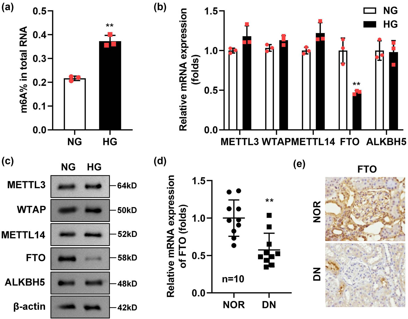
FTO was elevated in the HG-stimulated podocytes and renal tissues. (a) m6A methylation content was elevated in the HG-treated podocytes (n = 3). (b and c) The levels of m6A-related genes were determined with RT-qPCR and Western blot (n = 3). (d) FTO was elevated in the renal tissues of DN patients (n = 10). (e) The immunohistochemistry staining of FTO in the renal tissues of DN patients (n = 10). **P < 0.01.
3.2 Overexpressed FTO alleviated the injury of HG-stimulated podocytes
Given the significant reduction in FTO expression observed in DN, we hypothesized that enhancing FTO expression might alleviate the progression of DN. To test this hypothesis, we overexpressed FTO in a DN cell model to analyze its effects on cellular inflammatory responses and apoptosis levels. FTO was successfully overexpressed in podocytes, as evidenced by a substantial increase in FTO mRNA levels compared to control cells (Figure 2a). After HG stimulation, the LDH release (Figure 2b), mRNA expressions of MCP-1 (Figure 2c), IL-6 (Figure 2d), TNF-α (Figure 2e), apoptosis rate (Figure 2f and g), and TUNEL-positive cells (Figure 2h) were prominently elevated. The significant upregulation of MCP-1, IL-6, and TNF-α in the cells meant that HG has caused an inflammatory response in podocytes, which also explained the increase in apoptosis levels of podocytes may be caused by an inflammatory response. However, FTO overexpression prominently depleted the MCP-1 (Figure 2c), IL-6 (Figure 2d), TNF-α (Figure 2e) levels, and apoptosis rate (Figure 2f and g), as well as TUNEL-positive cells (Figure 2h). In addition, we obtained the podocytes from the renal tissues of DN patients and overexpressed FTO in the cells 2-fold. We found that FTO overexpression also significantly increased the cell viability and decreased the MCP-1, IL-6, TNF-α levels, apoptosis rate, and TUNEL-positive cells in the HG-treated podocytes (Figure S1).

Overexpressed FTO relieved the injury of podocytes induced by HG. (a) Validation of transfection efficiency of oe-FTO. (b) The cell viability was detected by CCK-8 assay. (c) The LDH release was assessed with a kit. (d–f) The mRNA levels of MCP-1, IL-6, and TNF-α were tested with RT-qPCR. (g and h) Flow cytometry was performed to detect the cell apoptosis. (i) TUNEL staining was conducted to detect cell death (n = 3). **P < 0.01 vs NG + oe-NC group, ## P < 0.01 vs HG + oe-FTO.
3.3 DACT1 was elevated in the HG-stimulated podocytes and renal tissues
As described in the introduction, DACT1 was significantly increased in diabetes. In the progression of osteosarcoma, the expression of DACT1 is regulated by the FTO-mediated m6A methylation modification. Therefore, we continued to analyze the correlation between DACT1 expression and FTO in DN. Compared with healthy renal tissues, DACT1 levels were markedly elevated in the renal tissues of DN patients (Figure 3a). Importantly, a negative correlation was observed between DACT1 and FTO expression in these tissues (Figure 3b), suggesting an inverse regulatory relationship between the two proteins. This finding was further supported by immunohistochemistry, which demonstrated high levels of DACT1 in the renal tissues of DN patients (Figure 3c). In the HG-stimulated podocytes, the mRNA (Figure 3d) and protein levels (Figure 3e) of DACT1 were prominently elevated.

DACT1 was elevated in the HG-stimulated podocytes and renal tissues. (a) DACT1 was elevated in the renal tissues of DN patients (n = 10). (b) DACT1 was negatively correlated with FTO (n = 10). (c) The immunohistochemistry staining of DACT1 in the renal tissues of DN patients (n = 10). (d and e) The levels of DACT1 were determined with RT-qPCR and Western blot (n = 3). **P < 0.01.
3.4 Overexpressed FTO declined the m6A methylation modification of DACT1
Given our finding that the expression of FTO and DACT1 in DN exhibits a negative correlation, we proceeded to investigate the m6A methylation modification mechanism of DACT1 by FTO, to clarify how FTO regulates the expression of DACT1. The SRAMP database (http://www.cuilab.cn/sramp) results showed that there were multiple methylation binding sites of DACT1 (Figure 4a). After FTO overexpression, the m6A methylation levels of DACT1 were significantly declined (Figure 4b). The luciferase activity of WT-DACT1 was significantly declined after FTO overexpression (Figure 4c). The RIP assay demonstrated that the FTO could bind to DACT1 mRNA (Figure 4d). The luciferase reporter and RIP assay results indicated that the interaction between FTO and DACT1 occurs at the m6A methylation modification site of DACT1. After FTO overexpression, the mRNA expression (Figure 4e) as well as stability (Figure 4f) of DACT1 were prominently declined. The protein levels of DACT1 were also declined after FTO overexpression (Figure 4g), the results of immunofluorescence showed the same results (Figure 4h).

Overexpressed FTO declined the m6A methylation and mRNA levels of DACT1. (a) The m6A methylation sites of DACT1 were predicted using the SRAMP database. (b) After FTO overexpression, the m6A methylation levels of DACT1 were prominently declined. (c) The luciferase activity of WT-DACT1 was prominently depleted after FTO overexpression. (d) The RIP assay showed the FTO antibody specifically enriched the DACT1 mRNA in podocytes. (e and f) After FTO overexpression, the mRNA expression and stability of DACT1 were prominently declined. (g and h) Western blot and immunofluorescence of DACT1 (n = 3). **P < 0.01.
3.5 Overexpressed DACT1 reversed the function of FTO in the HG-treated podocytes
Having understood the regulatory role of FTO in the m6A methylation modification and expression of DACT1, we subsequently carried out rescue experiments to demonstrate whether, in the DN cell model, FTO participates in the regulation of cellular inflammation and apoptosis by downregulating the expression of DACT1. The DACT1 transfected podocytes showed a high mRNA expression of DACT1 (Figure 5a). In the HG stimulated and FTO transfected podocytes, after DACT1 transfection, the LDH release (Figure 5b), mRNA expression of MCP-1 (Figure 5c), IL-6 (Figure 5d), TNF-α (Figure 5e), apoptosis rate (Figure 5f and g), and TUNEL-positive cells (Figure 5h) were prominently elevated.

Overexpressed DACT1 neutralized the role of FTO in the HG-stimulated podocytes. (a) Validation of transfection efficiency of oe-DACT1. (b) The cell viability was detected by CCK-8 assay. (c) The LDH release was assessed with a kit. (d–f) The mRNA levels of MCP-1, IL-6, and TNF-α were tested with RT-qPCR. (g and h) Flow cytometry was performed to detect the cell apoptosis. (i) TUNEL staining was conducted to detect the cell death (n = 3). **P < 0.01 vs oe-NC group, ## P < 0.01 vs HG + oe-NC group, && P < 0.01 vs HG + oe-NC + oe-FTO group.
3.6 IGF2BP1 was the m6A methylation modification recognition reader of DACT1
The final regulation of gene expression by m6A methylation modification depends on the reader’s recognition of m6A methylation modification. Finally, we further explored which reader could recognize the m6A methylation-modified reader of DCT1. After sh-RNAs’ transfection, the expressions of m6A methylation modification readers (IGF2BP1, IGF2BP2, IGF2BP3, YTHDF1, YTHDF2 and YTHDC1) were prominently declined (Figure 6a). Additionally, IGF2BP1 knockdown decreased the DACT1 levels (Figure 6b). Besides, sh-IGF2BP1 prominently depleted the mRNA stability of DACT1 (Figure 6c). The luciferase activity of WT-DACT1 was prominently depleted after IGF2BP1 knockdown (Figure 6d). The RIP assay demonstrated that the IGF2BP1 could bind to DACT1 mRNA (Figure 6e). Furthermore, sh-FTO prominently declined the FTO levels in the podocytes (Figure 6f). Finally, we confirmed that IGF2BP1 knockdown reversed the effects of sh-FTO on the expression levels of DACT1 (Figure 6g).

IGF2BP1-degraded DACT1 mRNAs in an m6A-dependent manner. (a) Validation of transfection efficiency of sh-RNAs. (b) After sh-IGF2BP1 transfection, the DACT1 levels were prominently declined. (c) Sh-IGF2BP1 also prominently depleted the mRNA stability of DACT1. (d) The luciferase activity of WT-DACT1 was prominently depleted after IGF2BP1 knockdown. (e) The RIP assay showed the IGF2BP1 antibody specifically enriched the DACT1 mRNA in podocytes. (f) Validation of transfection efficiency of sh-FTO. (g) After sh-FTO and sh-IGF2BP1 transfection, the DACT1 levels were assessed by RT-qPCR (n = 3). **P < 0.01.
4 Discussion
In this study, we demonstrated that FTO epigenetically inhibits DACT1 expression through an m6A-IGF2BP1-dependent mechanism. Moreover, we confirmed that FTO expression is significantly downregulated in podocytes stimulated by HG and in the kidneys of DN patients. FTO overexpression alleviated the injury of HG-stimulated podocytes.
Recent studies have increasingly highlighted the critical role of m6A methylation in RNA metabolism, influencing various cellular processes such as mRNA stability, splicing, and translation [23,24]. As the first identified m6A demethylase, FTO plays a pivotal role in removing m6A modifications from target RNAs, thereby regulating their expression and function [25]. FTO belongs to the AlkB family of non-heme iron(ii)-dependent and α-ketoglutarate-dependent dioxygenases, which are known to modulate various biological processes, including body mass and obesity development [26]. Previous research has shown that inhibition of FTO can prevent obesity and lead to growth retardation, underscoring its importance in metabolic regulation. Moreover, emerging evidence indicates that FTO exerts regulatory effects on mRNA expression in diverse pathological contexts. For instance, Niu et al. [27] demonstrated that FTO reduces the m6A methylation levels of BNIP3, accelerating its degradation and promoting breast cancer development. Shen et al. [28] found that FTO suppresses hypoxia/reoxygenation-induced apoptosis in myocardial cells by modulating the m6A methylation of Mhrt, suggesting its potential as a therapeutic target for heart failure. In DN progressions, Taira et al. [29] confirmed the dramatic relationship between rs56094641 in FTO and susceptibility to DN patients in Japan through genome-wide association studies. However, the specific mechanisms underlying FTO’s role in DN remain largely unexplored. In this research, we found that FTO was declined in the HG-stimulated podocytes and renal tissues of DN patients. FOT overexpression alleviated the injury, including LDH and inflammatory factors (MCP-1, IL-6, as well as TNF-α) release, induced by HG in the podocytes. These results indicated that increasing the FTO levels might be a promising therapeutic method for DN.
While numerous studies have demonstrated that FTO can promote the progression of obesity [30] and diabetes [31]. For the opposite phenomenon observed in this study, FTO expression was reduced in DN patients. We speculate that the downregulation of FTO observed in DN samples relative to normal (NOR) controls may be a consequence of chronic hyperglycemia and oxidative stress, which are hallmark features of diabetes. These conditions can lead to alterations in epigenetic modifiers as part of a compensatory response or due to impaired cellular homeostasis. While upregulation of FTO has been reported in some contexts related to diabetes, such as obesity, it is not universally observed across all diabetic complications. Therefore, the decrease in FTO expression in DN could reflect a specific aspect of this complication’s pathophysiology.
DACT1, recently identified as a tumor suppressor gene, has been demonstrated to be regulated by FTO through the m6A methylation modification [22]. Shi et al. [32] demonstrated that DACT1 overexpression depressed cell metastasis and accelerated cell death in cervical cancer. Similar antitumor effects of DACT1 have been reported in other malignancies, including breast cancer [33], lung cancer [34], gastric cancer [35], and so on. In the context of DN, the role of DACT1 has not been extensively studied. In our research, we found that DACT1 was overexpressed in the HG-stimulated podocytes and renal tissues of DN patients. Through the Double Luciferase Report and RIP experiment, we demonstrated the existence of interaction between FTO and DACT1. SRAMP database confirmed that multiple m6A methylation-modified sites existed on DACT1 mRNA. Meanwhile, we found that FTO overexpression depleted the m6A methylation levels of DACT1 and exacerbated the degradation of DACT1. Rescue experiments further illustrated that DACT1 overexpression reversed the role of FTO in HG-treated podocytes. All these findings suggested that FTO participated in the DN progression by modulating the m6A methylation levels of DACT1. As reported by Lv et al. [22], they also demonstrated that FTO decreased the mRNA expression and stability of DACT1 by regulating the m6A methylation levels in osteosarcoma. In this study, we have demonstrated for the first time that FTO regulates DACT1 modification in the context of DN, extending our understanding of the FTO/DACT1 signaling axis beyond its previously established role in tumors. In the future, we can continue to explore the role of this signaling axis in other diseases, to verify the universality of the FTO/DACT1 signaling axis and provide a solid theoretical basis for the development of clinical molecular targeted drugs.
The stability of target genes during m6A methylation modification is critically influenced by the recognition of m6A marks by “reader” proteins [36]. These readers play a pivotal role in regulating the expression of target genes, thereby participating in the progression of various diseases [37]. IGF2BP1, as a new family of m6A readers, has been confirmed to enhance the mRNA stability of target genes [38]. As an m6A reader, IGF2BP1 acts as an oncogenic factor in various cancer cells by stabilizing the expression of methylated mRNAs associated with oncogenic genes [39,40]. In addition, Huang et al. [41] demonstrated that IGF2BP1 promotes mRNA stability in an m6A-dependent manner, preferentially recognizing m6A methylation sites on mRNA. This selective recognition ultimately regulates the expression levels of related genes, underscoring the importance of IGF2BP1 in the post-transcriptional regulation of gene expression. In this study, through knockouting the expressions of readers, we found that IGF2BP1 knockdown declined the DACT1 expressions. The double luciferase report and RIP experiment confirmed the existence of interaction between IGF2BP1 and DACT1. Further experiments indicated that IGF2BP1 knockdown decreased the stability of DACT1 and neutralized the sh-FTO effects on DACT1 levels. Understanding the molecular mechanisms by which FTO regulates m6A methylation levels and interacts with IGF2BP1 provides a foundation for developing therapies that modulate these interactions rather than directly altering FTO expression. This approach could offer a safer alternative with reduced side effects.
However, there were still some limitations in this study. For RNA stability determination of DACT1, using only actinomycin D is not enough, and pulse tracking experiments are needed to further prove this. However, due to limited conditions, this will be the focus of our future research. On the other hand, the concern about the knockdown of DACT1 being potentially oncogenic is valid. However, it is important to note that the knockdown was performed in an in vitro model of DN, not in a cancer setting. The implications of DACT1 knockdown in DN need to be carefully evaluated in more specific models to ensure safety. Furthermore, any potential therapeutic strategy involving DACT1 would require extensive preclinical testing to assess both efficacy and safety. Exploring small molecules or RNA-based therapies that can selectively interfere with the DACT1–IGF2BP1 interaction might offer a safer alternative compared to direct knockdown.
To sum up, our study unveiled that low levels of FTO were a key factor for DN progression. Mechanistically, FTO overexpression inhibited the inflammatory reaction and cell death in the HG-stimulated podocytes by down-regulating the DACT1 levels. FTO-medicated m6A methylation modification of DACT1 was dependent on IGF2BP1.
-
Funding information: Authors state no funding involved.
-
Author contributions: All authors have accepted responsibility for the entire content of this manuscript and consented to its submission to the journal, reviewed all the results, and approved the final version of the manuscript. X.L. and X.Z. drafted the work and revised it critically for important intellectual content; Q.H. and S.G. were responsible for the acquisition, analysis, or interpretation of data for the work; and P.Z. and Z.M. made substantial contributions to the conception or design of the work and prepared the manuscript with contributions from all co-authors. All authors read and approved the final manuscript.
-
Conflict of interest: Authors state no conflict of interest.
-
Data availability statement: The datasets generated during and/or analyzed during the current study are available from the corresponding author on reasonable request.
References
[1] Flyvbjerg A. The role of the complement system in diabetic nephropathy. Nat Rev Nephrol. 2017;13(5):311–8.10.1038/nrneph.2017.31Search in Google Scholar PubMed
[2] Giorgino F, Vora J, Fenici P, Solini A. Renoprotection with SGLT2 inhibitors in type 2 diabetes over a spectrum of cardiovascular and renal risk. Cardiovasc Diabetol. 2020;19(1):196.10.1186/s12933-020-01163-9Search in Google Scholar PubMed PubMed Central
[3] Christ-Crain M, Winzeler B, Refardt J. Diagnosis and management of diabetes insipidus for the internist: an update. J Intern Med. 2021;290(1):73–87.10.1111/joim.13261Search in Google Scholar PubMed
[4] Hong Q, Zhang L, Das B, Li Z, Liu B, Cai G, et al. Increased podocyte Sirtuin-1 function attenuates diabetic kidney injury. Kidney Int. 2018;93(6):1330–43.10.1016/j.kint.2017.12.008Search in Google Scholar PubMed PubMed Central
[5] Bose M, Almas S. Wnt signaling and podocyte dysfunction in diabetic nephropathy. J Investig Med. 2017;65(8):1093–101.10.1136/jim-2017-000456Search in Google Scholar PubMed
[6] Tung CW, Hsu YC, Shih YH, Chang PJ, Lin CL. Glomerular mesangial cell and podocyte injuries in diabetic nephropathy. Nephrology (Carlton, Vic). 2018;23(S4):32–7.10.1111/nep.13451Search in Google Scholar PubMed
[7] Dai H, Liu Q, Liu B, Wei JL, Liu WJ. Research progress on mechanism of podocyte depletion in diabetic nephropathy. J Diabetes Res. 2017;2017:2615210–86.10.1155/2017/2615286Search in Google Scholar PubMed PubMed Central
[8] Stieger N, Worthmann K, Schiffer M. The role of metabolic and haemodynamic factors in podocyte injury in diabetes. Diabetes Metab Res Rev. 2011;27(3):207–15.10.1002/dmrr.1164Search in Google Scholar PubMed
[9] Singh R, Chandel S, Dey D, Ghosh A, Roy S, Ravichandiran V, et al. Epigenetic modification and therapeutic targets of diabetes mellitus. Biosci Rep. 2020;40(9):1.10.1042/BSR20202160Search in Google Scholar PubMed PubMed Central
[10] Chen Y, Hong T, Wang S, Mo J, Tian T, Zhou X. Epigenetic modification of nucleic acids: from basic studies to medical applications. Chem Soc Rev. 2017;46(10):2844–72.10.1039/C6CS00599CSearch in Google Scholar
[11] Zhang Y, Geng X, Li Q, Xu J, Tan Y, Xiao M, et al. m6A modification in RNA: biogenesis, functions and roles in gliomas. J Exp Clin Cancer Res. 2020;39(1):1–192.10.1186/s13046-020-01706-8Search in Google Scholar PubMed PubMed Central
[12] Wu X, Sang L, Gong Y. N6-methyladenine RNA modification and cancers. Am J Cancer Res. 2018;8(10):1957–66.Search in Google Scholar
[13] Li J, Zhu L, Shi Y, Liu J, Lin L, Chen X. m6A demethylase FTO promotes hepatocellular carcinoma tumorigenesis via mediating PKM2 demethylation. Am J Transl Res. 2019;11(9):6084–92.Search in Google Scholar
[14] Mizuno TM. Fat mass and obesity associated (FTO) gene and hepatic glucose and lipid metabolism. Nutrients. 2018;10(11):1600.10.3390/nu10111600Search in Google Scholar PubMed PubMed Central
[15] Yang Y, Shen F, Huang W, Qin S, Huang J, Sergi C, et al. Glucose is involved in the dynamic regulation of m6A in patients with type 2 diabetes. J Clin Endocrinol Metab. 2019;104(3):665–73.10.1210/jc.2018-00619Search in Google Scholar PubMed
[16] Wang S, Kang W, Go MY, Tong JH, Li L, Zhang N, et al. Dapper homolog 1 is a novel tumor suppressor in gastric cancer through inhibiting the nuclear factor-kappaB signaling pathway. Mol Med. 2012;18:1402–11.10.2119/molmed.2012.00243Search in Google Scholar PubMed PubMed Central
[17] Zeng L, Chen C, Yao C, Morelli MB. Histone deacetylation regulated by KDM1A to suppress DACT1 in proliferation and migration of cervical cancer. Anal Cell Pathol (Amst). 2021;2021:5555416–52.10.1155/2021/5555452Search in Google Scholar PubMed PubMed Central
[18] Yuan G, Wang C, Ma C, Chen N, Tian Q, Zhang T, et al. Oncogenic function of DACT1 in colon cancer through the regulation of beta-catenin. PLoS One. 2012;7(3):e34004.10.1371/journal.pone.0034004Search in Google Scholar PubMed PubMed Central
[19] Yang ZQ, Zhao Y, Liu Y, Zhang JY, Zhang S, Jiang GY, et al. Downregulation of HDPR1 is associated with poor prognosis and affects expression levels of p120-catenin and beta-catenin in nonsmall cell lung cancer. Mol Carcinog. 2010;49(5):508–19.10.1002/mc.20622Search in Google Scholar PubMed
[20] Contriciani RE, Da Veiga FC, Do Amaral MJ, Castelucci BG, de Sousa LM, de Jesus MB, et al. Dact1 is expressed during chicken and mouse skeletal myogenesis and modulated in human muscle diseases. Comp Biochem Physiol Part B: Biochem Mol Biol. 2021;256:110645.10.1016/j.cbpb.2021.110645Search in Google Scholar PubMed
[21] Yu C, Yang C, Rui Z. MicroRNA-125b-5p improves pancreatic β-cell function through inhibiting JNK signaling pathway by targeting DACT1 in mice with type 2 diabetes mellitus. Life Sci. 2019;224:67–75.10.1016/j.lfs.2019.01.031Search in Google Scholar PubMed
[22] Lv D, Ding S, Zhong L, Tu J, Li H, Yao H, et al. M 6 A demethylase FTO-mediated downregulation of DACT1 mRNA stability promotes Wnt signaling to facilitate osteosarcoma progression. Oncogene. 2022;41(12):1727.10.1038/s41388-022-02214-zSearch in Google Scholar PubMed
[23] Roignant J, Soller M. m 6 A in mRNA: An ancient mechanism for fine-tuning gene expression. Trends Genet. 2017;33(6):380–90.10.1016/j.tig.2017.04.003Search in Google Scholar PubMed
[24] Gilbert WV, Bell TA, Schaening C. Messenger RNA modifications: Form, distribution, and function. Science (American Association for the Advancement of Science). 2016;352(6292):1408–12.10.1126/science.aad8711Search in Google Scholar PubMed PubMed Central
[25] Niu Y, Zhao X, Wu YS, Li MM, Wang XJ, Yang YG. N6-methyl-adenosine (m6A) in RNA: An old modification with a novel epigenetic function. Genomics Proteom Bioinforma. 2013;11(1):8–17.10.1016/j.gpb.2012.12.002Search in Google Scholar PubMed PubMed Central
[26] Smemo S, Tena JJ, Kim K, Gamazon ER, Sakabe NJ, Gómez-Marín C, et al. Obesity-associated variants within FTO form long-range functional connections with IRX3. Nature (London). 2014;507(7492):371–5.10.1038/nature13138Search in Google Scholar PubMed PubMed Central
[27] Niu Y, Lin Z, Wan A, Chen H, Liang H, Sun L, et al. RNA N6-methyladenosine demethylase FTO promotes breast tumor progression through inhibiting BNIP3. Mol Cancer. 2019;18(1):46.10.1186/s12943-019-1004-4Search in Google Scholar PubMed PubMed Central
[28] Shen W, Li H, Su H, Chen K, Yan J. FTO overexpression inhibits apoptosis of hypoxia/reoxygenation-treated myocardial cells by regulating m6A modification of Mhrt. Mol Cell Biochem. 2021;476(5):2171–9.10.1007/s11010-021-04069-6Search in Google Scholar PubMed
[29] Taira M, Imamura M, Takahashi A, Kamatani Y, Yamauchi T, Araki SI, et al. A variant within the FTO confers susceptibility to diabetic nephropathy in Japanese patients with type 2 diabetes. PLoS One. 2018;13(12):e208654.10.1371/journal.pone.0208654Search in Google Scholar PubMed PubMed Central
[30] Chen J, Xiao WC, Zhao JJ, Heitkamp M, Chen DF, Shan R, et al. FTO genotype and body mass index reduction in childhood obesity interventions: A systematic review and meta‐analysis. Obes Rev. 2024;25(5):e13715.10.1111/obr.13715Search in Google Scholar PubMed
[31] Zhou C, She X, Gu C, Hu Y, Ma M, Qiu Q, et al. FTO fuels diabetes-induced vascular endothelial dysfunction associated with inflammation by erasing m6A methylation of TNIP1. J Clin Invest. 2023;133(19):1–16.10.1172/JCI160517Search in Google Scholar PubMed PubMed Central
[32] Shi X, Huo J, Gao X, Cai H, Zhu W. A newly identified lncRNA H1FX-AS1 targets DACT1 to inhibit cervical cancer via sponging miR-324-3p. Cancer Cell Int. 2020;20:358.10.1186/s12935-020-01385-7Search in Google Scholar PubMed PubMed Central
[33] Yin X, Xiang T, Li L, Su X, Shu X, Luo X, et al. DACT1, an antagonist to Wnt/beta-catenin signaling, suppresses tumor cell growth and is frequently silenced in breast cancer. Breast Cancer Res. 2013;15(2):R23.10.1186/bcr3399Search in Google Scholar PubMed PubMed Central
[34] Paschidis K, Zougros A, Chatziandreou I, Tsikalakis S, Korkolopoulou P, Kavantzas N, et al. Methylation analysis of APC, AXIN2, DACT1, RASSF1A and MGMT gene promoters in non-small cell lung cancer. Pathol Res Pract. 2022;234:153899.10.1016/j.prp.2022.153899Search in Google Scholar PubMed
[35] Deng J, Liang H, Zhang R, Ying G, Xie X, Yu J, et al. Methylated CpG site count of dapper homolog 1 (DACT1) promoter prediction the poor survival of gastric cancer. Am J Cancer Res. 2014;4(5):518–27.Search in Google Scholar
[36] Shi H, Wei J, He C. Where, When, and How: Context-dependent functions of RNA methylation writers, readers, and erasers. Mol Cell. 2019;74(4):640–50.10.1016/j.molcel.2019.04.025Search in Google Scholar PubMed PubMed Central
[37] Anita R, Paramasivam A, Priyadharsini JV, Chitra S. The m6A readers YTHDF1 and YTHDF3 aberrations associated with metastasis and predict poor prognosis in breast cancer patients. Am J Cancer Res. 2020;10(8):2546–54.Search in Google Scholar
[38] Bell JL, Wächter K, Mühleck B, Pazaitis N, Köhn M, Lederer M, et al. Insulin-like growth factor 2 mRNA-binding proteins (IGF2BPs): Post-transcriptional drivers of cancer progression? Cell Mol Life Sci: CMLS. 2012;70(15):2657–75.10.1007/s00018-012-1186-zSearch in Google Scholar PubMed PubMed Central
[39] Müller S, Glaß M, Singh AK, Haase J, Bley N, Fuchs T, et al. IGF2BP1 promotes SRF-dependent transcription in cancer in a m6A- and miRNA-dependent manner. Nucleic Acids Res. 2019;47(1):375–90.10.1093/nar/gky1012Search in Google Scholar PubMed PubMed Central
[40] Zhang L, Wan Y, Zhang Z, Jiang Y, Gu Z, Ma X, et al. IGF2BP1 overexpression stabilizes PEG10 mRNA in an m6A-dependent manner and promotes endometrial cancer progression. Theranostics. 2021;11(3):1100–14.10.7150/thno.49345Search in Google Scholar PubMed PubMed Central
[41] Huang H, Weng H, Sun W, Qin X, Shi H, Wu H, et al. Recognition of RNA N(6)-methyladenosine by IGF2BP proteins enhances mRNA stability and translation. Nat Cell Biol. 2018;20(3):285–95.10.1038/s41556-018-0045-zSearch in Google Scholar PubMed PubMed Central
© 2025 the author(s), published by De Gruyter
This work is licensed under the Creative Commons Attribution 4.0 International License.
Articles in the same Issue
- Biomedical Sciences
- Mechanism of triptolide regulating proliferation and apoptosis of hepatoma cells by inhibiting JAK/STAT pathway
- Maslinic acid improves mitochondrial function and inhibits oxidative stress and autophagy in human gastric smooth muscle cells
- Comparative analysis of inflammatory biomarkers for the diagnosis of neonatal sepsis: IL-6, IL-8, SAA, CRP, and PCT
- Post-pandemic insights on COVID-19 and premature ovarian insufficiency
- Proteome differences of dental stem cells between permanent and deciduous teeth by data-independent acquisition proteomics
- Optimizing a modified cetyltrimethylammonium bromide protocol for fungal DNA extraction: Insights from multilocus gene amplification
- Preliminary analysis of the role of small hepatitis B surface proteins mutations in the pathogenesis of occult hepatitis B infection via the endoplasmic reticulum stress-induced UPR-ERAD pathway
- Efficacy of alginate-coated gold nanoparticles against antibiotics-resistant Staphylococcus and Streptococcus pathogens of acne origins
- Battling COVID-19 leveraging nanobiotechnology: Gold and silver nanoparticle–B-escin conjugates as SARS-CoV-2 inhibitors
- Neurodegenerative diseases and neuroinflammation-induced apoptosis
- Impact of fracture fixation surgery on cognitive function and the gut microbiota in mice with a history of stroke
- COLEC10: A potential tumor suppressor and prognostic biomarker in hepatocellular carcinoma through modulation of EMT and PI3K-AKT pathways
- High-temperature requirement serine protease A2 inhibitor UCF-101 ameliorates damaged neurons in traumatic brain-injured rats by the AMPK/NF-κB pathway
- SIK1 inhibits IL-1β-stimulated cartilage apoptosis and inflammation in vitro through the CRTC2/CREB1 signaling
- Rutin–chitooligosaccharide complex: Comprehensive evaluation of its anti-inflammatory and analgesic properties in vitro and in vivo
- Knockdown of Aurora kinase B alleviates high glucose-triggered trophoblast cells damage and inflammation during gestational diabetes
- Calcium-sensing receptors promoted Homer1 expression and osteogenic differentiation in bone marrow mesenchymal stem cells
- ABI3BP can inhibit the proliferation, invasion, and epithelial–mesenchymal transition of non-small-cell lung cancer cells
- Changes in blood glucose and metabolism in hyperuricemia mice
- Rapid detection of the GJB2 c.235delC mutation based on CRISPR-Cas13a combined with lateral flow dipstick
- IL-11 promotes Ang II-induced autophagy inhibition and mitochondrial dysfunction in atrial fibroblasts
- Short-chain fatty acid attenuates intestinal inflammation by regulation of gut microbial composition in antibiotic-associated diarrhea
- Application of metagenomic next-generation sequencing in the diagnosis of pathogens in patients with diabetes complicated by community-acquired pneumonia
- NAT10 promotes radiotherapy resistance in non-small cell lung cancer by regulating KPNB1-mediated PD-L1 nuclear translocation
- Phytol-mixed micelles alleviate dexamethasone-induced osteoporosis in zebrafish: Activation of the MMP3–OPN–MAPK pathway-mediating bone remodeling
- Association between TGF-β1 and β-catenin expression in the vaginal wall of patients with pelvic organ prolapse
- Primary pleomorphic liposarcoma involving bilateral ovaries: Case report and literature review
- Effects of de novo donor-specific Class I and II antibodies on graft outcomes after liver transplantation: A pilot cohort study
- Sleep architecture in Alzheimer’s disease continuum: The deep sleep question
- Ephedra fragilis plant extract: A groundbreaking corrosion inhibitor for mild steel in acidic environments – electrochemical, EDX, DFT, and Monte Carlo studies
- Langerhans cell histiocytosis in an adult patient with upper jaw and pulmonary involvement: A case report
- Inhibition of mast cell activation by Jaranol-targeted Pirin ameliorates allergic responses in mouse allergic rhinitis
- Aeromonas veronii-induced septic arthritis of the hip in a child with acute lymphoblastic leukemia
- Clusterin activates the heat shock response via the PI3K/Akt pathway to protect cardiomyocytes from high-temperature-induced apoptosis
- Research progress on fecal microbiota transplantation in tumor prevention and treatment
- Low-pressure exposure influences the development of HAPE
- Stigmasterol alleviates endplate chondrocyte degeneration through inducing mitophagy by enhancing PINK1 mRNA acetylation via the ESR1/NAT10 axis
- AKAP12, mediated by transcription factor 21, inhibits cell proliferation, metastasis, and glycolysis in lung squamous cell carcinoma
- Association between PAX9 or MSX1 gene polymorphism and tooth agenesis risk: A meta-analysis
- A case of bloodstream infection caused by Neisseria gonorrhoeae
- Case of nasopharyngeal tuberculosis complicated with cervical lymph node and pulmonary tuberculosis
- p-Cymene inhibits pro-fibrotic and inflammatory mediators to prevent hepatic dysfunction
- GFPT2 promotes paclitaxel resistance in epithelial ovarian cancer cells via activating NF-κB signaling pathway
- Transfer RNA-derived fragment tRF-36 modulates varicose vein progression via human vascular smooth muscle cell Notch signaling
- RTA-408 attenuates the hepatic ischemia reperfusion injury in mice possibly by activating the Nrf2/HO-1 signaling pathway
- Decreased serum TIMP4 levels in patients with rheumatoid arthritis
- Sirt1 protects lupus nephritis by inhibiting the NLRP3 signaling pathway in human glomerular mesangial cells
- Sodium butyrate aids brain injury repair in neonatal rats
- Interaction of MTHFR polymorphism with PAX1 methylation in cervical cancer
- Convallatoxin inhibits proliferation and angiogenesis of glioma cells via regulating JAK/STAT3 pathway
- The effect of the PKR inhibitor, 2-aminopurine, on the replication of influenza A virus, and segment 8 mRNA splicing
- Effects of Ire1 gene on virulence and pathogenicity of Candida albicans
- Small cell lung cancer with small intestinal metastasis: Case report and literature review
- GRB14: A prognostic biomarker driving tumor progression in gastric cancer through the PI3K/AKT signaling pathway by interacting with COBLL1
- 15-Lipoxygenase-2 deficiency induces foam cell formation that can be restored by salidroside through the inhibition of arachidonic acid effects
- FTO alleviated the diabetic nephropathy progression by regulating the N6-methyladenosine levels of DACT1
- Clinical relevance of inflammatory markers in the evaluation of severity of ulcerative colitis: A retrospective study
- Zinc valproic acid complex promotes osteoblast differentiation and exhibits anti-osteoporotic potential
- Primary pulmonary synovial sarcoma in the bronchial cavity: A case report
- Metagenomic next-generation sequencing of alveolar lavage fluid improves the detection of pulmonary infection
- Uterine tumor resembling ovarian sex cord tumor with extensive rhabdoid differentiation: A case report
- Genomic analysis of a novel ST11(PR34365) Clostridioides difficile strain isolated from the human fecal of a CDI patient in Guizhou, China
- Effects of tiered cardiac rehabilitation on CRP, TNF-α, and physical endurance in older adults with coronary heart disease
- Changes in T-lymphocyte subpopulations in patients with colorectal cancer before and after acupoint catgut embedding acupuncture observation
- Modulating the tumor microenvironment: The role of traditional Chinese medicine in improving lung cancer treatment
- Alterations of metabolites related to microbiota–gut–brain axis in plasma of colon cancer, esophageal cancer, stomach cancer, and lung cancer patients
- Research on individualized drug sensitivity detection technology based on bio-3D printing technology for precision treatment of gastrointestinal stromal tumors
- CEBPB promotes ulcerative colitis-associated colorectal cancer by stimulating tumor growth and activating the NF-κB/STAT3 signaling pathway
- Oncolytic bacteria: A revolutionary approach to cancer therapy
- A de novo meningioma with rapid growth: A possible malignancy imposter?
- Diagnosis of secondary tuberculosis infection in an asymptomatic elderly with cancer using next-generation sequencing: Case report
- Hesperidin and its zinc(ii) complex enhance osteoblast differentiation and bone formation: In vitro and in vivo evaluations
- Research progress on the regulation of autophagy in cardiovascular diseases by chemokines
- Anti-arthritic, immunomodulatory, and inflammatory regulation by the benzimidazole derivative BMZ-AD: Insights from an FCA-induced rat model
- Immunoassay for pyruvate kinase M1/2 as an Alzheimer’s biomarker in CSF
- The role of HDAC11 in age-related hearing loss: Mechanisms and therapeutic implications
- Evaluation and application analysis of animal models of PIPNP based on data mining
- Therapeutic approaches for liver fibrosis/cirrhosis by targeting pyroptosis
- Fabrication of zinc oxide nanoparticles using Ruellia tuberosa leaf extract induces apoptosis through P53 and STAT3 signalling pathways in prostate cancer cells
- Haplo-hematopoietic stem cell transplantation and immunoradiotherapy for severe aplastic anemia complicated with nasopharyngeal carcinoma: A case report
- Modulation of the KEAP1-NRF2 pathway by Erianin: A novel approach to reduce psoriasiform inflammation and inflammatory signaling
- The expression of epidermal growth factor receptor 2 and its relationship with tumor-infiltrating lymphocytes and clinical pathological features in breast cancer patients
- Innovations in MALDI-TOF Mass Spectrometry: Bridging modern diagnostics and historical insights
- BAP1 complexes with YY1 and RBBP7 and its downstream targets in ccRCC cells
- Hypereosinophilic syndrome with elevated IgG4 and T-cell clonality: A report of two cases
- Electroacupuncture alleviates sciatic nerve injury in sciatica rats by regulating BDNF and NGF levels, myelin sheath degradation, and autophagy
- Polydatin prevents cholesterol gallstone formation by regulating cholesterol metabolism via PPAR-γ signaling
- RNF144A and RNF144B: Important molecules for health
- Analysis of the detection rate and related factors of thyroid nodules in the healthy population
- Artesunate inhibits hepatocellular carcinoma cell migration and invasion through OGA-mediated O-GlcNAcylation of ZEB1
- Endovascular management of post-pancreatectomy hemorrhage caused by a hepatic artery pseudoaneurysm: Case report and review of the literature
- Efficacy and safety of anti-PD-1/PD-L1 antibodies in patients with relapsed refractory diffuse large B-cell lymphoma: A meta-analysis
- SATB2 promotes humeral fracture healing in rats by activating the PI3K/AKT pathway
- Overexpression of the ferroptosis-related gene, NFS1, corresponds to gastric cancer growth and tumor immune infiltration
- Understanding risk factors and prognosis in diabetic foot ulcers
- Atractylenolide I alleviates the experimental allergic response in mice by suppressing TLR4/NF-kB/NLRP3 signalling
- FBXO31 inhibits the stemness characteristics of CD147 (+) melanoma stem cells
- Immune molecule diagnostics in colorectal cancer: CCL2 and CXCL11
- Inhibiting CXCR6 promotes senescence of activated hepatic stellate cells with limited proinflammatory SASP to attenuate hepatic fibrosis
- Cadmium toxicity, health risk and its remediation using low-cost biochar adsorbents
- Pulmonary cryptococcosis with headache as the first presentation: A case report
- Solitary pulmonary metastasis with cystic airspaces in colon cancer: A rare case report
- RUNX1 promotes denervation-induced muscle atrophy by activating the JUNB/NF-κB pathway and driving M1 macrophage polarization
- Morphometric analysis and immunobiological investigation of Indigofera oblongifolia on the infected lung with Plasmodium chabaudi
- The NuA4/TIP60 histone-modifying complex and Hr78 modulate the Lobe2 mutant eye phenotype
- Experimental study on salmon demineralized bone matrix loaded with recombinant human bone morphogenetic protein-2: In vitro and in vivo study
- A case of IgA nephropathy treated with a combination of telitacicept and half-dose glucocorticoids
- Analgesic and toxicological evaluation of cannabidiol-rich Moroccan Cannabis sativa L. (Khardala variety) extract: Evidence from an in vivo and in silico study
- Wound healing and signaling pathways
- Combination of immunotherapy and whole-brain radiotherapy on prognosis of patients with multiple brain metastases: A retrospective cohort study
- To explore the relationship between endometrial hyperemia and polycystic ovary syndrome
- Research progress on the impact of curcumin on immune responses in breast cancer
- Biogenic Cu/Ni nanotherapeutics from Descurainia sophia (L.) Webb ex Prantl seeds for the treatment of lung cancer
- Dapagliflozin attenuates atrial fibrosis via the HMGB1/RAGE pathway in atrial fibrillation rats
- Glycitein alleviates inflammation and apoptosis in keratinocytes via ROS-associated PI3K–Akt signalling pathway
- ADH5 inhibits proliferation but promotes EMT in non-small cell lung cancer cell through activating Smad2/Smad3
- Apoptotic efficacies of AgNPs formulated by Syzygium aromaticum leaf extract on 32D-FLT3-ITD human leukemia cell line with PI3K/AKT/mTOR signaling pathway
- Novel cuproptosis-related genes C1QBP and PFKP identified as prognostic and therapeutic targets in lung adenocarcinoma
- Bee venom promotes exosome secretion and alters miRNA cargo in T cells
- Treatment of pure red cell aplasia in a chronic kidney disease patient with roxadustat: A case report
- Comparative bioinformatics analysis of the Wnt pathway in breast cancer: Selection of novel biomarker panels associated with ER status
- Kynurenine facilitates renal cell carcinoma progression by suppressing M2 macrophage pyroptosis through inhibition of CASP1 cleavage
- RFX5 promotes the growth, motility, and inhibits apoptosis of gastric adenocarcinoma cells through the SIRT1/AMPK axis
- ALKBH5 exacerbates early cardiac damage after radiotherapy for breast cancer via m6A demethylation of TLR4
- Phytochemicals of Roman chamomile: Antioxidant, anti-aging, and whitening activities of distillation residues
- Circadian gene Cry1 inhibits the tumorigenicity of hepatocellular carcinoma by the BAX/BCL2-mediated apoptosis pathway
- The TNFR-RIPK1/RIPK3 signalling pathway mediates the effect of lanthanum on necroptosis of nerve cells
- Longitudinal monitoring of autoantibody dynamics in patients with early-stage non-small-cell lung cancer undergoing surgery
- The potential role of rutin, a flavonoid, in the management of cancer through modulation of cell signaling pathways
- Construction of pectinase gene engineering microbe and its application in tobacco sheets
- Construction of a microbial abundance prognostic scoring model based on intratumoral microbial data for predicting the prognosis of lung squamous cell carcinoma
- Sepsis complicated by haemophagocytic lymphohistiocytosis triggered by methicillin-resistant Staphylococcus aureus and human herpesvirus 8 in an immunocompromised elderly patient: A case report
- Sarcopenia in liver transplantation: A comprehensive bibliometric study of current research trends and future directions
- Advances in cancer immunotherapy and future directions in personalized medicine
- Can coronavirus disease 2019 affect male fertility or cause spontaneous abortion? A two-sample Mendelian randomization analysis
- Heat stroke associated with novel leukaemia inhibitory factor receptor gene variant in a Chinese infant
- PSME2 exacerbates ulcerative colitis by disrupting intestinal barrier function and promoting autophagy-dependent inflammation
- Hyperosmolar hyperglycemic state with severe hypernatremia coexisting with central diabetes insipidus: A case report and literature review
- Efficacy and mechanism of escin in improving the tissue microenvironment of blood vessel walls via anti-inflammatory and anticoagulant effects: Implications for clinical practice
- Merkel cell carcinoma: Clinicopathological analysis of three patients and literature review
- Genetic variants in VWF exon 26 and their implications for type 1 Von Willebrand disease in a Saudi Arabian population
- Lipoxin A4 improves myocardial ischemia/reperfusion injury through the Notch1-Nrf2 signaling pathway
- High levels of EPHB2 expression predict a poor prognosis and promote tumor progression in endometrial cancer
- Knockdown of SHP-2 delays renal tubular epithelial cell injury in diabetic nephropathy by inhibiting NLRP3 inflammasome-mediated pyroptosis
- Exploring the toxicity mechanisms and detoxification methods of Rhizoma Paridis
- Concomitant gastric carcinoma and primary hepatic angiosarcoma in a patient: A case report
- Ecology and Environmental Science
- Optimization and comparative study of Bacillus consortia for cellulolytic potential and cellulase enzyme activity
- The complete mitochondrial genome analysis of Haemaphysalis hystricis Supino, 1897 (Ixodida: Ixodidae) and its phylogenetic implications
- Epidemiological characteristics and risk factors analysis of multidrug-resistant tuberculosis among tuberculosis population in Huzhou City, Eastern China
- Indices of human impacts on landscapes: How do they reflect the proportions of natural habitats?
- Genetic analysis of the Siberian flying squirrel population in the northern Changbai Mountains, Northeast China: Insights into population status and conservation
- Diversity and environmental drivers of Suillus communities in Pinus sylvestris var. mongolica forests of Inner Mongolia
- Global assessment of the fate of nitrogen deposition in forest ecosystems: Insights from 15N tracer studies
- Fungal and bacterial pathogenic co-infections mainly lead to the assembly of microbial community in tobacco stems
- Influencing of coal industry related airborne particulate matter on ocular surface tear film injury and inflammatory factor expression in Sprague-Dawley rats
- Temperature-dependent development, predation, and life table of Sphaerophoria macrogaster (Thomson) (Diptera: Syrphidae) feeding on Myzus persicae (Sulzer) (Homoptera: Aphididae)
- Eleonora’s falcon trophic interactions with insects within its breeding range: A systematic review
- Agriculture
- Integrated analysis of transcriptome, sRNAome, and degradome involved in the drought-response of maize Zhengdan958
- Variation in flower frost tolerance among seven apple cultivars and transcriptome response patterns in two contrastingly frost-tolerant selected cultivars
- Heritability of durable resistance to stripe rust in bread wheat (Triticum aestivum L.)
- Molecular mechanism of follicular development in laying hens based on the regulation of water metabolism
- Animal Science
- Effect of sex ratio on the life history traits of an important invasive species, Spodoptera frugiperda
- Plant Sciences
- Hairpin in a haystack: In silico identification and characterization of plant-conserved microRNA in Rafflesiaceae
- Widely targeted metabolomics of different tissues in Rubus corchorifolius
- The complete chloroplast genome of Gerbera piloselloides (L.) Cass., 1820 (Carduoideae, Asteraceae) and its phylogenetic analysis
- Field trial to correlate mineral solubilization activity of Pseudomonas aeruginosa and biochemical content of groundnut plants
- Correlation analysis between semen routine parameters and sperm DNA fragmentation index in patients with semen non-liquefaction: A retrospective study
- Plasticity of the anatomical traits of Rhododendron L. (Ericaceae) leaves and its implications in adaptation to the plateau environment
- Effects of Piriformospora indica and arbuscular mycorrhizal fungus on growth and physiology of Moringa oleifera under low-temperature stress
- Effects of different sources of potassium fertiliser on yield, fruit quality and nutrient absorption in “Harward” kiwifruit (Actinidia deliciosa)
- Comparative efficiency and residue levels of spraying programs against powdery mildew in grape varieties
- The DREB7 transcription factor enhances salt tolerance in soybean plants under salt stress
- Using plant electrical signals of water hyacinth (Eichhornia crassipes) for water pollution monitoring
- Food Science
- Phytochemical analysis of Stachys iva: Discovering the optimal extract conditions and its bioactive compounds
- Review on role of honey in disease prevention and treatment through modulation of biological activities
- Computational analysis of polymorphic residues in maltose and maltotriose transporters of a wild Saccharomyces cerevisiae strain
- Optimization of phenolic compound extraction from Tunisian squash by-products: A sustainable approach for antioxidant and antibacterial applications
- Liupao tea aqueous extract alleviates dextran sulfate sodium-induced ulcerative colitis in rats by modulating the gut microbiota
- Toxicological qualities and detoxification trends of fruit by-products for valorization: A review
- Polyphenolic spectrum of cornelian cherry fruits and their health-promoting effect
- Optimizing the encapsulation of the refined extract of squash peels for functional food applications: A sustainable approach to reduce food waste
- Advancements in curcuminoid formulations: An update on bioavailability enhancement strategies curcuminoid bioavailability and formulations
- Impact of saline sprouting on antioxidant properties and bioactive compounds in chia seeds
- The dilemma of food genetics and improvement
- Bioengineering and Biotechnology
- Impact of hyaluronic acid-modified hafnium metalorganic frameworks containing rhynchophylline on Alzheimer’s disease
- Emerging patterns in nanoparticle-based therapeutic approaches for rheumatoid arthritis: A comprehensive bibliometric and visual analysis spanning two decades
- Application of CRISPR/Cas gene editing for infectious disease control in poultry
- Preparation of hafnium nitride-coated titanium implants by magnetron sputtering technology and evaluation of their antibacterial properties and biocompatibility
- Preparation and characterization of lemongrass oil nanoemulsion: Antimicrobial, antibiofilm, antioxidant, and anticancer activities
- Corrigendum
- Corrigendum to “Utilization of convolutional neural networks to analyze microscopic images for high-throughput screening of mesenchymal stem cells”
- Corrigendum to “Effects of Ire1 gene on virulence and pathogenicity of Candida albicans”
- Retraction
- Retraction of “Down-regulation of miR-539 indicates poor prognosis in patients with pancreatic cancer”