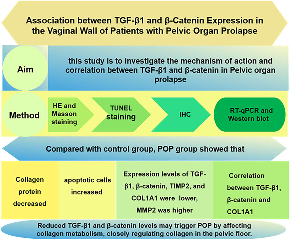
Association between TGF-β1 and β-catenin expression in the vaginal wall of patients with pelvic organ prolapse
-
Feng-Qin Xue


Abstract
The aim of this study is to investigate the mechanism of action and correlation between transforming growth factor beta 1 (TGF-β1) and β-catenin in pelvic organ prolapse (POP). The study compared vaginal wall tissues from two groups: 20 patients with POP (POP group) and 20 who had hysterectomies for benign conditions (control group). Hematoxylin and Eosin and Masson staining visualized collagen, while TUNEL staining detected apoptosis. Protein and mRNA expression levels of TGF-β1, β-catenin, matrix metallopeptidase 2 (MMP2), tissue inhibitor of metalloproteinases 2 (TIMP2), and collagen, type I, alpha 1 (COL1A1) were assessed using immunohistochemistry, quantitative real-time polymerase chain reaction, and western blot techniques. Relationships between the protein expressions of TGF-β1 and β-catenin, β-catenin and COL1A1, and TGF-β1 and COL1A1 were analyzed. In the POP group, vaginal wall collagen fibers were sparse, disorganized, and fragmented, with fewer fibers and more apoptotic cells compared to the control group. Protein and mRNA levels of TGF-β1, β-catenin, TIMP2, and COL1A1 were significantly lower, while MMP2 was higher (p < 0.05). Positive correlations were found between TGF-β1, β-catenin, and COL1A1. Reduced TGF-β1 and β-catenin levels may trigger POP by affecting pelvic floor collagen metabolism.
Graphical abstract
1 Introduction
Pelvic organ prolapse (POP) is a benign disorder caused by weakened pelvic floor support, characterized by the prolapse of the vaginal wall and/or uterus, often accompanied by fecal incontinence and dyspareunia [1]. Major risk factors for POP include vaginal delivery, aging, and menopause [2]. Although it rarely leads to serious morbidity and mortality, it has a significant impact on the physical and mental health of the patient. POP is most prevalent among individuals aged 60–69 [3]. Given the current national context in China, the incidence of POP is expected to rise, potentially exerting significant economic strain on the society. However, the underlying pathophysiology of POP has not been fully elucidated, and clinical management primarily focuses on symptomatic relief rather than addressing the root cause [4]. Therefore, we aim to investigate the molecular pathogenesis of POP to provide innovative insights for its accurate diagnosis and treatment.
The stability of pelvic organs relies on fibrous connective tissues. Consequently, any disruption or dysfunction within this connective tissue network can weaken the biomechanical properties of the pelvic floor support structures, potentially contributing to the development of POP.
Collagen is the primary structural component of the connective tissue [5], constituting 70–80% of connective tissue, and is the main element of the extracellular matrix. Collagen, which provides both strength and elasticity to tissues, serves as the molecular and biochemical basis for the biomechanical properties of pelvic floor support tissues. Recent research indicates that reduced collagen content and diminished cross-linking may play critical roles in the onset of POP. Collagen metabolism is closely influenced by matrix metalloproteinases (MMPs) and their inhibitors, known as tissue inhibitors of metalloproteinases (TIMPs). While MMPs primarily promote catabolism (collagen breakdown), TIMPs act as a counterbalance by inhibiting this process. The interplay between MMPs and TIMPs ensures the dynamic equilibrium of the extracellular matrix.
Fibrotic diseases are primarily caused by abnormalities in the connective tissue. The TGF-β/Smad signaling pathway and the Wnt/β-catenin signaling pathway are widely recognized as critical pathways in fibrotic metabolism. Hyperactivation of these pathways has been observed in diseases such as cardiac remodeling, pulmonary fibrosis, renal fibrosis, and systemic sclerosis. Additionally, there are multiple interactions between these two pathways. First, transforming growth factor beta 1 (TGF-β1) can promptly move β-catenin into the cell nucleus in a Smads3-dependent manner. This process regulates the differentiation of mesenchymal stem cells into osteoblasts [6]. Second, TGF-β has the ability to activate the classical Wnt signaling pathway by down-regulating the expression of DKK1, an antagonist of the Wnt pathway. In fibrotic processes regulated by TGF-β, the Wnt/β-catenin pathway is also involved [7]. During tumor metastasis, there is evidence that TGF-β promotes epithelial–mesenchymal transition of tumor cells, activating the Wnt/β-catenin pathway and leading to the expression of related pathway proteins in the tumor cells. In contrast, in diseases like POP, where connective tissue is weakened, several studies suggest that the TGF-β/Smad signaling pathway and the Wnt/β-catenin signaling pathway are inhibited. It is speculated that this inhibition may block collagen synthesis, thereby promoting POP. However, no study has yet reported the correlation between these two pathways and their molecular linkage mechanism in POP. Based on this information, we propose further exploration of the roles of the TGF-β/Smad signaling pathway and Wnt/β-catenin signaling pathway in POP through techniques such as immunohistochemistry, quantitative polymerase chain reaction (qPCR), and western blot analyses. Our goal is to establish a theoretical foundation for future molecular investigations into POP.
2 Materials and methods
2.1 Research patients
Twenty patients diagnosed with POP of stage III and above, as determined by the POP-Q score were selected as the POP group. Another 20 patients who underwent total hysterectomy for other benign lesions, such as uterine fibroids, benign endometrial lesions, and pre-cancerous cervical lesions, were chosen as the control group. All patients had no history of urinary incontinence, malignant tumors, or connective tissue disease. They had not used estrogen therapy for 3 months before surgery, had no acute or chronic pelvic inflammation, and were confirmed by postoperative pathology to have no endometriosis or adenomyosis. The research was carried out at Laboratory of Otolaryngology-Head and Neck Surgery, the First Hospital of Shanxi Medical University.
Twenty patients in the POP group underwent total hysterectomy + sacroiliac ligament suspension + anterior and posterior vaginal wall repair. Following hysterectomy, a 0.5 × 0.5 × 0.5 cm3 piece of vaginal wall tissue was extracted from the midline of the anterior vaginal wall near the fornix during the operation. The tissue was thoroughly rinsed with saline to remove any blood, and then the tissue block was halved using a scalpel. One portion of the tissue block was preserved in 10% neutral formaldehyde solution for histological staining (hematoxylin and Eosin [HE] and Masson staining) and immunohistochemistry experiments. The other portion of the tissue block was frozen at −80°C in a refrigerator for subsequent qPCR and western blot experiments.
-
Informed consent: Informed consent has been obtained from all individuals included in this study.
-
Ethical approval: The research related to human use has been complied with all the relevant national regulations, institutional policies and in accordance with the tenets of the Helsinki Declaration, and has been approved by the Ethics Committee of the First Hospital of Shanxi Medical University (Approval Number: KYLL-2024-132).
2.2 Main reagents and instruments
The main reagents and instruments used in the study were Masson trichrome staining kit (Batch No.: G1340-50, Solarbio), TUNEL cell apoptosis kit (Batch No.: MK1015, Boster), TGF-β1 (Batch No.: A16640, Abcolnal), β-catenin (Batch No.: BA0426, Boster), matrix metallopeptidase 2 (MMP2; Batch No.: 10373-2-AP. Wuhan Sanying), TIMP2 (Batch No.: A1558, Abcolnal), collagen type I alpha 1 chain (COL1A1; Batch No.: BA0325, Boster), reverse transcription kit (Batch No.: MF166-Plus-01, Juemei), fluorescence quantification kit (Batch No.: MF787-01, Juemei), enhanced RIPA lysate (Batch No.: AR0102-100, Boster), BCA protein quantification kit (Batch No.: AR0146, Boster), protease inhibitor (Batch No.: AR1178, Boster), 5X protein sampling buffer (Batch No.: AR1112-10, Boster), and electrochemiluminescence (ECL) luminescent liquid (Batch No.: MA0186, Meilun).
2.3 Experimental methods
2.3.1 HE and Masson staining
The freshly collected samples of the anterior vaginal wall tissues were immersed in a 10% neutral formaldehyde solution for 48 h. After fixation, 6 μm thick paraffin-embedded sections were prepared. These paraffin sections were dewaxed in xylene and dehydrated in a gradient of ethanol, and HE staining was performed using standard methods. Masson staining was conducted following the instructions provided with the Masson trichrome staining kit. The stained sections were observed using a light microscope.
2.3.2 TUNEL cell apoptosis assay
The sections were conventionally dewaxed and hydrated, then treated with 3% H2O2 for 10 min, followed by washing with distilled water for 2 min × 3 times. Subsequently, Proteinase K diluted at 1:200 with 0.01 M tris buffered saline (TBS) was applied to the specimens and incubated at 37°C for 10 min, after which the specimens were washed with 0.01 M TBS for 2 min × 3 times. An appropriate amount of labeling buffer was then applied to the tissue to keep it moist. One microliter each of terminal deoxynucleotide transferase and digoxin-d-UTP (digoxin labeled deoxyuridine triphosphate) was mixed with 18 μL of labeling buffer and applied to the sections. The sections were incubated at 37°C in a humid box for 2 h. They were further washed with 0.01 M TBS for 2 min × 3 times. A blocking solution was then applied and incubated at room temperature for 30 min, after which the sections were blotted dry without washing. Biotinylated anti-digoxin antibody, diluted as required, was applied to the tissue and incubated at 37°C for 30 min. The sections were washed with 0.01 M TBS for 2 min × 3 times. The diluted streptavidin-biotin-peroxidase complex was mixed and applied to the sections at 50 μL per slide, and the slides were incubated at 37°C for 30 min. After washing with 0.01 M TBS for 5 min × 4 times, the slides were finally developed with 3,3′-diaminobenzidine (DAB), counterstained with hematoxylin, and observed under a light microscope for staining results. Cells with brown-yellow granules in the nucleus were considered positive cells, indicating apoptotic cells.
2.3.3 Immunohistochemistry
After dewaxing and hydrating the paraffin sections, the antigen was retrieved using citrate buffer. The sections were then treated in the dark with a 3% solution of H2O2 methanol for 10 min, followed by blocking with 10% goat serum for 20 min. Subsequently, the sections were incubated with the primary antibody and stored overnight in the refrigerator at 4°C. The following day, the primary antibody was removed using phosphate-buffered saline, and the sections were incubated with the secondary antibody in an oven at 37°C for 20 min. The staining process involved using DAB for color development, and the cell nuclei were counterstained with hematoxylin. Finally, the sections were sealed with a neutral mounting medium. The staining was then visualized under a light microscope. The staining results were quantitatively analyzed using Image J software, and the average positive area of the sections was calculated.
Detected proteins: TGF-β1, β-catenin, MMP2, TIMP2, and COLIA1.
2.3.4 Quantitative real-time polymerase chain reaction
In step 1, RNA was extracted using the Trizol method. The concentration and purity of the RNA samples were assessed using a micro-nucleic acid analyzer, and the data were recorded. In step 2, reverse transcription was performed. The reaction system was prepared on ice, after which the reaction mixture was gently mixed and briefly centrifuged to the bottom of the tube, followed by incubation at 42°C for 2 min, and then cooled on ice. The reverse transcription reaction was carried out, and the reaction system was further prepared. The reaction mixture was gently mixed again, briefly centrifuged to collect the liquid at the bottom of the tube, and incubated at 37°C for 15 min; it was then heated at 85°C for 5 s to inactivate the enzyme; finally, it was cooled on ice for the next experiment or stored at −20°C for preservation. Step 3 involved fluorescence quantification using qPCR. The PCR reaction was conducted under the following conditions: initial denaturation at 95°C for 30 s, for one cycle; and 95°C for 3 s, 60°C for 30 s, for a total of 40 cycles. The internal control was human β-actin, and the relative expression of the target gene mRNA in the tissue was calculated using the 2−ΔΔCT method. The sample was centrifuged until all components settled at the bottom of the tube, and then machine detection was carried out. The primer sequences used are shown in Table 1.
RT-qPCR primer sequences
| Gene name | Sequence | |
|---|---|---|
| Upstream primer (5′–3′) | Downstream primer (5′–3′) | |
| TGF-β1 | TATTGAGCACCTTGGGCACT | ACCTCTCTGGGCTTGTTTCC |
| β-catenin | CGGGCAAGCCAGATGTTTAT | CGCCACCTTCTTTGTTCAGTTT |
| MMP2 | AGTTTCCATTCCGCTTCCAG | CGGTCGTAGTCCTCAGTGGT |
| TIMP2 | TCTGGAAACGACATTTATGG | GTTGGAGGCCTGCTTATGGG |
| COL1A1 | CAAGACGAAGACATCCCACCAATC | ACAGATCACGTCATCGCACAACA |
| β-actin | CCTGGCACCCAGCACAAT | GGGCCGGACTCGTCATAC |
2.3.5 Western blot
A solution containing enhanced RIPA lysate and protease inhibitor was prepared and pre-cooled at a volume ratio of 100:1. Fifty milligrams of tissue was weighed and placed in the pre-cooled lysate. The tissue was then ground in a grinder and lysed on ice for 30 min until the tissue mass morphology disappeared into a paste. Subsequently, the lysate was centrifuged at 10,000g for 10 min in a pre-cooled centrifuge at 4°C, and the supernatant was collected as the protein. Protein quantification was performed using the BCA protein quantification kit. The protein samples were then mixed with 5× protein sampling buffer at a volume ratio of 4:1 and the mixture was boiled at 100°C for 10 min. Subsequently, the samples were stored in the refrigerator at −20°C for future use. Before the experiment, the samples were thawed, uploaded, electrophoresed, and transferred to membranes. The membranes were then placed in 10% skimmed milk powder at room temperature in a closed shaker for 2 h. Following this, the primary antibody was incubated at 4°C overnight. The following day, the membranes were washed three times with TBST solution. The secondary antibody was then incubated at room temperature for 2 h, followed by another three washes with TBST solution. The proteins were visualized using the ECL luminescent solution, and the gray values of the images were analyzed with Image J software to create a bar graph.
The concentrations of primary antibodies used were as follows: TGF-β1 (1:1,000), β-catenin (1:1,000), MMP2 (1:1,000), TIMP2 (1:1,000), COL1A1 (1:1,000), and β-actin (1:8,000) and secondary antibody (1:15,000).
2.3.6 Statistical analysis
Statistical Package for the Social Sciences (SPSS) 27 software was used for data statistics, and GraphPad Prism 9.5.0 software was used for graphing. The measurements were expressed as mean ± standard deviation. Comparisons between groups were analyzed by t-test if they followed a normal distribution, and Mann–Whitney analysis was used if they did not follow a normal distribution. A significance level of p < 0.05 indicated statistically significant differences. Correlation tests were conducted based on the distribution of the data. If the data followed a normal distribution, Pearson’s correlation analysis was used. If the data did not follow a normal distribution, Spearman’s correlation analysis was employed. Significant differences in the graphs are indicated by p < 0.05 (*), p < 0.01 (**), p < 0.001 (***), and p < 0.0001 (****).
3 Results
3.1 General data
The general information of the 40 patients included in the study is summarized in Table 2. The age, number of pregnancies and births, and body mass index of the two groups were calculated. The differences between the respective comparisons were not statistically significant (p > 0.05).
Comparison of general information between the two groups
| Group | n | Age (years) | Number of pregnancies | Number of births | BMI (kg/m2) |
|---|---|---|---|---|---|
| POP group | 20 | 53.45 ± 6.67 | 2.85 ± 0.93 | 1.95 ± 1.00 | 23.75 ± 2.42 |
| Control group | 20 | 50.75 ± 6.80 | 2.50 ± 1.10 | 1.55 ± 0.69 | 23.23 ± 2.42 |
| t | 1.27 | 1.09 | 1.48 | 0.77 | |
| P | 0.213 | 0.285 | 0.148 | 0.448 |
3.2 HE and Masson staining
The staining results (Figure 1) revealed that the collagen fibers in the tissue of the anterior vaginal wall of the control group were arranged in an orderly manner and tightly aggregated into bundles. In contrast, the collagen fibers in the POP group appeared disorganized, loosely structured, and exhibited more breaks. Tissue components on the slide are indicated in Figure S1.
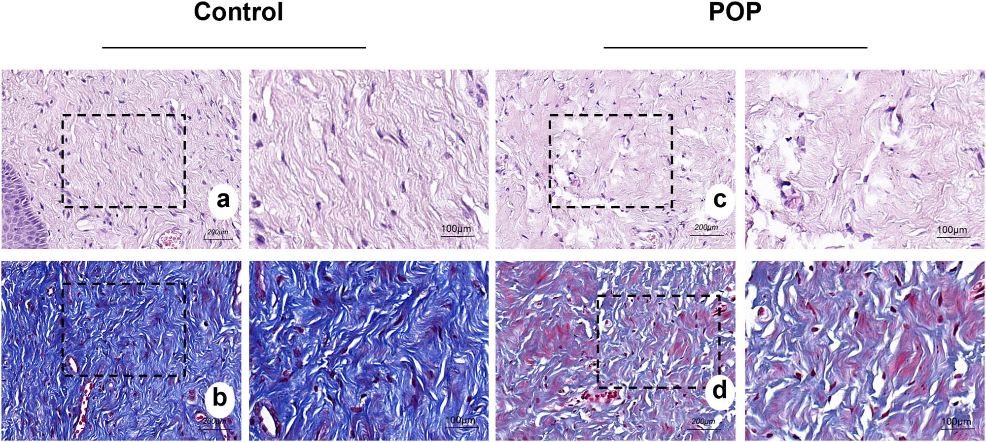
HE staining and Masson staining of the tissue of the anterior vaginal wall: (a) and (b) anterior vaginal wall of the control group; (c) and (d) anterior vaginal wall of the POP group. Collagen fibers were pink in HE staining and blue in Masson staining. (a)–(d) magnification 50×.
3.3 TUNEL cell apoptosis assay
The nuclei of apoptosis cells were brownish-yellow in color, and the staining results showed that there were significantly more apoptosis cells in the anterior vaginal wall tissue of the POP group than in that of the control group (Figure 2).
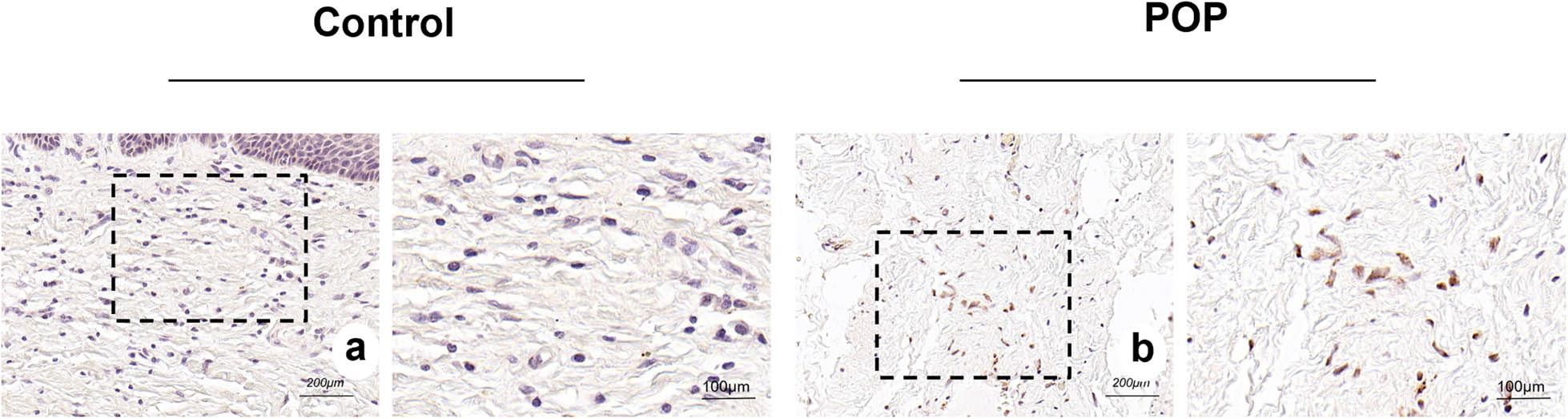
(a) and (b) TUNEL cell apoptosis staining of the tissue of the anterior vaginal wall in the control and POP groups, respectively. The brownish yellow granules are nuclei of the apoptosis cells. (a) and (b) magnification 50×.
3.4 Immunohistochemistry
The results showed that collagen I was mainly expressed in the intercellular matrix, and the expression of collagen I in the POP group was significantly lower than that in the control group. MMP2 and TIMP2 were primarily expressed in the interstitium. We observed a significantly higher expression of MMP2 in the POP group compared to the control group, whereas TIMP2 showed a significantly lower expression in the POP group than in the control group. β-catenin was expressed in both the nucleus and the cytoplasm, with predominant expression in the nucleus. The expression in the POP group was significantly lower than that in the control group. TGF-β1 was primarily expressed in the cytoplasm, and to a lesser extent in the nucleus. Its expression in the tissue of the anterior vaginal wall of the POP group was significantly lower than that of the control group. All the aforementioned differences were statistically significant (Figures 3 and 4).
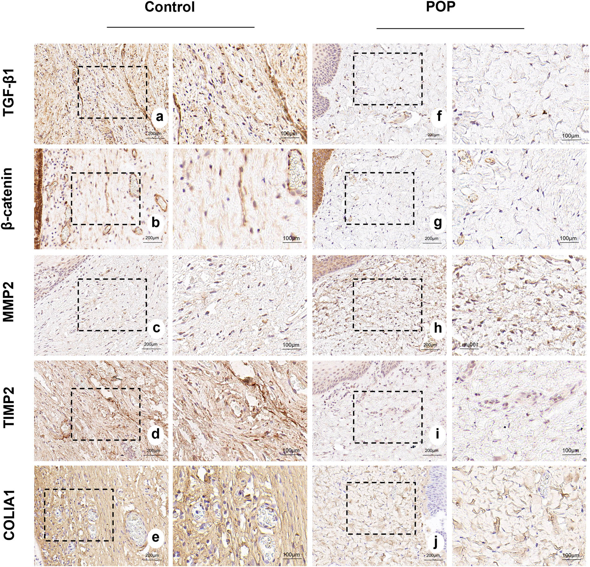
(a)–(e) Expression of TGF-β1, β-catenin, MMP2, TIMP2, and COL1A1 in the anterior vaginal wall tissues of patients in the control group. (f)–(j) Expression of TGF-β1, β-catenin, MMP2, TIMP2, and COL1A1 in the anterior vaginal wall tissues of patients in the POP group, respectively. (a)–(j) magnification 50×.
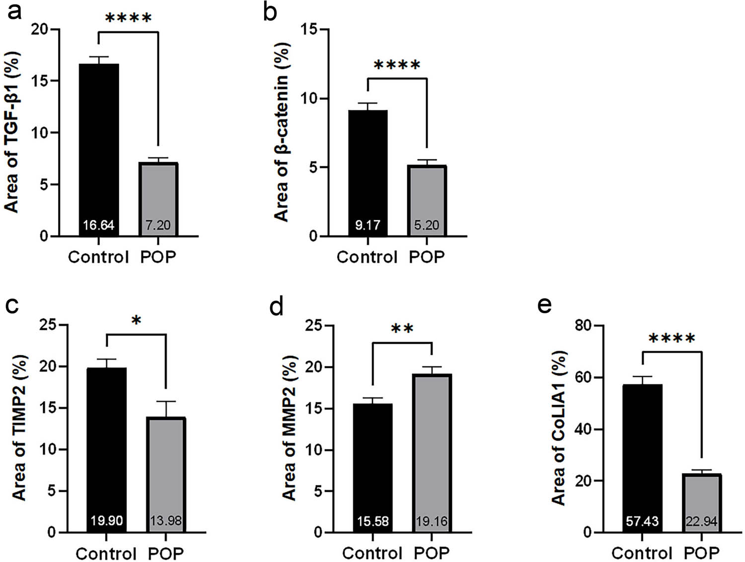
(a)–(e) Statistical results of immunohistochemical staining of TGF-β1, β-catenin, MMP2, TIMP2, and COL1A1, respectively.
3.5 Quantitative real-time polymerase chain reaction
The results revealed that the mRNA expression levels of TGF-β1, β-catenin, TIMP2, and COL1A1 in the POP group were significantly lower than those in the control group. In contrast, the expression of MMP2 was significantly higher in the POP group compared to the control group, and these differences were statistically significant (Figure 5).

(a)–(e) Statistical results of TGF-β1, β-catenin, MMP2, TIMP2, and COL1A1 mRNA expression levels in the anterior vaginal wall tissues of control and POP groups.
3.6 Western blot
The results indicated that the protein expression levels of TGF-β1, β-catenin, TIMP2, and COL1A1 in the tissue of the anterior vaginal wall of the POP group were significantly lower than those of the control group. The differences were statistically significant, with levels of difference at ***, **, **, and **, respectively. Although the expression of MMP2 was higher than that of the control group, the difference was not statistically significant (p > 0.05, Figure 6).
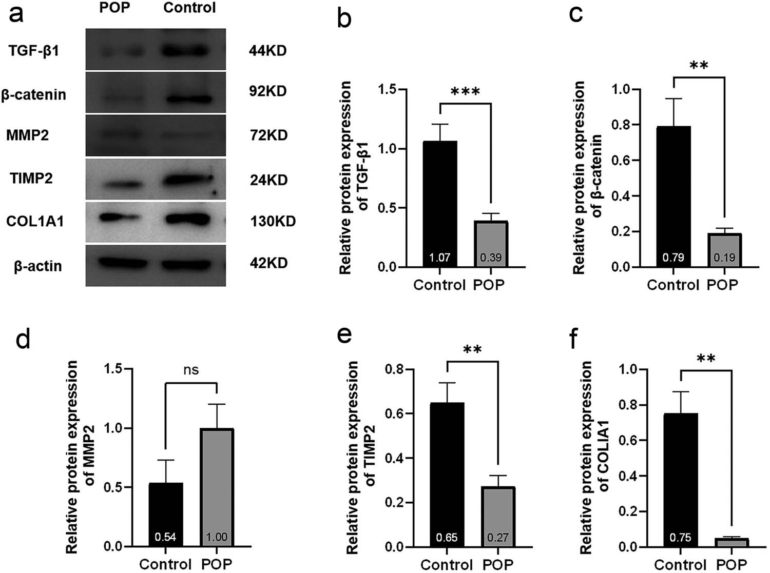
(a) Strip graph of the western blot and (b)–(f) statistical analysis results of western blot.
3.7 Correlation analysis
Examine the correlation of protein expression levels of TGF-β1, β-catenin, and COL1A1 in patients of the POP group. Considering the small sample size of the study, Spearman correlation test was used, revealing a significant positive correlation between TGF-β1 and β-catenin, β-catenin and COL1A1, as well as TGF-β1 and COL1A1 in the tissues of the anterior wall of the vagina (Figure 7).
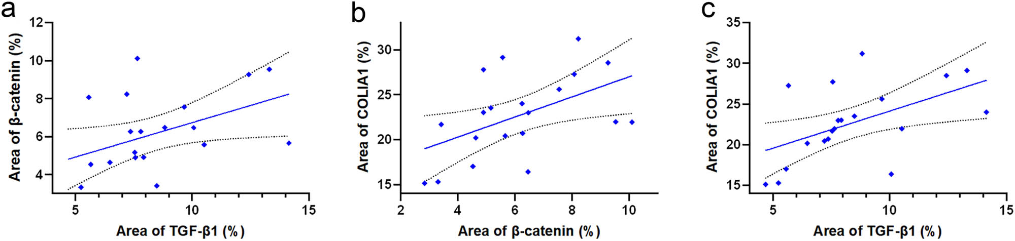
(a) Correlation test between the immunohistochemical staining results of TGF-β1 and β-catenin, r = 0.46, p < 0.05. (b) Correlation test between the immunohistochemical staining results of COL1A1 and β-catenin, r = 0.50, p < 0.05. (c) Correlation test between COL1A1 and TGF-β1 and immunohistochemical staining results, r = 0.62, p < 0.05.
4 Discussion
In this study, we found that collagen fibers in the tissue of the anterior vaginal wall of patients with POP were disorganized in orientation, diminished in quantity, and fractures were notably prevalent. Meanwhile, TUNEL cell apoptosis staining revealed a significantly higher number of apoptotic cells in the vaginal wall of the POP group compared to the control group. The results of immunohistochemistry, qPCR, and western blot all showed that patients in the POP group had significantly lower levels of type I collagen than those in non-POP patients. These results suggest that the decreased amount and structural abnormality of collagen may be related to the onset of POP. POP is commonly recognized as a condition characterized by diminished pelvic floor tissue support resulting from aging, childbirth, and connective tissue abnormalities. Collagen, an important component of connective tissue, plays a key role in supporting the pelvic floor. Zhu et al. [8] detected the expression of type I collagen and type III collagen in the uterine sacral ligament of 35 POP patients and 20 control patients. The experimental results showed that the expression levels of type I collagen and type III collagen in the sacral ligament tissues of the POP group were decreased, and the expression levels of the genes Bax and Bad related to apoptosis were up-regulated. It is suggested that POP is related to the remodeling of extracellular matrix in the uterine and sacral ligaments. The main pathological features are the decrease of collagen synthesis, the increase of collagen decomposition, and the increase of apoptosis level. This result is consistent with our experimental results, so it can be suggested that the occurrence of POP may be related to the imbalance of collagen metabolism in the pelvic floor supporting tissue. In addition, enzymes related to collagen metabolism, such as MMPs and TIMPs, play an important role in maintaining the dynamic remodeling of the extracellular matrix.
MMP2 is a crucial enzyme in the metabolism of the extracellular matrix and is closely associated with the remodeling of the extracellular matrix in both physiological and pathological conditions. MMP2 is also known as gelatinase A. The main hydrolysis products are collagen type IV and collagen type V, among others. The degradation function of MMP2 is regulated by its specific antagonist TIMP2. TIMP2 inhibits the degradation of extracellular matrix proteins by MMP2. It achieves this primarily by binding non-covalently to the activated MMP2 molecules in a 1:1 ratio, effectively inactivating them. Previous studies have shown that MMP2 expression is higher and TIMP2 expression is lower in sacral ligament tissues of patients with POP, the results indicated that the degradation of collagen in sacral ligament tissues of POP patients was higher than that of control group. In this study, the expression levels of MMP2 and TIMP2 in the anterior vaginal wall tissues were detected, and the same results were obtained. However, a recent study [9] found that the expression of MMP2 in the vaginal wall tissue of the POP group was significantly up-regulated, which was consistent with our results. However, although the expression of TIMP1 was reduced, there was no statistical difference between it and the control group, which we speculated might be caused by individual differences or insufficient sample size. It is hoped that this problem can be proved by establishing uniform sample acquisition standards and expanding sample size in the future. Combined with the fact that the expression of type I collagen is reduced and disorganized in its orientation in the vaginal wall of patients in the POP group, it is reasonable to hypothesize that the increased catabolism and decreased synthesis of collagen are important factors contributing to the development of POP.
TGF-β has been widely reported as an important regulator of fibrotic metabolism in fibrotic and degenerative diseases [10,11]. TGF-β is a multifunctional protein polypeptide. It exists in four isoforms: TGF-β1, TGF-β2, TGF-β3, and TGF-β1β2. These isoforms not only promote a large amount of collagen synthesis and secretion from fibroblasts, but also enhance collagen synthesis and reduce collagen degradation by up-regulating TIMPs and inhibiting the activity of MMPs. Research indicates that in early-passaged human gingival fibroblasts, TGF-β1 plays a role in regulating the activity of MMP2. Specifically, it decreases the mRNA level of MMP2 and increases the mRNA level of TIMP [12]. Another study found that the expression level of TGF-β1 in sacral ligament tissues of patients with POP was significantly lower compared to that in non-POP patients. Additionally, the expression level of TGF-β1 mRNA was partially negatively correlated with the severity of POP [13]. These findings align with our own experimental results. Our investigations, including immunohistochemistry, qPCR, and western blot analyses, consistently demonstrated that the expression of TGF-β1 in the tissue of the anterior vaginal wall of patients in the POP group was significantly lower than that in the control group. Based on these collective findings, we hypothesize that TGF-β1 may play a role in the development of POP by regulating the metabolism of the extracellular matrix. However, a recent study on the correlation between vaginal microecological changes and pelvic floor collagen metabolism in POP patients found [13] that mRNA expressions of Decorin, TGF-β1, and matrix metalloproteinase-3 were increased in sacrum ligament tissues of POP patients. The mRNA expression of type I and III collagen decreased. According to the different research results of TGF-β1, we speculated that the expression and function of TGF-β1 may be affected by factors such as research method, material location, study population, sample size, and genetic background.
The Wnt/β-catenin signaling pathway plays a significant role in a variety of tissue repair and fibrotic diseases. It is an important pathway in collagen synthesis [14]. β-catenin, a key member of this classical Wnt pathway, plays a crucial role in regulating cell differentiation, proliferation, and apoptosis. In a study by Gong et al. [15] silencing β-catenin in vaginal wall fibroblasts of non-POP patients resulted in a significant decrease in cell proliferation rate and a reduction in type I collagen expression. Conversely, activating the Wnt/β-catenin signaling pathway with lithium chloride yielded results opposite to the previous findings. Immunofluorescence results indicated an increase in the nuclear translocation of β-catenin. As a transcriptional co-activator, β-catenin initiates the expression of target genes downstream of the classical Wnt pathway by binding to transcription factors TCF/LEFs [16]. These factors form a transcriptional complex that can bind to the promoter sequence of the COL3a1 gene, which is directly involved in regulating extracellular matrix-related genes [17]. Our results demonstrated that the levels of β-catenin protein expression and gene transcription in the anterior vaginal wall tissues of patients with POP were significantly lower than those of non-POP patients. This suggests that the reduced expression of β-catenin may be another crucial factor contributing to the development of POP.
The TGF-β/Smad pathway and the Wnt/β-catenin pathway work together in various fibrotic diseases [18,19], including cardiac remodeling, pulmonary fibrosis, and renal fibrosis. They play a role in regulating cell fate and are involved in collagen metabolism. Increasing evidence suggests that TGF-β-induced fibrosis is closely related to the Wnt signaling pathway, and they are jointly involved not only in the regulation of fibrotic diseases but also in the regulation of tumor invasion and metastasis [20,21]. However, to the best of our knowledge, the interaction between the two has not been thoroughly studied in patients with POP. For this reason, we analyzed the correlation between the key factors of the two signaling pathways, TGF-β1 and β-catenin, as well as the protein expression of each with COL1A1 in the tissue of the anterior vaginal wall of patients with POP. The immunohistochemical staining results of the anterior vaginal wall of the patients were subjected to Spearman correlation test. The analysis revealed a significant positive correlation among the protein expression levels of TGF-β1, β-catenin, and COL1A1. This finding supports our hypothesis and establishes a solid theoretical foundation for our upcoming in vitro cellular experiments.
5 Conclusion
In this study, we demonstrated that the level of cellular necrosis in the vaginal wall tissues of patients with POP was higher than that of the control group. Additionally, we found that the expression level of type I collagen was significantly down-regulated. Moreover, the expression of MMP2, which promotes collagen degradation, had increased, while the expression of TIMP2, which inhibits collagen degradation, had decreased. In addition, we demonstrated that the expression of TGF-β1 and β-catenin in the vaginal wall tissue of patients with POP was significantly lower than that of the control patients, and the protein expression levels of the two showed a strong correlation. These data suggest that POP may occur due to the inhibition of the TGF-β/Smad signaling pathway and the Wnt/β-catenin signaling pathway. This inhibition leads to a decrease in collagenous components and an increase in cellular apoptosis in connective tissues. Additionally, the two pathways are interconnected and crosstalk with each other. Abnormalities in TGF and Wnt signaling pathways are commonly observed in various connective tissue diseases. By investigating the interaction between TGF-β and Wnt signaling pathways, we aim to develop targeted therapeutic drugs that effectively manage the progression of POP. This approach could potentially benefit women with POP, helping them regain confidence and improve their quality of life.
6 Limitations
It is essential to acknowledge the limitations of this study. First, the sample size is relatively small; hence, we plan to conduct multi-center clinical studies in the future to gather more robust data. Second, this study is retrospective, which makes it challenging to definitively establish whether the decrease in TGF-β1 and β-catenin expression directly causes POP or if POP itself leads to reduced expression of these factors based solely on this experiment. To address this, we intend to further investigate the causal relationship between these pathways through additional in vitro experiments.
-
Funding information: Authors state no funding involved.
-
Author contributions: FQX designed the study, drafted, and edited the manuscript. DX collected and analyzed the data. SRZ and YM performed the experiments and analyzed the data. YZ designed the study, collected the data, and prepared the manuscript for publication. All authors read and approved the final draft.
-
Conflict of interest: Authors state no conflict of interest.
-
Data availability statement: The datasets generated during and/or analyzed during the current study are available from the corresponding author on reasonable request.
References
[1] Li L, Ma Y, Yang H, Sun Z, Chen J, Zhu L. The polymorphisms of extracellular matrix-remodeling genes are associated with pelvic organ prolapse. Int Urogynecol J. 2022;33(2):267–74. 10.1007/s00192-021-04917-5.Search in Google Scholar PubMed PubMed Central
[2] Guler Z, Roovers JP. Role of fibroblasts and myofibroblasts on the pathogenesis and treatment of pelvic organ prolapse. Biomolecules. 2022;12(1):94. 10.3390/biom12010094.Search in Google Scholar PubMed PubMed Central
[3] Gardella B, Scatigno AL, Belli G, Gritti A, Visoná SD, Dominoni M. Aging of pelvic floor in animal models: a sistematic review of literature on the role of the extracellular matrix in the development of pelvic floor prolapse. Front Med (Lausanne). 2022;9:863945. 10.3389/fmed.2022.863945.Search in Google Scholar PubMed PubMed Central
[4] Silva MET, Bessa JNM, Parente MPL, Mascarenhas T, Natal Jorge RM, Fernandes AA. Effect of mesh anchoring technique in uterine prolapse repair surgery: a finite element analysis. J Biomech. 2021;127:110649. 10.1016/j.jbiomech.2021.110649.Search in Google Scholar PubMed
[5] Saputra AND, Rizal DM, Ayuandari S, Pangastuti N. The difference in collagen type-1 expression in women with and without pelvic organ prolapse: a systematic review and meta-analysis. Int Urogynecol J. 2022;33(7):1803–12. 10.1007/s00192-022-05229-y.Search in Google Scholar PubMed
[6] Jian H, Shen X, Liu I, Semenov M, He X, Wang XF. Smad3-dependent nuclear translocation of beta-catenin is required for TGF-beta1-induced proliferation of bone marrow-derived adult human mesenchymal stem cells. Genes Dev. 2006;20(6):666–74. 10.1101/gad.1388806.Search in Google Scholar PubMed PubMed Central
[7] Feng J, Zhang Q, Pu F, Zhu Z, Lu K, Lu WW, et al. Signalling interaction between β-catenin and other signalling molecules during osteoarthritis development. Cell Prolif. 2024;57(6):e13600. 10.1111/cpr.13600.Search in Google Scholar PubMed PubMed Central
[8] Zhu YP, Xie T, Guo T, Sun ZJ, Zhu L, Lang JH. Evaluation of extracellular matrix protein expression and apoptosis in the uterosacral ligaments of patients with or without pelvic organ prolapse. Int Urogynecol J. 2021;32(8):2273–81. 10.1007/s00192-020-04446-7.Search in Google Scholar PubMed
[9] Duan Y, Chen Y, He Y, Gong R, Xia Z. Expression of insulin-like growth factor binding protein 5 in the vaginal wall tissues of older women with pelvic organ prolapse. Sci Rep. 2024;14(1):18353. 10.1038/s41598-024-69098-9.Search in Google Scholar PubMed PubMed Central
[10] Yu XY, Sun Q, Zhang YM, Zou L, Zhao YY. TGF-β/smad signaling pathway in tubulointerstitial fibrosis. Front Pharmacol. 2022;13:860588. 10.3389/fphar.2022.860588.Search in Google Scholar PubMed PubMed Central
[11] Zeng Y, Hu R, Ma W, Ding Y, Zhou Y, Peng X, et al. New tricks for old drugs-praziquantel ameliorates bleomycin-induced pulmonary fibrosis in mice. BMC Pharmacol Toxicol. 2024;25(1):18. 10.1186/s40360-024-00737-7.Search in Google Scholar PubMed PubMed Central
[12] Costa CRR, Chalgoumi R, Baker A, Guillou C, Yamaguti PM, Simancas Escorcia V, et al. Gingival proteomics reveals the role of TGF beta and YAP/TAZ signaling in Raine syndrome fibrosis. Sci Rep. 2024;14(1):9497. 10.1038/s41598-024-59713-0.Search in Google Scholar PubMed PubMed Central
[13] Chen S, Zheng Q, Zhang L, Chen L, Wang J. Effect of vaginal microecological alterations on female pelvic organ prolapse. Int Urogynecol J. 2024;35(4):881–91. 10.1007/s00192-024-05759-7.Search in Google Scholar PubMed PubMed Central
[14] Uemura R, Tachibana D, Shiota M, Yoshida K, Kitada K, Hamuro A, et al. Upregulation of PTK7 and β-catenin after vaginal mechanical dilatation: an examination of fibulin-5 knockout mice. Int Urogynecol J. 2021;32(11):2993–9. 10.1007/s00192-021-04693-2.Search in Google Scholar PubMed
[15] Gong R, Xi Y, Jin X, Xu H, Feng J, Hu Q, et al. Effects of the decrease of β-catenin expression on human vaginal fibroblasts of women with pelvic organ prolapse. J Obstet Gynaecol Res. 2021;47(11):4014–22. 10.1111/jog.14946.Search in Google Scholar PubMed
[16] Vuong LM, Pan S, Sierra RA, Waterman ML, Gershon PD, Donovan PJ. Characterization of a chromatin-associated TCF7L1 complex in human embryonic stem cells. Proteomics. 2024;24(20):e2300641. 10.1002/pmic.202300641.Search in Google Scholar PubMed
[17] Wang R, Zhang H, Wang Y, Song F, Yuan Y. Inhibitory effects of quercetin on the progression of liver fibrosis through the regulation of NF-кB/IкBα, p38 MAPK, and Bcl-2/Bax signaling. Int Immunopharmacol. 2017;47:126–33. 10.1016/j.intimp.2017.03.029.Search in Google Scholar PubMed
[18] Chen X, Yan H, Chen Y, Li G, Bin Y, Zhou X. Moderate oxidative stress promotes epithelial-mesenchymal transition in the lens epithelial cells via the TGF-β/Smad and Wnt/β-catenin pathways. Mol Cell Biochem. 2021;476(3):1631–42. 10.1007/s11010-020-04034-9.Search in Google Scholar PubMed
[19] Hu L, Wang Y, Wan Y, Ma L, Zhao T, Li P. Tangshen formula improves diabetes-associated myocardial fibrosis by inhibiting TGF-β/smads and Wnt/β-catenin pathways. Front Med (Lausanne). 2021;8:732042. 10.3389/fmed.2021.732042.Search in Google Scholar PubMed PubMed Central
[20] Lüönd F, Pirkl M, Hisano M, Prestigiacomo V, Kalathur RK, Beerenwinkel N, et al. Hierarchy of TGFβ/SMAD, hippo/YAP/TAZ, and Wnt/β-catenin signaling in melanoma phenotype switching. Life Sci Alliance. 2021;5(2):e202101010. 10.26508/lsa.202101010.Search in Google Scholar PubMed PubMed Central
[21] Yayan J, Franke KJ, Berger M, Windisch W, Rasche K. Adhesion, metastasis, and inhibition of cancer cells: a comprehensive review. Mol Biol Rep. 2024;51(1):165. 10.1007/s11033-023-08920-5.Search in Google Scholar PubMed PubMed Central
© 2025 the author(s), published by De Gruyter
This work is licensed under the Creative Commons Attribution 4.0 International License.
Articles in the same Issue
- Biomedical Sciences
- Mechanism of triptolide regulating proliferation and apoptosis of hepatoma cells by inhibiting JAK/STAT pathway
- Maslinic acid improves mitochondrial function and inhibits oxidative stress and autophagy in human gastric smooth muscle cells
- Comparative analysis of inflammatory biomarkers for the diagnosis of neonatal sepsis: IL-6, IL-8, SAA, CRP, and PCT
- Post-pandemic insights on COVID-19 and premature ovarian insufficiency
- Proteome differences of dental stem cells between permanent and deciduous teeth by data-independent acquisition proteomics
- Optimizing a modified cetyltrimethylammonium bromide protocol for fungal DNA extraction: Insights from multilocus gene amplification
- Preliminary analysis of the role of small hepatitis B surface proteins mutations in the pathogenesis of occult hepatitis B infection via the endoplasmic reticulum stress-induced UPR-ERAD pathway
- Efficacy of alginate-coated gold nanoparticles against antibiotics-resistant Staphylococcus and Streptococcus pathogens of acne origins
- Battling COVID-19 leveraging nanobiotechnology: Gold and silver nanoparticle–B-escin conjugates as SARS-CoV-2 inhibitors
- Neurodegenerative diseases and neuroinflammation-induced apoptosis
- Impact of fracture fixation surgery on cognitive function and the gut microbiota in mice with a history of stroke
- COLEC10: A potential tumor suppressor and prognostic biomarker in hepatocellular carcinoma through modulation of EMT and PI3K-AKT pathways
- High-temperature requirement serine protease A2 inhibitor UCF-101 ameliorates damaged neurons in traumatic brain-injured rats by the AMPK/NF-κB pathway
- SIK1 inhibits IL-1β-stimulated cartilage apoptosis and inflammation in vitro through the CRTC2/CREB1 signaling
- Rutin–chitooligosaccharide complex: Comprehensive evaluation of its anti-inflammatory and analgesic properties in vitro and in vivo
- Knockdown of Aurora kinase B alleviates high glucose-triggered trophoblast cells damage and inflammation during gestational diabetes
- Calcium-sensing receptors promoted Homer1 expression and osteogenic differentiation in bone marrow mesenchymal stem cells
- ABI3BP can inhibit the proliferation, invasion, and epithelial–mesenchymal transition of non-small-cell lung cancer cells
- Changes in blood glucose and metabolism in hyperuricemia mice
- Rapid detection of the GJB2 c.235delC mutation based on CRISPR-Cas13a combined with lateral flow dipstick
- IL-11 promotes Ang II-induced autophagy inhibition and mitochondrial dysfunction in atrial fibroblasts
- Short-chain fatty acid attenuates intestinal inflammation by regulation of gut microbial composition in antibiotic-associated diarrhea
- Application of metagenomic next-generation sequencing in the diagnosis of pathogens in patients with diabetes complicated by community-acquired pneumonia
- NAT10 promotes radiotherapy resistance in non-small cell lung cancer by regulating KPNB1-mediated PD-L1 nuclear translocation
- Phytol-mixed micelles alleviate dexamethasone-induced osteoporosis in zebrafish: Activation of the MMP3–OPN–MAPK pathway-mediating bone remodeling
- Association between TGF-β1 and β-catenin expression in the vaginal wall of patients with pelvic organ prolapse
- Primary pleomorphic liposarcoma involving bilateral ovaries: Case report and literature review
- Effects of de novo donor-specific Class I and II antibodies on graft outcomes after liver transplantation: A pilot cohort study
- Sleep architecture in Alzheimer’s disease continuum: The deep sleep question
- Ephedra fragilis plant extract: A groundbreaking corrosion inhibitor for mild steel in acidic environments – electrochemical, EDX, DFT, and Monte Carlo studies
- Langerhans cell histiocytosis in an adult patient with upper jaw and pulmonary involvement: A case report
- Inhibition of mast cell activation by Jaranol-targeted Pirin ameliorates allergic responses in mouse allergic rhinitis
- Aeromonas veronii-induced septic arthritis of the hip in a child with acute lymphoblastic leukemia
- Clusterin activates the heat shock response via the PI3K/Akt pathway to protect cardiomyocytes from high-temperature-induced apoptosis
- Research progress on fecal microbiota transplantation in tumor prevention and treatment
- Low-pressure exposure influences the development of HAPE
- Stigmasterol alleviates endplate chondrocyte degeneration through inducing mitophagy by enhancing PINK1 mRNA acetylation via the ESR1/NAT10 axis
- AKAP12, mediated by transcription factor 21, inhibits cell proliferation, metastasis, and glycolysis in lung squamous cell carcinoma
- Association between PAX9 or MSX1 gene polymorphism and tooth agenesis risk: A meta-analysis
- A case of bloodstream infection caused by Neisseria gonorrhoeae
- Case of nasopharyngeal tuberculosis complicated with cervical lymph node and pulmonary tuberculosis
- p-Cymene inhibits pro-fibrotic and inflammatory mediators to prevent hepatic dysfunction
- GFPT2 promotes paclitaxel resistance in epithelial ovarian cancer cells via activating NF-κB signaling pathway
- Transfer RNA-derived fragment tRF-36 modulates varicose vein progression via human vascular smooth muscle cell Notch signaling
- RTA-408 attenuates the hepatic ischemia reperfusion injury in mice possibly by activating the Nrf2/HO-1 signaling pathway
- Decreased serum TIMP4 levels in patients with rheumatoid arthritis
- Sirt1 protects lupus nephritis by inhibiting the NLRP3 signaling pathway in human glomerular mesangial cells
- Sodium butyrate aids brain injury repair in neonatal rats
- Interaction of MTHFR polymorphism with PAX1 methylation in cervical cancer
- Convallatoxin inhibits proliferation and angiogenesis of glioma cells via regulating JAK/STAT3 pathway
- The effect of the PKR inhibitor, 2-aminopurine, on the replication of influenza A virus, and segment 8 mRNA splicing
- Effects of Ire1 gene on virulence and pathogenicity of Candida albicans
- Small cell lung cancer with small intestinal metastasis: Case report and literature review
- GRB14: A prognostic biomarker driving tumor progression in gastric cancer through the PI3K/AKT signaling pathway by interacting with COBLL1
- 15-Lipoxygenase-2 deficiency induces foam cell formation that can be restored by salidroside through the inhibition of arachidonic acid effects
- FTO alleviated the diabetic nephropathy progression by regulating the N6-methyladenosine levels of DACT1
- Clinical relevance of inflammatory markers in the evaluation of severity of ulcerative colitis: A retrospective study
- Zinc valproic acid complex promotes osteoblast differentiation and exhibits anti-osteoporotic potential
- Primary pulmonary synovial sarcoma in the bronchial cavity: A case report
- Metagenomic next-generation sequencing of alveolar lavage fluid improves the detection of pulmonary infection
- Uterine tumor resembling ovarian sex cord tumor with extensive rhabdoid differentiation: A case report
- Genomic analysis of a novel ST11(PR34365) Clostridioides difficile strain isolated from the human fecal of a CDI patient in Guizhou, China
- Effects of tiered cardiac rehabilitation on CRP, TNF-α, and physical endurance in older adults with coronary heart disease
- Changes in T-lymphocyte subpopulations in patients with colorectal cancer before and after acupoint catgut embedding acupuncture observation
- Modulating the tumor microenvironment: The role of traditional Chinese medicine in improving lung cancer treatment
- Alterations of metabolites related to microbiota–gut–brain axis in plasma of colon cancer, esophageal cancer, stomach cancer, and lung cancer patients
- Research on individualized drug sensitivity detection technology based on bio-3D printing technology for precision treatment of gastrointestinal stromal tumors
- CEBPB promotes ulcerative colitis-associated colorectal cancer by stimulating tumor growth and activating the NF-κB/STAT3 signaling pathway
- Oncolytic bacteria: A revolutionary approach to cancer therapy
- A de novo meningioma with rapid growth: A possible malignancy imposter?
- Diagnosis of secondary tuberculosis infection in an asymptomatic elderly with cancer using next-generation sequencing: Case report
- Hesperidin and its zinc(ii) complex enhance osteoblast differentiation and bone formation: In vitro and in vivo evaluations
- Research progress on the regulation of autophagy in cardiovascular diseases by chemokines
- Anti-arthritic, immunomodulatory, and inflammatory regulation by the benzimidazole derivative BMZ-AD: Insights from an FCA-induced rat model
- Immunoassay for pyruvate kinase M1/2 as an Alzheimer’s biomarker in CSF
- The role of HDAC11 in age-related hearing loss: Mechanisms and therapeutic implications
- Evaluation and application analysis of animal models of PIPNP based on data mining
- Therapeutic approaches for liver fibrosis/cirrhosis by targeting pyroptosis
- Fabrication of zinc oxide nanoparticles using Ruellia tuberosa leaf extract induces apoptosis through P53 and STAT3 signalling pathways in prostate cancer cells
- Haplo-hematopoietic stem cell transplantation and immunoradiotherapy for severe aplastic anemia complicated with nasopharyngeal carcinoma: A case report
- Modulation of the KEAP1-NRF2 pathway by Erianin: A novel approach to reduce psoriasiform inflammation and inflammatory signaling
- The expression of epidermal growth factor receptor 2 and its relationship with tumor-infiltrating lymphocytes and clinical pathological features in breast cancer patients
- Innovations in MALDI-TOF Mass Spectrometry: Bridging modern diagnostics and historical insights
- BAP1 complexes with YY1 and RBBP7 and its downstream targets in ccRCC cells
- Hypereosinophilic syndrome with elevated IgG4 and T-cell clonality: A report of two cases
- Electroacupuncture alleviates sciatic nerve injury in sciatica rats by regulating BDNF and NGF levels, myelin sheath degradation, and autophagy
- Polydatin prevents cholesterol gallstone formation by regulating cholesterol metabolism via PPAR-γ signaling
- RNF144A and RNF144B: Important molecules for health
- Analysis of the detection rate and related factors of thyroid nodules in the healthy population
- Artesunate inhibits hepatocellular carcinoma cell migration and invasion through OGA-mediated O-GlcNAcylation of ZEB1
- Endovascular management of post-pancreatectomy hemorrhage caused by a hepatic artery pseudoaneurysm: Case report and review of the literature
- Efficacy and safety of anti-PD-1/PD-L1 antibodies in patients with relapsed refractory diffuse large B-cell lymphoma: A meta-analysis
- SATB2 promotes humeral fracture healing in rats by activating the PI3K/AKT pathway
- Overexpression of the ferroptosis-related gene, NFS1, corresponds to gastric cancer growth and tumor immune infiltration
- Understanding risk factors and prognosis in diabetic foot ulcers
- Atractylenolide I alleviates the experimental allergic response in mice by suppressing TLR4/NF-kB/NLRP3 signalling
- FBXO31 inhibits the stemness characteristics of CD147 (+) melanoma stem cells
- Immune molecule diagnostics in colorectal cancer: CCL2 and CXCL11
- Inhibiting CXCR6 promotes senescence of activated hepatic stellate cells with limited proinflammatory SASP to attenuate hepatic fibrosis
- Cadmium toxicity, health risk and its remediation using low-cost biochar adsorbents
- Pulmonary cryptococcosis with headache as the first presentation: A case report
- Solitary pulmonary metastasis with cystic airspaces in colon cancer: A rare case report
- RUNX1 promotes denervation-induced muscle atrophy by activating the JUNB/NF-κB pathway and driving M1 macrophage polarization
- Morphometric analysis and immunobiological investigation of Indigofera oblongifolia on the infected lung with Plasmodium chabaudi
- The NuA4/TIP60 histone-modifying complex and Hr78 modulate the Lobe2 mutant eye phenotype
- Experimental study on salmon demineralized bone matrix loaded with recombinant human bone morphogenetic protein-2: In vitro and in vivo study
- A case of IgA nephropathy treated with a combination of telitacicept and half-dose glucocorticoids
- Analgesic and toxicological evaluation of cannabidiol-rich Moroccan Cannabis sativa L. (Khardala variety) extract: Evidence from an in vivo and in silico study
- Wound healing and signaling pathways
- Combination of immunotherapy and whole-brain radiotherapy on prognosis of patients with multiple brain metastases: A retrospective cohort study
- To explore the relationship between endometrial hyperemia and polycystic ovary syndrome
- Research progress on the impact of curcumin on immune responses in breast cancer
- Biogenic Cu/Ni nanotherapeutics from Descurainia sophia (L.) Webb ex Prantl seeds for the treatment of lung cancer
- Dapagliflozin attenuates atrial fibrosis via the HMGB1/RAGE pathway in atrial fibrillation rats
- Glycitein alleviates inflammation and apoptosis in keratinocytes via ROS-associated PI3K–Akt signalling pathway
- ADH5 inhibits proliferation but promotes EMT in non-small cell lung cancer cell through activating Smad2/Smad3
- Apoptotic efficacies of AgNPs formulated by Syzygium aromaticum leaf extract on 32D-FLT3-ITD human leukemia cell line with PI3K/AKT/mTOR signaling pathway
- Novel cuproptosis-related genes C1QBP and PFKP identified as prognostic and therapeutic targets in lung adenocarcinoma
- Bee venom promotes exosome secretion and alters miRNA cargo in T cells
- Treatment of pure red cell aplasia in a chronic kidney disease patient with roxadustat: A case report
- Comparative bioinformatics analysis of the Wnt pathway in breast cancer: Selection of novel biomarker panels associated with ER status
- Kynurenine facilitates renal cell carcinoma progression by suppressing M2 macrophage pyroptosis through inhibition of CASP1 cleavage
- RFX5 promotes the growth, motility, and inhibits apoptosis of gastric adenocarcinoma cells through the SIRT1/AMPK axis
- ALKBH5 exacerbates early cardiac damage after radiotherapy for breast cancer via m6A demethylation of TLR4
- Phytochemicals of Roman chamomile: Antioxidant, anti-aging, and whitening activities of distillation residues
- Circadian gene Cry1 inhibits the tumorigenicity of hepatocellular carcinoma by the BAX/BCL2-mediated apoptosis pathway
- The TNFR-RIPK1/RIPK3 signalling pathway mediates the effect of lanthanum on necroptosis of nerve cells
- Ecology and Environmental Science
- Optimization and comparative study of Bacillus consortia for cellulolytic potential and cellulase enzyme activity
- The complete mitochondrial genome analysis of Haemaphysalis hystricis Supino, 1897 (Ixodida: Ixodidae) and its phylogenetic implications
- Epidemiological characteristics and risk factors analysis of multidrug-resistant tuberculosis among tuberculosis population in Huzhou City, Eastern China
- Indices of human impacts on landscapes: How do they reflect the proportions of natural habitats?
- Genetic analysis of the Siberian flying squirrel population in the northern Changbai Mountains, Northeast China: Insights into population status and conservation
- Diversity and environmental drivers of Suillus communities in Pinus sylvestris var. mongolica forests of Inner Mongolia
- Global assessment of the fate of nitrogen deposition in forest ecosystems: Insights from 15N tracer studies
- Fungal and bacterial pathogenic co-infections mainly lead to the assembly of microbial community in tobacco stems
- Influencing of coal industry related airborne particulate matter on ocular surface tear film injury and inflammatory factor expression in Sprague-Dawley rats
- Agriculture
- Integrated analysis of transcriptome, sRNAome, and degradome involved in the drought-response of maize Zhengdan958
- Variation in flower frost tolerance among seven apple cultivars and transcriptome response patterns in two contrastingly frost-tolerant selected cultivars
- Heritability of durable resistance to stripe rust in bread wheat (Triticum aestivum L.)
- Animal Science
- Effect of sex ratio on the life history traits of an important invasive species, Spodoptera frugiperda
- Plant Sciences
- Hairpin in a haystack: In silico identification and characterization of plant-conserved microRNA in Rafflesiaceae
- Widely targeted metabolomics of different tissues in Rubus corchorifolius
- The complete chloroplast genome of Gerbera piloselloides (L.) Cass., 1820 (Carduoideae, Asteraceae) and its phylogenetic analysis
- Field trial to correlate mineral solubilization activity of Pseudomonas aeruginosa and biochemical content of groundnut plants
- Correlation analysis between semen routine parameters and sperm DNA fragmentation index in patients with semen non-liquefaction: A retrospective study
- Plasticity of the anatomical traits of Rhododendron L. (Ericaceae) leaves and its implications in adaptation to the plateau environment
- Effects of Piriformospora indica and arbuscular mycorrhizal fungus on growth and physiology of Moringa oleifera under low-temperature stress
- Effects of different sources of potassium fertiliser on yield, fruit quality and nutrient absorption in “Harward” kiwifruit (Actinidia deliciosa)
- Comparative efficiency and residue levels of spraying programs against powdery mildew in grape varieties
- The DREB7 transcription factor enhances salt tolerance in soybean plants under salt stress
- Food Science
- Phytochemical analysis of Stachys iva: Discovering the optimal extract conditions and its bioactive compounds
- Review on role of honey in disease prevention and treatment through modulation of biological activities
- Computational analysis of polymorphic residues in maltose and maltotriose transporters of a wild Saccharomyces cerevisiae strain
- Optimization of phenolic compound extraction from Tunisian squash by-products: A sustainable approach for antioxidant and antibacterial applications
- Liupao tea aqueous extract alleviates dextran sulfate sodium-induced ulcerative colitis in rats by modulating the gut microbiota
- Toxicological qualities and detoxification trends of fruit by-products for valorization: A review
- Polyphenolic spectrum of cornelian cherry fruits and their health-promoting effect
- Optimizing the encapsulation of the refined extract of squash peels for functional food applications: A sustainable approach to reduce food waste
- Advancements in curcuminoid formulations: An update on bioavailability enhancement strategies curcuminoid bioavailability and formulations
- Impact of saline sprouting on antioxidant properties and bioactive compounds in chia seeds
- The dilemma of food genetics and improvement
- Bioengineering and Biotechnology
- Impact of hyaluronic acid-modified hafnium metalorganic frameworks containing rhynchophylline on Alzheimer’s disease
- Emerging patterns in nanoparticle-based therapeutic approaches for rheumatoid arthritis: A comprehensive bibliometric and visual analysis spanning two decades
- Application of CRISPR/Cas gene editing for infectious disease control in poultry
- Preparation of hafnium nitride-coated titanium implants by magnetron sputtering technology and evaluation of their antibacterial properties and biocompatibility
- Preparation and characterization of lemongrass oil nanoemulsion: Antimicrobial, antibiofilm, antioxidant, and anticancer activities
- Corrigendum
- Corrigendum to “Utilization of convolutional neural networks to analyze microscopic images for high-throughput screening of mesenchymal stem cells”
- Corrigendum to “Effects of Ire1 gene on virulence and pathogenicity of Candida albicans”