Bioinspired ferromagnetic CoFe2O4 nanoparticles: Potential pharmaceutical and medical applications
-
 ,
,

Abstract
The primary goal of this work was to develop a cost-effective, non-toxic, eco-friendly, and simple approach for the green synthesis of CoFe2O4 nanoparticles (NPs) using Aloe vera leaf extract by the sol–gel auto-combustion method. In order to figure out their structural, morphological, and magnetic properties, the synthesized NPs were characterized using X-ray diffraction (XRD), Fourier transform infrared (FTIR), scanning electron microscope (SEM), energy-dispersive X-ray spectroscopy (EDX), transmission electron microscope (TEM), dynamic light scattering (DLS), zeta potential, and vibrating-sample magnetometer (VSM). XRD analysis showed that particles had a single-phase spinel crystalline structure with an average crystalline size of 33.5 nm. Under VSM studies, the produced NPs exhibit a soft ferromagnetic property. SEM revealed that the as-prepared NPs were agglomerated due to their magnetic behavior. To the best of our knowledge, the anticandidal, antibiofilm, antibacterial, and anticancer activities of CoFe2O4 NPs toward drug-resistant gram-positive and gram-negative bacteria, as well as fungal strains, have been comprehensively investigated for the first time. The synthesized NPs had a minimal inhibitory concentration of 0.25–0.75 mg/ml against the tested pathogens. CoFe2O4 NPs inhibited the biofilm formation by 37.3–61.8% in selected strains at concentrations of 0.125–0.5 mg/ml. It was observed that the NPs not only suppress biofilm formation but also eradicate established mature biofilms by 50.9–64.49% that was further supported by SEM. SEM analysis shows that NPs significantly inhibit the colonization and aggregation of tested biofilm strains. Light microscopic analysis revealed that NPs completely inhibit the development of hyphae and filaments in Candida albicans, which significantly attenuates their pathogenicity. 3-(4,5-Dimethylthiazol-2-yl)-2,5-diphenyltetrazolium bromide (MTT) assays and 4′,6-diamidino-2-phenylindole (DAPI) staining demonstrate that NPs significantly inhibit the proliferation of HCT-116 and HeLa cells. Furthermore, the SEM images of treated cells showed wrinkled and damaged cell walls, indicating the disruption and disorganization of the membrane. This study showed that the synthesized NPs were effective in inhibiting the growth of drug-resistant bacteria, candida, and their preformed biofilms as well. Thus, these NPs with broad-spectrum applications could be exploited in medical settings to diminish biofilm-based infections caused by these pathogenic strains.
1 Introduction
Ferrites with distinctive physical characteristics have been the focus of many studies in the pharmaceutical and industrial sectors. Within these nanomaterials, cobalt-ferrite nanoparticles (CoFe2O4 NPs) exhibited interesting properties such as strong overloaded magnetization without any favored ferromagnetic mechanism and good penetration [1,2]. Additionally, their potential to form magnetized nanospheres, high magnetostriction, and good coupling efficiency make them a commonly accepted alternative for many applications in industry and medicine [3,4]. Magnetic nanoparticles (NPs) have received a lot of interest over the past two decades because of their superior characteristics versus “traditional” materials with grain sizes >10 nm. They have a high potential for a broad variety of pharmaceutical industries because of their distinctive characteristics and microstructure, as well as their optical devices, powerful magnetic storage, catalytic, biomedical, and treatment of waste water [5]. As a result, they were used in biomedical industries, including drug administration, electromagnetic resonance imaging, magneto-thermo drug delivery, hyperthermia, nanosensors, enzymatic therapy of gases, gas detection, and alkane oxidation [6]. It is well known that the composition, structure, form, and general properties of magnetic ferrite NPs are greatly influenced by the production process. As a result, researchers are concentrating on creating NPs with known sizes and morphologies [7]. Numerous chemical production strategies, such as solvothermal, coprecipitation, hydrothermal, sol–gel, and combustion methods, are now used to produce CoFe2O4 NPs [8,9], but they may pose risks to ecosystems [10]. In the last decades, sustainable and green NP synthesis techniques have been developed to minimize such risks [11]. The green production strategy of CoFe2O4 NPs stands out as an easy and straightforward manufacturing process as they have several advantages over chemical methods, e.g., green route of NP synthesis is environmentally friendly, provides a non-hazardous compatible solvent system, eco-friendly reducing agent, and a non-hazardous gelling agent for stabilizing the nanostructures. The use of plant extracts from leaves, seeds, roots, or flowers in the biosynthetic method of NP manufacturing has already been reported [12,13]. Plant extracts have a number of secondary metabolites such as polyphenols, alkaloids, sugars, polysaccharides, amino acids, and vitamins [7,8,14]. The primary objective of this study was to synthesize CoFe2O4 NPs using a sustainable and environmentally friendly green chemistry technique that uses Aloe vera leaf extract as a fuel and reducing agent. It has been well established that A. vera contains a variety of phytochemicals, including polysaccharides, phenolic compounds, organic acids, alkaloids, tannins, flavonoids, vitamins, enzymes, carbohydrates, and plant steroids [8,15,16]. Aloe includes two types of aloin, A and B, which produce picric and oxalic acids of nitric acid and act as biological capping and reducing agents [17]. These compounds promote the development of NPs and alter their surface properties. Furthermore, these metabolites and bioactive compounds can effectively capture the metal ions by serving as capping agents, reducing agents, or stabilizing agents since they are non-toxic and environmentally friendly [18]. The second objective of this work was to characterize the biosynthesized CoFe2O4 NPs by X-ray diffraction (XRD), scanning electron microscope (SEM), transmission electron microscope (TEM), energy-dispersive X-ray spectroscopy (EDX), X-ray photoelectron spectroscopy (XPS), zeta potential, dynamic light scattering (DLS), Fourier transform infrared (FTIR), and vibrating-sample magnetometer (VSM). The structural, optical, photocatalytic, and magnetic properties of the biosynthesized CoFe2O4 NPs have been well documented in the literature [8,19,20,21,22,23,24]. Though, there are some studies that have reported the antibacterial activity of the biosynthesized CoFe2O4 NPs [20,21,25,26,27]. But there is still paucity in the broad-spectrum bioapplications of the biologically synthesized CoFe2O4 NPs, especially their anticandidal, antibiofilm, anticancer, and antibacterial activities against multidrug-resistant bacterial strains. Therefore, the third aim of this study was to preliminarily evaluate antibacterial, anticandidal, antibiofilm, and anticancer properties against Escherichia coli ATCC 25922, Pseudomonas aeruginosa ATCC 27853, Staphylococcus aureus ATCC 25923, methicillin-resistant S. aureus (MRSA ATCC 33591), multidrug-resistant P. aeruginosa (MDR-PA), Candida albicans (ATCC 14053), C. parapsilosis (ATCC 22019); and cancerous cell lines, i.e., human colorectal carcinoma (HCT-116 ATCC® CCL-247), human cervical carcinoma (HeLa ATCC®CRM-CCL-2), and normal embryonic kidney cell (HEK-293).
2 Materials and methods
2.1 Materials
Cobalt(ii) nitrate hexahydrate Co(NO3)2·6(H2O) and iron(iii) nitrate nonahydrate Fe(NO3)3·6(H2O)9 were purchased from Sigma Aldrich, St. Louis, MI, USA, as the starting materials. A. vera was obtained from local garden. Brain heart infusion (BHI), RPMI 1640, Mueller Hinton agar, Sabouraud dextrose agar, glutaraldehyde, osmium tetroxide, paraformaldehyde, trypticase soy broth (TSB), glucose, ethanol, 3-(4,5-dimethylthiazol-2-yl)-2,5-diphenyltetrazolium bromide (MTT) assay kit, Dulbecco’s modified Eagle medium (DMEM) penicillin–streptomycin (1%), l-glutamine, fetal bovine serum (FBS), 4′,6-diamidino-2-phenylindole (DAPI), phosphate-buffered saline (PBS), and Triton X-100 were purchased from Molequle-ON (New Zealand). All reagents were used without further purification.
2.1.1 Preparation of the leaf extract: green approach of spinel CoFe2O4 magnetic NPs
A. vera plants were chosen for the biosynthesis of CoFe2O4 NPs due to their accessibility, affordability, and medicinal benefits. Fresh A. vera leaves were cleaned in double-distilled water to eliminate dust. The leaves were then cut into small pieces and dried in the air. In 500 ml beakers with 200 ml of distilled water, 20 g of finely chopped leaves was added and boiled for 15 min at 80°C. After cooling to 37°C, these leaves were centrifuged at 10,000 rpm for 20 min at 4°C, and the filtrate was then filtered through Whatman No. 1 filter paper. The collected supernatant (solution A) was kept at 4–8°C and used as a capping agent in the biosynthetic pathways of CoFe2O4 NPs.
2.1.2 CoFe2O4 NP synthesis
Cobalt nitrate and iron nitrate in 1:2 ratios were mixed in 50 ml of double-distilled water and centrifuged to get a clear solution (solution B). Transferring 50 ml of solution A’s aqueous A. vera extract to solution B and vigorously stirring the mixture at 50°C for 4 h caused the mixture to turn a dark color, signifying the completion of the reduction process. The pH was adjusted to 10 using a NaOH solution. A dark precipitate started to form after being cooled to 37°C overnight, indicating the beginning of the formation of NPs [28]. The NPs were separated using centrifugation for 15 min at 4°C and an rpm of 12,000. Then, to remove any remaining biological molecules, they were washed with deionized water five times, followed by a single washing with 100% ethanol. A fine powder was formed after the purified NPs were dried in a hot air oven at 60°C. Additionally, the particles were treated with ultrasound to disperse the individual particles, which were then used for characterization [29].
2.1.3 Characterization
XRD analysis was used to characterize the phase composition, crystalline structure, and diameter of the NPs using CuKα radiation (λ = 1.54056) in the range of 20° ≤ 2θ ≤ 80° at 40 keV using an X-ray powder diffractometer (Shimadzu XRD-7,000) [30]. A FTIR spectrometer (Shimadzu IRSpirit, Shimadzu, Kyoto, Japan) was used to determine the functional group present in A. vera extract and synthesize NPs in the range of 4,000–500 cm−1 [30]. SEM (TESCAN VEGA3) and TEM (Morgagni 268) were used to further analyze the size and shape of the synthesized NPs. Furthermore, the elemental composition of bio-inspired NPs was carried out using EDX (TESCAN VEGA3) [30]. The hydrodynamic diameter and surface charge (zeta potential) were measured using a DLS and Nano-ZS Zetasizer (Malvern Instruments, UK). The electron binding energies for the elements were measured by XPS on an ESCALAB 250Xi X-ray photoelectron spectrometer. VSM (7410; USA) was used to create a magnetic field characteristic at 37°C with a maximum external magnetic field of 10,000 Oe [23].
2.2 Antibacterial, anticandidal, and antibiofilm activities of CoFe2O4 NPs
2.2.1 Strains
MRSA ATCC 33591, MDR-PA (clinical isolate), C. albicans ATCC 14053, C. parapsilosis (ATCC 22019), E. coli ATCC 25922, P. aeruginosa ATCC 27853, and S. aureus ATCC 25923 were used for antimicrobial testing of the synthesized CoFe2O4 NPs.
2.2.2 Antimicrobial activity by determining minimal inhibitory concentration (MIC)
The MIC of CoFe2O4 NPs was calculated using the MIC microbroth dilution method, as described by Qureshi et al. [31]. The bacteria and Candida cultures were incubated at 37°C for 24 h after being exposed to twofold serial dilutions of CoFe2O4 (i.e., 10−0.156 mg/ml). For testing the MIC of bacterial strains, BHI broth was used [31], whereas for C. albicans, RPMI 1,640 culture medium was used according to the guidelines of the Clinical and Laboratory Standards Institute (M27‐S4) [32]. The initial concentration of CoFe2O4 NPs at which no perceivable growth was noted is known as the MIC value.
2.3 Effect of CoFe2O4 NPs on biofilm-forming capabilities of bacteria and candida
The biofilm prevention of bacteria and Candida after treatment with CoFe2O4 NPs was examined by crystal violet bioassay [33]. The freshly harvested cultures were inoculated in a 96-well plate containing TSB + 2% glucose in the case of bacteria and RPMI + 2% glucose in the case of Candida, and then, each plate was incubated for 24 h at 37 and 28°C, respectively, after being exposed to various CoFe2O4 NP concentrations. The CoFe2O4 NP-free bacteria and Candida were used as controls. All of the contents were removed from the wells after the incubation time and delicately washed three times with PBS, and then the microtiter plate was left for air drying. Crystal violet (0.1% w/v) was used to stain the adhered biofilms for 30 min, and then, the dyes were decanted, cleaned with PBS, and allowed to dry. The stained biofilm was hydrolyzed with 95% ethanol after the wells were dried, and the absorption spectrum was recorded at 595 nm.
2.4 Removal of the prevailing biofilms by CoFe2O4 NPs
Additionally, the impact of CoFe2O4 NPs on preformed bacterial and Candida biofilms, inoculated in TSB + 2% glucose and RPMI + 2% glucose, respectively, was investigated. In this assay, the test cultures were incubated for 24 h without any intervention, resulting in the formation of biofilms in 96-well polystyrene plates. To remove the weakly adhered and planktonic bacteria, a gentle rinsing with PBS was applied to the wells. Then, fresh RPMI was once again poured into the wells, and CoFe2O4 NPs were then added to achieve the required concentrations. Another 24 h of static incubation was done on the microtiter plate. As previously mentioned, biofilms were stained, and the wells of polystyrene plates were washed. Using a microplate reader, the optical density (OD) of the wells was measured at 595 nm [34]. The data are displayed as the percentage of biofilms that are still observable in treatment groups when compared to untreated control groups.
2.5 SEM and light microscopic visualization of biofilm architecture
The impact of CoFe2O4 NPs on the architecture of the tested biofilm strains was further examined by SEM. In 12-well culture plate, 100 µl of fresh cultures of bacteria and Candida cells was grown on the coverslip for 24 h at 37 and 28°C, respectively. The coverslips were delicately removed after the incubation, washed with PBS to get rid of the unadherent cells, and then fixed with glutaraldehyde (2.5% v/v). Then, the coverslips were rinsed again in PBS and dehydrated with a series of ethanol, i.e., 20, 30, 40, 50, 60, 70, 80, and 90%, one time for each and twice in 100% for 10 min each, and then air-dried. Finally, SEM images of Candida and bacterial biofilm structures were taken at 20 kV [38].
2.6 SEM morphology of bacteria and Candida cells treated with CoFe2O4 NPs
SEM was used to analyze the topological changes in bacteria and Candida after treatment with 0.25 mg/ml of CoFe2O4 NPs. After treatment and incubation, the samples were centrifuged for 15 min, and the recovered pellets were then washed four times with PBS. Then, the samples were fixed with 2.5% glutaraldehyde and 1% osmium tetroxide, followed by dehydration with a series of ethanol, i.e., 20, 30, 40, 50, 60, 70, 80, and 90%, one time for each and twice at 100% for 10 min each. Then, the samples were put on the aluminum stubs, followed by a gold coating. Finally, the effects of CoFe2O4 NPs on bacteria and Candida structure were studied using a SEM at 20 kV [38].
2.7 Assessment of anticancer effectiveness
2.7.1 MTT assay
Two cancer cell lines, i.e., human colon adenocarcinoma HCT116 (ATCC® CCL-247) and cervical cancer HeLa (ATCC®CRM-CCL-2), and one normal non-cancerous cell line, i.e., human embryonic kidney HEK-293 (ATCC CRL-1573), were used to determine the cytotoxic effect of CoFe2O4 NPs as previously described [31]. The cells were maintained at 37°C in DMEM supplemented with penicillin–streptomycin (1%), l-glutamine (1%), and FBS (10%) in a CO2 (5%) humid chamber. The cells were then exposed to 2.0 µg to 20 µg/ml of CoFe2O4 NPs for 48 h and processed for the cell viability assay. In the untreated control, CoFe2O4 NPs were not added. After 4 h of incubation with MTT (5.0 mg/ml), the OD was measured at 570 nm and the cell viability was computed as follows:
2.7.2 Apoptotic assay by DAPI staining
To examine the effects of NPs on cancer cell DNA, a modified DAPI (1 μg/ml) staining test was used as described in a previous study [31]. Two groups of cells were produced, one as the control group, which received no CoFe2O4 NPs, and the other as the experimental group, which received 20 µg/ml of CoFe2O4 NPs. All these groups underwent ice-cold paraformaldehyde (4%) exposure after the 48 h treatment period, followed by Triton X-100 in PBS. A fluorescent confocal scanning microscope was used to investigate DAPI-stained cells (Zeiss, Germany).
2.8 Statistical analysis
The statistical analyses were performed using version 16 of SPSS. Using t-tests, comparisons were made between the control group and the treatment group. A value of p < 0.05 was used to determine a statistically significant variation.
3 Results and discussion
3.1 Synthesis of CoFe2O4 NPs
The green sol–gel process, followed by auto-combustion, was used to synthesize CoFe2O4 nanopowder. Initially, 50 ml of distilled water was used to dissolve a mixture of metal nitrate ions (2Fe3+:1Co2+) for 30 min at 45°C followed by the addition of 50 ml of the A. vera extract as an eco-friendly fuel and reducing agent. At a constant temperature, the obtained mixture was stirred until the resulting sol began to transform into a brown, viscous gel. The gel was then allowed to auto-combust by placing the sol on a hot plate. The gels initially melted and then spontaneously self-ignited, releasing magnetic foams as a byproduct. The magnetic foams were then calcined at 800°C/2 h to increase their crystallization. The obtained fine powders were then characterized by various techniques. The following procedure illustrates how A. vera leaf extract contributes to the synthesis of CoFe2O4 NPs. The polyphenolic compounds (PCs) present in the extract combine with Fe3+ and Co2+ ions to form brownish-green PCs-Fe-Co complex. Finally, CoFe2O4 NPs (brown-black) were obtained after the complex was decomposed via heat treatment.
3.2 Characterization
3.2.1 XRD analysis
The calcined powder spinel CoFe2O4NPs produced using an environmentally sustainable synthetic method is the end product, as shown by the XRD peaks (Figure 1). The peak positions at 31.24°, 36.74°, 37.38°, 44.81°, 54.28°, 58.16°, and 63.58° correspond to hkl values of (220), (311), (222), (400), (422), (511), and (440), respectively. These match well with the ICDD card no: 22-1086 of CoFe2O4 and belong to the Fd3m space group with an inverse spinel crystal structure [21]. The synthesized substance was found to be pure CoFe2O4 NP since no other phases or impurities were present. Our findings are fairly consistent with those from Routray et al.’s [22] study, which showed comparable findings. The lattice parameter was discovered to be a = 8.3572 Å, which is in good accord with the values published in other investigations [21,35]. The structural parameters were determined by the Rietveld refinement for XRD patterns through a full-proof program (Table 1) by the Scherrer formula [21] using the most intense (311) peak, and the average crystalline size was 33.5 nm, which is also supported by previous research findings on the synthesis of CoFe2O4 by olive leaf and hibiscus extracts [23,36,37].

XRD pattern of CoFe2O4 NPs synthesized via the green synthesis route.
Structural parameters of CoFe2O4 NPs synthesized via the green synthesis route
| a (Å) | V (Å)3 | D XRD (±0.05 nm) | χ 2 (chi2) | R Bragg | |
|---|---|---|---|---|---|
| CoFe2O4 | 8.3572 | 583.8452 | 33.5 | 5.3 | 35 |
3.2.2 FTIR spectroscopic study
The FTIR spectra of A. vera extract, the as-prepared NPs, NPs after drying at 60°C, and NPs after calcination at 800° C are represented in Figure 2. The presence of the Fe–O bond at about 557 cm−1 confirmed the formation of iron oxide NPs in the biosynthesized CoFe2O4 [38]. The peak at 557 cm−1 resembles Fe ions present in the tetrahedral sites, whereas the peak at 412–422 cm−1 is characteristic of the vibration of the metal–oxygen absorption band [21,22]. These results are consistent with those reported in the literature [21,22,25,38]. The aforementioned two prominent bands prove that the synthesized NPs have the characteristic of spinel structures [21,22,38]. The formation of CoFe2O4 NPs is also supported by the XRD analysis. The peaks in the area of 1,637 cm−1 (Figure 2a), 1,642 cm−1 (Figure 2b), and 1,633 cm−1 (Figure 2c) show the presence of deformation of the vibrations of phenolic hydroxyl group (−OH) and vibrations of C–O and C═O bonds in COO− groups of flavonoids [35] in plant extract, confirming the capping activity of polyphenols on the surface of the NPs [38]. Furthermore, the shifting of bands from 1,637 to 1,642 and from 1,054 to 1,046 cm−1 after capping may show that the extract was effective in acting as both a stabilizing and a capping agent during the green synthesis of the CoFe2O4 NPs [39]. The broad peak at ⁓3,200 cm−1 indicates more surface hydroxyl groups due to −OH stretching on the surface of molecules.

FTIR absorption spectra of CoFe2O4 NPs. (a) A. vera extract, (b) as-synthesized NPs before drying, (c) spectra after drying at 60°C, and (d) spectra after calcination at 800°C.
3.2.3 SEM and EDX studies
The magnetic CoFe2O4 NP microstructures were described using SEM. Figure 3 reveals the SEM image of CoFe2O4. The SEM image showed a highly agglomerated, nano-grained structure with irregular shapes due to its magnetic nature. The NPs were evenly distributed throughout the sample and were nearly spherical in size. According to the findings of SEM analysis, CoFe2O4 particles have a non-uniform and heterogeneous morphology because of the magnetic force’s agglomeration [37]. The EDX spectra and elemental mapping of CoFe2O4 are presented in Figure 3 along with the elements Co, Fe, and O. The elemental composition of CoFe2O4 NPs shows Fe as the major element with 44.80% weight, Co as the second main element with 34.52% weight, and oxygen as the final main element with 20.69% weight. These results confirmed the formation of the desired CoFe2O4 NP compositions. No additional elements have been observed, suggesting the high purity of the synthesized sample, and the elements were successfully incorporated into the CoFe2O4 samples. The findings of the XRD and the EDX studies were in good agreement.

SEM image, EDX spectrum, and elemental mapping of CoFe2O4 NPs synthesized via the green synthesis route.
3.2.4 TEM, zeta potential, and DLS analysis
TEM examination was used to further explore the morphology and structure of CoFe2O4 nanostructures. The TEM image and selected area electron diffraction (SAED) pattern of CoFe2O4 NPs are shown in Figure 4a and c, respectively. The image depicts NPs with a good degree of crystalline structure, a nearly narrow size distribution, a spherical form, and an average particle size of 8.68 nm. The corresponding SAED patterns (Figure 4c) of CoFe2O4 nanostructures exhibit spotty ring patterns, indicative of the typical nanocrystalline nature of the spinel ferrite structure, with no additional diffraction spots [40,41]. The SAED pattern exhibits seven discernible diffraction rings, the coordinates of which correspond well to standard CoFe2O4 powder XRD analysis [8,42]. Figure 5 depicts the zeta potential and mean hydrodynamic diameter. Zeta potential measurements reveal a surface charge of −13.3 mV, which suggests that CoFe2O4 NP has a significant quantity of negative charges on its surface, which help to keep NP stable. The DLS studies presented in Figure 5b showed that the average size of the green synthesized CoFe2O4 NPs was 621.1 ± 146.8 nm, which is quite comparable with the result of Kombaiah et al. [20], where the average size of the CoFe2O4 NPs synthesized by okra extract was 445 ± 514. When compared to DLS experiments, the average particle sizes found in TEM and XRD are different. This might be a result of the solvation attributes observed during DLS studies. Here, DLS only permits monitoring the material in its solvated condition, where the NPs will be bound to a solvent molecule. However, in the case of TEM and XRD analyses, the sample is seen in its most compact, dry state [43].

(a) TEM image of CoFe2O4 NPs (low magnification), (b) high magnification, and (c) SAED pattern CoFe2O4 NPs.

(a) Zeta potential and (b) DLS analysis of CoFe2O4 NPs.
3.2.5 XPS analysis
An XPS analysis of CoFe2O4 NPs was performed to ascertain the existence of each element and the related energy states. On the basis of the survey scan conducted over a broad energy range, as depicted in Figure 6a, each component of the sample was efficiently determined. The identified elements have been further deconvoluted and matched by fixing an inadvertent C–C bond energy to 284.8 eV. As illustrated in Figure 6b, the core spectra for Co 2p3/2 and Co 2p1/2 were found at 780.4 and 795.5 eV, respectively, coupled with two satellite peaks at 786 and 804 eV [44]. Figure 1c illustrates the core-level spectra of Fe 2p3/2 at 710.2 eV and Fe 2p1/2 at 723.5 eV. Furthermore, deconvolution of the Fe spectra did not reveal any peaks confirming the existence of Fe3+ ions at sites other than octahedral sites in the ferrite structure. Figure 6d depicts a broad peak of the O 1s spectrum that has been deconvoluted into three distinct peaks at 526.8, 530.4, and 534.1 eV. It is difficult to assign the exact oxidation number of O to molecules comprising transition metals that are complex [45]. According to Duplin’s interpretation [46], the O2− state has the lowest binding energy, the O1− state has the intermediate binding energy, and the adsorbed oxygen species have the highest binding energy [47].

(a) XPS analysis of CoFe2O4 sample with (a) survey scan and core-level spectrum of (b) Co 2p, (c) Fe 2p, and (d) O 1s.
3.2.6 Magnetic properties
VSM was used to reveal the M–H behavior of CoFe2O4 NPs synthesized via the green synthesis route. The applied magnetic field dependence of magnetization M (H) measurements was accomplished at 37°C in the magnetic force range of ±1 T. The field dependence of the magnetization exhibits a narrow hysteresis, as seen in Figure 7. From the examination of Figure 7, the saturation magnetization (M S) value was found to be 45.07 emu/g at 37°C, which shows a ferromagnetic behavior. This result (M S value) is similar to those reported by Banifatemi et al. [23] for CoFe2O4 magnetic NPs. This result (M S value) is smaller than the bulk value of 74.08 emu/g [48]. Modabberasl et al. [49] reported a maximum M S value of 69 emu/g for CoFe2O4 magnetic NPs. Another report by Kobylinska et al. [38] indicates an M S value of about 41.8–68.0 emu/g for CoFe2O4-NP synthesized by extracts of “hairy” roots. Because of their large specific surface area, ferrite NPs have significant surface energy and surface tension. As a result, antiorder defects become more common, and magnetization decreases [50].

M–H hysteresis loop of CoFe2O4 NPs synthesized via the green synthesis route.
3.3 Evaluation of antimicrobial activity
The antimicrobial activity of CoFe2O4 NPs against test pathogens was determined by standard broth dilution methods. The MIC results are shown in Figure 8. For bacterial isolates, the minimum MIC ranged from 0.25 to 0.5 mg/ml, whereas for Candida spp., it was 0.75–1 mg/ml. The current findings are quite consistent with Naghizadeh et al.’s [51] earlier study, where they found that the MIC values of CoFe@Zn-Ce MNC were 1.25 mg/ml for MDR-P. aeruginosa and 5 mg/ml for MRSA strains. In another study, it has been reported that CoFe2O4 NPs synthesized by the solvothermal method exhibit an MIC value of 0.06–0.24 mg/ml against E. coli, S. aureus, P. aeruginosa, and Bacillus cereus under visible light irradiation [37]. In a similar study, Ag-substituted CoFe2O4 NPs exhibit an MIC value of 1 mg/ml against gram-positive and gram-negative bacteria [52]. The MIC of CoFe2O4 NPs synthesized by Monascus purpureus exhibits a broad-spectrum antimicrobial activity against the E. coli, S. aureus, P. aeruginosa, K. pneumoniae, and C. albicans, and the MIC values were found in the range of 0.25–1 mg/ml [26]. Bacteria classified as gram-positive and gram-negative have different cellular wall compositions, which may have an impact on how susceptible they are to the antibacterial agents under experiment. The current study’s findings show a stronger antimicrobial activity at lower concentrations than what Gonelimali et al. [53] previously reported. All these studies indicate that gram-positive bacteria are less sensitive to CoFe2O4NPs, but this may be because of the different particle sizes. This current finding is consistent with the study of Sanpo et al. [54], where they found that CoFe2O4 NPs inhibit gram-negative bacteria E. coli more potently than gram-positive S. aureus. Previous research by Nahar et al. suggested that this impact was caused by the structural and chemical differences between gram-positive and gram-negative bacteria cell walls, and it also suggested that CoFe2O4 NPs were considerably less efficient against gram-positive bacteria [55]. In a recent study, CoFe2O4 NPs made by Hibiscus rosa-sinensis extract were also reported to be more sensitive to the gram-negative bacteria E. coli than to the gram-positive S. aureus [27].

MIC values of CoFe2O4 NPs against tested pathogens.
3.4 SEM observations of the impacts of CoFe2O4 NPs on test pathogens
The effects of CoFe2O4 NPs on the morphological and physiological structures of test pathogens were investigated using SEM. The untreated C. albicans cells had a smooth cellular structure with an intact oval shape (Figure 9). The C. albicans cells exposed to CoFe2O4 NPs had a rough and erratic cell surface, and there was severe cell damage. Additionally, the strictly damaged cells were no longer intact, which might eventually lead to cell death, and C. albicans cell numbers dropped sharply (Figure 9b). Our findings are in agreement with those of the Ansari et al.’s and Rehman et al.’s investigations [56,57]. According to research, the most plausible cause for the anticandidal action of NPs could be their size, their ability to adhere to the cell surface, and their interference with ergosterol, the main sterol that sustains the integrity of the fungal cell wall [58,59]. However, other researchers proposed that the NPs cause oxidative stress, lipid peroxidation, and hydrogen peroxide production, as well as the deactivation of cellular enzymes, which inhibit Candida growth or cause cell death [60].

SEM images displaying morphological changes in C. albicans cells (a, b), MRSA (c, d), and MDR-PA cells (e, f), in the absence (control) (a, c, e) and presence (b, d, f) of CoFe2O4 NPs.
Similarly, the untreated control MRSA cells had a smooth cell surface and were intact, regular, normal, and spherical (Figure 9c). However, CoFe2O4 NP treatment of MRSA cells revealed that the microbes were significantly damaged and their cell number was also reduced significantly. The cell wall and membrane were distorted, irregular, rough, and non-intact, indicating an absence of cellular membrane integrity that eventually leads to cell death (Figure 9d). In the case of MDR-PA, the untreated MDR-PA cells had an intact, typical, and rod-shaped structure with a smooth cytoplasmic membrane (Figure 9e). Nevertheless, MDR-PA cells exposed to CoFe2O4 NPs demonstrate that the cells were seriously damaged and the cell membrane and wall were non-intact, distorted, uneven, and rough, signifying an absence of cellular membrane integrity that eventually leads to the cell death (Figure 9f). The exact mechanism of cobalt-ferrite NPs against bacteria is not clear. Although a number of hypothesized mechanisms have been proposed, they remain speculative; consequently, further research may be required to determine the actual mechanism of action of NPs. Nonetheless, some recommendations can be drawn from the SEM results obtained in this investigation, and the possible mechanism of action might be due to (i) the direct physical contact of NPs to the cells and subsequent anchoring onto the cell walls; (ii) the production of various types of reactive oxygen species (ROS) and free radicals from the surface of metal oxide NPs; (iii) the generation of reactive oxide may cause the penetration of NPs inside the cell that may interfere with cell wall synthesis; (iv) penetration of NPs causes the rupturing of the cytoplasmic membrane that leads to the leakage of genetic materials, proteins, and minerals that cause the death of bacteria. According to a number of studies, the interaction of ferrite NPs with the cell membrane disrupts the fluid flow in the bacterial cell, resulting in cell membrane disruption that leads to the production of ROS, such as H2O2, superoxide anion, and free radicals that increase intracellular stress levels. Additionally, the Co and Fe ions enter the cell system through the interaction of the cell membrane with the CoFe2O4 NPs, interrupting normal cellular functions and causing an increase in DNA fragmentation and enzyme dysfunction [20,26,27,52,61,62,63,64]. The deactivation of bacterial proteins and enzymes by the interaction of Co ions liberated from ferrite NPs with the thiol groups of bacterial enzymes causes DNA damage, which ultimately results in bacterial cell death [65]. The possible antibacterial mechanism of CoFe2O4 NPs is illustrated in Figure 10.
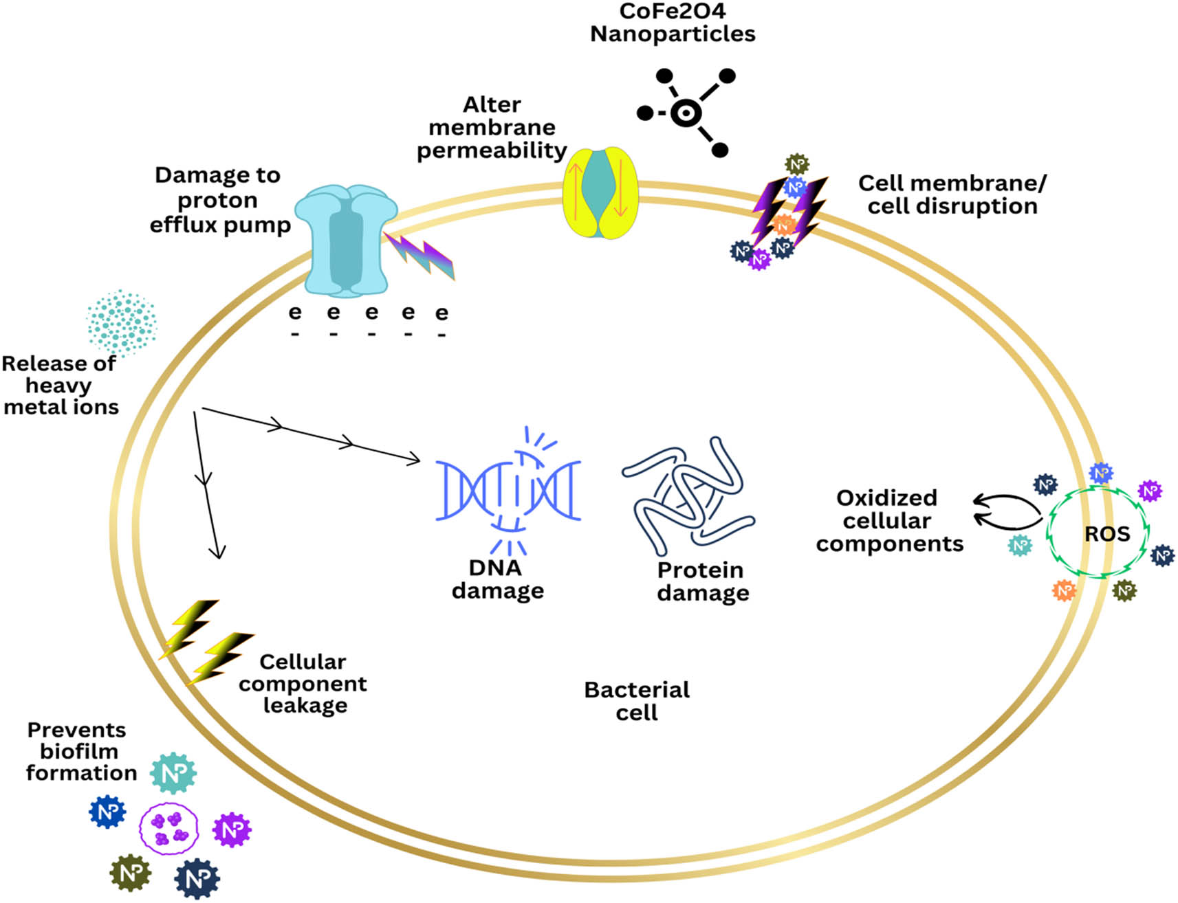
Possible antibacterial mechanism of CoFe2O4 NPs.
3.5 Effect of CoFe2O4 NPs on the biofilm-forming capabilities of bacteria and Candida
Biofilm formation is regarded as one of the key causes of antimicrobial resistance development. Biofilms serve as a barrier against antibiotics, and the increased metabolic rate of the bacteria encased within biofilm matrices also contributes to their antibiotic resistance [66]. In order to eradicate biofilms, antibacterial agents must enter the bacterial cells by penetrating the polysaccharide matrix. Nanotechnology may offer a solution to penetrate such biofilms and limit their development by applying nanoscale particles. In this study, it was observed that CoFe2O4 NPs at 0.125, 0.25, and 0.5 mg/ml inhibit the biofilm formation by 37.3, 43.3, and 37.8% for P. aeruginosa, 51.3, 55.2, and 49.9% for MRSA, and 59.8, 61.8, and 58.1% for C. albicans, respectively (Figure 11). Our results are in line with previously published research that found metal-substituted spinel cobalt ferrite NPs (Zn0.75Co0.25Fe2O4) to be effective in preventing the growth of biofilms in S. aureus, Aerococcus viridans, and Enterococcus columbae by 63.7, 57.9, and 45.5%, respectively, at concentrations of 20 ppm [67].

Effects of CoFe2O4 NPs on biofilm-forming abilities of test pathogens. (*p < 0.05, **p < 0.001).
3.6 Eradication of the established biofilms by CoFe2O4 NPs
Compared to their free-living counterparts, bacterial and candidal cells living in biofilms are more resistant to antimicrobial medications, biocides, and other chemical microcidal agents. Consequently, it is challenging to remove mature biofilms with antimicrobial treatments. In this study, the effects of CoFe2O4 NPs on the biofilm-forming abilities of test pathogens were assessed using a quantitative assay. The results observed that CoFe2O4 NPs at 0.125, 0.25, and 0.5 mg/ml inhibited the preformed biofilm by 24.7, 26.6, and 21.15% for P. aeruginosa, 32.8, 48.2, and 46.2% for MRSA, and 50.9, 57.4, and 64.49% for C. albicans, respectively (Figure 12). The present data clearly illustrate the capability of CoFe2O4 NPs to eradicate pre-existing biofilms. Based on the observed data, it is found that the CoFe2O4 NPs have penetrated the biofilm matrix and eliminated at least 50% of the preformed biofilm of all the test pathogens at a concentration of 0.5 mg/ml. This study shows, most likely for the first time, the destruction of C. albicans and bacterial preformed biofilms by CoFe2O4 NPs. Although the results of this analysis are very positive, additional in vivo research is needed to imitate the biofilms that develop during actual infections because in vivo biofilms exhibit a number of traits that are distinct from those found in most in vitro biofilms [26].

Effects of CoFe2O4 NPs on preformed biofilms of test pathogens. *p < 0.05, **p < 0.001; ***p < 0.0001).
3.7 SEM examination for visualizing the architecture of biofilms
SEM analysis was used to investigate how CoFe2O4 NPs affect the MDR-PA, MRSA, and C. albicans biofilms that had developed on the surface of the glass (Figure 13). Untreated (control) glass coverslips were discovered to be supportive of MDR-PA, MRSA, and C. albicans cell adhesion, colonization, and aggregation in large numbers (Figure 13a, c, and e). The adherence and colonization of cells in MDR-PA, MRSA, and C. albicans biofilms treated with CoFe2O4 NPs, however, significantly decreased (Figure 13b, d, and f). Additionally, dispersed cells that had lost their cellular structure and membrane proteins were also observed, illustrating the vicious destruction of the EPS matrix and biofilm architecture [56]. The SEM images agree well with the results of the biofilm inhibition assay, and it is clear that the CoFe2O4 NPs restrict bacteria and Candida from colonizing and aggregating, which led to a significant reduction of biofilm formation in the test pathogens.

SEM images showing effects of CoFe2O4 NPs on biofilm-forming abilities of MRSA (b), MDR-PA, (d) and C. albicans (f). (a), (c), and (e) are control. i.e., without CoFe2O4 NPs.
It has been suggested that the morphological transition of C. albicans between yeast and filamentous forms contributes substantially to its pathogenesis, and this transition is one of the prerequisites for biofilm formation [68]. Furthermore, it has been reported that the filamentous form also affects host tissue damage and invasion [69]. As a result, the anticandidal therapy can be boosted by targeting C. albicans’ morphological transition and dimorphism. SEM (Figure 13e and f) and optical light microscopy (Figure 14) were used in this study to determine the effect of various doses of CoFe2O4 NP on the biofilm architecture of C. albicans, specifically the transition from hyphae to yeast. Both SEM (Figure 13e) and light micrographs revealed a distinctively dense infrastructure biofilm matrix with highly accumulated yeast and hyphal cells in control (Figure 14a), whereas it greatly inhibited microcolony formation, which was primarily made up of dispersed individual cells, as was seen in CoFe2O4 NP treatment (Figure 14b–d). In addition, the true hyphae projections and hyphal network formation were also seen to drastically diminish in a dose-dependent manner (Figures 13f and 14b–d).

Effect of CoFe2O4 NPS on C. albicans phenotypic switching, surface adhesion, and hyphal development after 4 hours of incubation. (a) Control. (b), (c), and (d) micrographs illustrating the decrease in biofilm formation and hyphal development at concentrations of 0.25, 0.5, and 0.75 mg/ml CoFe2O4 NPs, respectively.
3.8 Anticancer activities
3.8.1 Impact of CoFe2O4 NPs on cancer cell viability
Based on their antibacterial and antifungal capabilities, CoFe2O4 NPs were further tested on two cancerous cell lines, i.e., HCT-116, and HeLa, and a normal non-cancerous cell, i.e., HEK-293, for their antiproliferative effects. It has been observed that CoFe2O4 NPs dose-dependently inhibit cancer growth and proliferation (Figure 15). The cell viability of HCT-116 cells was 59, 23, and 21% when treated with CoFe2O4 NPs at concentrations of 2, 10, and 20 µg/ml, respectively. The HeLa cells treated with CoFe2O4 NPs showed the cell viability of 81, 24, and 17% at concentrations of 2, 10, and 20 µg/ml, respectively. However, the cell viability of normal HEK-293 cells was found to be 81, 45, and 44% at similar doses. The findings of this investigation are in agreement with the study of El-Sayed et al. [26]. They found that CoFe2O4 NPs prepared by the fungus M. purpureus inhibited 50% of the cell proliferation of HepG-2 and MCF-7 at 45.21 and 61.86 µg/ml, respectively. In another earlier study, CoFe2O4 was found to be moderately toxic to A431 epidermoid carcinoma cells after 24 h. Then, it was gradually increased when incubated for 72 h [70]. In another investigation, 50–400 μg/ml CoFe2O4 NPs produced toxicity in HepG2 human liver cells due to ROS generation [71]. Our results suggest that CoFe2O4 NPs possess an anticancer activity against HCT-116 and HeLa cells.

Dose-dependent cytotoxic activity of CoFe2O4 NPs toward HCT-116, HeLa, and HEK- 293 cells analyzed by MTT assay. Significant variations in each cell’s viability are denoted by asterisks. (*p < 0.05; **p < 0.01; ***p < 0.001).
3.8.2 Cancer cell DNA disintegration
The exposure of CoFe2O4 NPs to HCT-116 cells produced a substantial reduction in colon cancer cells. We found that the number of DAPI-stained cells was lower in CoFe2O4 NPs-treated group compared to control cells (Figure 16b). The control cells (Figure 16a) showed normal and healthy cells, whereas the CoFe2O4 NP-treated cells showed nuclear disintegration and chromatic fragmentation, which are signs of apoptosis, or programmed cell death. When HCT-116 cells were treated with CoFe2O4 NPs, there were significantly fewer colon cancer cells than there were in the control cells. Cell death – likely from apoptosis or programmed cell death – is the cause of the decline in cancer cells. Apoptotic morphological changes, such as nuclear condensation and cellular structure damage, were visible in cells treated with CoFe2O4 NPs by DAPI staining. The control cells, on the other hand, displayed no inhibitory action and were undamaged, retaining their typical morphological and physiological structure. The results of this study are consistent with the results of the previous investigation [72]. Kanagesan et al. [73] reported that MnFe2O4 caused a number of cellular abnormalities and alterations that led to cell death after entering the breast cancer 4T1 cell [73].

The structure of cancer cells with DAPI staining after 48 h treatment. (a) Denotes untreated control, (b) denotes HCT-116 cells exposed with 20 μg/ml of CoFe2O4 NPs. Arrows show the chromatin condensation and DNA fragmentation as well as cell disintegration.
4 Conclusion
In this study, CoFe2O4 nano-spinel ferrites have been successfully synthesized by the green chemistry approach using A. vera leaf extract as a reducing and stabilizing agent. FTIR analysis revealed that polyphenolic and other functional groups present in the leaf extract play a vital role in the synthesis of NPs. The synthesized NPs showed potent antibacterial, antifungal, antibiofilm, and anticancer activities. The prepared CoFe2O4 NPs exhibited a soft ferromagnetic nature that substantially reduced the cell viability of HCT-116 and HeLa cancer cell lines, as well as the growth of yeast and drug-resistant bacteria. In addition to their antimicrobial properties, we investigated for the first time that CoFe2O4 NPs suppress the formation and colonization of preformed mature biofilms of MRSA, MDR-PA, and C. albicans. Intriguingly, light microscopic observations reveal that treatment with CoFe2O4 NPs totally inhibits the growth of true hyphae and filaments, suggesting an attenuation of C. albicans pathogenicity. The SEM analysis of C. albicans and bacterial strains reveals a noteworthy morphologic alteration in addition to substantial damage to the cellular membrane, indicating a major loss of membrane permeability that ultimately causes the cell death. The current findings highlight a novel and alternative method for producing NPs that have great biotechnological promise and could open a path for the production of NPs on an industrial scale using eco-friendly and cost-effective techniques. Therefore, the synthesized CoFe2O4 NPs can be better explored in the near future for various biomedical and therapeutic applications such as treating bacterial and fungal biofilm infections as well as the proliferation of cancer cells.
Acknowledgments
The authors extend their appreciation to the Deanship of Scientific Research at King Khalid University for funding this work through research group (large) [Project Number RGP2/54/44].
-
Funding information: This work was supported by the Deanship of Scientific Research at King Khalid University through Research Group (Large) [Project number RGP2/54/44].
-
Author contributions: All authors have accepted responsibility for the entire content of this manuscript and approved its submission.
-
Conflict of interest: The authors state no conflict of interest.
References
[1] El-Sayed ER, Mousa SA, Abdou DAM, Abo El-Seoud MA, Elmehlawy AA, Mohamed SS. Exploiting the exceptional biosynthetic potency of the endophytic Aspergillus terreus in enhancing production of Co3O4, CuO, Fe3O4, NiO, and ZnO nanoparticles using bioprocess optimization and gamma irradiation. Saudi J Biol Sci. 2022;29(4):2463–74.10.1016/j.sjbs.2021.12.019Search in Google Scholar PubMed PubMed Central
[2] Sharma RP, Raut SD, Mulani RM, Kadam AS, Mane RS. Sol–gel auto-combustion mediated cobalt ferrite nanoparticles: a potential material for antimicrobial applications. Int Nano Lett. 2019;9:141–7.10.1007/s40089-019-0268-4Search in Google Scholar
[3] Tomitaka A, Hirukawa A, Yamada T, Morishita S, Takemura Y. Biocompatibility of various ferrite nanoparticles evaluated by in vitro cytotoxicity assays using HeLa cells. J Magn Magn Mater. 2009;321(10):1482–4.10.1016/j.jmmm.2009.02.058Search in Google Scholar
[4] Tran N, Mir A, Mallik D, Sinha A, Nayar S, Webster TJ. Bactericidal effect of iron oxide nanoparticles on Staphylococcus aureus. Int J Nanomedicine. 2010;15:277–83.10.2147/IJN.S9220Search in Google Scholar PubMed PubMed Central
[5] Liu Z, Wang N, Ma L, Liu Y, Liu W, Li J, et al. Elaboration and photocatalytic properties of CoFe2O4/TiO2 composite nanowires with the side-by-side structure. Mater Res Bull. 2021;141:111354.10.1016/j.materresbull.2021.111354Search in Google Scholar
[6] Kumar CS, Mohammad F. Magnetic nanomaterials for hyperthermia-based therapy and controlled drug delivery. Adv Drug Deliv Rev. 2011;63(9):789–808. 10.1016/j.addr.2011.03.008 Search in Google Scholar PubMed PubMed Central
[7] Kumar Y, Sharma A, Shirage PM. Shape-controlled CoFe2O4 nanoparticles as an excellent material for humidity sensing. RSC Adv. 2017;7:55778–85.10.1039/C7RA11072CSearch in Google Scholar
[8] Manikandan A, Sridhar R, Antony SA, Ramakrishna S. A simple aloe vera plant-extracted microwave and conventional combustion synthesis: morphological, optical, magnetic and catalytic properties of CoFe2O4 nanostructures. J Mol Struct. 2014;5(1076):188–200.10.1016/j.molstruc.2014.07.054Search in Google Scholar
[9] Maksoud MA, El-Sayyad GS, Ashour AH, El-Batal AI, Abd-Elmonem MS, Hendawy HA, et al. Synthesis and characterization of metals-substituted cobalt ferrite [Mx Co(1-x) Fe2O4; (M = Zn, Cu and Mn; x = 0 and 0.5)] nanoparticles as antimicrobial agents and sensors for Anagrelide determination in biological samples. Mater Sci Eng C Mater Biol Appl. 2018;1(92):644–56.10.1016/j.msec.2018.07.007Search in Google Scholar PubMed
[10] Govindasamy R, Govindarasu M, Alharthi SS, Mani P, Bernaurdshaw N, Gomathi T, et al. Sustainable Green Synthesis of Yttrium Oxide (Y2O3) Nanoparticles Using Lantana camara Leaf Extracts: Physicochemical Characterization, Photocatalytic Degradation, Antibacterial, and Anticancer Potency. Nanomaterials (Basel). 2022;12(14):2393.10.3390/nano12142393Search in Google Scholar PubMed PubMed Central
[11] Hammad EN, Salem SS, Mohamed AA, El-Dougdoug W. Environmental impacts of ecofriendly iron oxide nanoparticles on dyes removal and antibacterial activity. Appl Biochem Biotechnol. 2022;194(12):6053–67.10.1007/s12010-022-04105-1Search in Google Scholar PubMed PubMed Central
[12] Salem SS, Fouda A. Green synthesis of metallic nanoparticles and their prospective biotechnological applications: an overview. Biol Trace Elem Res. 2021;199:344–70.10.1007/s12011-020-02138-3Search in Google Scholar PubMed
[13] Zhang D, Ma XL, Gu Y, Huang H, Zhang GW. Green Synthesis of Metallic Nanoparticles and Their Potential Applications to Treat Cancer. Front Chem. 2020;8:799.10.3389/fchem.2020.00799Search in Google Scholar PubMed PubMed Central
[14] Jia Z, Ren D, Zhu R. Synthesis, characterization and magnetic properties of CoFe2O4 nanorods. Mater Lett. 2012;66(1):128–31.10.1016/j.matlet.2011.08.056Search in Google Scholar
[15] Surjushe A, Vasani R, Saple DG. Aloe vera: a short review. Indian J Dermatol. 2008;53(4):163–6.10.4103/0019-5154.44785Search in Google Scholar PubMed PubMed Central
[16] Sánchez M, González-Burgos E, Iglesias I, Gómez-Serranillos MP. Pharmacological update properties of Aloe vera and its major active constituents. Molecules. 2020;25(6):1324.10.3390/molecules25061324Search in Google Scholar PubMed PubMed Central
[17] Lagashetty A, Pattar A, Ganiger SK. Synthesis, characterization and antibacterial study of Ag doped magnesium ferrite nanocomposite. Heliyon. 2019;5(5):e01760.10.1016/j.heliyon.2019.e01760Search in Google Scholar PubMed PubMed Central
[18] Al-Zahrani FA, Salem SS, Al-Ghamdi HA, Nhari LM, Lin L, El-Shishtawy RM. Green synthesis and antibacterial activity of Ag/Fe2O3 nanocomposite using Buddleja lindleyana extract. Bioengineering. 2022;9(9):452.10.3390/bioengineering9090452Search in Google Scholar PubMed PubMed Central
[19] Agnihotri S, Dhiman NK. Development of nano-antimicrobial biomaterials for biomedical applications. Advances in Biomaterials for Biomedical Applications. 2017;66:479–545.10.1007/978-981-10-3328-5_12Search in Google Scholar
[20] Kombaiah K, Vijaya JJ, Kennedy LJ, Bououdina M, Ramalingam RJ, Al-Lohedan HA. Okra extract-assisted green synthesis of CoFe2O4 nanoparticles and their optical, magnetic, and antimicrobial properties. Mater Chem Phys. 2018;204:410–9.10.1016/j.matchemphys.2017.10.077Search in Google Scholar
[21] Tatarchuk T, Liaskovska M, Kotsyubynsky V, Bououdina M. Green synthesis of cobalt ferrite nanoparticles using Cydonia oblonga extract: structural and mössbauer studies. Mol Cryst. 2018;672(1):54–66.10.1080/15421406.2018.1542107Search in Google Scholar
[22] Routray KL, Saha S, Behera D. Green synthesis approach for nano sized CoFe2O4 through aloe vera mediated sol-gel auto combustion method for high frequency devices. Mater Chem Phys. 2019;224:29–35.10.1016/j.matchemphys.2018.11.073Search in Google Scholar
[23] Banifatemi SS, Davar F, Aghabarari B, Segura JA, Alonso FJ, Ghoreishi SM. Green synthesis of CoFe2O4 nanoparticles using olive leaf extract and characterization of their magnetic properties. Ceram Int. 2021;47(13):19198–204.10.1016/j.ceramint.2021.03.267Search in Google Scholar
[24] Al-Zahrani FA, AL-Zahrani NA, Al-Ghamdi SN, Lin L, Salem SS, El-Shishtawy RM. Synthesis of Ag/Fe2O3 nanocomposite from essential oil of ginger via green method and its bactericidal activity. Biomass Convers. 2022;2:1–9.10.1007/s13399-022-03248-9Search in Google Scholar
[25] Gingasu D, Mindru I, Patron L, Calderon-Moreno JM, Mocioiu OC, Preda S, et al. Green synthesis methods of CoFe2O4 and Ag-CoFe2O4 nanoparticles using hibiscus extracts and their antimicrobial potential. J Nanomater. 2016;2016:23.10.1155/2016/2106756Search in Google Scholar
[26] El-Sayed ES, Abdelhakim HK, Zakaria Z. Extracellular biosynthesis of cobalt ferrite nanoparticles by Monascus purpureus and their antioxidant, anticancer and antimicrobial activities: yield enhancement by gamma irradiation. Mater Sci Eng C. 2020;107:110318.10.1016/j.msec.2019.110318Search in Google Scholar PubMed
[27] Velayutham L, Parvathiraja C, Anitha DC, Mahalakshmi K, Jenila M, Alasmary FA, et al. Photocatalytic and antibacterial activity of CoFe2O4 nanoparticles from Hibiscus rosa-sinensis plant extract. Nanomaterials. 2022;12(20):3668.10.3390/nano12203668Search in Google Scholar PubMed PubMed Central
[28] Ansari MA, Akhtar S, Rauf MA, Alomary MN, AlYahya S, Alghamdi S, et al. Sol–gel synthesis of dy-substituted Ni0.4Cu0.2Zn0.4(Fe2-xDyx)O4 nano spinel ferrites and evaluation of their antibacterial, antifungal, antibiofilm and anticancer potentialities for biomedical application. Int J Nanomedicine. 2021;16:5633–50.10.2147/IJN.S316471Search in Google Scholar PubMed PubMed Central
[29] Robertson JD, Rizzello L, Avila-Olias M, Gaitzsch J, Contini C, Magoń MS, et al. Purification of nanoparticles by size and shape. Sci Rep. 2016;6:27494.10.1038/srep27494Search in Google Scholar PubMed PubMed Central
[30] Shobha B, Ashwini BS, Ghazwani M, Hani U, Atwah B, Alhumaidi MS, et al. Trichoderma-mediated ZnO nanoparticles and their antibiofilm and antibacterial activities. J Fungi. 2023;9(2):133.10.3390/jof9020133Search in Google Scholar PubMed PubMed Central
[31] Qureshi F, Nawaz M, Ansari MA, Khan FA, Berekaa MM, Abubshait SA, et al. Synthesis of M-Ag3PO4,(M = Se, Ag, Ta) nanoparticles and their antibacterial and cytotoxicity study. Int JMol Sci. 2022;23(19):11403.10.3390/ijms231911403Search in Google Scholar PubMed PubMed Central
[32] Clinical and Laboratory Standards Institute (CLSI). Reference method for broth dilution antifungal susceptibility testing of yeasts; fourth informational supplement. Wayne: Clinical and Laboratory Standards Institute; 2012 (Document M27-S4).Search in Google Scholar
[33] Baig U, Gondal MA, Dastageer MA, Ansari MA, Sajid M, Falath WS. Synthesis of cadmium sulfide-tungsten trioxide nanocomposites for photo-catalytic degradation of organic pollutants and growth retardation of waterborne bacteria and biofilms. Colloids Surf A: Physicochem Eng Asp. 2020;606:125423.10.1016/j.colsurfa.2020.125423Search in Google Scholar
[34] Sahli C, Moya SE, Lomas JS, Gravier-Pelletier C, Briandet R, Hémadi M. Recent advances in nanotechnology for eradicating bacterial biofilm. Theranostics. 2022;12(5):2383–405.10.7150/thno.67296Search in Google Scholar PubMed PubMed Central
[35] GingaŞu D, Mindru I, Preda S, Calderon-Moreno JM, Daniela CC, Patron L, et al. Green synthesis of cobalt ferrite nanoparticles using plant extracts. Rev Roum Chim. 2017;62(8–9):645–53.Search in Google Scholar
[36] Kushwaha P, Chauhan P. Facile green synthesis of CoFe2O4 nanoparticles using hibiscus extract and their application in humidity sensing properties. Inorg Nano-Met Chem. 2021;10:1–8.10.1080/24701556.2021.1992432Search in Google Scholar
[37] Gheidari D, Mehrdad M, Maleki S, Hosseini S. Synthesis and potent antimicrobial activity of CoFe2O4 nanoparticles under visible light. Heliyon. 2020;6(10):e05058.10.1016/j.heliyon.2020.e05058Search in Google Scholar PubMed PubMed Central
[38] Kobylinska N, Klymchuk D, Shakhovsky A, Khainakova O, Ratushnyak Y, Duplij V, et al. Biosynthesis of magnetite and cobalt ferrite nanoparticles using extracts of “hairy” roots: preparation, characterization, estimation for environmental remediation and biological application. RSC Adv. 2021;11(43):26974–87.10.1039/D1RA04080DSearch in Google Scholar PubMed PubMed Central
[39] Yusefi M, Shameli K, Su Yee O, Teow SY, Hedayatnasab Z, Jahangirian H, et al. Green synthesis of Fe3O4 nanoparticles stabilized by a Garcinia mangostana fruit peel extract for hyperthermia and anticancer activities. Int J Nanomedicine. 2021;29:2515–32.10.2147/IJN.S284134Search in Google Scholar PubMed PubMed Central
[40] Kumar L, Kumar P, Narayan A, Kar M. Rietveld analysis of XRD patterns of different sizes of nanocrystalline cobalt ferrite. Int Nano Lett. 2013;3:8.10.1186/2228-5326-3-8Search in Google Scholar
[41] Sunny A, Aneesh Kumar KS, Karunakaran V, Aathira M, Mutta GR, Maiti KK, et al. Magnetic properties of biocompatible CoFe2O4 nanoparticles using a facile synthesis. Nano-Struct Nano-Objects. 2018;16:69–76.10.1016/j.nanoso.2018.04.003Search in Google Scholar
[42] Gu Z, Xiang X, Fan G, Li F. Facile synthesis and characterization of cobalt ferrite nanocrystals via a simple reduction−oxidation route. J Phys Chem C. 2008;112(47):18459–66.10.1021/jp806682qSearch in Google Scholar
[43] Ayyappan S, Mahadevan S, Chandramohan P, Srinivasan MP, Philip J, Raj B. Influence of Co2+ ion concentration on the size, magnetic properties, and purity of CoFe2O4 spinel ferrite nanoparticles. J Phys Chem C. 2010;114:6334.10.1021/jp911966pSearch in Google Scholar
[44] Zhang Z, Li W, Zou R, Kang W, San Chui Y, Yuen MF, et al. Layer-stacked cobalt ferrite (CoFe2O4) mesoporous platelets for high-performance lithium ion battery anodes. J Mater Chem A. 2015;3:6990–7.10.1039/C5TA00073DSearch in Google Scholar
[45] Wu LQ, Li YC, Li SQ, Li ZZ, Tang GD, Qi WH, et al. Method for estimating ionicities of oxides using O1s photoelectron spectra. AIP Adv. 2015;5(9):097210.10.1063/1.4931996Search in Google Scholar
[46] Dupin JC, Gonbeau D, Vinatier P, Levasseur A. Systematic XPS studies of metal oxides, hydroxides and peroxides. Phys Chem Chem Phys. 2000;2(6):1319–24.10.1039/a908800hSearch in Google Scholar
[47] Jiang QS, Li W, Wu J, Cheng W, Zhu J, Yan Z, et al. Electrodeposited cobalt and nickel selenides as high-performance electrocatalytic materials for dye-sensitized solar cells. J Mater Sci Mater Electron. 2019;30:9429–37.10.1007/s10854-019-01273-5Search in Google Scholar
[48] Eidi E, Kassaee MZ, Cummings PT. β-Enaminones over recyclable nano-CoFe2O4: a highly efficient solvent-free green protocol. Res Chem Intermed. 2018;44:5787–99.10.1007/s11164-018-3454-5Search in Google Scholar
[49] Modabberasl A, Pirhoushyaran T, Esmaeili-Faraj SH. Synthesis of CoFe2O4 magnetic nanoparticles for application in photocatalytic removal of azithromycin from wastewater. Sci Rep. 2022;12(1):19171.10.1038/s41598-022-21231-2Search in Google Scholar PubMed PubMed Central
[50] Shawky A, Alshaikh H. Cobalt ferrite-modified sol-gel synthesized ZnO nanoplatelets for fast and bearable visible light remediation of ciprofloxacin in water. Environ Res. 2022;205:112462.10.1016/j.envres.2021.112462Search in Google Scholar PubMed
[51] Naghizadeh A, Mohammadi-Aghdam S, Mortazavi-Derazkola S. Novel CoFe2O4@ ZnO-CeO2 ternary nanocomposite: Sonochemical green synthesis using Crataegus microphylla extract, characterization and their application in catalytic and antibacterial activities. Bioorg Chem. 2020;103:104194.10.1016/j.bioorg.2020.104194Search in Google Scholar PubMed
[52] Xavier S, Cleetus H, Nimila P, Thankachan S, Sebastian R, Mohammed EM. Structural and antibacterial properties of silver substituted cobalt ferrite nanoparticles. Res J Pharm Bio Chem Sci. 2014;5(5):364–71.Search in Google Scholar
[53] Gonelimali FD, Lin J, Miao W, Xuan J, Charles F, Chen M, et al. Antimicrobial properties and mechanism of action of some plant extracts against food pathogens and spoilage microorganisms. Front Microbiol. 2018;9:1639.10.3389/fmicb.2018.01639Search in Google Scholar PubMed PubMed Central
[54] Sanpo N, Berndt CC, Wen C, Wang J. Transition metal-substituted cobalt ferrite nanoparticles for biomedical applications. Acta Biomater. 2013;9(3):5830–7.10.1016/j.actbio.2012.10.037Search in Google Scholar PubMed
[55] Nahar A, Maria KH, Liba SI, Anwaruzzaman M, Khan MN, Islam A, et al. Surface-modified CoFe2O4 nanoparticles using Folate-Chitosan for cytotoxicity Studies, hyperthermia applications and Positive/Negative contrast of MRI. J Magn Magn Mater. 2022;554:169282.10.1016/j.jmmm.2022.169282Search in Google Scholar
[56] Ansari MA, Baykal A, Asiri S, Rehman S. Synthesis and characterization of antibacterial activity of spinel chromium-substituted copper ferrite nanoparticles for biomedical application. J Inorg Organomet Polym Mater. 2018;28(6):2316–27.10.1007/s10904-018-0889-5Search in Google Scholar
[57] Rehman S, Ansari MA, Alzohairy MA, Alomary MN, Jermy BR, Shahzad R, et al. Antibacterial and antifungal activity of novel synthesized neodymium-substituted cobalt ferrite nanoparticles for biomedical application. Processes. 2019;7(10):714.10.3390/pr7100714Search in Google Scholar
[58] Patil JS, Dhadde SB, Chandakavathe BN. Nanostructure drug delivery system is an option to solve antimicrobial drug resistance: perspective review. In: Characterization and Biology of Nanomaterials for Drug Delivery. Amsterdam: Elsevier; 2019. p. 165–97.10.1016/B978-0-12-814031-4.00007-6Search in Google Scholar
[59] Singh R, Jaisingh A, Maurya IK, Salunke DB. Design, synthesis and bio-evaluation of C-1 alkylated tetrahydro-β-carboline derivatives as novel antifungal lead compounds. Bioorg Med Chem Lett. 2020;30(3):126869.10.1016/j.bmcl.2019.126869Search in Google Scholar PubMed
[60] Rahdar A, Aliahmad M, Samani M, HeidariMajd M, Susan MA. Synthesis and characterization of highly efficacious Fe-doped ceria nanoparticles for cytotoxic and antifungal activity. Ceram Int. 2019;45(6):7950–5.10.1016/j.ceramint.2019.01.108Search in Google Scholar
[61] Mahajan P, Sharma A, Kaur B, Goyal N, Gautam S. Green synthesized (Ocimum sanctum and Allium sativum) Ag-doped cobalt ferrite nanoparticles for antibacterial application. Vacuum. 2019;161:389–97.10.1016/j.vacuum.2018.12.021Search in Google Scholar
[62] Satheeshkumar MK, Kumar ER, Srinivas C, Suriyanarayanan N, Deepty M, Prajapat CL, et al. Study of structural, morphological and magnetic properties of Ag substituted cobalt ferrite nanoparticles prepared by honey assisted combustion method and evaluation of their antibacterial activity. J Magn Magn Mater. 2018;469:691–7.10.1016/j.jmmm.2018.09.039Search in Google Scholar
[63] Naik MM, Naik HB, Nagaraju G, Vinuth M, Vinu K, Viswanath R. Green synthesis of zinc doped cobalt ferrite nanoparticles: Structural, optical, photocatalytic and antibacterial studies. Nano-Struct Nano-Objects. 2019;19:100322.10.1016/j.nanoso.2019.100322Search in Google Scholar
[64] Hashim M, Shirsath SE, Meena SS, Kotnala RK, Parveen A, Roy AS, et al. Investigation of structural, dielectric, magnetic and antibacterial activity of Cu–Cd–Ni–FeO4 nanoparticles. J Magn Magn Mater. 2013;341:148–57.10.1016/j.jmmm.2013.04.024Search in Google Scholar
[65] Alahmadi NS, Betts JW, Cheng F, Francesconi MG, Kelly SM, Kornherr A, et al. Synthesis and antibacterial effects of cobalt–cellulose magnetic Nanocomposites. RSC Adv. 2017;7:20020–6.10.1039/C7RA00920HSearch in Google Scholar
[66] Zubair M, Husain FM, Qais FA, Alam P, Ahmad I, Albalawi T, et al. Bio-fabrication of titanium oxide nanoparticles from Ochradenus arabicus to obliterate biofilms of drug-resistant Staphylococcus aureus and Pseudomonas aeruginosa isolated from diabetic foot infections. Appl Nanosci. 2021;11(2):375–87.10.1007/s13204-020-01630-5Search in Google Scholar
[67] Maksoud MA, El-Sayyad GS, Ashour AH, El-Batal AI, Elsayed MA, Gobara M, et al. Antibacterial, antibiofilm, and photocatalytic activities of metals-substituted spinel cobalt ferrite nanoparticles. Microb Pathog. 2019;127:144–58.10.1016/j.micpath.2018.11.045Search in Google Scholar PubMed
[68] Gulati M, Nobile CJ. Candida albicans biofilms: development, regulation, and molecular mechanisms. Microbes Infect. 2016;18(5):310–21.10.1016/j.micinf.2016.01.002Search in Google Scholar PubMed PubMed Central
[69] Vila T, Romo JA, Pierce CG, McHardy SF, Saville SP, Lopez-Ribot JL. Targeting Candida albicans filamentation for antifungal drug development. Virulence. 2017;8:150–8. 10.1080/21505594.2016.1197444 Search in Google Scholar PubMed PubMed Central
[70] Balakrishnan PB, Silvestri N, Fernandez-Cabada T, Marinaro F, Fernandes S, Fiorito S, et al. Exploiting unique alignment of cobalt ferrite nanoparticles, mild hyperthermia, and controlled intrinsic cobalt toxicity for cancer therapy. Adv Mater. 2020;32(45):2003712.10.1002/adma.202003712Search in Google Scholar PubMed
[71] Ahamed M, Akhtar MJ, Khan MM, Alhadlaq HA, Alshamsan A. Cobalt iron oxide nanoparticles induce cytotoxicity and regulate the apoptotic genes through ROS in human liver cells (HepG2). Colloids Surf B. 2016;148:665–73.10.1016/j.colsurfb.2016.09.047Search in Google Scholar PubMed
[72] Alfareed TM, Slimani Y, Almessiere MA, Nawaz M, Khan FA, Baykal A, et al. Biocompatibility and colorectal anti-cancer activity study of nanosized BaTiO3 coated spinel ferrites. Sci Rep. 2022;12(1):1–8.10.1038/s41598-022-18306-5Search in Google Scholar PubMed PubMed Central
[73] Kanagesan S, Aziz SB, Hashim M, Ismail I, Tamilselvan S, Alitheen NB, et al. Synthesis, characterization and in vitro evaluation of manganese ferrite (MnFe2O4) nanoparticles for their biocompatibility with murine breast cancer cells (4T1). Molecules. 2016;21(3):312.10.3390/molecules21030312Search in Google Scholar PubMed PubMed Central
© 2023 the author(s), published by De Gruyter
This work is licensed under the Creative Commons Attribution 4.0 International License.
Articles in the same Issue
- Research Articles
- Preparation of CdS–Ag2S nanocomposites by ultrasound-assisted UV photolysis treatment and its visible light photocatalysis activity
- Significance of nanoparticle radius and inter-particle spacing toward the radiative water-based alumina nanofluid flow over a rotating disk
- Aptamer-based detection of serotonin based on the rapid in situ synthesis of colorimetric gold nanoparticles
- Investigation of the nucleation and growth behavior of Ti2AlC and Ti3AlC nano-precipitates in TiAl alloys
- Dynamic recrystallization behavior and nucleation mechanism of dual-scale SiCp/A356 composites processed by P/M method
- High mechanical performance of 3-aminopropyl triethoxy silane/epoxy cured in a sandwich construction of 3D carbon felts foam and woven basalt fibers
- Applying solution of spray polyurea elastomer in asphalt binder: Feasibility analysis and DSR study based on the MSCR and LAS tests
- Study on the chronic toxicity and carcinogenicity of iron-based bioabsorbable stents
- Influence of microalloying with B on the microstructure and properties of brazed joints with Ag–Cu–Zn–Sn filler metal
- Thermohydraulic performance of thermal system integrated with twisted turbulator inserts using ternary hybrid nanofluids
- Study of mechanical properties of epoxy/graphene and epoxy/halloysite nanocomposites
- Effects of CaO addition on the CuW composite containing micro- and nano-sized tungsten particles synthesized via aluminothermic coupling with silicothermic reduction
- Cu and Al2O3-based hybrid nanofluid flow through a porous cavity
- Design of functional vancomycin-embedded bio-derived extracellular matrix hydrogels for repairing infectious bone defects
- Study on nanocrystalline coating prepared by electro-spraying 316L metal wire and its corrosion performance
- Axial compression performance of CFST columns reinforced by ultra-high-performance nano-concrete under long-term loading
- Tungsten trioxide nanocomposite for conventional soliton and noise-like pulse generation in anomalous dispersion laser cavity
- Microstructure and electrical contact behavior of the nano-yttria-modified Cu-Al2O3/30Mo/3SiC composite
- Melting rheology in thermally stratified graphene-mineral oil reservoir (third-grade nanofluid) with slip condition
- Re-examination of nonlinear vibration and nonlinear bending of porous sandwich cylindrical panels reinforced by graphene platelets
- Parametric simulation of hybrid nanofluid flow consisting of cobalt ferrite nanoparticles with second-order slip and variable viscosity over an extending surface
- Chitosan-capped silver nanoparticles with potent and selective intrinsic activity against the breast cancer cells
- Multi-core/shell SiO2@Al2O3 nanostructures deposited on Ti3AlC2 to enhance high-temperature stability and microwave absorption properties
- Solution-processed Bi2S3/BiVO4/TiO2 ternary heterojunction photoanode with enhanced photoelectrochemical performance
- Electroporation effect of ZnO nanoarrays under low voltage for water disinfection
- NIR-II window absorbing graphene oxide-coated gold nanorods and graphene quantum dot-coupled gold nanorods for photothermal cancer therapy
- Nonlinear three-dimensional stability characteristics of geometrically imperfect nanoshells under axial compression and surface residual stress
- Investigation of different nanoparticles properties on the thermal conductivity and viscosity of nanofluids by molecular dynamics simulation
- Optimized Cu2O-{100} facet for generation of different reactive oxidative species via peroxymonosulfate activation at specific pH values to efficient acetaminophen removal
- Brownian and thermal diffusivity impact due to the Maxwell nanofluid (graphene/engine oil) flow with motile microorganisms and Joule heating
- Appraising the dielectric properties and the effectiveness of electromagnetic shielding of graphene reinforced silicone rubber nanocomposite
- Synthesis of Ag and Cu nanoparticles by plasma discharge in inorganic salt solutions
- Low-cost and large-scale preparation of ultrafine TiO2@C hybrids for high-performance degradation of methyl orange and formaldehyde under visible light
- Utilization of waste glass with natural pozzolan in the production of self-glazed glass-ceramic materials
- Mechanical performance of date palm fiber-reinforced concrete modified with nano-activated carbon
- Melting point of dried gold nanoparticles prepared with ultrasonic spray pyrolysis and lyophilisation
- Graphene nanofibers: A modern approach towards tailored gypsum composites
- Role of localized magnetic field in vortex generation in tri-hybrid nanofluid flow: A numerical approach
- Intelligent computing for the double-diffusive peristaltic rheology of magneto couple stress nanomaterials
- Bioconvection transport of upper convected Maxwell nanoliquid with gyrotactic microorganism, nonlinear thermal radiation, and chemical reaction
- 3D printing of porous Ti6Al4V bone tissue engineering scaffold and surface anodization preparation of nanotubes to enhance its biological property
- Bioinspired ferromagnetic CoFe2O4 nanoparticles: Potential pharmaceutical and medical applications
- Significance of gyrotactic microorganisms on the MHD tangent hyperbolic nanofluid flow across an elastic slender surface: Numerical analysis
- Performance of polycarboxylate superplasticisers in seawater-blended cement: Effect from chemical structure and nano modification
- Entropy minimization of GO–Ag/KO cross-hybrid nanofluid over a convectively heated surface
- Oxygen plasma assisted room temperature bonding for manufacturing SU-8 polymer micro/nanoscale nozzle
- Performance and mechanism of CO2 reduction by DBD-coupled mesoporous SiO2
- Polyarylene ether nitrile dielectric films modified by HNTs@PDA hybrids for high-temperature resistant organic electronics field
- Exploration of generalized two-phase free convection magnetohydrodynamic flow of dusty tetra-hybrid Casson nanofluid between parallel microplates
- Hygrothermal bending analysis of sandwich nanoplates with FG porous core and piezomagnetic faces via nonlocal strain gradient theory
- Design and optimization of a TiO2/RGO-supported epoxy multilayer microwave absorber by the modified local best particle swarm optimization algorithm
- Mechanical properties and frost resistance of recycled brick aggregate concrete modified by nano-SiO2
- Self-template synthesis of hollow flower-like NiCo2O4 nanoparticles as an efficient bifunctional catalyst for oxygen reduction and oxygen evolution in alkaline media
- High-performance wearable flexible strain sensors based on an AgNWs/rGO/TPU electrospun nanofiber film for monitoring human activities
- High-performance lithium–selenium batteries enabled by nitrogen-doped porous carbon from peanut meal
- Investigating effects of Lorentz forces and convective heating on ternary hybrid nanofluid flow over a curved surface using homotopy analysis method
- Exploring the potential of biogenic magnesium oxide nanoparticles for cytotoxicity: In vitro and in silico studies on HCT116 and HT29 cells and DPPH radical scavenging
- Enhanced visible-light-driven photocatalytic degradation of azo dyes by heteroatom-doped nickel tungstate nanoparticles
- A facile method to synthesize nZVI-doped polypyrrole-based carbon nanotube for Ag(i) removal
- Improved osseointegration of dental titanium implants by TiO2 nanotube arrays with self-assembled recombinant IGF-1 in type 2 diabetes mellitus rat model
- Functionalized SWCNTs@Ag–TiO2 nanocomposites induce ROS-mediated apoptosis and autophagy in liver cancer cells
- Triboelectric nanogenerator based on a water droplet spring with a concave spherical surface for harvesting wave energy and detecting pressure
- A mathematical approach for modeling the blood flow containing nanoparticles by employing the Buongiorno’s model
- Molecular dynamics study on dynamic interlayer friction of graphene and its strain effect
- Induction of apoptosis and autophagy via regulation of AKT and JNK mitogen-activated protein kinase pathways in breast cancer cell lines exposed to gold nanoparticles loaded with TNF-α and combined with doxorubicin
- Effect of PVA fibers on durability of nano-SiO2-reinforced cement-based composites subjected to wet-thermal and chloride salt-coupled environment
- Effect of polyvinyl alcohol fibers on mechanical properties of nano-SiO2-reinforced geopolymer composites under a complex environment
- In vitro studies of titanium dioxide nanoparticles modified with glutathione as a potential drug delivery system
- Comparative investigations of Ag/H2O nanofluid and Ag-CuO/H2O hybrid nanofluid with Darcy-Forchheimer flow over a curved surface
- Study on deformation characteristics of multi-pass continuous drawing of micro copper wire based on crystal plasticity finite element method
- Properties of ultra-high-performance self-compacting fiber-reinforced concrete modified with nanomaterials
- Prediction of lap shear strength of GNP and TiO2/epoxy nanocomposite adhesives
- A novel exploration of how localized magnetic field affects vortex generation of trihybrid nanofluids
- Fabrication and physicochemical characterization of copper oxide–pyrrhotite nanocomposites for the cytotoxic effects on HepG2 cells and the mechanism
- Thermal radiative flow of cross nanofluid due to a stretched cylinder containing microorganisms
- In vitro study of the biphasic calcium phosphate/chitosan hybrid biomaterial scaffold fabricated via solvent casting and evaporation technique for bone regeneration
- Insights into the thermal characteristics and dynamics of stagnant blood conveying titanium oxide, alumina, and silver nanoparticles subject to Lorentz force and internal heating over a curved surface
- Effects of nano-SiO2 additives on carbon fiber-reinforced fly ash–slag geopolymer composites performance: Workability, mechanical properties, and microstructure
- Energy bandgap and thermal characteristics of non-Darcian MHD rotating hybridity nanofluid thin film flow: Nanotechnology application
- Green synthesis and characterization of ginger-extract-based oxali-palladium nanoparticles for colorectal cancer: Downregulation of REG4 and apoptosis induction
- Abnormal evolution of resistivity and microstructure of annealed Ag nanoparticles/Ag–Mo films
- Preparation of water-based dextran-coated Fe3O4 magnetic fluid for magnetic hyperthermia
- Statistical investigations and morphological aspects of cross-rheological material suspended in transportation of alumina, silica, titanium, and ethylene glycol via the Galerkin algorithm
- Effect of CNT film interleaves on the flexural properties and strength after impact of CFRP composites
- Self-assembled nanoscale entities: Preparative process optimization, payload release, and enhanced bioavailability of thymoquinone natural product
- Structure–mechanical property relationships of 3D-printed porous polydimethylsiloxane films
- Nonlinear thermal radiation and the slip effect on a 3D bioconvection flow of the Casson nanofluid in a rotating frame via a homotopy analysis mechanism
- Residual mechanical properties of concrete incorporated with nano supplementary cementitious materials exposed to elevated temperature
- Time-independent three-dimensional flow of a water-based hybrid nanofluid past a Riga plate with slips and convective conditions: A homotopic solution
- Lightweight and high-strength polyarylene ether nitrile-based composites for efficient electromagnetic interference shielding
- Review Articles
- Recycling waste sources into nanocomposites of graphene materials: Overview from an energy-focused perspective
- Hybrid nanofiller reinforcement in thermoset and biothermoset applications: A review
- Current state-of-the-art review of nanotechnology-based therapeutics for viral pandemics: Special attention to COVID-19
- Solid lipid nanoparticles for targeted natural and synthetic drugs delivery in high-incidence cancers, and other diseases: Roles of preparation methods, lipid composition, transitional stability, and release profiles in nanocarriers’ development
- Critical review on experimental and theoretical studies of elastic properties of wurtzite-structured ZnO nanowires
- Polyurea micro-/nano-capsule applications in construction industry: A review
- A comprehensive review and clinical guide to molecular and serological diagnostic tests and future development: In vitro diagnostic testing for COVID-19
- Recent advances in electrocatalytic oxidation of 5-hydroxymethylfurfural to 2,5-furandicarboxylic acid: Mechanism, catalyst, coupling system
- Research progress and prospect of silica-based polymer nanofluids in enhanced oil recovery
- Review of the pharmacokinetics of nanodrugs
- Engineered nanoflowers, nanotrees, nanostars, nanodendrites, and nanoleaves for biomedical applications
- Research progress of biopolymers combined with stem cells in the repair of intrauterine adhesions
- Progress in FEM modeling on mechanical and electromechanical properties of carbon nanotube cement-based composites
- Antifouling induced by surface wettability of poly(dimethyl siloxane) and its nanocomposites
- TiO2 aerogel composite high-efficiency photocatalysts for environmental treatment and hydrogen energy production
- Structural properties of alumina surfaces and their roles in the synthesis of environmentally persistent free radicals (EPFRs)
- Nanoparticles for the potential treatment of Alzheimer’s disease: A physiopathological approach
- Current status of synthesis and consolidation strategies for thermo-resistant nanoalloys and their general applications
- Recent research progress on the stimuli-responsive smart membrane: A review
- Dispersion of carbon nanotubes in aqueous cementitious materials: A review
- Applications of DNA tetrahedron nanostructure in cancer diagnosis and anticancer drugs delivery
- Magnetic nanoparticles in 3D-printed scaffolds for biomedical applications
- An overview of the synthesis of silicon carbide–boron carbide composite powders
- Organolead halide perovskites: Synthetic routes, structural features, and their potential in the development of photovoltaic
- Recent advancements in nanotechnology application on wood and bamboo materials: A review
- Application of aptamer-functionalized nanomaterials in molecular imaging of tumors
- Recent progress on corrosion mechanisms of graphene-reinforced metal matrix composites
- Research progress on preparation, modification, and application of phenolic aerogel
- Application of nanomaterials in early diagnosis of cancer
- Plant mediated-green synthesis of zinc oxide nanoparticles: An insight into biomedical applications
- Recent developments in terahertz quantum cascade lasers for practical applications
- Recent progress in dielectric/metal/dielectric electrodes for foldable light-emitting devices
- Nanocoatings for ballistic applications: A review
- A mini-review on MoS2 membrane for water desalination: Recent development and challenges
- Recent updates in nanotechnological advances for wound healing: A narrative review
- Recent advances in DNA nanomaterials for cancer diagnosis and treatment
- Electrochemical micro- and nanobiosensors for in vivo reactive oxygen/nitrogen species measurement in the brain
- Advances in organic–inorganic nanocomposites for cancer imaging and therapy
- Advancements in aluminum matrix composites reinforced with carbides and graphene: A comprehensive review
- Modification effects of nanosilica on asphalt binders: A review
- Decellularized extracellular matrix as a promising biomaterial for musculoskeletal tissue regeneration
- Review of the sol–gel method in preparing nano TiO2 for advanced oxidation process
- Micro/nano manufacturing aircraft surface with anti-icing and deicing performances: An overview
- Cell type-targeting nanoparticles in treating central nervous system diseases: Challenges and hopes
- An overview of hydrogen production from Al-based materials
- A review of application, modification, and prospect of melamine foam
- A review of the performance of fibre-reinforced composite laminates with carbon nanotubes
- Research on AFM tip-related nanofabrication of two-dimensional materials
- Advances in phase change building materials: An overview
- Development of graphene and graphene quantum dots toward biomedical engineering applications: A review
- Nanoremediation approaches for the mitigation of heavy metal contamination in vegetables: An overview
- Photodynamic therapy empowered by nanotechnology for oral and dental science: Progress and perspectives
- Biosynthesis of metal nanoparticles: Bioreduction and biomineralization
- Current diagnostic and therapeutic approaches for severe acute respiratory syndrome coronavirus-2 (SARS-COV-2) and the role of nanomaterial-based theragnosis in combating the pandemic
- Application of two-dimensional black phosphorus material in wound healing
- Special Issue on Advanced Nanomaterials and Composites for Energy Conversion and Storage - Part I
- Helical fluorinated carbon nanotubes/iron(iii) fluoride hybrid with multilevel transportation channels and rich active sites for lithium/fluorinated carbon primary battery
- The progress of cathode materials in aqueous zinc-ion batteries
- Special Issue on Advanced Nanomaterials for Carbon Capture, Environment and Utilization for Energy Sustainability - Part I
- Effect of polypropylene fiber and nano-silica on the compressive strength and frost resistance of recycled brick aggregate concrete
- Mechanochemical design of nanomaterials for catalytic applications with a benign-by-design focus