
Major ozonated autohemotherapy promoted functional recovery following spinal cord injury in adult rats via the inhibition of oxidative stress and inflammation
-
Liwei Xia


Abstract
This study sought to explore the value of major ozonated autohemotherapy (MOA) as a treatment for spinal cord injury (SCI) in a rat model system. In total, 54 female Sprague-Dawley rats were randomized into sham-operated, SCI model, and MOA treatment groups. We found that relative to the SCI model group, rats that underwent MOA treatment exhibited improved locomotor scores on days 14, 21, and 28 after injury (p < 0.05) together with reduced residual urine on days 5, 7, 14, and 21 after injury (p < 0.05). MOA treatment also lowered proinflammatory TNF-α, IL-1α, and C1q levels on day 3 post-injury (p < 0.05), decreased malondialdehyde levels, and enhanced superoxide dismutase activity (p < 0.001). Activated astrocytes in MOA-treated rats exhibited larger soma and higher levels of extracellular matrix secretion, whereas reactive microglia in the MOA group presented with a ramified morphology in contrast to the amoeboid morphology exhibited by these cells in SCI model rats. MOA offers potential value as a means of protecting spinal cord integrity, potentially through anti-inflammatory, antioxidant, and regulatory effects that shape the polarization of astrocytes and microglia.
1 Introduction
Spinal cord injury (SCI) represents an intractable medical issue that remains difficult to effectively manage. While countless studies have explored the pathogenesis of SCI and associated molecular mechanisms, treatment options for SCI patients remain limited and there is no clinical consensus regarding their application [1]. Following initial injury, inflammatory and oxidative stress in response to spinal trauma can induce the apoptotic or necrotic death of neurons while driving the formation of a spinal cord cavity, ultimately compromising spinal cord functionality [2,3]. In this pathological context, microglia can differentiate into deleterious M1-type microglia that secrete inflammatory factors including TNF-α, IL-1β, proteolytic enzymes, and nitric oxide synthase, ultimately inducing unrestrained inflammation that aggravates neuronal death and associated tissue damage [4,5]. Efforts to prevent severe inflammatory responses and oxidative stress induction following SCI have thus emerged as the preferred approach to treating SCI cases.
Medical ozone has been reported to be a safe and effective non-pharmacological treatment option with antioxidant, anti-inflammatory, and antibacterial properties [6,7,8,9]. Major ozonated autohemotherapy (MOA) is a treatment approach wherein autologous blood samples are mixed with medical ozone (containing O2 and O3) followed by gradual infusion back into the host. MOA has been used to treat a range of conditions including osteonecrosis, acute cerebral infarction, lumbar disc herniation, vascular, and immunological diseases [8,9,10,11,12].
MOA therapy can effectively alleviate oxidative stress in emergency settings, and can also reportedly protect against cytokine storm induction when treating COVID-19, thereby providing a promising means of preventing hospitalized COVID-19 patient mortality [13,14]. Ozone can also alleviate neuronal damage following acute cerebral infarction through oxygen free radical scavenging and the activation of antioxidant enzymes. Yu et al. [15] found that a patient who was paraplegic following a spontaneous spinal epidural hematoma exhibited significant lower extremity muscle contractions 5 days following MOA treatment, ultimately achieving 3/5 strength in their lower extremities after two rounds of treatment. Ameli et al. [16] suggested that ozone therapy may be an effective therapeutic option for patients with multiple sclerosis owing to its ability to alleviate oxidative stress and inflammation while increasing blood oxygen concentrations. Tural Emon et al. [17] further observed accelerated SCI healing in rodents that were subjected to ozone therapy, suggesting that it may represent a valuable complementary means of treating SCI patients. However, the ability of MOA to promote the repair of spinal cord damage through the suppression of inflammation and oxidative stress remains to be studied and documented in detail.
In the present study, MOA was found to effectively contribute to motor function recovery in adult rats following SCI. At the mechanistic level, MOA suppressed inflammation, prevented the M1 differentiation of microglia, and effectively promoted the growth of neuronal axons and dendrites. As such, MOA holds promise as a novel treatment for SCI.
2 Materials and methods
2.1 Establishment of the cellular OGD model
Primary spinal cord neurons were isolated and cultured. Female SD rats were obtained from the Animal Center of the University of Guangxi Medical University. Fourteen days after the start of pregnancy, the rats were anesthetized in a sterile environment. The spinal cord of the embryos was harvested, digested with 0.125% trypsin at 37°C for 15 min, and then digested with an equal volume of an inoculant solution (Dulbecco’s modified eagle medium (DMEM) containing 10% fetal bovine serum and 10% equine serum; Sigma-Aldrich, St Louis, MO, USA). The cells were then centrifuged at 1,000 rpm for 5 min (centrifugation radius 10 cm) and the middle layer containing single cells was collected. These cells were inoculated into polylysine-coated sterile six-well plates (2 ml/well) at a density of 1 × 10⁵ cells/ml. When the purity of the neuron cells was greater than 90%, the culture supernatant was removed and the cells were rinsed three times with normal saline, then added 5% CO2–95% N2 sugar-free Earle’s balanced salt solution. Lastly, the cells were placed in an anoxic incubator at 37°C (5% CO2–95% N2) and incubated for 2 h, after which they were returned to the neuron culture medium and grown at 37°C with 5% CO2–95% air. The cells were then divided into the control, OGD, and OGD + MOA groups.
-
Ethical approval: The research related to animal use has been complied with all the relevant national regulations and institutional policies for the care and use of animals and has been approved by the Ethics Committee of the Second Affiliated Hospital of Soochow University.
2.2 Animals model establishment
Adult female Sprague-Dawley rats (250–270 g) were obtained from the Animal Center of the University of Guangxi Medical University in Nanning, China. An experimental model of SCI was established using Allen’s method. Briefly, rats were intraperitoneally injected with 1% pentobarbital sodium (60 mg/kg) and fixed in the prone position, after which a longitudinal incision was used to expose the T10 spinous process, followed by the excision of the T10 lamina and the exposure of the spinal cord. The exposed spinal cord (approximately 4 mm × 10 mm) was then impacted using a multi-center animal spinal cord injury study (MASCIS) impounder to hit the exposed spinal cord (10 g × 6 cm). Successful SCI modeling was indicated by rapid dural swelling, bruising, lower extremity convulsions, and a tail-wagging reflex. After surgery, rats received intramuscular injections of penicillin (0.8 mg/g) for 3 days. The bladder of each rat was emptied twice per day until autonomous urination had resumed. Rats in the sham control group underwent T9-10 laminectomy without SCI. The procedures were approved by the Ethics Committee of the Second Affiliated Hospital of Soochow University.
2.3 Ozonated autohemotherapy
Pure oxygen was used to produce ozone with a medical ozone generator (Kasener-Praxisbedarf Gmbh, Rastatt, Germany). Samples of blood (1 ml) were collected from the tail vein and immediately mixed with the produced ozone for 5 min, followed by the re-infusion of the ozonated blood through the tail vein. MOA was performed once per day on 3 consecutive days.
2.4 Behavioral testing
Two investigators independently performed rat behavioral assessments with the BBB Locomotor Rating Scale prior to surgery and on days 1, 3, 7, 14, 21, and 28 post-SCI. Each rat was analyzed three times, with the average value being reported.
2.5 Bladder function analyses
The bladders of injured rats were manually emptied twice per day after injury, with the obtained urine being collected as a measure of bladder function. Residual urine volumes were measured for each rat on days 1, 2, 3, 5, 6, 14, and 21 post-injury.
2.6 MOA treatment of RAW264.7 cells
The mouse macrophage cell line RAW264.7 was purchased from Wuhan Procell Life Science & Technology Co., Ltd. The cells (2.0 × 10⁴/well) were plated in 24-well plates in DMEM containing 10% fetal bovine serum, divided into the control, lipopolysaccharide (LPS) + phosphate buffered saline (PBS), and LPS + MOA groups, and treated with anti-CD86 (ab239075, Abcam, UK) and anti-CD206 (anti-mannoreceptor antibody, ab125028, Abcam). Macrophage polarization was observed by confocal laser scanning microscopy (CLSM, LSM800 with Airyscan, Zeiss, Germany).
2.7 Histological staining
Sections of spinal cord tissue from the 10th thoracic segment were collected, fixed using paraformaldehyde, paraffin-embedded, and cut into serial 5 µm sections followed by hematoxylin and eosin (H&E) staining. Sections were then imaged using a light microscope (XSP-C204, CIC).
2.8 NeuN/4′,6-diamidino-2-phenylindole (DAPI) staining
After OGD treatment, NSC cells were fixed with 4% paraformaldehyde solution for 30 min and then permeabilized with 0.1% Triton X-100 for 10 min. FITC-phalloidin (Yeasen, Shanghai, China) and βIII Tubulin (Yeasen) were used to stain actin in the cytoskeleton, and the nuclei were counterstained with DAPI for 5 min at 37°C. The cells were examined and imaged using a laser-scanning confocal microscope. Cell numbers and morphology were analyzed by ImageJ software.
2.9 Western immunoblotting
Samples of spinal cord tissue (1 cm long) were isolated and lysed on ice in a lysis buffer supplemented with protease and phosphatase inhibitors. A bicinchoninic acid kit was used to assess protein concentrations, after which proteins were separated via 10 or 12.5% SDS-PAGE and transferred onto polyvinylidene fluoride membranes. Blots were then blocked for 1 h using 5% BSA followed by incubation overnight with antibodies specific for TNF-α (1:500; Novusbio), IL-1α (1:1,000; Novusbio), C1q (1:1,000; CST), anti-HO-1 (1:10,000; Abcam), anti-Nrf2 (1:500; Thermo), or SOD (1:1,000; CST) at 4°C. After probing for 1 h at room temperature using HRP-conjugated secondary antibodies, ImageJ (v1.48) was used to analyze densitometric values for detected protein bands, with β-actin serving as a loading control.
2.10 PCR
TRIzol (Thermo Fisher Scientific, MA, USA) was used to extract RNA from spinal cord tissue samples based on provided directions. The QuantiNova SYBR Green PCR Kit (QIAGEN) and an ABI 7500 platform (Applied Biosystems, USA) were used for qPCR analyses, with β-actin as a normalization control. The reaction conditions were: 95°C for 10 min, 95°C for 10 s, 60°C for 20 s, 72°C for 30 s, 40 cycles. The 2–ΔΔCt method was used to compare relative gene expression levels across treatment groups. Primer sequences used for this study are listed below (Table 1).
Primer sequences of TNF-α, Rat-IL-1α, Rat-IL-1β, Rat-IL6, HO-1, Nrf2, Rat-C1q, and Rat-β-actin
| Gene | Forward | Reverse |
|---|---|---|
| TNF-α | ATGGGCTCCCTCTCATCAGT | TGGTGGTTTGCTACGACGTG |
| Rat-IL-1α | CGCTTGAGTCGGCAAAGAAATC | AGAGACAGATGGTCAATGGCAG |
| Rat-IL-1β | GATGATGACGACCTGCTAGTGTGT | TTGGCTTATGTTCTGTCCATTGAG |
| Rat-IL6 | CTTCCAGCCAGTTGCCTTCTT | GGTCTGTTGTGGGTGGTATCCT |
| HO-1 | TCAAGGCCTCAGACAAATCC | ACAACCAGTGAGTGGAGCCT |
| Nrf2 | TGCCTCCAAAGGATGTCAAT | CCTCTGCTGCAAGTAGCCTC |
| Rat-C1q | GACCACGGAGGCAGGAACATC | AATTCCTGCAACCCCGTCCT |
| Rat-β-actin | GGTGGGGCGCCCCAGGCACCA | GCTCCTTAATGTCACGCACGA |
2.11 Malondialdehyde (MDA) and superoxide dismutase (SOD) activity assays
Levels of MDA (nmol/l) and SOD activity (U/mg) in spinal cord tissue were assessed with commercial kits (Nanjing Jiancheng Biotech, Nanjing, China). These spinal cord samples were collected 3 days following SCI, suspended in PBS (pH 7.4), homogenized completely with a homogenizer, and centrifuged at 3,000 rpm for 20 min, and supernatants were collected for analysis.
2.12 Immunofluorescent and immunohistochemical staining
Changes in astrocyte and microglial morphology were assessed on day 3 post-SCI via GFPA and Iba immunostaining. Briefly, 4-µm tissue sections were rinsed with PBS, blocked with 3% BSA, and treated with an autofluorescence quenching kit (G1221, ServieBio). These sections were then incubated overnight with primary rabbit anti-GFAP (Abcam, ab33922) or rabbit anti-Iba1 antibody (Abcam, ab178847) for immunofluorescent or immunohistochemical staining, respectively. Sections were then proved with secondary antibody goat Anti-Rabbit IgG (HRP) (1:500, GB23303, ServiceBio) for 50 min at room temperature, followed by visualization with a fluorescence microscope (Nikon Eclipse C1, Japan). Changes in NSC morphology were assessed via GFPA and Iba immunostaining.
2.13 Statistical analysis
In this experiment, the data were statistically analyzed by Graphpad Prism, and the mean ± standard deviation was used to represent the data. The t-test was used for the comparison between the two groups, and the data conforming to the normal distribution were used. The non-parametric Wilcoxon rank-sum test was used for the non-parametric data that did not conform to the normal distribution. p < 0.05, which was considered statistically significant. In this experiment, the western blot band plot was processed using ImageJ image analysis software.
3 Results
3.1 MOA treatment restores locomotor function and bladder function following SCI
Locomotor recovery after SCI was initially assessed in experimental rats using BBB scores. BBB scores for rats in the SCI group were very low after injury with no significant differences between the MOA and SCI groups for the first week post-injury. However, these scores were significantly higher for rats in the MOA treatment group on days 7, 14, 21, and 28 post-injury (9.4 ± 1.7, 13.8 ± 1.5, 14.1 ± 1.6, 14.9 ± 1.9) relative to the SCI group (5.6 ± 1.0, 10.0 ± 1.6, 11.1 ± 1.9, 11.8 ± 1.3) (Figure 1a). This suggests that MOA treatment significantly enhanced locomotor recovery in these rats (p < 0.05, n = 8/group).

(a) BBB scores of each group, *p < 0.05 versus the SCI group, n = 8. (b) Residual urine volumes in each group, *p < 0.05 versus the SCI group, n = 8. (c) HE staining of spinal cord sections in the sham, SCI, and MOA groups. (d) Cavity area of HE staining in each group, *p < 0.05 versus the SCI group.
The recovery of bladder function in all experimental groups was assessed based on residual urine volume. At 1 day post-injury, the residual urine volume in the SCI and MOA groups was 5 ml, with this value decreasing over time, particularly in the MOA group (Figure 1b). While no significant differences in residual urine volume were evident between the MOA and SCI groups at early time points, on days 5, 7, 14, and 21 post-injury this volume was significantly larger in the SCI group relative to the MOA group (p < 0.05).
3.2 MOA protects against necrotic tissue damage in the spinal cord
On day 3 post-injury, histologic analyses of rat spinal cord tissue samples were performed. Morphological analyses revealed extensive edema, hemorrhage, necrosis, and cavity formation in the SCI model group, whereas these necrotic phenotypes were markedly blunted in the MOA treatment group, suggesting that MOA can protect against severe damage following SCI (Figure 1c and d).
3.3 MOA suppresses proinflammatory cytokine expression
Proinflammatory cytokines serve as important mediators of the pathogenesis of SCI. Accordingly, Western immunoblotting and qPCR were used to evaluate TNF-α, IL-1α, C1q, and IL-6 expression in these rats. All of these cytokines were upregulated in the spinal cord of SCI model rats, while they were effectively suppressed by MOA treatment (Figure 2).
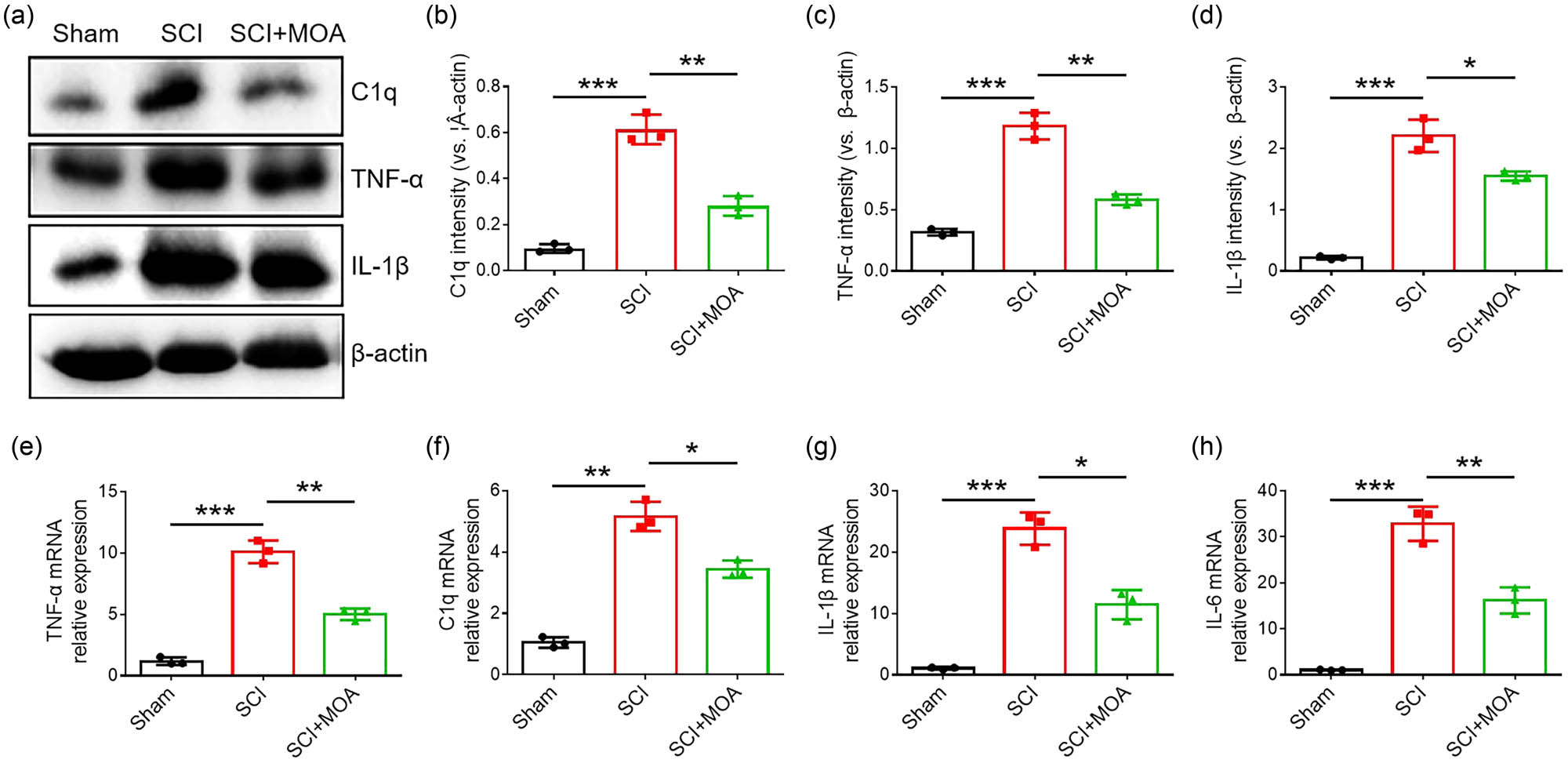
(a) Protein expression of C1q, TNF-α, and IL-1α in the spinal cords of rats in the three groups at 3 dpi. (b) The relative level of C1q protein expression in the three groups at 3 dpi, **p < 0.05. (c) The relative level of TNF-α protein expression in the three groups at 3 dpi, **p < 0.05. (d) The relative level of IL-1β protein expression in the three groups at 3 dpi, **p < 0.01. (e) TNF-α mRNA expression in the three groups at 3 dpi, **p < 0.05. (f) C1q mRNA expression in the three groups at 3 dpi, **p < 0.05. (g) IL-1β mRNA expression in the three groups at 3 dpi, **p < 0.05. (h) IL-6 mRNA expression in the three groups at 3 dpi, **p < 0.05.
3.4 MOA suppresses the maturation of M1 microglia in SCI model rats
The expression of CD86 was increased while that of CD206 was decreased significantly after treatment of macrophages with lipopolysaccharide (Figure 3a and b), MOA thus effectively regulated polarization of the macrophages. Under basal conditions, the microglia found within the spinal cord were ramified, exhibiting a characteristic branched morphology with small spherical cell bodies. After SCI, these native microglia were activated and differentiated into M1 microglia with amoeboid-like cell bodies with few or no ramified processes (Figure 3c). Relative to the SCI model group, significantly fewer amoeboid microglia were evident in the MOA-treated group, with M2-like microglia with more ramified processes instead evident in this group (Figure 3c and d). These results indicated the ability of MOA to suppress M1 maturation of microglia in the spinal cord.
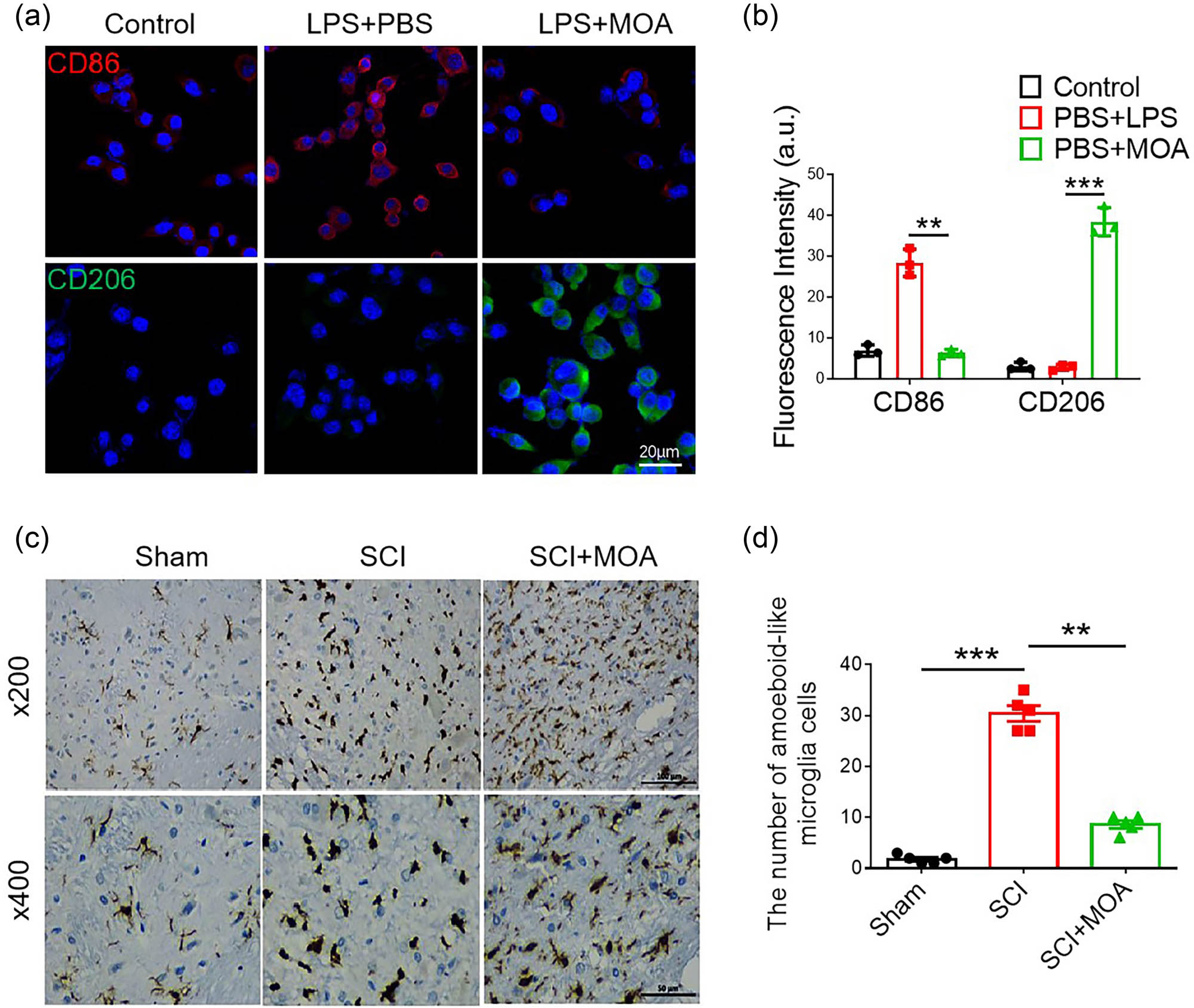
(a) Immunofluorescence-stained macrophages in the control, LPS + PBS, and LPS + MOA groups. M1 macrophage (CD86: red), M2 macrophage (CD206: green). (b) Fluorescence intensity of the control, LPS + PBS, and LPS + MOA groups, **, ***p < 0.05. (c) Immunohistochemical staining of Iba1 in Sham, SCI, and MOA groups 3 day following contusion. (d) The number of amoeboid-like microglia 3 day following contusion. **, ***p < 0.01, n = 5. Scale bar = 100 µm, scale bar = 50 µm.
3.5 MOA alleviates oxidative stress within the spinal cord following SCI
A significant increase in HO-1 levels was evident in the MOA group relative to the SCI group (p < 0.01) (Figure 4a and b), while SOD levels were reduced in the MOA group (p < 0.01) (Figure 4a and c). The respective SOD vigor levels in the sham, SCI, and MOA groups were 66.51 ± 3.68, 11.97 ± 2.49, and 53.42 ± 3.19 U/mg, with significant suppression of SOD activity in the SCI and MOA groups relative to the sham group (p < 0.01) but a significantly higher level of SOD activity in the MOA group relative to the SCI group (p < 0.01) (Figure 4d). Respective MDA levels in the sham, SCI, and MOA groups were 4.49 ± 0.21, 1.55 ± 0.29, and 3.58 ± 0.23 nmol/mg. These levels were highest in the SCI group, while in the MOA group, these levels were significantly below those in the SCI group but above those in the sham control group (p < 0.01) (Figure 4e). The mRNA expression of HO-1 was increased significantly in the SCI group and decreased significantly after MOA treatment (p < 0.05) (Figure 4f). The mRNA expression of SOD and Nrf2 was decreased significantly in the SCI group and increased significantly after MOA treatment (p < 0.05) ( Figure 4g and h).
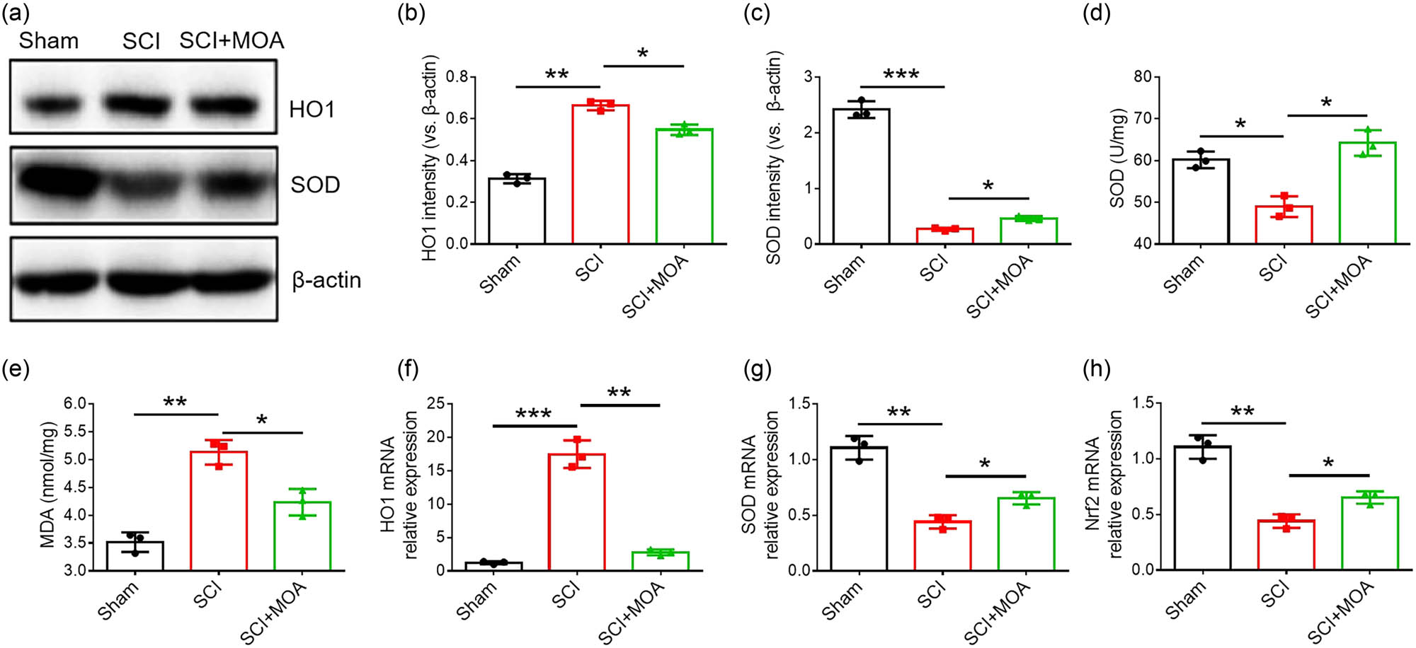
(a) Levels of HO-1 and SOD in the spinal cords of rats in the three groups at 3 dpi. (b) Relative levels of HO-1 level in the three groups at 3 dpi, *p < 0.01. (c) Relative levels of SOD expression in the three groups at 3 dpi, *p < 0.01. (d) SOD vigor level comparison at 3 day after contusion, *p < 0.05. (e) MDA vigor level comparison at 3 day after contusion, *p < 0.01 versus sham group and **p < 0.05 versus SCI group. (f) HO-1 mRNA expression in the three groups at 3 dpi, *p < 0.05. (g) SOD mRNA expression in the three groups at 3 dpi, *p < 0.05. (h) Nrf2 mRNA expression in the three groups at 3 dpi, *p < 0.05.
3.6 MOA promotes the growth of neurons and inhibits the formation of glial scar
Immunofluorescent staining showed that the number of neurons was significantly decreased after SCI but was increased significantly after MOA (Figure 5a and b). The numbers of branches and primary dendrites, as well as the length of the longest dendrites, were significantly reduced in neurons in the OGD group, while showing significant increases after MOA treatment (Figure 5c and d). At baseline, astrocytes in the spinal cord of rats in the sham control group exhibited small cell bodies and thin processes. Following SCI, these astrocytes were activated and exhibited pronounced morphological changes. Immunofluorescent staining indicated that astrocytes began undergoing polarization 3 days post-SCI, with accompanying cell body swelling and the thickening of cell processes. No significant morphological differences were observed between the SCI and MOA groups at 3 days post-injury. By day 21, there were significantly more astrocytes and significantly higher GFAP expression levels consistent with glial scar formation. Relative to the SCI group, astrocytes from MOA-treated rats exhibited larger cell bodies and elevated intracellular matrix levels (Figure 5e and f).
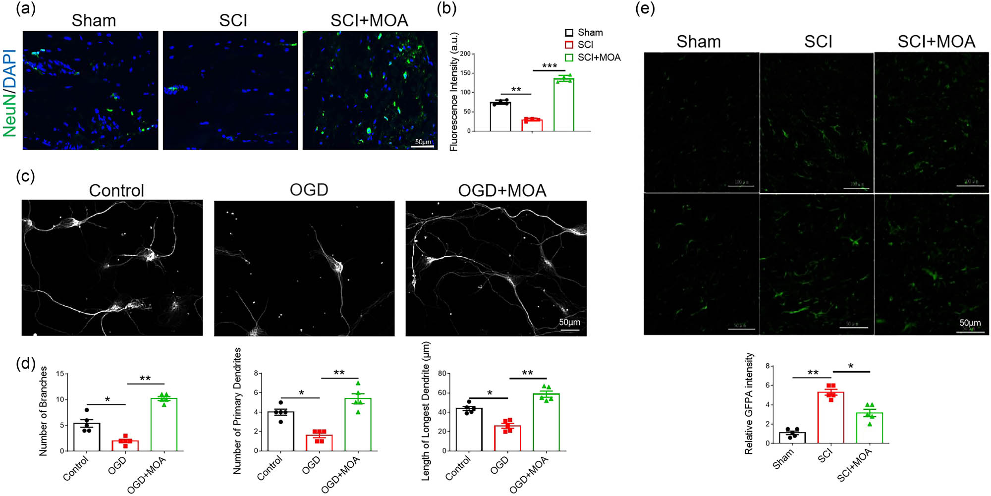
(a) Immunofluorescence-stained primary NSCs in the control, SCI, and SCI + MOA groups, Neurons (NeuN: green), Nuclei (DAPI: blue). (b) Fluorescence intensities in the control, SCI, and SCI + MOA groups, **p < 0.05, ***p < 0.05. (c) Neurons are labeled with anti-βIII-tubulin antibody for neuronal microtubules. (d) Numbers of branches in the control, OGD, and OGD + MOA groups, *p < 0.05, **p < 0.05. Numbers of primary dendrites in the control, OGD, and OGD + MOA groups, *p < 0.05, **p < 0.05. Lengths of longest dendrites in the control, OGD, and OGD + MOA groups, *p < 0.05, **p < 0.05. (e) Resting astrocytes in the Sham group, activated astrocytes in the SCI group at 3 d after contusion, and activated astrocytes in the MOA group at 21 d after contusion. Scale bar = 100 µm, scale bar = 50 µm. (f) Relative GFAP intensities in the three groups at 21 d after contusion, *p < 0.05, **p < 0.05.
4 Discussion
Prior reports have found ozone to provide therapeutic benefits in the context of SCI, but the underlying molecular mechanisms have yet to be established [17]. While one previous study intraperitoneally injected rats with ozone [18], an MOA treatment strategy was instead employed in the present study given that MOA is widely used to administer ozone in clinical settings and is an established, safe procedure [19]. This study demonstrated that (1) MOA promoted the recovery of movement and bladder function, as well as the repair of local spinal cord tissue structure in the model group, (2) MOA alleviated oxidative stress resulting from SCI and reduced the expression of inflammatory factors such as TNF-α, IL-1β, and IL-6, as well as C1q, and (3) MOA inhibited the maturation of M1 microglia and the formation of glial scars while promoting the growth of neurons.
Relative to the SCI model group, rats that underwent MOA treatment exhibited significant improvements in BBB scores (p < 0.05), suggesting that MOA can promote motor recovery following SCI. Histological staining confirmed that while rats in the SCI group exhibited extensive spinal hemorrhage, edema, and cavity formation, these effects were significantly blunted in MOA-treated rats. MOA thus appears to protect against spinal tissue necrosis following traumatic SCI.
Christie et al. [20] found that acute oxidative stress can persist for 120 h following acute SCI in a rat model system. In clinical settings, methylprednisolone is prescribed within 24 h as a treatment for SCI, but other therapeutic options are vital to mitigate such oxidative stress after this 24 h post-injury interval has elapsed. Here, MOA treatment was found to enhance SOD activity while suppressing MDA levels. The transcription factor Nrf2 is associated with processes such as exogenous and endogenous metabolism, the inflammatory response, and oxidative stress. Huang et al. [21] found that up-regulation of the Nrf2/HO-1 pathway in diabetic rats could enhance antioxidant capacity. In this study, Nrf2 and HO-1expression was found to be significantly increased after MOA treatment in SCI rats, which indicated that MOA increased SOD activity and reduced the membrane lipid peroxidation possibly through up-regulating the expression of the Nrf2/HO-1 pathway. With the enhancement of the antioxidant capacity in the spinal cord, the secondary injury of spinal cord tissue was alleviated.
Following SCI incidence, microglia can rapidly proliferate and differentiate into neurotoxic M1 cells [22]. In the SCI model group, microglia presented with amoeboid morphology whereas in the MOA treatment group, these cells exhibited ramified processes. This suggests that the neuroprotective benefits of MOA treatment are associated with the M2 differentiation of microglia. M1 microglia can induce the development of A1 astrocytes by secreting proinflammatory factors including C1q, TNF-α, IL-6, and IL-1β, thereby modulating neuronal apoptotic activity [23]. In this study, CD86 levels were significantly increased in LPS-treated macrophages, with the macrophages exhibiting the M1 phenotype, while significant increases in CD206 accompanying the M2 phenotype were seen in the MOA-treated group. These results indicated that MOA could promote the differentiation of microglia into M2 phenotype, then the expression of TNF-α, IL-1, IL-6, and C1q were all significantly downregulated after the decline of M1 microglia differentiation.
In the study, the number of axons in the primary cultured neurons decreased significantly after OGD treatment, while axon numbers increased significantly in the MOA group. The number of neurons was also increased significantly in the MOA-treated group in vivo. These results indicate that MOA has a neuroprotective effect in SCI. Besides, reactive astrocyte polarization in vivo occurs 2–3 days following SCI, and the impact of MOA on astrocytes was thus analyzed in this experimental model system [24]. Analyses of astrocytes in the spinal tissue samples from these rats revealed that SCI models exhibited higher levels of GFPA expression and consistent astrocyte hypertrophy relative to the sham control group, in line with prior reports [25]. Reactive astrocytes in MOA-treated rats were more hypertrophic and exhibited a denser intracellular matrix as compared to those in the SCI model group. These changes may be associated with beneficial improvements in the astrocytes located proximal to the spinal lesion, given that MOA can improve the local oxygen supply and associated microcirculatory activity [26,27], further study still should be needed to reveal the mechanism.
At present, no regenerative therapies have been approved for the clinical treatment of SCI [28]. Hyperbaric oxygen therapy is generally initiated immediately following surgical decompression and methylprednisolone treatment in the clinic when managing SCI patients. However, some hospitals lack the necessary hyperbaric oxygen chambers, and other patients may be unable to undergo such treatment for a range of reasons. Ozone represents an easily obtainable and cost-effective alternative therapeutic tool that is not associated with any severe side effects such that it can readily be administered to patients. In this study, MOA effectively inhibited oxidative stress and inflammation. If further studies can validate the therapeutic benefits of MOA in an SCI patient cohort, then such treatment may help revolutionize the treatment of this extremely debilitating and intractable condition.
5 Conclusion
In summary, this study demonstrated that MOA has the potential to protect spinal cord integrity in the context of traumatic injury, potentially through anti-inflammatory, antioxidant, and regulatory effects that modulate the polarization of astrocytes and microglia. These beneficial effects indicate that MOA treatment represents an effective candidate for the clinical treatment of SCI.
-
Funding information: This work was supported by the Gusu Health Talent Program for Suzhou Health Commission (No. GSWS2021019) and the Science and Technology Project of Jiangsu Chinese Medicine Bureau (No. MS2023172).
-
Author contributions: L.X., D.L., and J.H. designed research. L.X., D.L., Y.Z., Q.Y., and J.H. performed research. D.L. and Y.S. provided the funding. L.X., D.L., and Y.S. analyzed data. L.X. and D.L. prepared the draft manuscript. All authors read and approved the final manuscript.
-
Conflict of interest: Authors state no conflict of interest.
-
Data availability statement: The datasets generated during and/or analyzed during the current study are available from the corresponding author on reasonable request.
References
[1] Donovan J, Kirshblum S. Clinical trials in traumatic spinal cord injury. Neurotherapeutics. 2018;15(3):654–68.10.1007/s13311-018-0632-5Search in Google Scholar PubMed PubMed Central
[2] Lin S, Xu C, Lin J, Hu H, Zhang C, Mei X. Regulation of inflammatory cytokines for spinal cord injury recovery. Histol Histopathol. 2021;36(2):137–42.Search in Google Scholar
[3] Fatima G, Sharma VP, Das SK, Mahdi AA. Oxidative stress and antioxidative parameters in patients with spinal cord injury: Implications in the pathogenesis of disease. Spinal Cord. 2015;53(1):3–6.10.1038/sc.2014.178Search in Google Scholar PubMed
[4] Block ML, Hong JS. Microglia and inflammation-mediated neurodegeneration: multiple triggers with a common mechanism. Prog Neurobiol. 2005;76:77–98.10.1016/j.pneurobio.2005.06.004Search in Google Scholar PubMed
[5] Miron VE, Franklin RJ. Macrophages and CNS remyelination. J Neurochem. 2014;130:165–71.10.1111/jnc.12705Search in Google Scholar PubMed
[6] Hu B, Zheng J, Liu Q, Yang Y, Ying Z. The effect and safety of ozone autohemotherapy combined with pharmacological therapy in postherpetic neuralgia. J Pain Res. 2018;11:1637.Search in Google Scholar
[7] Zhang C, Ma S, Zhao X, Wen B, Sun P. Upregulation of antioxidant and autophagy pathways via NRF2 activation protects spinal cord neurons from ozone damage. Mol Med Rep. 2021;23(6):428.10.3892/mmr.2021.12067Search in Google Scholar PubMed PubMed Central
[8] Ma S, Zhao X, Zhang C, Sun P, Li Y, Lin X, et al. Ozone exposure induces metabolic disorders and NAD+ depletion through PARP1 activation in spinal cord neurons. Front Med. 2020;7:617321.10.3389/fmed.2020.617321Search in Google Scholar PubMed PubMed Central
[9] Düzgün E, Sahin T, Yesiltas SK, Alkan AA, Dikmen NT. Ozone-induced retinal vascular reactivity as assessed by optical coherence tomography angiography. Photodiagn Photodyn Ther. 2022;39:102957.10.1016/j.pdpdt.2022.102957Search in Google Scholar PubMed
[10] Deng L, Meng W, Li D, Qiu D, Wang S, Liu H. The effect of ozone on hypoxia, hemolysis and morphological change of blood from patients with aortic dissection (AD): A preliminary in vitro experiment of ozonated autohemotherapy for treating AD. Am J Transl Res. 2018;10(6):1829–40.Search in Google Scholar
[11] Ogut E, Yildirim FB, Sarikcioglu L, Aydin MA, Demir N. Neuroprotective effects of ozone therapy after sciatic nerve cut injury. Kurume Med J. 2020;65(4):137–44.10.2739/kurumemedj.MS654002Search in Google Scholar PubMed
[12] Wu X, Zhang T, Wang J, Liu XY, Li ZS, Xiang W, et al. Magnetic resonance diffusion tensor imaging following major ozonated autohemotherapy for treatment of acute cerebral infarction. Neural Regen Res. 2016;11(7):1115–21.10.4103/1673-5374.187046Search in Google Scholar PubMed PubMed Central
[13] Çolak Ş, Genç Yavuz B, Yavuz M, Yavuz M, Özçelik B, Öner M, et al. Effectiveness of ozone therapy in addition to conventional treatment on mortality in patients with COVID-19. Int J Clin Pract. 2021;75(8):14321.10.1111/ijcp.14321Search in Google Scholar PubMed PubMed Central
[14] Cattel F, Giordano S, Bertiond C, Lupia T, Corcione S, Scaldaferri M, et al. Ozone therapy in COVID-19: A narrative review. Virus Res. 2021;291:198207.10.1016/j.virusres.2020.198207Search in Google Scholar PubMed PubMed Central
[15] Yu L, Lu X, Shi H, Wang Q. Does ozone autohemotherapy have positive effect on neurologic recovery in spontaneous spinal epidural hematoma? Am J Emerg Med. 2014;32(8):949.10.1016/j.ajem.2014.01.039Search in Google Scholar PubMed
[16] Ameli J, Banki A, Khorvash F, Simonetti V, Jafari NJ, Izadi M. Mechanisms of pathophysiology of blood vessels in patients with multiple sclerosis treated with ozone therapy: A systematic review. Acta Biomed. 2019;90(3):213–7.Search in Google Scholar
[17] Tural Emon S, Uslu S, Ilgaz Aydınlar E, Irban A, Ince U, Orakdogen M, et al. Effects of ozone on spinal cord recovery via the Wnt/beta-catenin pathway following spinal cord injury in Rats. Neurosurg. 2017;27(6):946–51.10.5137/1019-5149.JTN.17508-16.1Search in Google Scholar PubMed
[18] Gürkan G, Sayin M, Kizmazoglu C, Erdogan MA, Yigitturk G, Erbak Yilmaz H, et al. Evaluation of the neuroprotective effects of ozone in an experimental spine injury model. J Neurosurg Spine. 2020;33(3):406–14.10.3171/2020.2.SPINE191439Search in Google Scholar PubMed
[19] Hu B, Zheng J, Liu Q, Yang Y, Zhang Y. The effect and safety of ozone autohemotherapy combined with pharmacological therapy in postherpetic neuralgia. J Pain Res. 2018;11:1637.10.2147/JPR.S154154Search in Google Scholar PubMed PubMed Central
[20] Yang J, Wang M, Zheng S, Huang R, Wen G, Zhou P, et al. Mesoporous polydopamine delivering 8-gingerol for the target and synergistic treatment to the spinal cord injury. J Nanobiotechnol. 2023;21(1):192.10.1186/s12951-023-01896-1Search in Google Scholar PubMed PubMed Central
[21] Huang Y, He B, Song C, Long X, He J, Huang Y, et al. Oxymatrine ameliorates myocardial injury by inhibiting oxidative stress and apoptosis via the Nrf2/HO-1 and JAK/STAT pathways in type 2 diabetic rats. BMC Complementary Med Ther. 2023;23:2.10.1186/s12906-022-03818-4Search in Google Scholar PubMed PubMed Central
[22] Kroner A, Greenhalgh AD, Zarruk JG, Passos Dos Santos R, Gaestel M, David S. TNF and increased intracellular iron alter macrophage polarization to a detrimental M1 phenotype in the injured spinal cord. Neuron. 2014;83(5):1098–16.10.1016/j.neuron.2014.07.027Search in Google Scholar PubMed
[23] Liddelow SA, Guttenplan KA, Clarke LE, Bennett FC, Bohlen CJ, Schirmer L, et al. Neurotoxic reactive astrocytes are induced by activated microglia. Nature. 2017;541(7638):481–7.10.1038/nature21029Search in Google Scholar PubMed PubMed Central
[24] Sonn I, Nakamura M, Renault-Mihara F, Okano H. Polarization of reactive astrocytes in response to spinal cord injury is enhanced by M2 macrophage–mediated activation of Wnt/β-catenin pathway. Mol Neurobiol. 2020;57(4):1847–62.10.1007/s12035-019-01851-ySearch in Google Scholar PubMed
[25] Xu Y, Geng Y, Wang H, Zhang H, Qi J, Li F, et al. Cyclic helix B peptide alleviates proinflammatory cell death and improves functional recovery after traumatic spinal cord injury. Redox Biol. 2023;64:102767.10.1016/j.redox.2023.102767Search in Google Scholar PubMed PubMed Central
[26] Sycheva EI, Khodasevich LS, Solomina OE, Zubareva MI. The influence of ozone therapy on oxygen metabolism kinetics and the microcirculation system during spa and resort treatment of the post-infarction patients. Vopr Kurortol Fizioter Lech Fiz Kult. 2013;6:9–13.Search in Google Scholar
[27] Bocci V, Zanardi I, Huijberts MSP, Travagli V. Diabetes and chronic oxidative stress. A perspective based on the possible usefulness of ozone therapy. Diabetes Metab Syndr. 2011;5(1):45–9.10.1016/j.dsx.2010.05.014Search in Google Scholar PubMed
[28] Cornea CM, Silva NA, Marble WS, Hooten K, Sindelar B. Evolution of spinal cord injury treatment in military neurosurgery. Neurosurg Focus. 2022;53(3):E11.10.3171/2022.6.FOCUS22255Search in Google Scholar PubMed
© 2024 the author(s), published by De Gruyter
This work is licensed under the Creative Commons Attribution 4.0 International License.
Articles in the same Issue
- Biomedical Sciences
- Constitutive and evoked release of ATP in adult mouse olfactory epithelium
- LARP1 knockdown inhibits cultured gastric carcinoma cell cycle progression and metastatic behavior
- PEGylated porcine–human recombinant uricase: A novel fusion protein with improved efficacy and safety for the treatment of hyperuricemia and renal complications
- Research progress on ocular complications caused by type 2 diabetes mellitus and the function of tears and blepharons
- The role and mechanism of esketamine in preventing and treating remifentanil-induced hyperalgesia based on the NMDA receptor–CaMKII pathway
- Brucella infection combined with Nocardia infection: A case report and literature review
- Detection of serum interleukin-18 level and neutrophil/lymphocyte ratio in patients with antineutrophil cytoplasmic antibody-associated vasculitis and its clinical significance
- Ang-1, Ang-2, and Tie2 are diagnostic biomarkers for Henoch-Schönlein purpura and pediatric-onset systemic lupus erythematous
- PTTG1 induces pancreatic cancer cell proliferation and promotes aerobic glycolysis by regulating c-myc
- Role of serum B-cell-activating factor and interleukin-17 as biomarkers in the classification of interstitial pneumonia with autoimmune features
- Effectiveness and safety of a mumps containing vaccine in preventing laboratory-confirmed mumps cases from 2002 to 2017: A meta-analysis
- Low levels of sex hormone-binding globulin predict an increased breast cancer risk and its underlying molecular mechanisms
- A case of Trousseau syndrome: Screening, detection and complication
- Application of the integrated airway humidification device enhances the humidification effect of the rabbit tracheotomy model
- Preparation of Cu2+/TA/HAP composite coating with anti-bacterial and osteogenic potential on 3D-printed porous Ti alloy scaffolds for orthopedic applications
- Aquaporin-8 promotes human dermal fibroblasts to counteract hydrogen peroxide-induced oxidative damage: A novel target for management of skin aging
- Current research and evidence gaps on placental development in iron deficiency anemia
- Single-nucleotide polymorphism rs2910829 in PDE4D is related to stroke susceptibility in Chinese populations: The results of a meta-analysis
- Pheochromocytoma-induced myocardial infarction: A case report
- Kaempferol regulates apoptosis and migration of neural stem cells to attenuate cerebral infarction by O‐GlcNAcylation of β-catenin
- Sirtuin 5 regulates acute myeloid leukemia cell viability and apoptosis by succinylation modification of glycine decarboxylase
- Apigenin 7-glucoside impedes hypoxia-induced malignant phenotypes of cervical cancer cells in a p16-dependent manner
- KAT2A changes the function of endometrial stromal cells via regulating the succinylation of ENO1
- Current state of research on copper complexes in the treatment of breast cancer
- Exploring antioxidant strategies in the pathogenesis of ALS
- Helicobacter pylori causes gastric dysbacteriosis in chronic gastritis patients
- IL-33/soluble ST2 axis is associated with radiation-induced cardiac injury
- The predictive value of serum NLR, SII, and OPNI for lymph node metastasis in breast cancer patients with internal mammary lymph nodes after thoracoscopic surgery
- Carrying SNP rs17506395 (T > G) in TP63 gene and CCR5Δ32 mutation associated with the occurrence of breast cancer in Burkina Faso
- P2X7 receptor: A receptor closely linked with sepsis-associated encephalopathy
- Probiotics for inflammatory bowel disease: Is there sufficient evidence?
- Identification of KDM4C as a gene conferring drug resistance in multiple myeloma
- Microbial perspective on the skin–gut axis and atopic dermatitis
- Thymosin α1 combined with XELOX improves immune function and reduces serum tumor markers in colorectal cancer patients after radical surgery
- Highly specific vaginal microbiome signature for gynecological cancers
- Sample size estimation for AQP4-IgG seropositive optic neuritis: Retinal damage detection by optical coherence tomography
- The effects of SDF-1 combined application with VEGF on femoral distraction osteogenesis in rats
- Fabrication and characterization of gold nanoparticles using alginate: In vitro and in vivo assessment of its administration effects with swimming exercise on diabetic rats
- Mitigating digestive disorders: Action mechanisms of Mediterranean herbal active compounds
- Distribution of CYP2D6 and CYP2C19 gene polymorphisms in Han and Uygur populations with breast cancer in Xinjiang, China
- VSP-2 attenuates secretion of inflammatory cytokines induced by LPS in BV2 cells by mediating the PPARγ/NF-κB signaling pathway
- Factors influencing spontaneous hypothermia after emergency trauma and the construction of a predictive model
- Long-term administration of morphine specifically alters the level of protein expression in different brain regions and affects the redox state
- Application of metagenomic next-generation sequencing technology in the etiological diagnosis of peritoneal dialysis-associated peritonitis
- Clinical diagnosis, prevention, and treatment of neurodyspepsia syndrome using intelligent medicine
- Case report: Successful bronchoscopic interventional treatment of endobronchial leiomyomas
- Preliminary investigation into the genetic etiology of short stature in children through whole exon sequencing of the core family
- Cystic adenomyoma of the uterus: Case report and literature review
- Mesoporous silica nanoparticles as a drug delivery mechanism
- Dynamic changes in autophagy activity in different degrees of pulmonary fibrosis in mice
- Vitamin D deficiency and inflammatory markers in type 2 diabetes: Big data insights
- Lactate-induced IGF1R protein lactylation promotes proliferation and metabolic reprogramming of lung cancer cells
- Meta-analysis on the efficacy of allogeneic hematopoietic stem cell transplantation to treat malignant lymphoma
- Mitochondrial DNA drives neuroinflammation through the cGAS-IFN signaling pathway in the spinal cord of neuropathic pain mice
- Application value of artificial intelligence algorithm-based magnetic resonance multi-sequence imaging in staging diagnosis of cervical cancer
- Embedded monitoring system and teaching of artificial intelligence online drug component recognition
- Investigation into the association of FNDC1 and ADAMTS12 gene expression with plumage coloration in Muscovy ducks
- Yak meat content in feed and its impact on the growth of rats
- A rare case of Richter transformation with breast involvement: A case report and literature review
- First report of Nocardia wallacei infection in an immunocompetent patient in Zhejiang province
- Rhodococcus equi and Brucella pulmonary mass in immunocompetent: A case report and literature review
- Downregulation of RIP3 ameliorates the left ventricular mechanics and function after myocardial infarction via modulating NF-κB/NLRP3 pathway
- Evaluation of the role of some non-enzymatic antioxidants among Iraqi patients with non-alcoholic fatty liver disease
- The role of Phafin proteins in cell signaling pathways and diseases
- Ten-year anemia as initial manifestation of Castleman disease in the abdominal cavity: A case report
- Coexistence of hereditary spherocytosis with SPTB P.Trp1150 gene variant and Gilbert syndrome: A case report and literature review
- Utilization of convolutional neural networks to analyze microscopic images for high-throughput screening of mesenchymal stem cells
- Exploratory evaluation supported by experimental and modeling approaches of Inula viscosa root extract as a potent corrosion inhibitor for mild steel in a 1 M HCl solution
- Imaging manifestations of ductal adenoma of the breast: A case report
- Gut microbiota and sleep: Interaction mechanisms and therapeutic prospects
- Isomangiferin promotes the migration and osteogenic differentiation of rat bone marrow mesenchymal stem cells
- Prognostic value and microenvironmental crosstalk of exosome-related signatures in human epidermal growth factor receptor 2 positive breast cancer
- Circular RNAs as potential biomarkers for male severe sepsis
- Knockdown of Stanniocalcin-1 inhibits growth and glycolysis in oral squamous cell carcinoma cells
- The expression and biological role of complement C1s in esophageal squamous cell carcinoma
- A novel GNAS mutation in pseudohypoparathyroidism type 1a with articular flexion deformity: A case report
- Predictive value of serum magnesium levels for prognosis in patients with non-small cell lung cancer undergoing EGFR-TKI therapy
- HSPB1 alleviates acute-on-chronic liver failure via the P53/Bax pathway
- IgG4-related disease complicated by PLA2R-associated membranous nephropathy: A case report
- Baculovirus-mediated endostatin and angiostatin activation of autophagy through the AMPK/AKT/mTOR pathway inhibits angiogenesis in hepatocellular carcinoma
- Metformin mitigates osteoarthritis progression by modulating the PI3K/AKT/mTOR signaling pathway and enhancing chondrocyte autophagy
- Evaluation of the activity of antimicrobial peptides against bacterial vaginosis
- Atypical presentation of γ/δ mycosis fungoides with an unusual phenotype and SOCS1 mutation
- Analysis of the microecological mechanism of diabetic kidney disease based on the theory of “gut–kidney axis”: A systematic review
- Omega-3 fatty acids prevent gestational diabetes mellitus via modulation of lipid metabolism
- Refractory hypertension complicated with Turner syndrome: A case report
- Interaction of ncRNAs and the PI3K/AKT/mTOR pathway: Implications for osteosarcoma
- Association of low attenuation area scores with pulmonary function and clinical prognosis in patients with chronic obstructive pulmonary disease
- Long non-coding RNAs in bone formation: Key regulators and therapeutic prospects
- The deubiquitinating enzyme USP35 regulates the stability of NRF2 protein
- Neutrophil-to-lymphocyte ratio and platelet-to-lymphocyte ratio as potential diagnostic markers for rebleeding in patients with esophagogastric variceal bleeding
- G protein-coupled receptor 1 participating in the mechanism of mediating gestational diabetes mellitus by phosphorylating the AKT pathway
- LL37-mtDNA regulates viability, apoptosis, inflammation, and autophagy in lipopolysaccharide-treated RLE-6TN cells by targeting Hsp90aa1
- The analgesic effect of paeoniflorin: A focused review
- Chemical composition’s effect on Solanum nigrum Linn.’s antioxidant capacity and erythrocyte protection: Bioactive components and molecular docking analysis
- Knockdown of HCK promotes HREC cell viability and inner blood–retinal barrier integrity by regulating the AMPK signaling pathway
- The role of rapamycin in the PINK1/Parkin signaling pathway in mitophagy in podocytes
- Laryngeal non-Hodgkin lymphoma: Report of four cases and review of the literature
- Clinical value of macrogenome next-generation sequencing on infections
- Overview of dendritic cells and related pathways in autoimmune uveitis
- TAK-242 alleviates diabetic cardiomyopathy via inhibiting pyroptosis and TLR4/CaMKII/NLRP3 pathway
- Hypomethylation in promoters of PGC-1α involved in exercise-driven skeletal muscular alterations in old age
- Profile and antimicrobial susceptibility patterns of bacteria isolated from effluents of Kolladiba and Debark hospitals
- The expression and clinical significance of syncytin-1 in serum exosomes of hepatocellular carcinoma patients
- A histomorphometric study to evaluate the therapeutic effects of biosynthesized silver nanoparticles on the kidneys infected with Plasmodium chabaudi
- PGRMC1 and PAQR4 are promising molecular targets for a rare subtype of ovarian cancer
- Analysis of MDA, SOD, TAOC, MNCV, SNCV, and TSS scores in patients with diabetes peripheral neuropathy
- SLIT3 deficiency promotes non-small cell lung cancer progression by modulating UBE2C/WNT signaling
- The relationship between TMCO1 and CALR in the pathological characteristics of prostate cancer and its effect on the metastasis of prostate cancer cells
- Heterogeneous nuclear ribonucleoprotein K is a potential target for enhancing the chemosensitivity of nasopharyngeal carcinoma
- PHB2 alleviates retinal pigment epithelium cell fibrosis by suppressing the AGE–RAGE pathway
- Anti-γ-aminobutyric acid-B receptor autoimmune encephalitis with syncope as the initial symptom: Case report and literature review
- Comparative analysis of chloroplast genome of Lonicera japonica cv. Damaohua
- Human umbilical cord mesenchymal stem cells regulate glutathione metabolism depending on the ERK–Nrf2–HO-1 signal pathway to repair phosphoramide mustard-induced ovarian cancer cells
- Electroacupuncture on GB acupoints improves osteoporosis via the estradiol–PI3K–Akt signaling pathway
- Renalase protects against podocyte injury by inhibiting oxidative stress and apoptosis in diabetic nephropathy
- Review: Dicranostigma leptopodum: A peculiar plant of Papaveraceae
- Combination effect of flavonoids attenuates lung cancer cell proliferation by inhibiting the STAT3 and FAK signaling pathway
- Renal microangiopathy and immune complex glomerulonephritis induced by anti-tumour agents: A case report
- Correlation analysis of AVPR1a and AVPR2 with abnormal water and sodium and potassium metabolism in rats
- Gastrointestinal health anti-diarrheal mixture relieves spleen deficiency-induced diarrhea through regulating gut microbiota
- Myriad factors and pathways influencing tumor radiotherapy resistance
- Exploring the effects of culture conditions on Yapsin (YPS) gene expression in Nakaseomyces glabratus
- Screening of prognostic core genes based on cell–cell interaction in the peripheral blood of patients with sepsis
- Coagulation factor II thrombin receptor as a promising biomarker in breast cancer management
- Ileocecal mucinous carcinoma misdiagnosed as incarcerated hernia: A case report
- Methyltransferase like 13 promotes malignant behaviors of bladder cancer cells through targeting PI3K/ATK signaling pathway
- The debate between electricity and heat, efficacy and safety of irreversible electroporation and radiofrequency ablation in the treatment of liver cancer: A meta-analysis
- ZAG promotes colorectal cancer cell proliferation and epithelial–mesenchymal transition by promoting lipid synthesis
- Baicalein inhibits NLRP3 inflammasome activation and mitigates placental inflammation and oxidative stress in gestational diabetes mellitus
- Impact of SWCNT-conjugated senna leaf extract on breast cancer cells: A potential apoptotic therapeutic strategy
- MFAP5 inhibits the malignant progression of endometrial cancer cells in vitro
- Major ozonated autohemotherapy promoted functional recovery following spinal cord injury in adult rats via the inhibition of oxidative stress and inflammation
- Axodendritic targeting of TAU and MAP2 and microtubule polarization in iPSC-derived versus SH-SY5Y-derived human neurons
- Differential expression of phosphoinositide 3-kinase/protein kinase B and Toll-like receptor/nuclear factor kappa B signaling pathways in experimental obesity Wistar rat model
- The therapeutic potential of targeting Oncostatin M and the interleukin-6 family in retinal diseases: A comprehensive review
- BA inhibits LPS-stimulated inflammatory response and apoptosis in human middle ear epithelial cells by regulating the Nf-Kb/Iκbα axis
- Role of circRMRP and circRPL27 in chronic obstructive pulmonary disease
- Investigating the role of hyperexpressed HCN1 in inducing myocardial infarction through activation of the NF-κB signaling pathway
- Characterization of phenolic compounds and evaluation of anti-diabetic potential in Cannabis sativa L. seeds: In vivo, in vitro, and in silico studies
- Quantitative immunohistochemistry analysis of breast Ki67 based on artificial intelligence
- Ecology and Environmental Science
- Screening of different growth conditions of Bacillus subtilis isolated from membrane-less microbial fuel cell toward antimicrobial activity profiling
- Degradation of a mixture of 13 polycyclic aromatic hydrocarbons by commercial effective microorganisms
- Evaluation of the impact of two citrus plants on the variation of Panonychus citri (Acari: Tetranychidae) and beneficial phytoseiid mites
- Prediction of present and future distribution areas of Juniperus drupacea Labill and determination of ethnobotany properties in Antalya Province, Türkiye
- Population genetics of Todarodes pacificus (Cephalopoda: Ommastrephidae) in the northwest Pacific Ocean via GBS sequencing
- A comparative analysis of dendrometric, macromorphological, and micromorphological characteristics of Pistacia atlantica subsp. atlantica and Pistacia terebinthus in the middle Atlas region of Morocco
- Macrofungal sporocarp community in the lichen Scots pine forests
- Assessing the proximate compositions of indigenous forage species in Yemen’s pastoral rangelands
- Food Science
- Gut microbiota changes associated with low-carbohydrate diet intervention for obesity
- Reexamination of Aspergillus cristatus phylogeny in dark tea: Characteristics of the mitochondrial genome
- Differences in the flavonoid composition of the leaves, fruits, and branches of mulberry are distinguished based on a plant metabolomics approach
- Investigating the impact of wet rendering (solventless method) on PUFA-rich oil from catfish (Clarias magur) viscera
- Non-linear associations between cardiovascular metabolic indices and metabolic-associated fatty liver disease: A cross-sectional study in the US population (2017–2020)
- Knockdown of USP7 alleviates atherosclerosis in ApoE-deficient mice by regulating EZH2 expression
- Utility of dairy microbiome as a tool for authentication and traceability
- Agriculture
- Enhancing faba bean (Vicia faba L.) productivity through establishing the area-specific fertilizer rate recommendation in southwest Ethiopia
- Impact of novel herbicide based on synthetic auxins and ALS inhibitor on weed control
- Perspectives of pteridophytes microbiome for bioremediation in agricultural applications
- Fertilizer application parameters for drip-irrigated peanut based on the fertilizer effect function established from a “3414” field trial
- Improving the productivity and profitability of maize (Zea mays L.) using optimum blended inorganic fertilization
- Application of leaf multispectral analyzer in comparison to hyperspectral device to assess the diversity of spectral reflectance indices in wheat genotypes
- Animal Sciences
- Knockdown of ANP32E inhibits colorectal cancer cell growth and glycolysis by regulating the AKT/mTOR pathway
- Development of a detection chip for major pathogenic drug-resistant genes and drug targets in bovine respiratory system diseases
- Exploration of the genetic influence of MYOT and MB genes on the plumage coloration of Muscovy ducks
- Transcriptome analysis of adipose tissue in grazing cattle: Identifying key regulators of fat metabolism
- Comparison of nutritional value of the wild and cultivated spiny loaches at three growth stages
- Transcriptomic analysis of liver immune response in Chinese spiny frog (Quasipaa spinosa) infected with Proteus mirabilis
- Disruption of BCAA degradation is a critical characteristic of diabetic cardiomyopathy revealed by integrated transcriptome and metabolome analysis
- Plant Sciences
- Effect of long-term in-row branch covering on soil microorganisms in pear orchards
- Photosynthetic physiological characteristics, growth performance, and element concentrations reveal the calcicole–calcifuge behaviors of three Camellia species
- Transcriptome analysis reveals the mechanism of NaHCO3 promoting tobacco leaf maturation
- Bioinformatics, expression analysis, and functional verification of allene oxide synthase gene HvnAOS1 and HvnAOS2 in qingke
- Water, nitrogen, and phosphorus coupling improves gray jujube fruit quality and yield
- Improving grape fruit quality through soil conditioner: Insights from RNA-seq analysis of Cabernet Sauvignon roots
- Role of Embinin in the reabsorption of nucleus pulposus in lumbar disc herniation: Promotion of nucleus pulposus neovascularization and apoptosis of nucleus pulposus cells
- Revealing the effects of amino acid, organic acid, and phytohormones on the germination of tomato seeds under salinity stress
- Combined effects of nitrogen fertilizer and biochar on the growth, yield, and quality of pepper
- Comprehensive phytochemical and toxicological analysis of Chenopodium ambrosioides (L.) fractions
- Impact of “3414” fertilization on the yield and quality of greenhouse tomatoes
- Exploring the coupling mode of water and fertilizer for improving growth, fruit quality, and yield of the pear in the arid region
- Metagenomic analysis of endophytic bacteria in seed potato (Solanum tuberosum)
- Antibacterial, antifungal, and phytochemical properties of Salsola kali ethanolic extract
- Exploring the hepatoprotective properties of citronellol: In vitro and in silico studies on ethanol-induced damage in HepG2 cells
- Enhanced osmotic dehydration of watermelon rind using honey–sucrose solutions: A study on pre-treatment efficacy and mass transfer kinetics
- Effects of exogenous 2,4-epibrassinolide on photosynthetic traits of 53 cowpea varieties under NaCl stress
- Comparative transcriptome analysis of maize (Zea mays L.) seedlings in response to copper stress
- An optimization method for measuring the stomata in cassava (Manihot esculenta Crantz) under multiple abiotic stresses
- Fosinopril inhibits Ang II-induced VSMC proliferation, phenotype transformation, migration, and oxidative stress through the TGF-β1/Smad signaling pathway
- Antioxidant and antimicrobial activities of Salsola imbricata methanolic extract and its phytochemical characterization
- Bioengineering and Biotechnology
- Absorbable calcium and phosphorus bioactive membranes promote bone marrow mesenchymal stem cells osteogenic differentiation for bone regeneration
- New advances in protein engineering for industrial applications: Key takeaways
- An overview of the production and use of Bacillus thuringiensis toxin
- Research progress of nanoparticles in diagnosis and treatment of hepatocellular carcinoma
- Bioelectrochemical biosensors for water quality assessment and wastewater monitoring
- PEI/MMNs@LNA-542 nanoparticles alleviate ICU-acquired weakness through targeted autophagy inhibition and mitochondrial protection
- Unleashing of cytotoxic effects of thymoquinone-bovine serum albumin nanoparticles on A549 lung cancer cells
- Erratum
- Erratum to “Investigating the association between dietary patterns and glycemic control among children and adolescents with T1DM”
- Erratum to “Activation of hypermethylated P2RY1 mitigates gastric cancer by promoting apoptosis and inhibiting proliferation”
- Retraction
- Retraction to “MiR-223-3p regulates cell viability, migration, invasion, and apoptosis of non-small cell lung cancer cells by targeting RHOB”
- Retraction to “A data mining technique for detecting malignant mesothelioma cancer using multiple regression analysis”
- Special Issue on Advances in Neurodegenerative Disease Research and Treatment
- Transplantation of human neural stem cell prevents symptomatic motor behavior disability in a rat model of Parkinson’s disease
- Special Issue on Multi-omics
- Inflammasome complex genes with clinical relevance suggest potential as therapeutic targets for anti-tumor drugs in clear cell renal cell carcinoma
- Gastroesophageal varices in primary biliary cholangitis with anti-centromere antibody positivity: Early onset?