Long non-coding RNAs in bone formation: Key regulators and therapeutic prospects
-
Chun Jiang


Abstract
Recent scientific investigations have revealed the intricate mechanisms underlying bone formation, emphasizing the essential role of long non-coding RNAs (lncRNAs) as critical regulators. This process, essential for skeletal strength and functionality, involves the transformation of mesenchymal stem cells into osteoblasts and subsequent deposition of bone matrix. lncRNAs, including HOX transcript antisense RNA (HOTAIR), metastasis-associated lung adenocarcinoma transcript 1 (MALAT1), differentiation antagonizing non-coding RNA (DANCR), and maternally expressed gene 3 (MEG3), have emerged as prominent players in this regulatory network. HOTAIR modulates osteoblast differentiation by interacting with chromatin-modifying enzymes, while MALAT1 regulates osteogenic differentiation through microRNA interactions. DANCR collaborates with Runx2 to fine-tune osteoblast differentiation, and MEG3 orchestrates multiple signaling pathways crucial for bone formation. Moreover, other lncRNAs such as H19, lncRNA for enhancing osteogenesis 3, rhabdomyosarcoma 2-associated transcript, urothelial cancer associated 1, taurine up-regulated gene 1, and nuclear enriched abundant transcript 1 contribute to the complex regulatory network governing osteoblast activities. Understanding the precise roles of these lncRNAs offers promising avenues for developing innovative therapeutic strategies targeting bone-related disorders like osteoporosis. Overall, this review summarizes the pivotal role of lncRNAs in bone formation, highlighting their potential as targets for future research endeavors aimed at advancing therapeutic interventions in bone diseases.
1 Introduction
The process of bone formation requires the precise coordination of osteoblast differentiation, matrix mineralization, and bone remodeling. Essential factors in this process include Runx2 and Osterix, which are well-recognized for their roles in osteoblast differentiation and bone formation. Recent studies have identified various long non-coding RNAs (lncRNAs) that serve as regulators for these transcription factors and other genes involved in bone formation [1,2]. lncRNAs, defined as RNAs exceeding 200 nucleotides in length but lacking protein-coding capacity, were initially disregarded as mere transcriptional noise generated during RNA polymerase II transcription. However, accumulating evidence now highlights the crucial functions of lncRNAs as integral regulators of nuclear chromatin structure and gene expression, both in the context of development and during various pathological conditions.
In the past, lncRNAs were often dismissed as inconsequential transcriptional byproducts. However, contemporary research has unveiled the substantial roles that lncRNAs play in a wide spectrum of cellular functions. These functions encompass pivotal activities such as genomic imprinting, manipulation of chromosomal structure, and the allosteric modulation of enzyme behavior [3]. The influence of lncRNA expression extends to a diverse array of cellular processes, including the regulation of the cell cycle, cell proliferation, metastasis, immunobiological responses, and cellular differentiation [4]. lncRNAs employ a versatile array of mechanisms, functioning as scaffolds, decoys, signals, and guides. These fundamental prototypes represent the molecular underpinnings of lncRNA activities, which may operate independently or in combination. Currently, lncRNAs are predominantly recognized for their roles in regulating cellular processes through RNA-protein, RNA-RNA, and RNA-DNA interactions [5]. In addition to facilitating the precise localization of chromatin regulatory proteins, lncRNAs also orchestrate the formation of RNA-protein complexes, thereby recruiting functional proteins possessing diverse protein interaction domains [6]. lncRNAs are known to modulate an assortment of processes, including pre-mRNA splicing, RNA editing, regulation of mRNA stability, activation of translation, and acting as sponges for miRNAs [7]. Furthermore, lncRNAs exert regulatory control over biological processes by selectively targeting specific DNA sequences through the formation of RNA–DNA triplexes. A mounting body of research [8] underscores the pivotal roles played by lncRNAs in the onset and progression of various human disorders.
Although most lncRNAs are produced only at specific times during the life cycle of a given cell or tissue, their partial transcripts are widely and constitutively expressed. lncRNAs are produced at lower levels than mRNAs and can be found in both the nucleus and cytoplasm of a cell. However, lncRNAs do exhibit some degree of conservation in their promoter and exon regions, suggesting that their functions are relatively conserved, unlike miRNAs, which display much higher interspecies homology. In contrast to the 1% of the human genome that codes for proteins, lncRNAs constitute 49% of the genome’s sequence. The biogenesis and mechanisms of lncRNAs have been extensively studied in previous literature [7,9]. This review focuses on exploring the potential of specific lncRNAs as targets for promoting osteogenic differentiation and bone regeneration.
2 HOX transcript antisense RNA (HOTAIR)
On chromosome 12, HOXC transcribes HOTAIR. In breast cancer, it recruits chromatin-modifying enzymes to gene loci. HOTAIR is now known to be involved in stem cell differentiation, epigenetic control, and immune system function. By utilizing chromatin-modifying enzymes, this lncRNA regulates osteoblast differentiation and bone formation. Several studies have shown that HOTAIR controls osteoblast differentiation and the accumulation of bone matrix during bone formation. The expression of HOTAIR has been reported to be elevated in various cancers, and its overexpression is associated with tumor growth, invasion, and metastasis. It has been discovered that HOTAIR regulates the expression of important transcription factors and signaling pathways related to osteoblast development. Additionally, HOTAIR has the ability to function as a molecular scaffold, aiding in the formation of protein complexes necessary for osteoblast development. Gaining an understanding of the molecular aspects of HOTAIR’s role in this process could lead to the development of innovative treatment approaches for conditions connected to bone. However, recent studies have also revealed HOTAIR’s involvement in other diseases, such as osteonecrosis and developmental disorders.
Li et al. demonstrated that targeted disruption of Hotair in mice led to homeotic transformation and gene derepression [10]. The researchers found that Hotair knockout mice exhibited developmental defects, including a shift in the expression of Hox genes, which are critical regulators of developmental processes. The study provided evidence that HOTAIR plays a crucial role in regulating gene expression during development. In addition to its role in development, HOTAIR has been implicated in the pathogenesis of osteonecrosis, a condition characterized by the death of bone tissue due to a lack of blood supply. In the case of non-traumatic osteonecrosis of the femoral head, Wei et al. found that HOTAIR suppresses miR-17-5p to control osteogenic differentiation and proliferation [11]. The researchers observed that HOTAIR expression was upregulated in osteonecrosis tissues compared to normal tissues. They also noted that HOTAIR knockdown promoted osteogenic differentiation and inhibited cell proliferation in osteonecrosis-derived bone marrow mesenchymal stem cells (BMSCs). These findings suggest that HOTAIR plays a role in the pathogenesis of osteonecrosis by regulating osteogenic differentiation and proliferation. Shen et al. investigated the role of HOTAIR in the osteogenic differentiation of BMSCs [12]. By modulating the Wnt/beta-catenin pathway, the researchers found that HOTAIR suppressed BMSCs’ ability to differentiate into osteoblasts. They also observed that HOTAIR knockdown promoted osteogenic differentiation of BMSCs, while HOTAIR overexpression had the opposite effect. These findings suggest that HOTAIR plays a negative role in the osteogenic differentiation of BMSCs and may contribute to the development of osteoporosis and other bone-related diseases.
In conclusion, HOTAIR is an lncRNA involved in various biological processes, including development, gene expression, and disease pathogenesis. The studies reviewed here suggest that HOTAIR plays a role in the development of various diseases, including cancer, osteonecrosis, and osteoporosis. While much remains to be learned about the precise mechanisms underlying HOTAIR’s functions, these findings highlight the molecule’s importance in regulating cellular processes and disease pathogenesis. Osteoblast differentiation and bone formation are impacted by HOTAIR’s essential function in epigenetically controlling osteogenic gene expression. Knowing its mechanisms can help develop new treatment strategies to address bone problems. These strategies may allow for targeted interventions to modify the processes that lead to bone remodeling and treat conditions such as osteoporosis and fractures. Further research on HOTAIR and its functions may lead to the development of novel therapeutic strategies for a wide range of diseases.
3 Metastasis-associated lung adenocarcinoma transcript 1 (MALAT1)
The lncRNA MALAT1 was initially associated with predicting lung cancer metastasis. Since then, MALAT1 has been implicated in numerous cellular processes, including cell growth, migration, differentiation, and diseases such as cancer, cardiovascular diseases, and neurodegenerative conditions. Recent research has also linked MALAT1 to bone growth and suggested its involvement in osteogenic differentiation, potentially contributing to the development of osteoporosis. MALAT1 controls chromatin architecture, signaling pathway modulation, gene expression regulation, and osteoblast maturation during osteogenic differentiation. Recognizing its function helps to clarify the processes controlling bone homeostasis and skeletal development.
Zhou et al. delved into MALAT1’s role in osteogenic differentiation in osteoporosis [13]. Their study revealed that MALAT1 mediates osteogenic differentiation by regulating the miR-485-5p/WNT7B axis. MALAT1 expression was found to be elevated in osteoporotic bone tissues, and its knockdown inhibited the osteogenic differentiation of BMSCs. Furthermore, MALAT1 knockdown reduced the expression of WNT7B, a key osteogenic regulator, through miR-485-5p mediation. These findings suggest MALAT1’s involvement in osteoporosis pathogenesis via the miR-485-5p/WNT7B axis. In human BMSCs, MALAT1 was shown to upregulate osterix expression, thereby positively influencing osteogenic differentiation [14]. During BMSC osteogenic differentiation, MALAT1 expression increased, and its knockdown hindered this process. Additionally, MALAT1 regulated osterix expression by targeting miRNA-143, which acts as a negative regulator of osterix. This evidence suggests that MALAT1 plays a constructive role in osteogenic differentiation by modulating osterix expression.
Zhang et al. explored the function of MALAT1 in regulating osteo-lineage differentiation of BMSCs by investigating its interaction with microRNA-124 (miR-124) [15]. Their study revealed that MALAT1 binds to miR-124 and modulates the expression of Runx2, a pivotal regulator of osteogenic differentiation. MALAT1 knockdown was observed to hinder BMSC osteogenic differentiation, with this effect being mediated by miR-124. These findings suggest that MALAT1 contributes to osteogenic differentiation by regulating the interplay between miR-124 and Runx2. Altogether, MALAT1 is an lncRNA implicated in osteogenic differentiation and potentially linked to the pathogenesis of osteoporosis. The studies discussed here propose that MALAT1 governs osteogenic differentiation by targeting key regulators of this process, including WNT7B, osterix, and Runx2. Osteoporosis and bone metastases are two bone disorders that are correlated with MALAT1 dysregulation. Bone homeostasis and osteogenic differentiation are disturbed by its abnormal expression. A possible therapeutic approach to improve bone health, regulate bone remodeling processes, and maybe slow the advancement of bone-related illnesses is to target MALAT1, opening up new treatment options. Further research into the precise mechanisms governing MALAT1’s functions may pave the way for novel therapeutic approaches in managing bone diseases.
4 Maternally expressed gene 3 (MEG3)
The article herein presents a comprehensive review of the role played by MEG3 in osteogenic differentiation and bone-related processes. The MEG3 is involved in the differentiation of osteoblasts. It affects osteogenic gene expression and bone production by controlling gene expression, influencing signaling pathways, and interacting with chromatin modifiers. Knowing the function of MEG3 helps us to better understand the molecular processes that control bone homeostasis and skeletal development. To facilitate clearer understanding and enhanced precision, grammatical corrections are hereby proffered. MEG3 contributes to bone formation in addition to other cellular functions. The mother gene responsible for MEG3 regulates cellular growth, mitosis, and apoptosis. Numerous studies indicate that MEG3 plays a critical role in osteogenic differentiation and bone formation. As an lncRNA, MEG3 has been implicated in both osteogenic differentiation and bone diseases. This article examines various studies that have explored the functionalities and mechanisms of MEG3 in bone-related processes. Sun et al. conducted a comprehensive literature review on MEG3, concluding that it regulates the expression of key genes and signaling pathways related to osteogenic differentiation, including BMP signaling, Wnt signaling, and Runx2. The review also revealed that dysregulation of MEG3 is prevalent in a variety of bone diseases such as osteoporosis, osteoarthritis, and bone cancer [16].
Zhao et al. found that MEG3 inhibits osteogenic differentiation of human dental pulp stem cells through regulation of the miR-543/SURF-1/Runx2 axis [17]. Their findings indicated that knocking down MEG3 expression promoted osteogenic differentiation, whereas overexpression of MEG3 yielded the opposite effect. The authors further demonstrated that MEG3 acts as a competing endogenous RNA (ceRNA) by sponging miR-543, thereby regulating SURF-1 and Runx2 expression. Liu et al. demonstrated that downregulation of MEG3 enhanced osteogenic differentiation of BMSCs and facilitated bone repair through activation of the Wnt/β-catenin signaling pathway [18]. Their in vitro and in vivo experiments showed that MEG3 knockdown increased BMSC proliferation and osteogenic differentiation, whereas its overexpression had the reverse effect. The study posited that MEG3 could serve as a promising therapeutic target for bone regeneration and the treatment of bone-related diseases. In another set of studies by Liu et al., MEG3 was found to regulate osteogenic differentiation in periodontal ligament stem cells (PDLSCs) through the miR-27a-3p/IGF1 axis and in periodontal ligament cells through the Wnt/β-catenin signaling pathway) [19,20].
Separately, studies on MALAT1 have indicated its role in promoting osteogenic differentiation. Huang et al. showed that MALAT1 upregulates activating transcription factor 4 by sponging miR-214, thereby enhancing osteogenic differentiation [21]. Yang et al. discovered that MALAT1 elevates the expression of Runx2 through miR-30, promoting osteoblast differentiation [22]. Exosomal MALAT1, as found by Yi et al., enhances osteogenic differentiation through the miR-34c/SATB2 axis [23]. In summation, the reviewed studies underscore the pivotal role that MEG3 plays in regulating osteogenic differentiation and bone pathology. Dysregulation of MEG3 is implicated in various bone diseases, offering a potential therapeutic target for such conditions. Bone disorders like osteoporosis and bone cancers are associated with MEG3 dysregulation. The mechanisms of osteoblast development and bone remodeling are disrupted by aberrant expression. By specifically targeting MEG3, the therapeutic potential is presented for the restoration of normal bone metabolism, the modulation of bone-related signaling pathways, and the development of novel treatments for a variety of bone illnesses, such as osteoporosis and bone cancer. Further research is imperative for a comprehensive understanding of MEG3’s roles in bone biology (Figure 1).
![Figure 1
In vivo experiments demonstrated that downregulation of MEG3 in BMSCs promoted bone repair. The researchers used micro-CT imaging to compare the bone-repairing ability of PHMG scaffold with BMSCs that had MEG3 knocked down (PHMG + sh-Meg3 group) to control groups (PHMG and PHMG + sh-Ctrl) at 8 weeks. The micro-CT images (a1–c3) showed the representative bone repair in each group. Additionally, (d and e) the bar graph displayed the quantitative analysis of vessel volume measured by micro-CT. Furthermore, the researchers analyzed the bone mineral density and bone volume/total volume (BV/TV) in each group using morphometric analysis, with the results demonstrating significant improvements in the PHMG + sh-Meg3 group compared to the control groups after 8 weeks. Images obtained from Liu et al. [18].](/document/doi/10.1515/biol-2022-0908/asset/graphic/j_biol-2022-0908_fig_001.jpg)
In vivo experiments demonstrated that downregulation of MEG3 in BMSCs promoted bone repair. The researchers used micro-CT imaging to compare the bone-repairing ability of PHMG scaffold with BMSCs that had MEG3 knocked down (PHMG + sh-Meg3 group) to control groups (PHMG and PHMG + sh-Ctrl) at 8 weeks. The micro-CT images (a1–c3) showed the representative bone repair in each group. Additionally, (d and e) the bar graph displayed the quantitative analysis of vessel volume measured by micro-CT. Furthermore, the researchers analyzed the bone mineral density and bone volume/total volume (BV/TV) in each group using morphometric analysis, with the results demonstrating significant improvements in the PHMG + sh-Meg3 group compared to the control groups after 8 weeks. Images obtained from Liu et al. [18].
5 Differentiation antagonizing non-coding RNA (DANCR)
DANCR is a lncRNA that has been implicated in regulating cell differentiation. DANCR influences the decision of cell destiny during differentiation by controlling gene expression, altering signaling pathways, and interacting with chromatin modifiers. Knowing the function of DANCR helps us understand the mechanisms controlling cellular differentiation in the contexts of development and illness. Its role in the suppression of progenitor differentiation was first described by Kretz et al. [24]. In this study, the researchers found that DANCR is expressed in a tissue-specific manner in progenitor cells, and its overexpression inhibits their differentiation into mature cells. Conversely, the knockdown of DANCR promotes differentiation. The researchers proposed that DANCR acts as a "molecular sponge" for microRNAs involved in the differentiation process, thereby blocking their activity and maintaining the progenitor state.
Subsequent to this initial study, several others have explored the role of DANCR in various differentiation processes. For instance, a 2017 study by Zhang et al. discovered that DANCR promotes chondrogenic differentiation of human synovium-derived mesenchymal stem cells (hSMSCs) by regulating the miR-1305-Smad4 axis. The researchers found that DANCR knockdown inhibited hSMSC differentiation into chondrocytes, while DANCR overexpression yielded the opposite effect. Moreover, they demonstrated that DANCR regulates the expression of miR-1305, which in turn controls the expression of the transcription factor Smad4, a key regulator of chondrogenic differentiation [25]. Wang et al. published a study investigating the role of DANCR in the osteogenic differentiation of PDLSCs. They observed a significant decrease in DANCR expression during osteogenic differentiation of PDLSCs, and that the knockdown of DANCR promoted osteogenic differentiation as evidenced by increased expression of osteogenic markers. They concluded that DANCR serves as a negative regulator of osteogenic differentiation in PDLSCs [26].
Collectively, these studies suggest that DANCR plays a significant role in the regulation of differentiation processes across various cell types. Its tissue-specific expression and its capacity to act as a molecular sponge for microRNAs render it a compelling target for further research and potential therapeutic intervention. Furthermore, comprehending the mechanisms through which DANCR regulates differentiation may offer broader insights into the role of non-coding RNAs in cellular differentiation and development.
A study by Zhu and Xu established that DANCR regulates osteoblast differentiation by targeting the epigenetic regulator EZH2 and modulating the expression of Runx2, a pivotal transcription factor in osteoblast differentiation. The researchers found that the knockdown of DANCR inhibited osteoblast differentiation and diminished the expression of Runx2, while the overexpression of DANCR elicited the opposite effect. They posited that DANCR acts as a negative regulator of EZH2, thereby promoting osteoblast differentiation [27]. Similarly, a study by Jia et al. examined the role of DANCR in the osteogenic differentiation of PDLSCs. They found that the knockdown of DANCR enhanced the expression of osteogenic markers and boosted the osteogenic differentiation of PDLSCs, while DANCR overexpression had an inverse effect. They concluded that DANCR acts as a negative regulator of osteogenic differentiation in PDLSCs [28].
Human bone marrow mesenchymal stem cells (hBMSCs) were studied to discern the role DANCR plays in their growth and osteogenic differentiation. The researchers discovered that DANCR knockdown inhibited both the proliferation and osteogenic differentiation of hBMSCs, while overexpression of DANCR had the opposite impact. They proposed that DANCR fosters osteogenic differentiation by activating the p38 MAPK signaling pathway [29]. In sum, these studies collectively suggest that DANCR plays a critical role in regulating osteoblast and mesenchymal stem cell (MSC) differentiation. Its capability to control the expression of key transcription factors and signaling pathways makes it an attractive candidate for further exploration and possible therapeutic interventions. Additionally, understanding the molecular mechanisms by which DANCR governs osteoblast and MSC differentiation could have far-reaching implications for our comprehension of how non-coding RNAs contribute to bone development and homeostasis. Bone disorders such as osteoporosis and bone metastases are linked to DANCR dysregulation. Bone homeostasis and osteogenic differentiation are disturbed by aberrant expression. By specifically targeting DANCR, therapeutic potential is shown for the restoration of normal bone metabolism, the modulation of signaling pathways associated with bone, and the development of novel treatments for a variety of bone illnesses, such as osteoporosis and bone malignancies.
6 H19
lncRNA H19, located on human chromosome 11p15.5, serves as an essential regulator in osteogenesis and bone regeneration. It is involved in numerous biological processes, including but not limited to development, differentiation, and cellular growth. Recent research has unveiled H19’s pivotal role in osteoporosis and bone tissue regeneration. Elevated H19 levels have been observed in serum or plasma samples of osteoporotic patients. These findings imply a potential involvement of H19 in osteoporosis pathogenesis and bone metabolism regulation.
A study showed the upregulation of H19 in patients with osteoporosis [30]. It was observed that the overexpression of H19 could both encourage osteoclast formation and inhibit osteoblast differentiation. Conversely, the downregulation of H19 ameliorated osteoporosis by promoting osteoblast differentiation and restraining osteoclast formation. These findings lead to the inference that H19 serves as a prospective therapeutic target for the treatment of osteoporosis. Zhou et al. corroborated the role of H19 in regulating osteogenic differentiation and bone regeneration [31]. Their research indicated an upsurge in H19 expression during osteogenic differentiation in MSCs Knocking down H19 expression was found to hinder osteogenic differentiation and suppress bone regeneration [32].
Moreover, H19 advances the differentiation of stem cells from the apical papilla (SCAPs) into odontoblasts responsible for dentin formation. Li et al. identified this effect as mediated via the miR-141/SPAG9 pathway [33]. Experimental data, inclusive of histological examinations and OCN abundance comparisons, corroborated these observations. Behera et al. disclosed that exosomal H19 promotes both osteogenesis and angiogenesis in CBS-heterozygous mice [34]. These exosomes could be assimilated by osteoblasts and endothelial cells, thus bolstering their proliferation and migration. Mechanistically, H19 regulated angiopoietin-1/Tie2-NO signaling, culminating in the activation of osteogenesis and angiogenesis pathways.
Diverging into the realm of oncology, H19 has been implicated in various cancer types and has been studied for its dual roles as a tumor suppressor and a cancer promoter [35,36,37,38]. Multiple studies have revealed its regulatory effect on various microRNAs and signaling pathways that could make it a potential therapeutic target for cancer treatment as well [39]. Therefore, while H19 offers promising avenues for therapeutic applications in osteoporosis and bone regeneration, its role remains complex and not fully delineated. Studies present a dichotomy where H19 both inhibits and promotes osteoblast differentiation, depending on the context. Chen et al. have even suggested that its dysregulation may be integral to the pathogenesis of osteoporosis [30]. H19 affects bone metabolism and osteoblast development in a number of ways. It regulates microRNA activity by acting as a competing endogenous RNA (ceRNA) [40], which affects the expression of osteogenic genes. H19 affects osteoblast development pathways through its interactions with transcription factors and chromatin modifiers. Knowing these pathways could reveal possible osteoporosis treatment targets. When treating osteoporosis, targeting H19 may have unintended consequences, such as interfering with regular cell functions that H19 regulates in other tissues. Furthermore, modifying H19 expression may have an impact on the ratio of osteoblast to osteoclast activity, which could have unanticipated consequences for the dynamics of bone remodeling and overall bone health. Overall, H19 emerges as a crucial regulatory molecule in osteogenesis and bone tissue regeneration. Its precise molecular mechanisms remain an area ripe for further investigation, with the ultimate objective of developing efficacious therapeutic strategies predicated on H19 regulation (Figure 2).
![Figure 2
For the in vivo experiments, subcutaneous transplantation was performed in 5-week-old BALB/c homozygous nude mice for 8 weeks, with stem cells from the apical papilla (SCAPs) in NC and H19 groups. (a) Three-dimensional micro-CT imaging was used to reconstruct the tissue-engineered bone constructs, and the percentage of new bone volume to tissue volume (BV/TV) was calculated for each group. (b and c) The results showed significant differences in BV/TV between the NC and H19 groups, with higher values observed in the H19 group. Histological analysis was also performed, including H&E staining, Masson staining, and immunohistochemical staining of osteocalcin, which revealed the presence of bone/dentin-like tissues around the scaffold in both groups, with higher levels of osteocalcin staining in the H19 group. II. These findings suggest that H19 may enhance bone regeneration and have potential therapeutic applications in bone tissue engineering. Images obtained from Li et al. [33].](/document/doi/10.1515/biol-2022-0908/asset/graphic/j_biol-2022-0908_fig_002.jpg)
For the in vivo experiments, subcutaneous transplantation was performed in 5-week-old BALB/c homozygous nude mice for 8 weeks, with stem cells from the apical papilla (SCAPs) in NC and H19 groups. (a) Three-dimensional micro-CT imaging was used to reconstruct the tissue-engineered bone constructs, and the percentage of new bone volume to tissue volume (BV/TV) was calculated for each group. (b and c) The results showed significant differences in BV/TV between the NC and H19 groups, with higher values observed in the H19 group. Histological analysis was also performed, including H&E staining, Masson staining, and immunohistochemical staining of osteocalcin, which revealed the presence of bone/dentin-like tissues around the scaffold in both groups, with higher levels of osteocalcin staining in the H19 group. II. These findings suggest that H19 may enhance bone regeneration and have potential therapeutic applications in bone tissue engineering. Images obtained from Li et al. [33].
7 lncRNA for enhancing osteogenesis 3 (lncRNA-ES3) and rhabdomyosarcoma 2-associated transcript (RMST)
This lncRNA helps bone cells grow and mature. These transcripts control gene translation and cellular activities in many biological processes, including bone development. lncRNA-ES3 is one of several bone-developing lncRNAs. lncRNA-ES3, a 1,874-base-pair gene, was found in a genome-wide study of human mesenchymal stem cell osteogenic development. lncRNA-ES3 has been found to play a role in regulating the calcification and senescence of vascular smooth muscle cells (VSMCs) induced by high glucose levels [41]. The lncRNA interacts with miR-34c-5p to target the pro-apoptotic protein BMF, ultimately leading to reduced apoptosis and increased calcification and senescence of VSMCs. This study suggests that lncRNA-ES3 may serve as a potential therapeutic target for treating vascular calcification associated with diabetes. RMST affects bone and osteoblast formation. RMST is an lncRNA that plays a critical role in regulating BMP9-induced osteogenic differentiation of MSCs. The study by Zhang et al. found that RMST acts as an important mediator of BMP9-induced osteogenesis by antagonizing Notch-targeting microRNAs [42]. lncRNA-ES3 controls the development of osteoblasts via modifying the BMP and Wnt/β-catenin signaling pathways. Through the control of Sox9 and Runx2, essential transcription factors in chondrogenesis and osteogenesis, respectively, RMST affects the development of bones. Comprehending these pathways clarifies the mechanics behind lncRNA-mediated regulation of bone growth and equilibrium [43]. It may be possible to determine whether lncRNA-ES3, RMST, and recognized osteogenic regulators including miRNAs, transcription factors, and signaling molecules work in concert or against one another to create bones by investigating possible interactions between these entities. This thorough comprehension would disclose complex regulatory networks controlling osteogenesis and bone homeostasis. This implies that RMST might be a useful therapeutic target to encourage the growth and repair of new bone. Since they alter important regulatory pathways, targeting lncRNA-ES3 and RMST may lead to more targeted therapeutic approaches and better patient outcomes in the treatment of bone ailments.
8 Urothelial cancer associated 1 (UCA1)
UCA1 controls the growth and hardening of osteoblasts during bone remodeling. This lncRNA affects cell division, death, movement, and infiltration. Recently, UCA1 has been linked to bone development and remodeling, suggesting it could be a target for treating various bone disorders. UCA1, a 1.4-kb long lncRNA, was first identified as a cancer gene in bladder urothelial cancer. It is active in many organs and plays roles in various cell processes, new studies show. Among them, lncRNA UCA1 has a critical role in controlling the growth and change of osteoblasts, the transformation of hBMSCs into cartilage cells, and cell growth in bone cancer.
In a study by Zhang et al. UCA1 was found to influence osteoblast growth and change by regulating BMP-2 expression. BMP-2 is a key growth factor important for bone formation [44]. The researchers showed that reducing UCA1 lowered BMP-2 levels and held back osteoblast growth and change. On the other hand, increasing UCA1 boosted BMP-2 levels and encouraged osteoblast growth and change. Mechanistically, UCA1 controls BMP-2 levels by binding to and holding back miR-204-5p, a molecule that usually reduces BMP-2 levels. So, UCA1 helps bone formation by increasing BMP-2 levels. Likewise, a study by Shu et al. showed that UCA1 helps the cartilage-making process of hBMSCs by controlling certain pathways [45]. When UCA1 levels were raised, cartilage formation improved, and when they were lowered, it got worse. Through a number of methods, UCA1 controls the proliferation and differentiation of osteoblasts. By sponging miR-204-5p, it increases osteogenic differentiation and upregulates Runx2 expression. In addition, it stimulates the PI3K/Akt pathway, which increases osteoblast proliferation. These mechanistic findings highlight the critical function of UCA1 in bone homeostasis and development [45].
In a study by Li et al., UCA1 was found to boost cell growth in bone cancer by turning off the PTEN/AKT signaling pathway, which is important for controlling cell growth and survival [46]. Reducing UCA1 held back cell growth and caused cell death while increasing UCA1 boosted cell growth and prevented cell death. Mechanistically, UCA1 did this by interacting with HIF-1α, a molecule that controls the activity of many genes, including UCA1 itself. In summary, these studies show that UCA1 has a key role in bone formation and could be a target for new treatments. Clinical applications for treating bone diseases could benefit from targeting UCA1. Its potential as a therapeutic target is suggested by preclinical research indicating its regulatory involvement in osteoblast function. To achieve its therapeutic effects in human bone problems, clinical studies must be conducted after comprehensive confirmation of safety profiles, efficacy, and delivery strategy modification. Targeting UCA1 for bone disorders presents a number of challenges, including the necessity for effective delivery mechanisms, off-target effects brought on by its involvement in numerous cellular processes, and potential individual differences in expression levels. Before starting a therapy, it is also necessary to thoroughly evaluate the complex regulatory networks including UCA1 and long-term safety issues. More research is needed to fully understand how UCA1 works and to see if it can be used for treating bone diseases.
9 Taurine up-regulated gene 1 (TUG1)
TUG1, which influences bone cell development and mineralization, may affect osteoporosis and other bone diseases. This lncRNA has multiple roles, including bone development. Recent research has highlighted the function of lncRNAs like TUG1 in controlling MSC differentiation into osteoblasts. The 7.1-kilobase lncRNA TUG1 has been implicated in controlling cell growth, apoptosis, and differentiation across a wide range of cell types and tissues. In the context of bone formation, TUG1 has been found to promote osteogenic differentiation of human periodontal ligament stem cells (hPDLSCs) by sponging microRNA-222-3p (miR-222-3p) to negatively regulate the Smad2/7 signaling pathway. By interacting with miRNAs like miR-204-5p and releasing their repression on osteogenic transcription factors like Runx2, TUG1 controls osteogenic differentiation and bone production. Furthermore, TUG1 may influence Wnt/β-catenin and other signaling pathways. These revelations deepen our understanding of TUG1’s function in bone biology. Some studies looked into how TUG1 affects osteogenic development in hPDLSCs. During osteogenic differentiation of hPDLSCs [47,48], TUG1 expression was observed to be upregulated, and knockdown of TUG1 significantly suppressed osteogenic differentiation. Mechanistically, TUG1 was shown to act as a sponge for miR-222-3p, which led to the upregulation of Smad2/7, key regulators of the osteogenic differentiation pathway. Further experiments showed that overexpression of miR-222-3p could reverse the effect of TUG1 knockdown on osteogenic differentiation.
The impact of TUG1 reduction on osteoblast viability, migration, and differentiation was studied [47]. They found that TUG1 knockdown significantly reduced cell viability and migration, and inhibited osteoblast differentiation. Mechanistically, TUG1 was shown to act as a sponge for miR-214, which led to the downregulation of the osteogenic transcription factor Runx2. Further experiments showed that overexpression of miR-214 could reverse the effect of TUG1 knockdown on osteoblast differentiation. Hao et al. investigated the role of TUG1 in promoting osteoblast proliferation and differentiation by regulating the miR-545-3p/cannabinoid receptor 2 (CNR2) axis. They found that TUG1 expression was upregulated during osteogenic differentiation of hBMSCs, and knockdown of TUG1 significantly reduced osteogenic differentiation. Mechanistically, TUG1 was shown to act as a sponge for miR-545-3p, which led to the upregulation of CNR2 expression. Further experiments showed that overexpression of miR-545-3p could reverse the effect of TUG1 knockdown on osteogenic differentiation [49]. In a study by Özgür et al., the authors investigated the differential expression of lncRNAs during genotoxic stress-induced apoptosis in HeLa and MCF-7 cells. They found that TUG1 expression was upregulated during apoptosis in both cell lines, suggesting a potential role for TUG1 in regulating apoptosis [50]. While this study did not specifically investigate the role of TUG1 in bone formation, it highlights the potential importance of TUG1 in regulating cell survival and apoptosis, which are critical processes during bone formation.
The study by Teng et al. aimed to investigate the role of TUG1 in osteoporosis and its effect on the osteogenic differentiation of BMSCs. The researchers found that TUG1 was downregulated in osteoporotic patients, and overexpression of TUG1 promoted osteogenic differentiation of BMSCs [51]. This suggests that TUG1 might be a potential target for treating osteoporosis. Another study by Zhang et al. explored the role of TUG1 in inhibiting osteogenesis of BMSCs after irradiation. The researchers found that TUG1 inhibited the osteogenic differentiation of BMSCs by suppressing the expression of Smad5, a critical mediator of bone formation. This study suggests that TUG1 might be a potential target for preventing radiation-induced bone loss [52]. In a study by Liu et al., the researchers investigated the potential use of lncRNAs, including TUG1, as biomarkers for regulating the osteogenic differentiation process in bone defect management. The researchers found that TUG1 was significantly upregulated during the osteogenic differentiation of BMSCs and that its overexpression enhanced the osteogenic differentiation of BMSCs. This study suggests that TUG1 might be a potential biomarker for bone defect management [53].
One such study by He et al. investigated the role of TUG1 in the osteogenic differentiation of PDLSCs. The researchers found that TUG1 facilitated the osteogenic differentiation of PDLSCs by interacting with Lin28A, a critical regulator of stem cell differentiation. This study suggests that TUG1 might be a potential therapeutic target for promoting bone regeneration in periodontal diseases [54]. According to Liu et al. scientists looked into how TUG1 controls osteoblast proliferation and differentiation via the Wnt/-catenin signaling system. By activating the Wnt/-catenin signaling pathway, TUG1 was discovered to stimulate osteoblast proliferation and differentiation, as previously hypothesized. This study suggests that TUG1 might be a potential target for treating osteoporosis and other bone-related disorders by promoting bone formation [55].
Additionally, Wang et al. studied the role of TUG1 in regulating osteoblast proliferation and apoptosis under fluid shear stress. The researchers found that TUG1 regulated osteoblast proliferation and apoptosis via the TUG1/miR-34a/FGFR1 axis under fluid shear stress. This study suggests that TUG1 might be a potential target for promoting bone formation under mechanical loading conditions. Overall, these studies indicate that TUG1 plays a crucial role in bone formation and its regulation [56]. TUG1 might be a potential therapeutic target for promoting bone regeneration in periodontal diseases and treating osteoporosis and other bone-related disorders by promoting bone formation. Moreover, TUG1 might be a potential target for promoting bone formation under mechanical loading conditions. However, further studies are needed to explore the precise mechanisms by which TUG1 regulates bone formation and its potential use as a therapeutic target for treating bone-related disorders. Additionally, the development of novel strategies for delivering TUG1 to bone tissue would be necessary for its potential clinical use as a therapeutic target. The potential for TUG1-targeted treatments to be used in the treatment of bone diseases in a clinical setting appears intriguing. Transitioning from preclinical research to clinical trials necessitates meticulous validation of effectiveness, safety profiles, and refinement of administration techniques. These improvements show great potential in meeting the unfulfilled requirements of managing bone diseases. Targeting TUG1 for therapeutic intervention in bone diseases presents a number of challenges, including the need for effective delivery mechanisms, off-target effects resulting from its diverse activities, and individual variations in expression levels. The complex regulatory networks around TUG1 and long-term safety issues make a full review necessary before clinical deployment.
Overall, these studies suggest that TUG1 plays a crucial role in bone formation and its regulation. TUG1 might be a potential target for treating osteoporosis and preventing radiation-induced bone loss. Moreover, TUG1 might be a potential biomarker for bone defect management. However, further studies are needed to explore the precise mechanisms by which TUG1 regulates bone formation and its potential use as a therapeutic target or biomarker for bone-related disorders.
10 Nuclear enriched abundant transcript 1 (NEAT1)
NEAT1 controls osteoblast formation and mineralization, indicating it helps bone growth and maintenance. NEAT1 is increasingly important in gene translation and other biological functions. Nuclear lncRNA NEAT1 creates paraspeckles and is highly conserved across species. Recent studies have examined NEAT1’s role in osteogenesis and bone formation. NEAT1, an lncRNA, has been reported to play a crucial role in bone formation. Multiple studies have indicated the involvement of NEAT1 in the regulation of osteoblast function and bone regeneration. NEAT1 modulates osteoblastogenesis and bone mineralization through several pathways. It engages with miRNAs, such as miR-204-5p, to regulate important osteogenic transcription factors including Runx2. Furthermore, NEAT1 has the potential to affect signaling pathways such as Wnt/β-catenin, which in turn can have an impact on the differentiation of osteoblasts and the mineralization of bones. This enhances our understanding of NEAT1’s significance in the field of bone biology. In this article, we will review the latest findings related to the function of NEAT1 in bone formation.
One study by Zhang et al. explored the role of NEAT1 in hBMSCs and found that it promotes osteogenic differentiation by regulating the BMP1 signaling pathway. The study showed that NEAT1 positively regulates BMP1 expression by targeting miR-29b-3p, which is a negative regulator of BMP1. The results showed that the overexpression of NEAT1 could enhance the osteogenic differentiation of hBMSCs, as evidenced by the increased expression of osteogenic markers such as ALP, COL1A1, and RUNX2. The study also demonstrated that the knockdown of NEAT1 led to decreased osteogenic differentiation of hBMSCs [43].
In another study, Liu et al. investigated the mechanosensitive function of NEAT1 in osteoblasts. The study showed that NEAT1 is involved in the regulation of osteoblast function through the paraspeckle-dependent Smurf1 mRNA retention. The results showed that NEAT1 positively regulates the expression of Smurf1 by promoting its mRNA retention in paraspeckles. Smurf1 is a negative regulator of BMP signaling, and its upregulation can inhibit osteoblast differentiation. The study demonstrated that the knockdown of NEAT1 led to decreased osteoblast function, as evidenced by the reduced expression of osteogenic markers such as ALP, OCN, and RUNX2 [57]. The article "The mechanosensitive lncRNA Neat1 promotes osteoblast function through paraspeckle-dependent Smurf1 mRNA retention" explores the role of the lncRNA Neat1 in promoting osteoblast function. The study found that Neat1 is upregulated in response to mechanical stimulation and promotes osteoblast differentiation and mineralization through the regulation of Smurf1 mRNA stability. The authors suggest that Neat1 may be a potential therapeutic target for treating bone-related diseases such as osteoporosis [57]. This proposed model suggests that Neat1-containing paraspeckles play a crucial role in the regulation of osteoblasts in response to mechanical stimulation. These paraspeckles act as mechanotransducers and help in the regulation of bone formation (Figure 3). Chen et al. studied the role of exosomal NEAT1 in bone regeneration. The study showed that NEAT1 derived from endothelial cells could be transported to macrophages through exosomes, promoting their polarization towards an M2 phenotype. The study demonstrated that NEAT1 positively regulates the expression of DDX3X, which is involved in the NLRP3 inflammasome pathway, and promotes M2 macrophage polarization. The results showed that the overexpression of NEAT1 led to enhanced bone regeneration in a rat calvarial defect model [58].
![Figure 3
This proposed model suggests that Neat1-containing paraspeckles play a crucial role in the regulation of osteoblasts in response to mechanical stimulation. These paraspeckles act as mechanotransducers and help in the regulation of bone formation. Images obtained from Liu et al. [57].](/document/doi/10.1515/biol-2022-0908/asset/graphic/j_biol-2022-0908_fig_003.jpg)
This proposed model suggests that Neat1-containing paraspeckles play a crucial role in the regulation of osteoblasts in response to mechanical stimulation. These paraspeckles act as mechanotransducers and help in the regulation of bone formation. Images obtained from Liu et al. [57].
Zhao et al. explored a novel ceRNA regulatory network involving NEAT1 in osteoblast autophagy and osteoporosis. The study demonstrated that NEAT1 acts as a sponge for miR-466f-3p and regulates the expression of its mRNA target, Atp6v0d2, which is involved in autophagy regulation. The study showed that the overexpression of NEAT1 could promote autophagy in osteoblasts and enhance osteoblast differentiation, as evidenced by the increased expression of osteogenic markers such as ALP, OCN, and RUNX2. The study also demonstrated that the knockdown of NEAT1 led to decreased bone mass and increased bone resorption in an ovariectomized mouse model of osteoporosis [59]. The lncRNA NEAT1 plays an important role in bone formation by regulating the lineage fate of bone marrow-derived mesenchymal stem cells (BMSCs). Therapies that specifically target NEAT1 have the potential to be used in the treatment of bone diseases. The transition from preclinical research to clinical trials requires thorough validation of both efficacy and safety profiles, as well as the optimization of delivery strategies. These improvements could provide new ways to treat diseases, highlighting the importance of NEAT1 in bone disorders and medical treatment. The challenges in targeting NEAT1 for therapeutic intervention in bone illnesses encompass off-target effects arising from its multifunctional activities, heterogeneity in expression levels between people, and the requirement for effective delivery mechanisms. A thorough assessment of safety profiles and refinement of therapeutic approaches are essential for the successful implementation of clinical translation. NEAT1 can impair the mitochondrial function and pluripotency maintenance of BMSCs during skeletal aging, leading to a reduction in their osteogenic differentiation potential. This study suggests that NEAT1 could be a potential target for therapeutic interventions to enhance bone formation and prevent age-related bone loss [60].
11 Conclusion
In conclusion, the expanding field of lncRNAs in bone formation underscores their crucial roles in maintaining bone strength and health. The studies reviewed demonstrate the diverse ways in which lncRNAs contribute to bone growth, hardening, and maintenance. Notably, lncRNAs such as HOTAIR, MALAT1, DANCR, MEG3, H19, TUG1, UCA1, RMST, and NEAT1 have been extensively investigated for their involvement in bone regulation, acting through pathways like BMP, Wnt, Notch, and MAPK (Figure 4). These findings suggest the potential utility of lncRNAs in treating bone diseases like osteoporosis. However, further research is imperative to fully elucidate the mechanisms by which these molecules function and to develop targeted therapeutic interventions. The reviewed studies emphasize the crucial functions of lncRNAs in the process of bone formation and regulation. The authors clarify the processes by which lncRNAs are involved in the differentiation of osteoblasts, the process of bone remodeling, and the development of diseases. Comprehending these molecular interactions provides opportunities for novel therapeutic tactics and diagnostic methods in the field of bone biology, emphasizing the importance of lncRNAs as essential regulators in maintaining skeletal balance. Continued exploration of lncRNAs in bone development holds promise for improving care for individuals with bone diseases. Novel treatments for conditions such as osteoporosis may emerge from this research endeavor. Future studies could identify additional lncRNAs involved in bone growth and elucidate their mechanisms of action. Furthermore, understanding how lncRNAs interact with other pathways and factors to regulate bone cell growth and hardening is essential for developing targeted therapies to enhance bone growth and prevent bone loss. Moreover, lncRNAs may serve as diagnostic markers for early detection and improved treatment of bone diseases. Overall, advancing research in lncRNAs and bone growth holds significant potential for substantial improvements in the treatment of bone disorders and ultimately enhancing bone health and quality of life. Potential future investigations on lncRNAs in bone formation and disease could examine their functions in the differentiation of skeletal cells, the process of bone remodeling, and the pathogenesis of diseases related to the skeletal system. Exploring the relationships between lncRNA molecules and epigenetic regulators and signaling pathways has the potential to provide new targets for therapy and diagnostic biomarkers, which can contribute to the development of customized treatments for bone-related illnesses.
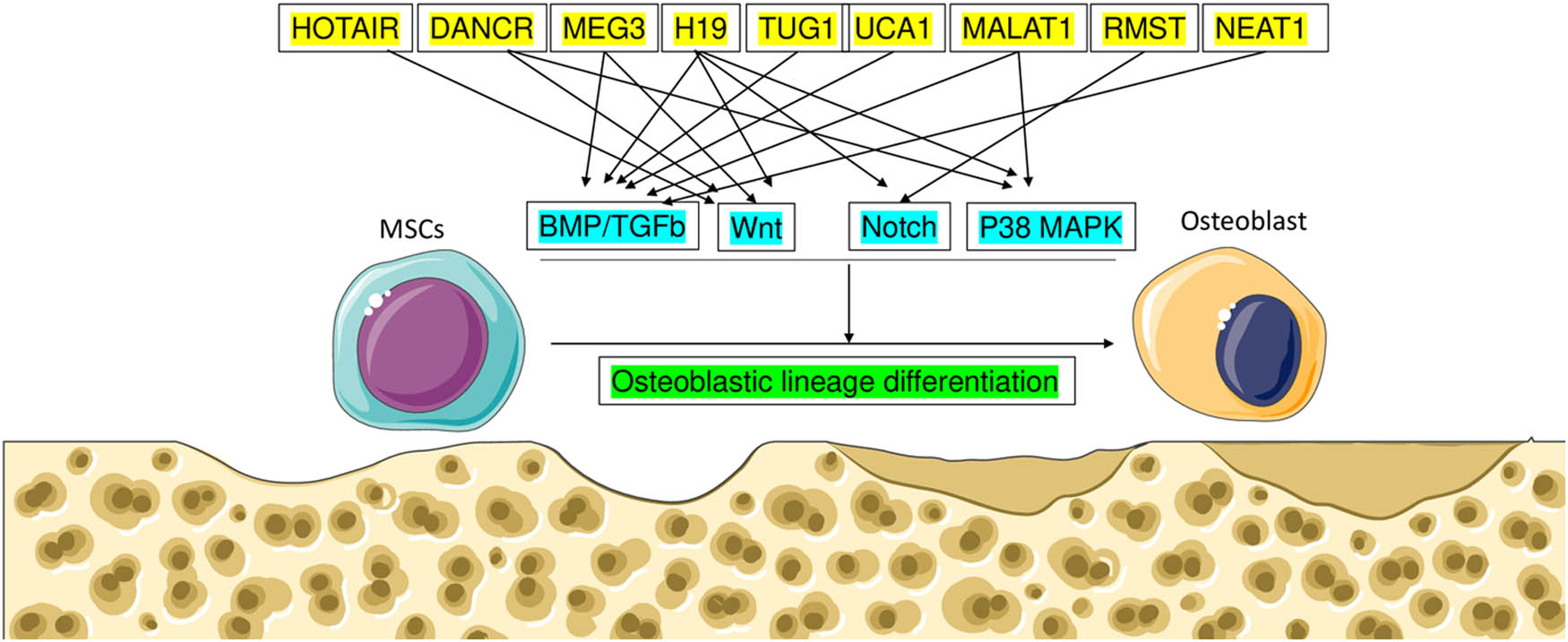
The osteogenic process is controlled by lncRNA through BMP, Wnt, Notch, and MAPK signaling.
-
Funding information: Authors state no funding involved.
-
Author contributions: Chun Jiang and Peng Wang were involved in the preparation and validation of the manuscript. ZhenWei Tan was involved in the preparation of the manuscript. Yin Zhang played a role in designing the work, preparing and validating the manuscript, as well as participating in the revision process.
-
Conflict of interest: Authors state no conflict of interest.
-
Data availability statement: The datasets generated during and/or analyzed during the current study are available from the corresponding author on reasonable request.
References
[1] Vimalraj S. Alkaline phosphatase: Structure, expression and its function in bone mineralization. Gene. 2020;754:144855. 10.1016/j.gene.2020.144855. PMID: 32522695.Suche in Google Scholar PubMed
[2] Vimalraj S, Arumugam B, Miranda PJ, Selvamurugan N. Runx2: Structure, function, and phosphorylation in osteoblast differentiation. Int J Biol Macromol. 2015;78:202–8. 10.1016/j.ijbiomac.2015.04.008. PMID: 25881954.Suche in Google Scholar PubMed
[3] Lin C, Yang L. Long noncoding RNA in cancer: wiring signaling circuitry. Trends Cell Biol. 2018;28(4):287–301.10.1016/j.tcb.2017.11.008Suche in Google Scholar PubMed PubMed Central
[4] Fatica A, Bozzoni I. Long non-coding RNAs: new players in cell differentiation and development. Nat Rev Genet. 2014;15(1):7–21.10.1038/nrg3606Suche in Google Scholar PubMed
[5] Li Y, Syed J, Sugiyama H. RNA-DNA triplex formation by long noncoding RNAs. Cell. Chem Biol. 2016;23(11):1325–33.10.1016/j.chembiol.2016.09.011Suche in Google Scholar PubMed
[6] Tsai MC, Manor O, Wan Y, Mosammaparast N, Wang JK, Lan F, et al. Long noncoding RNA as modular scaffold of histone modification complexes. Science. 2010;329(5992):689–93. 10.1126/science.1192002.Suche in Google Scholar PubMed PubMed Central
[7] Mattick JS, Amaral PP, Carninci P, Carpenter S, Chang HY, Chen LL, et al. Long non-coding RNAs: definitions, functions, challenges and recommendations. Nat Rev Mol Cell Biol. 2023 Jan;24(6):430–47. 10.1038/s41580-022-00566-8. Epub ahead of print. PMID: 36596869.Suche in Google Scholar PubMed PubMed Central
[8] Statello L, Guo CJ, Chen LL, Huarte M. Gene regulation by long non-coding RNAs and its biological functions. Nat Rev Mol Cell Biol. 2021 Feb;22(2):96–118. 10.1038/s41580-020-00315-9.Suche in Google Scholar PubMed PubMed Central
[9] Nojima T, Proudfoot NJ. Mechanisms of lncRNA biogenesis as revealed by nascent transcriptomics. Nat Rev Mol Cell Biol. 2022 Jun;23(6):389–406. 10.1038/s41580-021-00447-6. Epub 2022 Jan 25. Erratum in: Nat Rev Mol Cell Biol. 2022 Dec;23(12):853. PMID: 35079163.Suche in Google Scholar PubMed
[10] Li L, Liu B, Wapinski OL, Tsai MC, Qu K, Zhang J, et al. Targeted disruption of Hotair leads to homeotic transformation and gene derepression. Cell Rep. 2013;5(1):3–12. 10.1016/j.celrep.2013.09.003.Suche in Google Scholar PubMed PubMed Central
[11] Wei B, Wei W, Zhao B, Guo X, Liu S. Long non-coding RNA HOTAIR inhibits miR-17-5p to regulate osteogenic differentiation and proliferation in nontraumatic osteonecrosis of the femoral head. PLoS One. 2017;12(2):e0169097. 10.1371/journal.pone.0169097. PMID: 28152035; PMCID: PMC5289500.Suche in Google Scholar PubMed PubMed Central
[12] Shen JJ, Zhang CH, Chen ZW, Wang ZX, Yang DC, Zhang FL, et al. LncRNA HOTAIR inhibited osteogenic differentiation of BMSCs by regulating Wnt/β-catenin pathway. Eur Rev Med Pharmacol Sci. 2019;23(17):7232–46. 10.26355/eurrev_201909_18984.Suche in Google Scholar PubMed
[13] Zhou Y, Xu Z, Wang Y, Song Q, Yin R. LncRNA MALAT1 mediates osteogenic differentiation in osteoporosis by regulating the miR-485-5p/WNT7B axis. Front Endocrinol (Lausanne). 2023;13:922560. 10.3389/fendo.2022.922560. PMID: 36760811; PMCID: PMC9904362.Suche in Google Scholar PubMed PubMed Central
[14] Gao Y, Xiao F, Wang C, Wang C, Cui P, Zhang X, et al. Long non-coding RNA MALAT1 promotes osterix expression to regulate osteogenic differentiation by targeting miRNA-143 in human bone marrow-derived mesenchymal stem cells. J Cell Biochem. 2018;119(8):6986–96.10.1002/jcb.26907Suche in Google Scholar PubMed
[15] Zhang Y, Guo H, Ma L, Zhu J, Guo A, He Y. Study on adsorption of microRNA-124 by long-chain non-coding RNA MALAT1 regulates osteogenic differentiation of mesenchymal stem cells. Zhongguo Xiu Fu Chong Jian Wai Ke Za Zhi. 2020;34(2):240–5. 10.7507/1002-1892.201909042.Suche in Google Scholar
[16] Sun H, Peng G, Wu H, Liu M, Mao G, Ning X, et al. Long noncoding RNA MEG3 is involved in osteogenic differentiation and bone diseases (review). Biomed Rep. 2020;13(1):15–21.10.3892/br.2020.1305Suche in Google Scholar PubMed PubMed Central
[17] Zhao LD, Xu WC, Cui J, Liang YC, Cheng WQ, Xin BC, et al. Long noncoding RNA maternally expressed gene 3 inhibits osteogenic differentiation of human dental pulp stem cells via microRNA-543/Smad ubiquitin regulatory factor 1/runt-related transcription factor 2 axis. Arch Oral Biol. 2020;118:104838. 10.1016/j.archoralbio.2020.104838. PMID: 32160148.Suche in Google Scholar PubMed
[18] Liu J, Qi X, Wang XH, Miao HS, Xue ZC, Zhang LL, et al. Downregulation of the LncRNA MEG3 promotes osteogenic differentiation of BMSCs and bone repairing by activating Wnt/β-catenin signaling pathway. J Clin Med. 2022 Jan;11(2):395. 10.3390/jcm11020395. PMID: 35054086; PMCID: PMC8781453.Suche in Google Scholar PubMed PubMed Central
[19] Liu Y, Liu C, Zhang A, Yin S, Wang T, Wang Y, et al. Down-regulation of long non-coding RNA MEG3 suppresses osteogenic differentiation of periodontal ligament stem cells (PDLSCs) through miR-27a-3p/IGF1 axis in periodontitis. Aging (Albany NY). 2019;11(15):5334–50.10.18632/aging.102105Suche in Google Scholar PubMed PubMed Central
[20] Liu Y, Zeng X, Miao J, Liu C, Wei F, Liu D, et al. Upregulation of long non-coding RNA MEG3 inhibits the osteogenic differentiation of periodontal ligament cells. J Cell Physiol. 2019;234(4):4617–26.10.1002/jcp.27248Suche in Google Scholar PubMed
[21] Huang XZ, Huang J, Li WZ, Wang JJ, Song DY, Ni JD. LncRNA-MALAT1 promotes osteogenic differentiation through regulating ATF4 by sponging miR-214: implication of steroid-induced avascular necrosis of the femoral head. Steroids. 2020;154:108533.10.1016/j.steroids.2019.108533Suche in Google Scholar PubMed
[22] Yang X, Yang J, Lei P, Wen T. LncRNA MALAT1 shuttled by bone marrow-derived mesenchymal stem cells-secreted exosomes alleviates osteoporosis through mediating microRNA-34c/SATB2 axis. Aging (Albany NY). 2019;11(20):8777–91. 10.18632/aging.102403. PMID: 31693539; PMCID: PMC6854825.Suche in Google Scholar PubMed PubMed Central
[23] Yi J, Liu D, Xiao J. LncRNA MALAT1 sponges miR-30 to promote osteoblast differentiation of adipose-derived mesenchymal stem cells by the promotion of Runx2 expression. Cell Tissue Res. 2019;376(1):113–21. 10.1007/s00441-018-2966-5.Suche in Google Scholar
[24] Kretz M, Webster DE, Flockhart RJ, Lee CS, Zehnder A, Lopez-Pajares V, et al. Suppression of progenitor differentiation requires the long non-coding RNA ANCR. Genes Dev. 2012;26(4):338–43.10.1101/gad.182121.111Suche in Google Scholar PubMed PubMed Central
[25] Zhang L, Sun X, Chen S, Yang C, Shi B, Zhou L, et al. Long non-coding RNA DANCR regulates miR-1305-Smad4 axis to promote chondrogenic differentiation of human synovium-derived mesenchymal stem cells. Biosci Rep. 2017;37(4):BSR20170347. 10.1042/BSR20170347. PMID: 28760952.Suche in Google Scholar PubMed PubMed Central
[26] Wang Z, Huang Y, Tan L. Downregulation of lncRNA DANCR promotes osteogenic differentiation of periodontal ligament stem cells. BMC Dev Biol. 2020;20(1):2. 10.1186/s12861-019-0209-0.Suche in Google Scholar
[27] Zhu L, Xu PC. Downregulated LncRNA-ANCR promotes osteoblast differentiation by targeting EZH2 and regulating Runx2 expression. Biochem Biophys Res Commun. 2013;432(4):612–7. 10.1016/j.bbrc.2013.02.035. PMID: 23428552.Suche in Google Scholar PubMed
[28] Jia Q, Jiang W, Ni L. Down-regulated non-coding RNA (lncRNA-ANCR) promotes osteogenic differentiation of periodontal ligament stem cells. Arch Oral Biol. 2015;60(2):234–41.10.1016/j.archoralbio.2014.10.007Suche in Google Scholar PubMed
[29] Zhang J, Tao Z, Wang Y. Long non-coding RNA DANCR regulates the proliferation and osteogenic differentiation of human bone-derived marrow mesenchymal stem cells via the p38 MAPK pathway. Int J Mol Med. 2018;41(1):213–9. 10.3892/ijmm.2017.3240. PMID: 29115578.Suche in Google Scholar PubMed PubMed Central
[30] Chen S, Liu D, Zhou Z, Qin S. Role of long non-coding RNA H19 in the development of osteoporosis. Mol Med. 2021;27(1):122. 10.1186/s10020-021-00386-0. PMID: 34583640; PMCID: PMC8480040.Suche in Google Scholar PubMed PubMed Central
[31] Zhou Z, Hossain MS, Liu D. Involvement of the long noncoding RNA H19 in osteogenic differentiation and bone regeneration. Stem Cell Res Ther. 2021;12(1):74. 10.1186/s13287-021-02149-4. PMID: 33478579; PMCID: PMC7819155.Suche in Google Scholar PubMed PubMed Central
[32] Liao J, Xiao H, Dai G, He T, Huang W. Recombinant adenovirus (AdEasy system) mediated exogenous expression of long non-coding RNA H19 (lncRNA H19) biphasic regulating osteogenic differentiation of mesenchymal stem cells (MSCs). Am J Transl Res. 2020;12(5):1700–13.Suche in Google Scholar
[33] Li Z, Yan M, Yu Y, Wang Y, Lei G, Pan Y, et al. LncRNA H19 promotes the committed differentiation of stem cells from apical papilla via miR-141/SPAG9 pathway. Cell Death Dis. 2019 Feb;10(2):130.10.1038/s41419-019-1337-3Suche in Google Scholar PubMed PubMed Central
[34] Behera J, Kumar A, Voor MJ, Tyagi N. Exosomal lncRNA-H19 promotes osteogenesis and angiogenesis through mediating Angpt1/Tie2-NO signaling in CBS-heterozygous mice. Theranostics. 2021;11(16):7715–34. 10.7150/thno.58410. PMID: 34335960; PMCID: PMC8315071.Suche in Google Scholar PubMed PubMed Central
[35] Dugimont T, Curgy JJ, Wernert N, Delobelle A, Raes MB, Joubel A, et al. The H19 gene is expressed within both epithelial and stromal components of human invasive adenocarcinomas. Biol Cell. 1995;85(2–3):117–24. 10.1016/0248-4900(96)82997-6.Suche in Google Scholar
[36] Elkin M, Shevelev A, Schulze E, Tykocinsky M, Cooper M, Ariel I, et al. The expression of the imprinted H19 and IGF-2 genes in human bladder carcinoma. FEBS Lett. 1995;374(1):57–61. 10.1016/0014-5793(95)01111-4.Suche in Google Scholar
[37] Verhaegh GW, Verkleij L, Vermeulen SH, den Heijer M, Witjes JA, Kiemeney LA. Polymorphisms in the H19 gene and the risk of bladder cancer. Eur Urol. 2008;54(5):1118–26. 10.1016/j.eururo.2008.06.028.Suche in Google Scholar PubMed
[38] Yoshimizu T, Miroglio A, Ripoche MA, Gabory A, Vernucci M, Riccio A, et al. The H19 locus acts in vivo as a tumor suppressor. Proc Natl Acad Sci U S A. 2008;105(34):12417–22. 10.1073/pnas.0801540105.Suche in Google Scholar PubMed PubMed Central
[39] Ghafouri-Fard S, Esmaeili M, Taheri M. H19 lncRNA: roles in tumorigenesis. Biomed Pharmacother. 2020;123:109774.10.1016/j.biopha.2019.109774Suche in Google Scholar PubMed
[40] Liang WC, Fu WM, Wang YB, Sun YX, Xu LL, Wong CW, et al. H19 activates Wnt signaling and promotes osteoblast differentiation by functioning as a competing endogenous RNA. Sci Rep. 2016;6:20121.10.1038/srep20121Suche in Google Scholar PubMed PubMed Central
[41] Lin X, Zhan JK, Zhong JY, Wang YJ, Wang Y, Li S, et al. lncRNA-ES3/miR-34c-5p/BMF axis is involved in regulating high-glucose-induced calcification/senescence of VSMCs. Aging (Albany NY). 2019 Jan;11(2):523–35. 10.18632/aging.101758. PMID: 30654331; PMCID: PMC6366973.Suche in Google Scholar PubMed PubMed Central
[42] Zhang Z, Liu J, Zeng Z, Fan J, Huang S, Zhang L, et al. lncRNA Rmst acts as an important mediator of BMP9-induced osteogenic differentiation of mesenchymal stem cells (MSCs) by antagonizing Notch-targeting microRNAs. Aging (Albany NY). 2019;11(24):12476–96. 10.18632/aging.102583. PMID: 31825894; PMCID: PMC6949095.Suche in Google Scholar PubMed PubMed Central
[43] Zhang Y, Chen B, Li D, Zhou X, Chen Z. LncRNA NEAT1/miR-29b-3p/BMP1 axis promotes osteogenic differentiation in human bone marrow-derived mesenchymal stem cells. Pathol Res Pract. 2019;215(3):525–31. 10.1016/j.prp.2018.12.034. PMID: 30638953.Suche in Google Scholar PubMed
[44] Zhang RF, Liu JW, Yu SP, Sun D, Wang XH, Fu JS, et al. LncRNA UCA1 affects osteoblast proliferation and differentiation by regulating BMP-2 expression. Eur Rev Med Pharmacol Sci. 2019;23(16):6774–82. 10.26355/eurrev_201908_18715. PMID: 31486475.Suche in Google Scholar PubMed
[45] Shu T, He L, Wang X, Pang M, Yang B, Feng F, et al. Long noncoding RNA UCA1 promotes chondrogenic differentiation of human bone marrow mesenchymal stem cells via miRNA-145-5p/SMAD5 and miRNA-124-3p/SMAD4 axis. Biochem Biophys Res Commun. 2019 Jun;514(1):316–22. 10.1016/j.bbrc.2019.04.140. Epub 2019 Apr 26. PMID: 31036320.Suche in Google Scholar PubMed
[46] Li T, Xiao Y, Huang T. HIF 1α induced upregulation of lncRNA UCA1 promotes cell growth in osteosarcoma by inactivating the PTEN/AKT signaling pathway. Oncol Rep. 2018 Mar;39(3):1072–80. 10.3892/or.2018.6182. Epub 2018 Jan 3. PMID: 29328452; PMCID: PMC5802028.Suche in Google Scholar PubMed PubMed Central
[47] Yao Z, An W, Moming A, Tuerdi M. Long non-coding RNA TUG1 knockdown repressed the viability, migration and differentiation of osteoblasts by sponging miR-214. Exp Ther Med. 2022;23(3):203. 10.3892/etm.2022.11126. PMID: 35126706; PMCID: PMC8794542.Suche in Google Scholar PubMed PubMed Central
[48] Wu D, Zhu Q, Luo X, Zhu W, Gong S. Long noncoding RNA TUG1 promotes osteogenic differentiation of human periodontal ligament stem cell through sponging microRNA-222-3p to negatively regulate Smad2/7. Arch Oral Biol. 2020;117:104814. 10.1016/j.archoralbio.2020.104814. PMID: 31932213.Suche in Google Scholar PubMed
[49] Hao R, Wang B, Wang H, Huo Y, Lu Y. lncRNA TUG1 promotes proliferation and differentiation of osteoblasts by regulating the miR-545-3p/CNR2 axis. Braz J Med Biol Res. 2020 Oct;53(11):e9798. 10.1590/1414-431X20209798. PMID: 33053117; PMCID: PMC7552904.Suche in Google Scholar PubMed PubMed Central
[50] Özgür E, Mert U, Isin M, Okutan M, Dalay N, Gezer U. Differential expression of long non-coding RNAs during genotoxic stress-induced apoptosis in HeLa and MCF-7 cells. Clin Exp Med. 2013;13:119–26. 10.1007/s10238-012-0181-x.Suche in Google Scholar PubMed
[51] Teng Z, Zhu Y, Hao Q, Yu X, Teng Y, Yue Q, et al. Long non-coding RNA taurine upregulated gene 1 is downregulated in osteoporosis and influences the osteogenic differentiation of bone marrow mesenchymal stem cells. PeerJ. 2021 Apr;9:e11251.10.7717/peerj.11251Suche in Google Scholar PubMed PubMed Central
[52] Zhang W, Chen L, Wu J, Li J, Zhang X, Xiang Y, et al. Long noncoding RNA TUG1 inhibits osteogenesis of bone marrow mesenchymal stem cells via Smad5 after irradiation. Theranostics. 2019;9(8):2198–208. 10.7150/thno.30798. PMID: 31149038; PMCID: PMC6531293.Suche in Google Scholar PubMed PubMed Central
[53] Liu JL, Liu YS, Zheng MJ, He HY. The management of bone defect using long non-coding RNA as a potential biomarker for regulating the osteogenic differentiation process. Mol Biol Rep. 2022 Mar;49(3):2443–53. 10.1007/s11033-021-07013-5. Epub 2022 Jan 1. PMID: 34973122; PMCID: PMC8863721.Suche in Google Scholar PubMed PubMed Central
[54] He Q, Yang S, Gu X, Li M, Wang C, Wei F. Long noncoding RNA TUG1 facilitates osteogenic differentiation of periodontal ligament stem cells via interacting with Lin28A. Cell Death Dis. 2018 May;9(5):455. 10.1038/s41419-018-0484-2. Erratum in: Cell Death Dis. 2018 Jun 15;9(7):710. PMID: 29674645; PMCID: PMC5908786.Suche in Google Scholar PubMed PubMed Central
[55] Liu SC, Sun QZ, Qiao XF, Li XG, Yang JH, Wang TQ, et al. LncRNA TUG1 influences osteoblast proliferation and differentiation through the Wnt/β-catenin signaling pathway. Eur Rev Med Pharmacol Sci. 2019 Jun;23(11):4584–90. 10.26355/eurrev_201906_18035. PMID: 31210284.Suche in Google Scholar PubMed
[56] Wang X, Yan L, Zhang H, Wang X, Zhang J, Yang R, et al. Fluid shear stress regulates osteoblast proliferation and apoptosis via the lncRNA TUG1/miR-34a/FGFR1 axis. J Cell Mol Med. 2021;25:8734–47. 10.1111/jcmm.16829.Suche in Google Scholar PubMed PubMed Central
[57] Liu C, Gao X, Li Y, Sun W, Xu Y, Tan Y, et al. The mechanosensitive lncRNA Neat1 promotes osteoblast function through paraspeckle-dependent Smurf1 mRNA retention. Bone Res. 2022 Feb;10(1):18. 10.1038/s41413-022-00191-3. PMID: 35210394; PMCID: PMC8873336.Suche in Google Scholar PubMed PubMed Central
[58] Chen Y, Wu Y, Guo L, Yuan S, Sun J, Zhao K, et al. Exosomal Lnc NEAT1 from endothelial cells promotes bone regeneration by regulating macrophage polarization via DDX3X/NLRP3 axis. J Nanobiotechnol. 2023;21(1):98. 10.1186/s12951-023-01855-w. PMID: 36941678; PMCID: PMC10029245.Suche in Google Scholar PubMed PubMed Central
[59] Zhao X, Zhao D, Geng B, Wang Y, Xia Y. A novel ceRNA regulatory network involving the long noncoding NEAT1, miRNA-466f-3p and its mRNA target in osteoblast autophagy and osteoporosis. J Mol Med (Berl). 2022;100(11):1629–46. 10.1007/s00109-022-02255-7. PMID: 36169673.Suche in Google Scholar PubMed
[60] Zhang H, Xu R, Li B, Xin Z, Ling Z, Zhu W, et al. LncRNA NEAT1 controls the lineage fates of BMSCs during skeletal aging by impairing mitochondrial function and pluripotency maintenance. Cell Death Differ. 2022;29(2):351–65. 10.1038/s41418-021-00858-0. PMID: 34497381; PMCID: PMC8816946.Suche in Google Scholar PubMed PubMed Central
© 2024 the author(s), published by De Gruyter
This work is licensed under the Creative Commons Attribution 4.0 International License.
Artikel in diesem Heft
- Biomedical Sciences
- Constitutive and evoked release of ATP in adult mouse olfactory epithelium
- LARP1 knockdown inhibits cultured gastric carcinoma cell cycle progression and metastatic behavior
- PEGylated porcine–human recombinant uricase: A novel fusion protein with improved efficacy and safety for the treatment of hyperuricemia and renal complications
- Research progress on ocular complications caused by type 2 diabetes mellitus and the function of tears and blepharons
- The role and mechanism of esketamine in preventing and treating remifentanil-induced hyperalgesia based on the NMDA receptor–CaMKII pathway
- Brucella infection combined with Nocardia infection: A case report and literature review
- Detection of serum interleukin-18 level and neutrophil/lymphocyte ratio in patients with antineutrophil cytoplasmic antibody-associated vasculitis and its clinical significance
- Ang-1, Ang-2, and Tie2 are diagnostic biomarkers for Henoch-Schönlein purpura and pediatric-onset systemic lupus erythematous
- PTTG1 induces pancreatic cancer cell proliferation and promotes aerobic glycolysis by regulating c-myc
- Role of serum B-cell-activating factor and interleukin-17 as biomarkers in the classification of interstitial pneumonia with autoimmune features
- Effectiveness and safety of a mumps containing vaccine in preventing laboratory-confirmed mumps cases from 2002 to 2017: A meta-analysis
- Low levels of sex hormone-binding globulin predict an increased breast cancer risk and its underlying molecular mechanisms
- A case of Trousseau syndrome: Screening, detection and complication
- Application of the integrated airway humidification device enhances the humidification effect of the rabbit tracheotomy model
- Preparation of Cu2+/TA/HAP composite coating with anti-bacterial and osteogenic potential on 3D-printed porous Ti alloy scaffolds for orthopedic applications
- Aquaporin-8 promotes human dermal fibroblasts to counteract hydrogen peroxide-induced oxidative damage: A novel target for management of skin aging
- Current research and evidence gaps on placental development in iron deficiency anemia
- Single-nucleotide polymorphism rs2910829 in PDE4D is related to stroke susceptibility in Chinese populations: The results of a meta-analysis
- Pheochromocytoma-induced myocardial infarction: A case report
- Kaempferol regulates apoptosis and migration of neural stem cells to attenuate cerebral infarction by O‐GlcNAcylation of β-catenin
- Sirtuin 5 regulates acute myeloid leukemia cell viability and apoptosis by succinylation modification of glycine decarboxylase
- Apigenin 7-glucoside impedes hypoxia-induced malignant phenotypes of cervical cancer cells in a p16-dependent manner
- KAT2A changes the function of endometrial stromal cells via regulating the succinylation of ENO1
- Current state of research on copper complexes in the treatment of breast cancer
- Exploring antioxidant strategies in the pathogenesis of ALS
- Helicobacter pylori causes gastric dysbacteriosis in chronic gastritis patients
- IL-33/soluble ST2 axis is associated with radiation-induced cardiac injury
- The predictive value of serum NLR, SII, and OPNI for lymph node metastasis in breast cancer patients with internal mammary lymph nodes after thoracoscopic surgery
- Carrying SNP rs17506395 (T > G) in TP63 gene and CCR5Δ32 mutation associated with the occurrence of breast cancer in Burkina Faso
- P2X7 receptor: A receptor closely linked with sepsis-associated encephalopathy
- Probiotics for inflammatory bowel disease: Is there sufficient evidence?
- Identification of KDM4C as a gene conferring drug resistance in multiple myeloma
- Microbial perspective on the skin–gut axis and atopic dermatitis
- Thymosin α1 combined with XELOX improves immune function and reduces serum tumor markers in colorectal cancer patients after radical surgery
- Highly specific vaginal microbiome signature for gynecological cancers
- Sample size estimation for AQP4-IgG seropositive optic neuritis: Retinal damage detection by optical coherence tomography
- The effects of SDF-1 combined application with VEGF on femoral distraction osteogenesis in rats
- Fabrication and characterization of gold nanoparticles using alginate: In vitro and in vivo assessment of its administration effects with swimming exercise on diabetic rats
- Mitigating digestive disorders: Action mechanisms of Mediterranean herbal active compounds
- Distribution of CYP2D6 and CYP2C19 gene polymorphisms in Han and Uygur populations with breast cancer in Xinjiang, China
- VSP-2 attenuates secretion of inflammatory cytokines induced by LPS in BV2 cells by mediating the PPARγ/NF-κB signaling pathway
- Factors influencing spontaneous hypothermia after emergency trauma and the construction of a predictive model
- Long-term administration of morphine specifically alters the level of protein expression in different brain regions and affects the redox state
- Application of metagenomic next-generation sequencing technology in the etiological diagnosis of peritoneal dialysis-associated peritonitis
- Clinical diagnosis, prevention, and treatment of neurodyspepsia syndrome using intelligent medicine
- Case report: Successful bronchoscopic interventional treatment of endobronchial leiomyomas
- Preliminary investigation into the genetic etiology of short stature in children through whole exon sequencing of the core family
- Cystic adenomyoma of the uterus: Case report and literature review
- Mesoporous silica nanoparticles as a drug delivery mechanism
- Dynamic changes in autophagy activity in different degrees of pulmonary fibrosis in mice
- Vitamin D deficiency and inflammatory markers in type 2 diabetes: Big data insights
- Lactate-induced IGF1R protein lactylation promotes proliferation and metabolic reprogramming of lung cancer cells
- Meta-analysis on the efficacy of allogeneic hematopoietic stem cell transplantation to treat malignant lymphoma
- Mitochondrial DNA drives neuroinflammation through the cGAS-IFN signaling pathway in the spinal cord of neuropathic pain mice
- Application value of artificial intelligence algorithm-based magnetic resonance multi-sequence imaging in staging diagnosis of cervical cancer
- Embedded monitoring system and teaching of artificial intelligence online drug component recognition
- Investigation into the association of FNDC1 and ADAMTS12 gene expression with plumage coloration in Muscovy ducks
- Yak meat content in feed and its impact on the growth of rats
- A rare case of Richter transformation with breast involvement: A case report and literature review
- First report of Nocardia wallacei infection in an immunocompetent patient in Zhejiang province
- Rhodococcus equi and Brucella pulmonary mass in immunocompetent: A case report and literature review
- Downregulation of RIP3 ameliorates the left ventricular mechanics and function after myocardial infarction via modulating NF-κB/NLRP3 pathway
- Evaluation of the role of some non-enzymatic antioxidants among Iraqi patients with non-alcoholic fatty liver disease
- The role of Phafin proteins in cell signaling pathways and diseases
- Ten-year anemia as initial manifestation of Castleman disease in the abdominal cavity: A case report
- Coexistence of hereditary spherocytosis with SPTB P.Trp1150 gene variant and Gilbert syndrome: A case report and literature review
- Utilization of convolutional neural networks to analyze microscopic images for high-throughput screening of mesenchymal stem cells
- Exploratory evaluation supported by experimental and modeling approaches of Inula viscosa root extract as a potent corrosion inhibitor for mild steel in a 1 M HCl solution
- Imaging manifestations of ductal adenoma of the breast: A case report
- Gut microbiota and sleep: Interaction mechanisms and therapeutic prospects
- Isomangiferin promotes the migration and osteogenic differentiation of rat bone marrow mesenchymal stem cells
- Prognostic value and microenvironmental crosstalk of exosome-related signatures in human epidermal growth factor receptor 2 positive breast cancer
- Circular RNAs as potential biomarkers for male severe sepsis
- Knockdown of Stanniocalcin-1 inhibits growth and glycolysis in oral squamous cell carcinoma cells
- The expression and biological role of complement C1s in esophageal squamous cell carcinoma
- A novel GNAS mutation in pseudohypoparathyroidism type 1a with articular flexion deformity: A case report
- Predictive value of serum magnesium levels for prognosis in patients with non-small cell lung cancer undergoing EGFR-TKI therapy
- HSPB1 alleviates acute-on-chronic liver failure via the P53/Bax pathway
- IgG4-related disease complicated by PLA2R-associated membranous nephropathy: A case report
- Baculovirus-mediated endostatin and angiostatin activation of autophagy through the AMPK/AKT/mTOR pathway inhibits angiogenesis in hepatocellular carcinoma
- Metformin mitigates osteoarthritis progression by modulating the PI3K/AKT/mTOR signaling pathway and enhancing chondrocyte autophagy
- Evaluation of the activity of antimicrobial peptides against bacterial vaginosis
- Atypical presentation of γ/δ mycosis fungoides with an unusual phenotype and SOCS1 mutation
- Analysis of the microecological mechanism of diabetic kidney disease based on the theory of “gut–kidney axis”: A systematic review
- Omega-3 fatty acids prevent gestational diabetes mellitus via modulation of lipid metabolism
- Refractory hypertension complicated with Turner syndrome: A case report
- Interaction of ncRNAs and the PI3K/AKT/mTOR pathway: Implications for osteosarcoma
- Association of low attenuation area scores with pulmonary function and clinical prognosis in patients with chronic obstructive pulmonary disease
- Long non-coding RNAs in bone formation: Key regulators and therapeutic prospects
- The deubiquitinating enzyme USP35 regulates the stability of NRF2 protein
- Neutrophil-to-lymphocyte ratio and platelet-to-lymphocyte ratio as potential diagnostic markers for rebleeding in patients with esophagogastric variceal bleeding
- G protein-coupled receptor 1 participating in the mechanism of mediating gestational diabetes mellitus by phosphorylating the AKT pathway
- LL37-mtDNA regulates viability, apoptosis, inflammation, and autophagy in lipopolysaccharide-treated RLE-6TN cells by targeting Hsp90aa1
- The analgesic effect of paeoniflorin: A focused review
- Chemical composition’s effect on Solanum nigrum Linn.’s antioxidant capacity and erythrocyte protection: Bioactive components and molecular docking analysis
- Knockdown of HCK promotes HREC cell viability and inner blood–retinal barrier integrity by regulating the AMPK signaling pathway
- The role of rapamycin in the PINK1/Parkin signaling pathway in mitophagy in podocytes
- Laryngeal non-Hodgkin lymphoma: Report of four cases and review of the literature
- Clinical value of macrogenome next-generation sequencing on infections
- Overview of dendritic cells and related pathways in autoimmune uveitis
- TAK-242 alleviates diabetic cardiomyopathy via inhibiting pyroptosis and TLR4/CaMKII/NLRP3 pathway
- Hypomethylation in promoters of PGC-1α involved in exercise-driven skeletal muscular alterations in old age
- Profile and antimicrobial susceptibility patterns of bacteria isolated from effluents of Kolladiba and Debark hospitals
- The expression and clinical significance of syncytin-1 in serum exosomes of hepatocellular carcinoma patients
- A histomorphometric study to evaluate the therapeutic effects of biosynthesized silver nanoparticles on the kidneys infected with Plasmodium chabaudi
- PGRMC1 and PAQR4 are promising molecular targets for a rare subtype of ovarian cancer
- Analysis of MDA, SOD, TAOC, MNCV, SNCV, and TSS scores in patients with diabetes peripheral neuropathy
- SLIT3 deficiency promotes non-small cell lung cancer progression by modulating UBE2C/WNT signaling
- The relationship between TMCO1 and CALR in the pathological characteristics of prostate cancer and its effect on the metastasis of prostate cancer cells
- Heterogeneous nuclear ribonucleoprotein K is a potential target for enhancing the chemosensitivity of nasopharyngeal carcinoma
- PHB2 alleviates retinal pigment epithelium cell fibrosis by suppressing the AGE–RAGE pathway
- Anti-γ-aminobutyric acid-B receptor autoimmune encephalitis with syncope as the initial symptom: Case report and literature review
- Comparative analysis of chloroplast genome of Lonicera japonica cv. Damaohua
- Human umbilical cord mesenchymal stem cells regulate glutathione metabolism depending on the ERK–Nrf2–HO-1 signal pathway to repair phosphoramide mustard-induced ovarian cancer cells
- Electroacupuncture on GB acupoints improves osteoporosis via the estradiol–PI3K–Akt signaling pathway
- Renalase protects against podocyte injury by inhibiting oxidative stress and apoptosis in diabetic nephropathy
- Review: Dicranostigma leptopodum: A peculiar plant of Papaveraceae
- Combination effect of flavonoids attenuates lung cancer cell proliferation by inhibiting the STAT3 and FAK signaling pathway
- Renal microangiopathy and immune complex glomerulonephritis induced by anti-tumour agents: A case report
- Correlation analysis of AVPR1a and AVPR2 with abnormal water and sodium and potassium metabolism in rats
- Gastrointestinal health anti-diarrheal mixture relieves spleen deficiency-induced diarrhea through regulating gut microbiota
- Myriad factors and pathways influencing tumor radiotherapy resistance
- Exploring the effects of culture conditions on Yapsin (YPS) gene expression in Nakaseomyces glabratus
- Screening of prognostic core genes based on cell–cell interaction in the peripheral blood of patients with sepsis
- Coagulation factor II thrombin receptor as a promising biomarker in breast cancer management
- Ileocecal mucinous carcinoma misdiagnosed as incarcerated hernia: A case report
- Methyltransferase like 13 promotes malignant behaviors of bladder cancer cells through targeting PI3K/ATK signaling pathway
- The debate between electricity and heat, efficacy and safety of irreversible electroporation and radiofrequency ablation in the treatment of liver cancer: A meta-analysis
- ZAG promotes colorectal cancer cell proliferation and epithelial–mesenchymal transition by promoting lipid synthesis
- Baicalein inhibits NLRP3 inflammasome activation and mitigates placental inflammation and oxidative stress in gestational diabetes mellitus
- Impact of SWCNT-conjugated senna leaf extract on breast cancer cells: A potential apoptotic therapeutic strategy
- MFAP5 inhibits the malignant progression of endometrial cancer cells in vitro
- Major ozonated autohemotherapy promoted functional recovery following spinal cord injury in adult rats via the inhibition of oxidative stress and inflammation
- Axodendritic targeting of TAU and MAP2 and microtubule polarization in iPSC-derived versus SH-SY5Y-derived human neurons
- Differential expression of phosphoinositide 3-kinase/protein kinase B and Toll-like receptor/nuclear factor kappa B signaling pathways in experimental obesity Wistar rat model
- The therapeutic potential of targeting Oncostatin M and the interleukin-6 family in retinal diseases: A comprehensive review
- BA inhibits LPS-stimulated inflammatory response and apoptosis in human middle ear epithelial cells by regulating the Nf-Kb/Iκbα axis
- Role of circRMRP and circRPL27 in chronic obstructive pulmonary disease
- Investigating the role of hyperexpressed HCN1 in inducing myocardial infarction through activation of the NF-κB signaling pathway
- Characterization of phenolic compounds and evaluation of anti-diabetic potential in Cannabis sativa L. seeds: In vivo, in vitro, and in silico studies
- Quantitative immunohistochemistry analysis of breast Ki67 based on artificial intelligence
- Ecology and Environmental Science
- Screening of different growth conditions of Bacillus subtilis isolated from membrane-less microbial fuel cell toward antimicrobial activity profiling
- Degradation of a mixture of 13 polycyclic aromatic hydrocarbons by commercial effective microorganisms
- Evaluation of the impact of two citrus plants on the variation of Panonychus citri (Acari: Tetranychidae) and beneficial phytoseiid mites
- Prediction of present and future distribution areas of Juniperus drupacea Labill and determination of ethnobotany properties in Antalya Province, Türkiye
- Population genetics of Todarodes pacificus (Cephalopoda: Ommastrephidae) in the northwest Pacific Ocean via GBS sequencing
- A comparative analysis of dendrometric, macromorphological, and micromorphological characteristics of Pistacia atlantica subsp. atlantica and Pistacia terebinthus in the middle Atlas region of Morocco
- Macrofungal sporocarp community in the lichen Scots pine forests
- Assessing the proximate compositions of indigenous forage species in Yemen’s pastoral rangelands
- Food Science
- Gut microbiota changes associated with low-carbohydrate diet intervention for obesity
- Reexamination of Aspergillus cristatus phylogeny in dark tea: Characteristics of the mitochondrial genome
- Differences in the flavonoid composition of the leaves, fruits, and branches of mulberry are distinguished based on a plant metabolomics approach
- Investigating the impact of wet rendering (solventless method) on PUFA-rich oil from catfish (Clarias magur) viscera
- Non-linear associations between cardiovascular metabolic indices and metabolic-associated fatty liver disease: A cross-sectional study in the US population (2017–2020)
- Knockdown of USP7 alleviates atherosclerosis in ApoE-deficient mice by regulating EZH2 expression
- Utility of dairy microbiome as a tool for authentication and traceability
- Agriculture
- Enhancing faba bean (Vicia faba L.) productivity through establishing the area-specific fertilizer rate recommendation in southwest Ethiopia
- Impact of novel herbicide based on synthetic auxins and ALS inhibitor on weed control
- Perspectives of pteridophytes microbiome for bioremediation in agricultural applications
- Fertilizer application parameters for drip-irrigated peanut based on the fertilizer effect function established from a “3414” field trial
- Improving the productivity and profitability of maize (Zea mays L.) using optimum blended inorganic fertilization
- Application of leaf multispectral analyzer in comparison to hyperspectral device to assess the diversity of spectral reflectance indices in wheat genotypes
- Animal Sciences
- Knockdown of ANP32E inhibits colorectal cancer cell growth and glycolysis by regulating the AKT/mTOR pathway
- Development of a detection chip for major pathogenic drug-resistant genes and drug targets in bovine respiratory system diseases
- Exploration of the genetic influence of MYOT and MB genes on the plumage coloration of Muscovy ducks
- Transcriptome analysis of adipose tissue in grazing cattle: Identifying key regulators of fat metabolism
- Comparison of nutritional value of the wild and cultivated spiny loaches at three growth stages
- Transcriptomic analysis of liver immune response in Chinese spiny frog (Quasipaa spinosa) infected with Proteus mirabilis
- Disruption of BCAA degradation is a critical characteristic of diabetic cardiomyopathy revealed by integrated transcriptome and metabolome analysis
- Plant Sciences
- Effect of long-term in-row branch covering on soil microorganisms in pear orchards
- Photosynthetic physiological characteristics, growth performance, and element concentrations reveal the calcicole–calcifuge behaviors of three Camellia species
- Transcriptome analysis reveals the mechanism of NaHCO3 promoting tobacco leaf maturation
- Bioinformatics, expression analysis, and functional verification of allene oxide synthase gene HvnAOS1 and HvnAOS2 in qingke
- Water, nitrogen, and phosphorus coupling improves gray jujube fruit quality and yield
- Improving grape fruit quality through soil conditioner: Insights from RNA-seq analysis of Cabernet Sauvignon roots
- Role of Embinin in the reabsorption of nucleus pulposus in lumbar disc herniation: Promotion of nucleus pulposus neovascularization and apoptosis of nucleus pulposus cells
- Revealing the effects of amino acid, organic acid, and phytohormones on the germination of tomato seeds under salinity stress
- Combined effects of nitrogen fertilizer and biochar on the growth, yield, and quality of pepper
- Comprehensive phytochemical and toxicological analysis of Chenopodium ambrosioides (L.) fractions
- Impact of “3414” fertilization on the yield and quality of greenhouse tomatoes
- Exploring the coupling mode of water and fertilizer for improving growth, fruit quality, and yield of the pear in the arid region
- Metagenomic analysis of endophytic bacteria in seed potato (Solanum tuberosum)
- Antibacterial, antifungal, and phytochemical properties of Salsola kali ethanolic extract
- Exploring the hepatoprotective properties of citronellol: In vitro and in silico studies on ethanol-induced damage in HepG2 cells
- Enhanced osmotic dehydration of watermelon rind using honey–sucrose solutions: A study on pre-treatment efficacy and mass transfer kinetics
- Effects of exogenous 2,4-epibrassinolide on photosynthetic traits of 53 cowpea varieties under NaCl stress
- Comparative transcriptome analysis of maize (Zea mays L.) seedlings in response to copper stress
- An optimization method for measuring the stomata in cassava (Manihot esculenta Crantz) under multiple abiotic stresses
- Fosinopril inhibits Ang II-induced VSMC proliferation, phenotype transformation, migration, and oxidative stress through the TGF-β1/Smad signaling pathway
- Antioxidant and antimicrobial activities of Salsola imbricata methanolic extract and its phytochemical characterization
- Bioengineering and Biotechnology
- Absorbable calcium and phosphorus bioactive membranes promote bone marrow mesenchymal stem cells osteogenic differentiation for bone regeneration
- New advances in protein engineering for industrial applications: Key takeaways
- An overview of the production and use of Bacillus thuringiensis toxin
- Research progress of nanoparticles in diagnosis and treatment of hepatocellular carcinoma
- Bioelectrochemical biosensors for water quality assessment and wastewater monitoring
- PEI/MMNs@LNA-542 nanoparticles alleviate ICU-acquired weakness through targeted autophagy inhibition and mitochondrial protection
- Unleashing of cytotoxic effects of thymoquinone-bovine serum albumin nanoparticles on A549 lung cancer cells
- Erratum
- Erratum to “Investigating the association between dietary patterns and glycemic control among children and adolescents with T1DM”
- Erratum to “Activation of hypermethylated P2RY1 mitigates gastric cancer by promoting apoptosis and inhibiting proliferation”
- Retraction
- Retraction to “MiR-223-3p regulates cell viability, migration, invasion, and apoptosis of non-small cell lung cancer cells by targeting RHOB”
- Retraction to “A data mining technique for detecting malignant mesothelioma cancer using multiple regression analysis”
- Special Issue on Advances in Neurodegenerative Disease Research and Treatment
- Transplantation of human neural stem cell prevents symptomatic motor behavior disability in a rat model of Parkinson’s disease
- Special Issue on Multi-omics
- Inflammasome complex genes with clinical relevance suggest potential as therapeutic targets for anti-tumor drugs in clear cell renal cell carcinoma
- Gastroesophageal varices in primary biliary cholangitis with anti-centromere antibody positivity: Early onset?