LARP1 knockdown inhibits cultured gastric carcinoma cell cycle progression and metastatic behavior
-
Xin Liu


Abstract
This study aimed to clarify the role of la-related protein 1 (LARP1) in cell cycle progression and metastatic behavior of cultured gastric carcinoma (GC) cells. To do that, LARP1 expression was detected in clinical GC tissues and cell lines using quantitative real-time polymerase chain reaction (qRT-PCR) and western blotting. The cell viability, apoptosis, cell cycle, migration, invasion, and cell growth were examined using a Cell Counting Kit-8, Annexin V-FITC staining, propidium iodide staining, Transwell migration and invasion assays, and colony formation assays after LARP1 knockdown. Phosphatidyl inositol 3-kinase (PI3K) and AKT1 mRNA and protein expression levels of PI3K, p-AKT1, AKT1, p-BAD, p-mTOR, and p21 in si-LARP1 transfected GC cells were determined using qRT-PCR and western blotting. Here, we've shown that LARP1 expression was upregulated in human GC tissues and KATO III cells. LARP1 knockdown inhibited GC cell proliferation, cell cycle progression, migration, invasion, and colony formation and promoted apoptosis. In si-LARP1-transfected KATO III cells, the mRNA expression levels of PI3K and AKT1, PI3K protein expression, and the p-AKT1/AKT1 ratio were significantly suppressed. p-mTOR and p-BAD were significantly decreased, whereas p21 was significantly increased in si-LARP1-transfected KATO III cells. In conclusion LARP1 knockdown induces apoptosis and inhibits cell cycle progression and metastatic behavior via PI3K/AKT1 signaling in GC cells.
1 Introduction
Gastric carcinoma (GC) is one of the five most common cancers worldwide and is a major contributor to cancer-related deaths [1]. Despite the introduction of new diagnostic and therapeutic strategies, the prognosis remains poor, and the mortality rate is high [2]. Widely explored chemotherapeutic agents have reached a plateau, where the median overall survival is no more than 1 year [3], and the main research direction is to understand the molecular biology of tumorigenesis. Understanding the cell signaling mechanisms that promote carcinogenesis can offer valuable information for the development of new and efficient therapies [4].
As a multifactorial disease, cancer can be influenced by both environmental and genetic factors, and the multiple stages of carcinogenesis are controlled by the gradual development of mutations in gene expression and epigenetic changes. These genetic alterations lead to cancer progression by affecting the fate of cancer cells (e.g., proliferation, apoptosis, and metastasis) [5]. With the recent advancements in understanding the complex genetic changes underlying human cancers, identifying molecularly targeted therapies that will benefit patients is significant [6]. However, the mechanisms underlying GC cell survival, invasion, and metastasis are not fully understood.
As a highly evolutionarily conserved RNA-binding protein, la-related protein 1 (LARP1) is well known as a member of the LARP family (including LARP1, LARP3, and LARP7), each of which carries a conserved RNA-binding La domain and an RNA recognition mode-like domain [7]. Functionally, LARP1 primarily plays the role of post-transcriptional regulator of genes by recognizing the 5′ terminal oligopyrimidine (5′ TOP) motif characterization or 3′ untranslated region of mRNA [8,9]. Abnormal levels and functions of LARP1 are associated with disease progression [10]. Changes in LARP1 levels are associated with cancer progression. Upregulated LARP1 expression in hepatocellular carcinoma and lung carcinoma is correlated with poor prognosis [11]. Increased levels of LARP1 in cervical cancer facilitate the migration and invasion of cancer cells by acting on mRNAs rich in carcinogenic transcripts [12]. LARP1 is essential for the survival of epithelial ovarian cancer cells and promotes ovarian cancer tension and chemotherapy resistance [13]. These studies indicate that LARP1 acts as an oncogene and is involved in carcinoma progression. To date, limited evidence has demonstrated the role of LARP1 in GC.
This study determined the expression pattern of LARP1 in clinical GC tissues and investigated the LARP1 function in GC cell survival and metastasis in vitro. The discovery of new therapeutic targets is expected to improve the therapeutic effects in GC.
2 Materials and methods
2.1 Clinical GC tissues
Six patients (two females and four males) with GC at the antrum of the stomach were hospitalized between January 2020 and August 2020 and underwent resection. Paired GC and para-carcinoma tissues were obtained after obtaining informed consent prior to sample collection. A part of the tissue was used for pathological examination and a part was used for quantitative real-time polymerase chain reaction (qRT-PCR) detection. Tissues for qRT-PCR detection were placed in liquid nitrogen immediately after excision and stored at −80°C before analysis. Pathological results showed that the samples were in TNM stages I and II. Sample acquisition and experimental procedures were conducted in accordance with the Ethics Committee of Wuming Hospital Affiliated to Guangxi Medical University.
-
Informed consent: Informed consent has been obtained from all individuals included in this study.
-
Ethical approval: The research related to human use has been complied with all the relevant national regulations, institutional policies and in accordance with the tenets of the Helsinki Declaration, and has been approved by the Ethical Committee of Wuming Hospital Affiliated to Guanxi Medical University.
2.2 Cell lines
A normal human gastric mucosa cell line (GES-1) and three human GC cell lines (SNU-1, NCI-N87, and KATO III) were purchased from the Cell Bank of the Chinese Academy of Sciences (Shanghai, China). All cells were maintained in 37°C and 5% CO2 cell incubator using RPMI-1640 medium (11875101, Thermo Fisher Scientific, Waltham, MA, USA), supplemented with 10% fetal bovine serum (10091148, Thermo Fisher Scientific) and 1% penicillin–streptomycin (15140122, Thermo Fisher Scientific).
2.3 qRT-PCR
Before RNA isolation, tissue samples were homogenized, and cells with various treatments were collected by washing with phosphate-buffered saline (PBS) (B548117-0500, Sangon Biotech, Shanghai, China). RNAiso Plus (TaKaRa, Kyoto, Japan) was used to lyse the cells, and the chloroform isoamyl alcohol method was used to extract total RNA from the lysate. The purity and concentration of RNA were determined using a microplate reader (Thermo Fisher Scientific). For reverse transcription, RNA was mixed with primeScript RT Master MIX (RR036A, TaKaRa), and a reaction procedure was performed at 37°C for 60 min and at 85°C for 5 s. The cDNA product was amplified using Power SYBR Green PCR Master Mix (4367659, Thermo Fisher Scientific). The primers used are listed in Table 1. Relative mRNA expression was calculated using the 2‒ΔΔCT method with GAPDH as an internal reference.
Primers used in qRT-PCR
| Gene | Direction | Sequence (5′−3′) |
|---|---|---|
| LARP1 | Forward | ACACAAGTGGGTTCCATTACAAA |
| Reverse | CTCCGCGATTGGCAGGTAT | |
| PI3K | Forward | CCACGACCATCATCAGGTGAA |
| Reverse | CCTCACGGAGGCATTCTAAAGT | |
| AKT1 | Forward | AGCGACGTGGCTATTGTGAAG |
| Reverse | GCCATCATTCTTGAGGAGGAAGT | |
| GAPDH | Forward | TGACAACTTTGGTATCGTGGAAGG |
| Reverse | AGGCAGGGATGATGTTCTGGAGAG |
2.4 siRNA transfection
Three LARP1 siRNAs were designed and synthesized by BioTen Co., Ltd. (Shanghai, China). Before transfection, approximately 6 × 10⁵/well of GC cells were seeded in a six-well plate and kept at 37°C and 5% CO2 for 24 h. LARP1 siRNA mix with Lipofectamine 2000 (11668027, Thermo Fisher Scientific) was added to each well and incubated for 6 h. After removing the siRNA mixture solution, the cell culture was continued in a complete medium. After 48 h of transfection, the cells were harvested to determine the effect of LARP1 siRNA.
2.5 Cell viability assay
A total of 5 × 10³/well of GC cells were seeded in a 96-well plate and incubated at 37°C and 5% CO2 for 24 h. Cells were harvested after 24, 48, and 72 h of transfection. Cell viability was detected using a cell counting kit-8 (C0037, Beyotime) based on the protocols recommended by the manufacturer.
2.6 Apoptosis detection
Cultured cells were digested using trypsin and collected after centrifugation at 1,000 × g for 5 min. Apoptosis was determined using the FITC Annexin V Apoptosis Detection Kit (556420; BD Biosciences). In brief, after washing with PBS, cells were resuspended with 195 μL of Annexin V-FITC binding buffer. Cells were then added with 5 μL of Annexin V-FITC and 10 μL of propidium iodide (PI) staining solution. Incubation staining was performed for 20 min at room temperature (20–25°C) away from light. The cells were subjected to apoptosis detection using a FACSCalibur flow cytometer (BD Biosciences).
2.7 Cell cycle distribution detection
The cultured cells were resuspended in PBS and mixed with 4 mL of 70% ethanol (−20°C precooled). Cells were fixed overnight in a 4°C refrigerator. After washing with PBS, the fixed cells were resuspended in PBS supplemented with 50 μg/mL RNase A and kept in a 37°C water bath for 30 min. PI at a final concentration of 50 μg/mL (ST512, Beyotime) was added to stain the cells for 30 min away from light. The cells were subjected to cell cycle distribution analysis using a FACSCalibur flow cytometer (BD Biosciences, USA).
2.8 Transwell assay
Cultured cells were collected after trypsin digestion and resuspended in 5 mL of sterile PBS for cell counting. The cells were maintained in a serum-free medium to adjust the cell density to 5 × 10⁵/mL. The upper transwell was supplemented with 200 μL of cell suspension and cultured for 48 h. The migrated and invaded cells were fixed in 4% paraformaldehyde for 20 min. Staining was performed using a crystal violet staining solution (C0121, Beyotime). For the Transwell invasion assay, the transwell chamber was pre-coated with Matrigel (354234, Corning, MA, USA).
2.9 Colony formation
After 24 h of transfection, the cells were digested with trypsin and resuspended. The cell suspension was seeded in a six-well plate at 200 cells/well and cultured for 14 days. Colonies on the plates were fixed with 4% paraformaldehyde (80096618; Sinopharm, Shanghai, China) for 4 min. Staining was performed using a crystal violet staining solution (C0121, Beyotime). After the unstained cells were washed away, the plates were photographed, and colonies (containing >50 cells) were counted.
2.10 Western blot
Radio immunoprecipitation assay lysis (P0013B, Beyotime) buffer containing 1 mM phenylmethanesulfonylfluoride (ST506, Beyotime) was added to GC cells and centrifuged at 12,000 × g and 4°C for 10 min. Proteins were quantified using a bicinchoninic acid kit (PL212989, Thermo Fisher Scientific). After adding the loading buffer, the proteins were separated via sodium dodecyl sulfate-polyacrylamide gel electrophoresis and transferred onto a polyvinylidene fluoride (PVDF) membrane (IPVH00010, Millipore, Boston, MA, USA). Subsequently, the PVDF membrane was blocked by 5% defatted milk for 1 h at 37°C and incubated with primary antibodies of LARP1 (ab86359; Abcam, Cambridge, UK; 1:2,000), phosphatidyl inositol 3-kinase (PI3K) (20584-1-AP; Proteintech, Wuhan, Hubei, China; 1:500), p-AKT (28731-1-AP; Proteintech; 1:1,000), AKT1 (60203-2-Ig; Proteintech; 1:5,000), p-BAD (5284T; CST, Boston, MA, USA; 1:1,000), p-mTOR (5536T; CST; 1:1,000), and GAPDH (10494-1-AP; Proteintech; 1:5,000) overnight at 4°C. On the second day, (H + L)-HRP secondary antibody (115-035-003; Jackson ImmunoResearch, West Grove, PA, USA; 1:5,000) was added and incubated at 37°C for 2 h. Protein expression was determined using an ECL system (Millipore) and quantified using ImageJ software.
2.11 Statistical analysis
GraphPad Prism 7.00 version was used for statistics and analysis of data. An unpaired t-test was used to analyze differences between two groups, and a one-way analysis of variance following Tukey’s multiple comparison test was used for analyzing differences among multiple groups. P values <0.05 were considered statistically significant.
3 Results
3.1 LARP1 expression was upregulated in human GC tissues and GC cell lines
To determine the expression pattern of LARP1 in GC, we first compared its expression in GC and normal tissues using the ENCORI (Starbase, https://rnasysu.com/encori/index.php). As shown in Figure 1a, the expression level of LARP1 was significantly higher in stomach cancer samples than in normal samples (P = 5.6 × 10⁻²⁰). To further verify the results, we measured its levels in six pairs of GC and para-carcinoma tissue samples in our cohort samples. qRT-PCR analysis showed that LARP1 mRNA was expressed at higher levels in GC tissues than in paracarcinoma tissues (Figure 1b). The LARP expression pattern was further verified in three GC cell lines. As shown in Figure 1c, compared to the normal gastric mucosa cell line (GES-1), LARP1 mRNA levels were significantly increased in KATO III cells (P < 0.01) but not in NCI-N87 and SNU-1 cells (P > 0.05). The protein expression of LARP1 was significantly higher in the three GC cell lines than in the normal gastric mucosal cell line (Figure 1d, P < 0.05). Therefore, the KATO-III cells were selected for subsequent experiments.
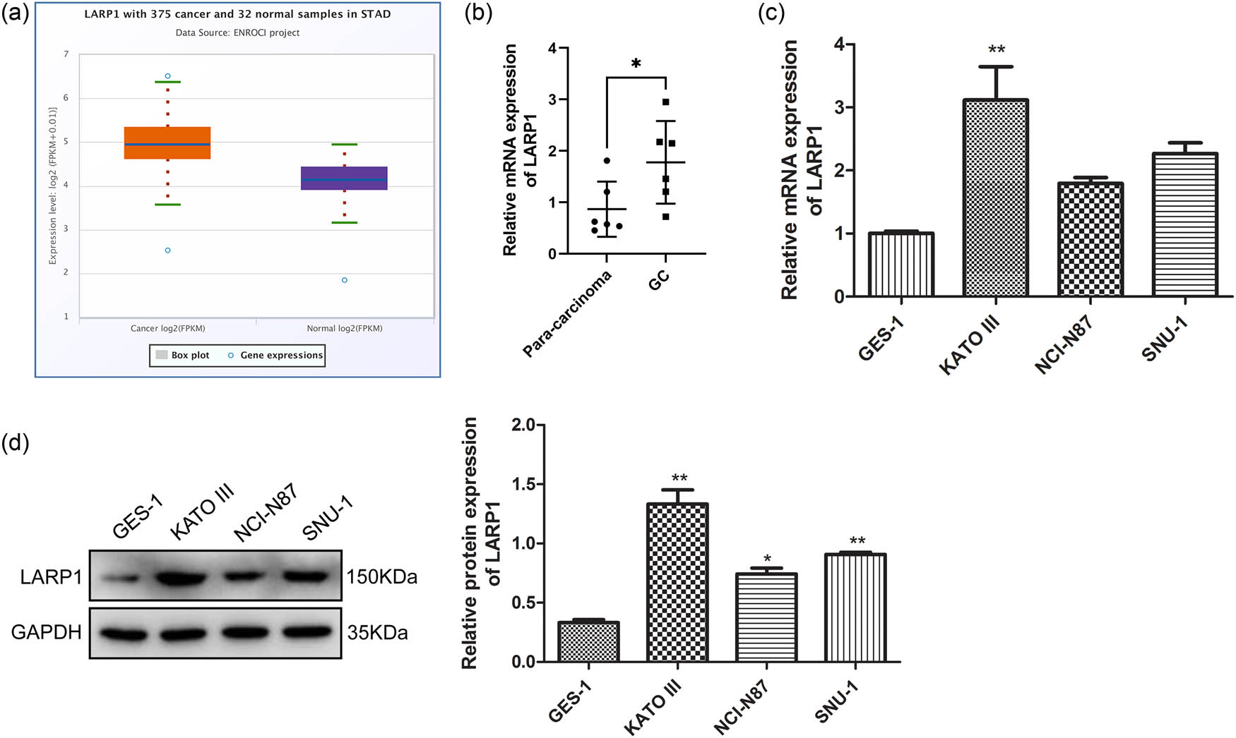
LARP1 expression level in clinical GC tissues and GC cell lines. (a) mRNA expression levels of LAPR1 in stomach cancer (STAD) and normal samples were searched in the ENCORI platform. (b) LARP1 level was compared between six pairs of GC tissues and para-carcinoma tissues using qRT-PCR. (c) The mRNA expression of LARP1 was compared between a normal gastric mucosa cell line (GES-1) and three GC cell lines (KATO III, NCI-N87, and SNU-1). (d) The protein expression of LARP1 was compared between GES-1 and three GC cell lines (KATO III, NCI-N87, and SNU-1) using a western blot. *P < 0.05, **P < 0.01 compared with para-carcinoma tissue or GES-1.
3.2 LARP1 knockdown inhibited GC cell survival and promoted cell apoptosis
Given the high levels of LARP1 in GC, the next step was to establish a knockdown LARP1 cell line and verify the function of LARP1 in GC cell survival. Three RNA-interfering sequences for LARP1 (si-LARP1-1, si-LARP1-2, and si-LARP1-3) were used to transfect KATO III cells, and si-LARP1-1 and si-LARP1-2 were effective in knocking down LARP1 expression (Figure 2a and b). si-LARP1-1, with the greatest interfering effect, was selected for subsequent experiments. In KATO III cells, at 48 and 72 h after si-LARP1 transfection, cell viability was remarkably decreased (Figure 2c), suggesting that LARP1 knockdown had an inhibitory effect on cell viability. Forty-eight hours of si-LARP1 transfection were used in subsequent experiments. Further experiments explored the cell apoptosis of si-LARP1-transfected KATO III cells. As shown in Figure 2d, compared with si-NC, LARP1 knockdown immensely increased the percentage of apoptotic cells. Cell cycle distribution analysis showed that cells with LARP1 knockdown had an increased G2/M phase fraction and a decreased S phase fraction, suggesting that the cell cycle was arrested in the G2/M phase fraction (Figure 2e). Overall, LARP1 knockdown inhibits GC cell survival and promotes cell apoptosis.
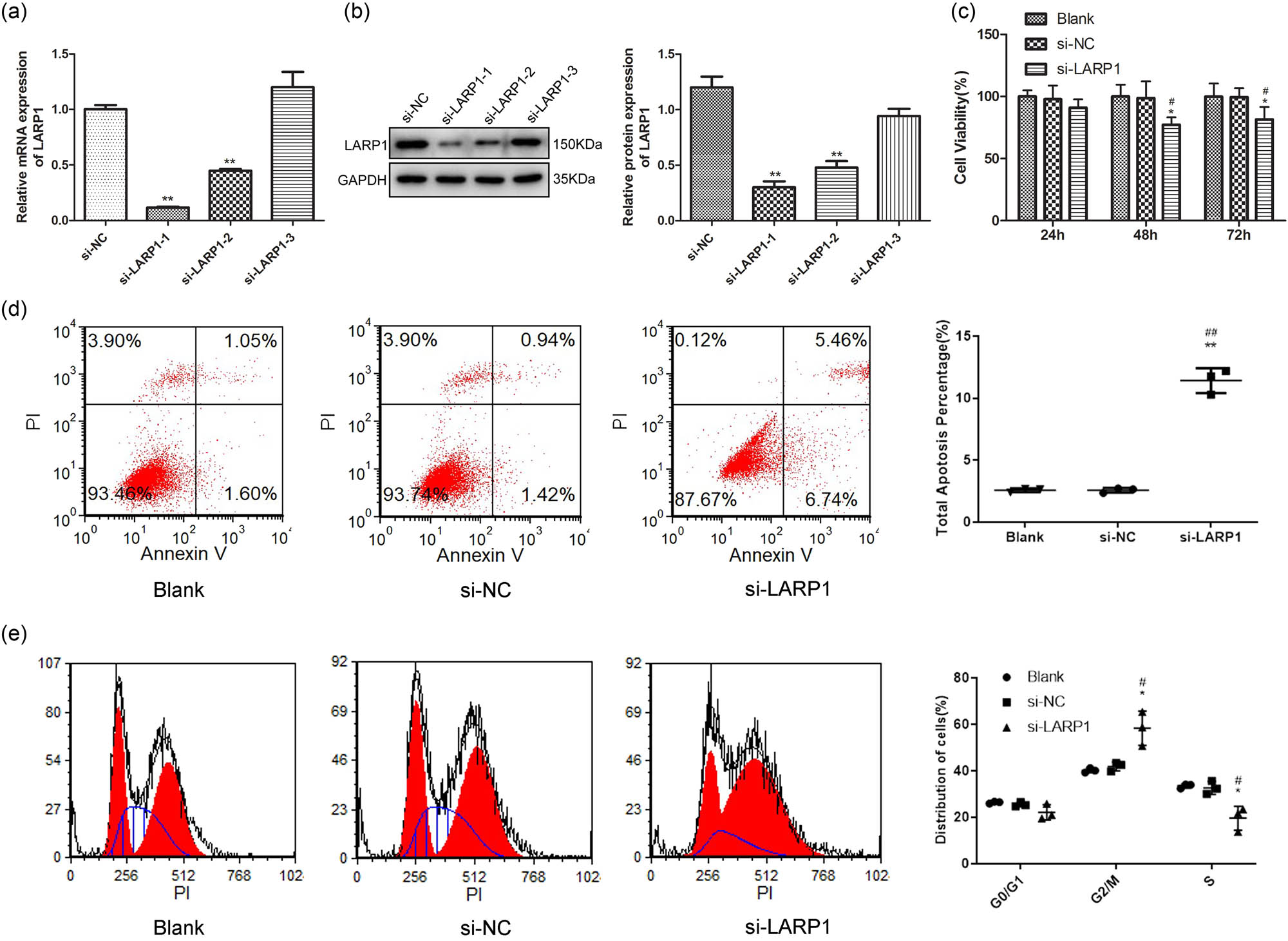
Effect of LARP1 knockdown on GC cell viability, apoptosis, and cell cycle distribution. (a) LARP1 mRNA expression was determined in KATO III cells transfected with three RNA interfering sequences. (b) LARP1 protein expression was determined in KATO III cells transfected with three RNA interfering sequences. (c) Cell viability was analyzed in KATO III cells 24, 48, and 72 h after si- LARP1 transfection. (d) Cell apoptosis was determined using Annexin V-FITC flow cytometric analysis in KATO III cells after 48 h of si-LARP1 transfection. (e) Cell cycle distribution was determined using propidium iodide (PI) flow cytometric analysis in KATO III cells after 48 h of si-LARP1 transfection. *P < 0.05, **P < 0.01 compared with the blank group. # P < 0.05, ## P < 0.01 compared with the si-NC group.
3.3 LARP1 knockdown restrained GC cell metastasis
To further investigate the function of LARP1 in GC cell metastasis, cell invasion, migration, and colony formation abilities were determined in KATO III cells transfected with si-LARP1. Figure 3a showed that compared with si-NC, LARP1 knockdown markedly reduced the number of migrating cells. In the transwell invasion analysis, compared with si-NC, LARP1 knockdown reduced the number of invaded cells (Figure 3b). Regarding colony formation, KATO III cells with LARP1 knockdown had fewer colonies (Figure 3c). These data indicate that LARP1 knockdown restrained the metastasis and growth of GC cells.
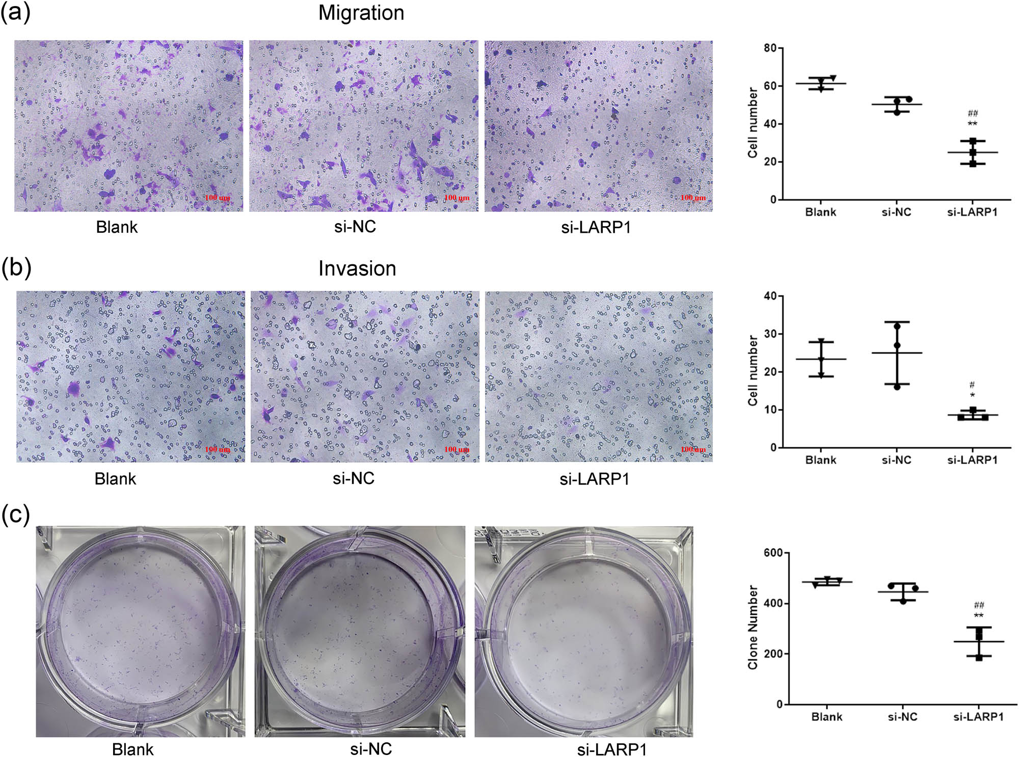
LARP1 knockdown inhibits GC cell migration, invasion, and colony formation. Transwell migration assay (a) and transwell invasion assay (b) were used to detect cell metastasis ability in si-LARP1-transfected KATO III cells. (c) Colony formation assay was used to evaluate cell growth in si-LARP1-transfected KATO III cells. *P < 0.05, **P < 0.01 compared with the blank group. # P < 0.05, ## P < 0.01 compared with the si-NC group.
3.4 LARP1 knockdown suppressed the PI3K/AKT1 signaling pathway
The PI3K/AKT1 signaling pathway is known to be involved in GC progression [14], and its inhibition promotes apoptosis and inhibits the metastasis of GC cells [15,16]. Next, we investigated whether the PI3K/AKT1 signaling pathway is the downstream target of LARP1 knockdown in KATO III cells. In si-LARP1 transfected KATO III cells, PI3K and AKT1 mRNA expression were significantly suppressed (Figure 4a). In addition, western blotting results suggested that PI3K and p-AKT1/AKT1 expression was significantly decreased in si-LARP1-transfected KATO III cells (P < 0.01). Given that LARP is involved in mTOR signaling, the downregulation of PI3K/AKT1 alone may not be equal to a reduction in cell viability. We further detected the expression of the PI3K/AKT downstream effectors involved in cell survival and proliferation, including p-mTOR, p-BAD, and p21. As shown in Figure 4c, p-mTOR and p-BAD were significantly decreased, whereas p21 was significantly increased in si-LARP1-transfected KATO III cells (P < 0.01).
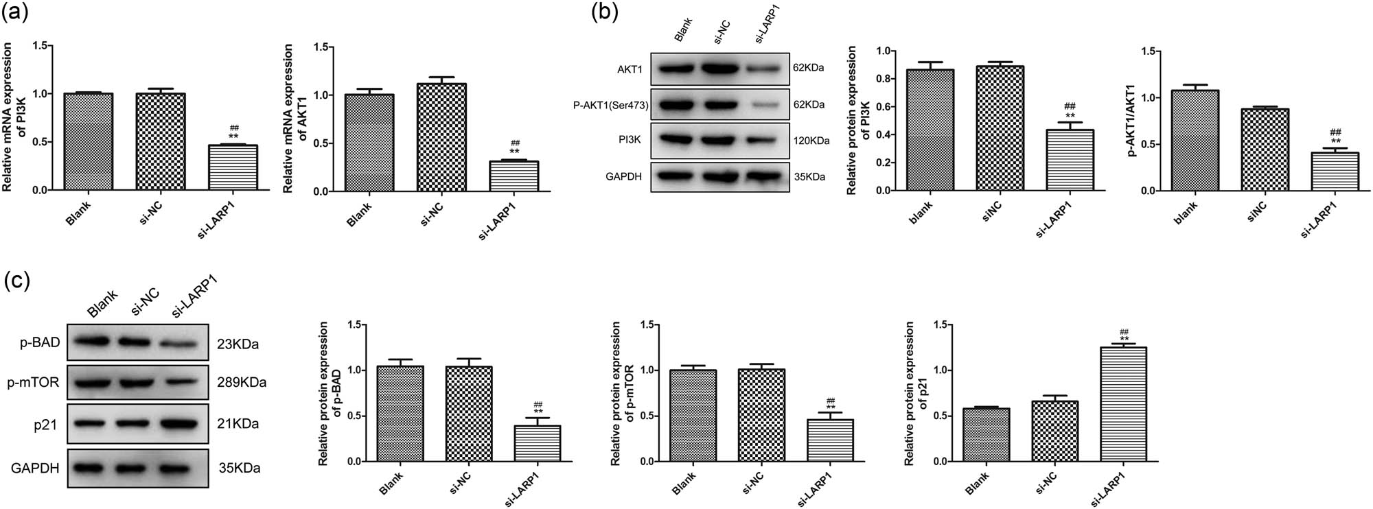
LARP1 knockdown suppresses the PI3K/AKT1 signaling pathway. (a) PI3K and AKT1 mRNA expression levels were determined using qRT-PCR. (b) Protein expression levels of PI3K, p-AKT1, and AKT1 were determined via western blotting. (c) Protein expression levels of p-BAD, p-mTOR, and p21 were determined via western blotting. **P < 0.01 compared with the blank group. ## P < 0.01 compared with the si-NC group.
4 Discussion
GC is a highly aggressive malignant tumor that constitutes a global health concern, and understanding its molecular characteristics will help identify more effective control strategies [17]. The current study elucidated the role of the RNA-binding protein LARP1 in GC cell cycle progression and metastatic behavior using in vitro cell experiments and demonstrated that LARP1 knockdown promoted cell apoptosis, arrested the cell cycle at the G2/M phase, and suppressed cell viability, migration, invasion, and colony formation in GC cells.
This study found that LARP1 was highly expressed in GC cells, and silencing of LARP1 inhibited GC cell viability and proliferation and promoted cell apoptosis and G2/M phase arrest. These results suggested that LARP1 plays an important role in regulating the malignant activity of cultured GC cells. LARP1 is an RNA-binding protein that can bind to mRNA with a 5′ TOP and regulate its stability and translation efficiency [18]. 5′ TOP mRNA mainly encodes proteins involved in translation and ribosome biogenesis. Therefore, LARP1 may affect protein synthesis and growth of GC cells by influencing the expression of 5′ TOP mRNA.
Additionally, LARP1 interacts with the mTORC1 complex in response to nutrient and growth factor signals. mTORC1 is a key regulator of cell growth, whose activity is regulated by LARP1 in pancreatic β-cells and hepatocytes [12]. Therefore, LARP1 may regulate the metabolism and proliferation of GC cells by participating in the mTORC1 signaling pathway. Moreover, this study found that silencing LARP1 caused G2/M phase arrest in GC cells, which may be related to the effect of LARP1 on cell cycle regulatory proteins. Some studies have shown that G2/M phase arrest is a mechanism of cell death induced by antitumor drugs that can trigger apoptosis or mitotic catastrophe [19–21]. Therefore, LARP1 may affect the fate of GC cells by regulating G2/M phase transition. However, how LARP1 senses and responds to cell cycle signals and how LARP1 interacts with other cell cycle regulators remain unclear, and further experiments are needed to investigate these issues.
As an oncogenic RNA-binding protein, LARP1 alters gene function and drives cancer development through abnormal changes in post-transcriptional regulation. Some studies have reported that metastasis, a crucial event in cancer progression, is affected by LARP1 expression. For example, Burrows et al. found that LARP1 regulates cell migration by facilitating the synthesis of proteins [22]. Mura et al. showed that LARP1 promotes cell growth, metastasis, and in vivo tumorigenesis in cervical cancer [12]. In the present study, GC cell invasion and migration were suppressed by LARP1 knockdown, indicating that LARP1 facilitates the motor ability of GC cells to invade and metastasize. LARP1 knockdown also inhibits colony formation in GC cells, suggesting that LARP1 plays an active role in proliferation and tumorigenesis. Therefore, we believe that LARP1 is critical for the aggressiveness of GC. However, the specific mechanisms require further investigation in animal studies and clinical trials.
Oxidative stress can lead to chronic inflammation, which, in turn, can cause multiple diseases, including cancer [23]. Natural products and their derived biomolecules that inhibit oxidative stress and inflammation are potential agents against cancer progression [24–27]. Inhibition of the PI3K/AKT signaling pathway was found to be effective in inhibiting angiogenesis, which plays a key role in cancer progression [28]. The present study revealed that PI3K and AKT1 expression was inhibited by the knockdown of LARP1. Activation of the PI3K and AKT1 cascades in many types of human cancers, including GC, drives cancer cell growth and survival, leading to tumor invasiveness and drug resistance [29,30]. The activation of PI3K and AKT1 and their roles in GC progression have been well described, including the promotion of survival and metastasis [31,32]. Activation of the PI3K/AKT1 pathway is involved in inhibiting the GC development of some anti-cancer substances, including drugs, functional proteins, and non-coding RNAs [33,34]. Although PI3K/AKT is a promising therapeutic target, AKT’s complex cellular functions of AKT and the activation of potential feedback signals limit AKT’s potency of AKT as a single drug [35]. A strategy targeting AKT in tumors with alterations in the PI3K pathway may benefit patients [36]. Our data showed that LARP1 knockdown suppressed the expression of PI3K and AKT1. Notably, there is limited evidence regarding the effect of LARP1 on the expression of PI3K and AKT1, and the specific regulatory mechanisms require further study.
Akt regulates cell growth by acting on the TSC1/TSC2 complex and mTORC signal transduction [37,38]. Akt affects cell proliferation by phosphorylating CDK inhibitors p21 and p27 [38]. Akt is a major regulator of cell survival and is regulated either directly by inhibiting pro-apoptotic proteins (such as BAD) or by inhibiting the production of pro-apoptotic signals via transcription factors [39]. Given that LARP is involved in mTOR signaling, the downregulation of PI3K/AKT1 alone may not be equal to a reduction in cell viability. We further detected the expression of the PI3K/AKT downstream effectors involved in cell survival and proliferation, including p-mTOR, p-BAD, and p21. The results indicated that p-mTOR and p-BAD were significantly decreased, whereas p21 was significantly increased in si-LARP1-transfected KATO III cells.
This study had some limitations. First, this study is only a preliminary investigation of the molecular mechanism of LARP1 in GC. The mechanistic analyses of the pathways involved in LARP1 expression are limited. Second, in vivo evidence is needed to confirm the role of LARP1 in the inhibition of metastatic behavior. Third, due to restrictions on funds and tissues, some experiments were lacking, such as repeated experiments in other cell lines or with another siRNA and immunohistochemical analysis of LARP1 in our cohort tissues.
5 Conclusion
Our findings indicate that LARP1 is upregulated in GC and that LARP1 knockdown induces apoptosis and inhibits cell cycle progression and metastatic behavior in cultured GC cells, mediated by the PI3K/AKT pathway (Figure 5). Thus, LARP1 is a potential therapeutic target for the treatment of GC.

Molecular mechanism underlying the regulation of GC cell cycle progression and metastatic behavior by LARP1.
-
Funding information: This project was supported by the Scientific Research Project of Guangxi Health Commission (No. Z2015357).
-
Author contributions: G.D.T. designed the experiments. X.L., W.M.Z., and N.M. carried out the experiments. L.J.L. provided the clinical samples and statistical analysis. X.L. prepared the manuscript with contributions from all co-authors.
-
Conflict of interest: Authors state no conflict of interest.
-
Data availability statement: The datasets generated during and/or analyzed during the current study are available from the corresponding author on reasonable request.
References
[1] Smyth EC, Nilsson M, Grabsch HI, van Grieken NC, Lordick F. Gastric cancer. Lancet. 2020;396(10251):635–48.10.1016/S0140-6736(20)31288-5Search in Google Scholar PubMed
[2] Joshi SS, Badgwell BD. Current treatment and recent progress in gastric cancer. CA Cancer J Clin. 2021;71(3):264–79.10.3322/caac.21657Search in Google Scholar PubMed PubMed Central
[3] Schinzari G, Cassano A, Orlandi A, Basso M, Barone C. Targeted therapy in advanced gastric carcinoma: the future is beginning. Curr Med Chem. 2014;21(8):1026–38.10.2174/0929867321666131129124054Search in Google Scholar PubMed
[4] Yang Y, Akashi Y, Shimomura O, Tateno H, Saito S, Hiemori K, et al. Glycan expression profile of signet ring cell gastric cancer cells and potential applicability of rBC2LCN-targeted lectin drug conjugate therapy. Gastric Cancer. 2022;25(5):896–905.10.1007/s10120-022-01312-xSearch in Google Scholar PubMed
[5] Novikov NM, Zolotaryova SY, Gautreau AM, Denisov EV. Mutational drivers of cancer cell migration and invasion. Br J Cancer. 2021;124(1):102–14.10.1038/s41416-020-01149-0Search in Google Scholar PubMed PubMed Central
[6] Reddavid R, Dagatti S, Franco C, Puca L, Tomatis M, Corso S, et al. Molecularly targeted therapies for gastric cancer. State of the art. Cancers (Basel). 2021;13(16):4094.10.3390/cancers13164094Search in Google Scholar PubMed PubMed Central
[7] Deragon JM, Bousquet-Antonelli C. The role of LARP1 in translation and beyond. Wiley Interdiscip Rev RNA. 2015;6(4):399–417.10.1002/wrna.1282Search in Google Scholar PubMed
[8] Tcherkezian J, Cargnello M, Romeo Y, Huttlin EL, Lavoie G, Gygi SP, et al. Proteomic analysis of cap-dependent translation identifies LARP1 as a key regulator of 5’TOP mRNA translation. Genes Dev. 2014;28(4):357–71.10.1101/gad.231407.113Search in Google Scholar PubMed PubMed Central
[9] Desi N, Tong QY, Teh V, Chan JJ, Zhang B, Tabatabaeian H, et al. Global analysis of RNA-binding proteins identifies a positive feedback loop between LARP1 and MYC that promotes tumorigenesis. Cell Mol Life Sci. 2022;79(3):147.10.1007/s00018-021-04093-1Search in Google Scholar PubMed
[10] Berman AJ, Thoreen CC, Dedeic Z, Chettle J, Roux PP, Blagden SP. Controversies around the function of LARP1. RNA Biol. 2021;18(2):207–17.10.1080/15476286.2020.1733787Search in Google Scholar PubMed PubMed Central
[11] Xie C, Huang L, Xie S, Xie D, Zhang G, Wang P, et al. LARP1 predict the prognosis for early-stage and AFP-normal hepatocellular carcinoma. J Transl Med. 2013;11:272.10.1186/1479-5876-11-272Search in Google Scholar PubMed PubMed Central
[12] Mura M, Hopkins TG, Michael T, Abd-Latip N, Weir J, Aboagye E, et al. LARP1 post-transcriptionally regulates mTOR and contributes to cancer progression. Oncogene. 2015;34(39):5025–36.10.1038/onc.2014.428Search in Google Scholar PubMed PubMed Central
[13] Hopkins TG, Mura M, Al-Ashtal HA, Lahr RM, Abd-Latip N, Sweeney K, et al. The RNA-binding protein LARP1 is a post-transcriptional regulator of survival and tumorigenesis in ovarian cancer. Nucleic Acids Res. 2016;44(3):1227–46.10.1093/nar/gkv1515Search in Google Scholar PubMed PubMed Central
[14] Fattahi S, Amjadi-Moheb F, Tabaripour R, Ashrafi GH, Akhavan-Niaki H. PI3K/AKT/mTOR signaling in gastric cancer: Epigenetics and beyond. Life Sci. 2020;262:118513.10.1016/j.lfs.2020.118513Search in Google Scholar PubMed
[15] Wu S, Chen M, Huang J, Zhang F, Lv Z, Jia Y, et al. ORAI2 promotes gastric cancer tumorigenicity and metastasis through PI3K/Akt signaling and MAPK-dependent focal adhesion disassembly. Cancer Res. 2021;81(4):986–1000.10.1158/0008-5472.CAN-20-0049Search in Google Scholar PubMed
[16] Dai J, Liu D, Chen L, Sun L. Effect of Ag-1031 on apoptosis in gastric cancer AGS cells and its effects on the PI3K/AKT/mTOR signaling pathway. Biotechnol Lett. 2020;42(11):2447–52.10.1007/s10529-020-02954-6Search in Google Scholar PubMed
[17] Gao JP, Xu W, Liu WT, Yan M, Zhu ZG. Tumor heterogeneity of gastric cancer: From the perspective of tumor-initiating cell. World J Gastroenterol. 2018;24(24):2567–81.10.3748/wjg.v24.i24.2567Search in Google Scholar PubMed PubMed Central
[18] Schwenzer H, Abdel Mouti M, Neubert P, Morris J, Stockton J, Bonham S, et al. LARP1 isoform expression in human cancer cell lines. RNA Biol. 2021;18(2):237–47.10.1080/15476286.2020.1744320Search in Google Scholar PubMed PubMed Central
[19] Khing TM, Choi WS, Kim DM, Po WW, Thein W, Shin CY, et al. The effect of paclitaxel on apoptosis, autophagy and mitotic catastrophe in AGS cells. Sci Rep. 2021;11(1):23490.10.1038/s41598-021-02503-9Search in Google Scholar PubMed PubMed Central
[20] Mehdizadeh R, Madjid Ansari A, Forouzesh F, Shahriari F, Shariatpanahi SP, Salaritabar A, et al. P53 status, and G2/M cell cycle arrest, are determining factors in cell-death induction mediated by ELF-EMF in glioblastoma. Sci Rep. 2023;13(1):10845.10.1038/s41598-023-38021-zSearch in Google Scholar PubMed PubMed Central
[21] Sun J, Li M, Lin T, Wang D, Chen J, Zhang Y, et al. Cell cycle arrest is an important mechanism of action of compound Kushen injection in the prevention of colorectal cancer. Sci Rep. 2022;12(1):4384.10.1038/s41598-022-08336-4Search in Google Scholar PubMed PubMed Central
[22] Burrows C, Abd Latip N, Lam SJ, Carpenter L, Sawicka K, Tzolovsky G, et al. The RNA binding protein Larp1 regulates cell division, apoptosis and cell migration. Nucleic Acids Res. 2010;38(16):5542–53.10.1093/nar/gkq294Search in Google Scholar PubMed PubMed Central
[23] Reuter S, Gupta SC, Chaturvedi MM, Aggarwal BB. Oxidative stress, inflammation, and cancer: how are they linked? Free Radic Biol Med. 2010;49(11):1603–16.10.1016/j.freeradbiomed.2010.09.006Search in Google Scholar PubMed PubMed Central
[24] Abdalla Y, Abdalla A, Hamza AA, Amin A. Safranal prevents liver cancer through inhibiting oxidative stress and alleviating inflammation. Front Pharmacol. 2021;12:777500.10.3389/fphar.2021.777500Search in Google Scholar PubMed PubMed Central
[25] Nelson DR, Hrout AA, Alzahmi AS, Chaiboonchoe A, Amin A, Salehi-Ashtiani K. Molecular mechanisms behind Safranal’s toxicity to HepG2 cells from dual Omics. Antioxidants (Basel). 2022;11(6):1125.10.3390/antiox11061125Search in Google Scholar PubMed PubMed Central
[26] Hamza AA, Heeba GH, Hassanin SO, Elwy HM, Bekhit AA, Amin A. Hibiscus-cisplatin combination treatment decreases liver toxicity in rats while increasing toxicity in lung cancer cells via oxidative stress- apoptosis pathway. Biomed Pharmacother. 2023;165:115148.10.1016/j.biopha.2023.115148Search in Google Scholar PubMed
[27] Abdu S, Juaid N, Amin A, Moulay M, Miled N. Therapeutic effects of crocin alone or in combination with sorafenib against hepatocellular carcinoma: In vivo & in vitro insights. Antioxidants (Basel). 2022;11(9):1645.10.3390/antiox11091645Search in Google Scholar PubMed PubMed Central
[28] Abdalla A, Murali C, Amin A. Safranal inhibits angiogenesis via targeting HIF-1α/VEGF machinery: In vitro and ex vivo insights. Front Oncol. 2021;11:789172.10.3389/fonc.2021.789172Search in Google Scholar PubMed PubMed Central
[29] Popova NV, Jucker M. The role of mTOR signaling as a therapeutic target in cancer. Int J Mol Sci. 2021;22(4):1743.10.3390/ijms22041743Search in Google Scholar PubMed PubMed Central
[30] Osaki M, Oshimura M, Ito H. PI3K-Akt pathway: its functions and alterations in human cancer. Apoptosis. 2004;9(6):667–76.10.1023/B:APPT.0000045801.15585.ddSearch in Google Scholar PubMed
[31] Wang LL, Zhang L, Cui XF. Downregulation of long noncoding RNA LINC01419 inhibits cell migration, invasion, and tumor growth and promotes autophagy via inactivation of the PI3K/Akt1/mTOR pathway in gastric cancer. Ther Adv Med Oncol. 2019;11:1758835919874651.10.1177/1758835919874651Search in Google Scholar PubMed PubMed Central
[32] Riquelme I, Tapia O, Espinoza JA, Leal P, Buchegger K, Sandoval A, et al. The gene expression status of the PI3K/AKT/mTOR pathway in gastric cancer tissues and cell lines. Pathol Oncol Res. 2016;22(4):797–805.10.1007/s12253-016-0066-5Search in Google Scholar PubMed PubMed Central
[33] Lin JX, Xie XS, Weng XF, Qiu SL, Yoon C, Lian NZ, et al. UFM1 suppresses invasive activities of gastric cancer cells by attenuating the expres7sion of PDK1 through PI3K/AKT signaling. J Exp Clin Cancer Res. 2019;38(1):410.10.1186/s13046-019-1416-4Search in Google Scholar PubMed PubMed Central
[34] Gu Y, Fei Z, Zhu R. miR-21 modulates cisplatin resistance of gastric cancer cells by inhibiting autophagy via the PI3K/Akt/mTOR pathway. Anticancer Drugs. 2020;31(4):385–93.10.1097/CAD.0000000000000886Search in Google Scholar PubMed
[35] Nunnery SE, Mayer IA. Targeting the PI3K/AKT/mTOR pathway in hormone-positive breast cancer. Drugs. 2020;80(16):1685–97.10.1007/s40265-020-01394-wSearch in Google Scholar PubMed PubMed Central
[36] Kim SB, Dent R, Im SA, Espie M, Blau S, Tan AR, et al. Ipatasertib plus paclitaxel versus placebo plus paclitaxel as first-line therapy for metastatic triple-negative breast cancer (LOTUS): a multicentre, randomised, double-blind, placebo-controlled, phase 2 trial. Lancet Oncol. 2017;18(10):1360–72.10.1016/S1470-2045(17)30450-3Search in Google Scholar PubMed PubMed Central
[37] Potter CJ, Pedraza LG, Xu T. Akt regulates growth by directly phosphorylating Tsc2. Nat Cell Biol. 2002;4(9):658–65.10.1038/ncb840Search in Google Scholar PubMed
[38] Glaviano A, Foo ASC, Lam HY, Yap KCH, Jacot W, Jones RH, et al. PI3K/AKT/mTOR signaling transduction pathway and targeted therapies in cancer. Mol Cancer. 2023;22(1):138.10.1186/s12943-023-01827-6Search in Google Scholar PubMed PubMed Central
[39] Xu Y, Jiang Y, Wang Y, Zhao Z, Li T. LINC00473 rescues human bone marrow mesenchymal stem cells from apoptosis induced by dexamethasone through the PEBP1‑mediated Akt/Bad/Bcl‑2 signaling pathway. Int J Mol Med. 2021;47(1):171–82.10.3892/ijmm.2020.4788Search in Google Scholar PubMed PubMed Central
© 2024 the author(s), published by De Gruyter
This work is licensed under the Creative Commons Attribution 4.0 International License.
Articles in the same Issue
- Biomedical Sciences
- Constitutive and evoked release of ATP in adult mouse olfactory epithelium
- LARP1 knockdown inhibits cultured gastric carcinoma cell cycle progression and metastatic behavior
- PEGylated porcine–human recombinant uricase: A novel fusion protein with improved efficacy and safety for the treatment of hyperuricemia and renal complications
- Research progress on ocular complications caused by type 2 diabetes mellitus and the function of tears and blepharons
- The role and mechanism of esketamine in preventing and treating remifentanil-induced hyperalgesia based on the NMDA receptor–CaMKII pathway
- Brucella infection combined with Nocardia infection: A case report and literature review
- Detection of serum interleukin-18 level and neutrophil/lymphocyte ratio in patients with antineutrophil cytoplasmic antibody-associated vasculitis and its clinical significance
- Ang-1, Ang-2, and Tie2 are diagnostic biomarkers for Henoch-Schönlein purpura and pediatric-onset systemic lupus erythematous
- PTTG1 induces pancreatic cancer cell proliferation and promotes aerobic glycolysis by regulating c-myc
- Role of serum B-cell-activating factor and interleukin-17 as biomarkers in the classification of interstitial pneumonia with autoimmune features
- Effectiveness and safety of a mumps containing vaccine in preventing laboratory-confirmed mumps cases from 2002 to 2017: A meta-analysis
- Low levels of sex hormone-binding globulin predict an increased breast cancer risk and its underlying molecular mechanisms
- A case of Trousseau syndrome: Screening, detection and complication
- Application of the integrated airway humidification device enhances the humidification effect of the rabbit tracheotomy model
- Preparation of Cu2+/TA/HAP composite coating with anti-bacterial and osteogenic potential on 3D-printed porous Ti alloy scaffolds for orthopedic applications
- Aquaporin-8 promotes human dermal fibroblasts to counteract hydrogen peroxide-induced oxidative damage: A novel target for management of skin aging
- Current research and evidence gaps on placental development in iron deficiency anemia
- Single-nucleotide polymorphism rs2910829 in PDE4D is related to stroke susceptibility in Chinese populations: The results of a meta-analysis
- Pheochromocytoma-induced myocardial infarction: A case report
- Kaempferol regulates apoptosis and migration of neural stem cells to attenuate cerebral infarction by O‐GlcNAcylation of β-catenin
- Sirtuin 5 regulates acute myeloid leukemia cell viability and apoptosis by succinylation modification of glycine decarboxylase
- Apigenin 7-glucoside impedes hypoxia-induced malignant phenotypes of cervical cancer cells in a p16-dependent manner
- KAT2A changes the function of endometrial stromal cells via regulating the succinylation of ENO1
- Current state of research on copper complexes in the treatment of breast cancer
- Exploring antioxidant strategies in the pathogenesis of ALS
- Helicobacter pylori causes gastric dysbacteriosis in chronic gastritis patients
- IL-33/soluble ST2 axis is associated with radiation-induced cardiac injury
- The predictive value of serum NLR, SII, and OPNI for lymph node metastasis in breast cancer patients with internal mammary lymph nodes after thoracoscopic surgery
- Carrying SNP rs17506395 (T > G) in TP63 gene and CCR5Δ32 mutation associated with the occurrence of breast cancer in Burkina Faso
- P2X7 receptor: A receptor closely linked with sepsis-associated encephalopathy
- Probiotics for inflammatory bowel disease: Is there sufficient evidence?
- Identification of KDM4C as a gene conferring drug resistance in multiple myeloma
- Microbial perspective on the skin–gut axis and atopic dermatitis
- Thymosin α1 combined with XELOX improves immune function and reduces serum tumor markers in colorectal cancer patients after radical surgery
- Highly specific vaginal microbiome signature for gynecological cancers
- Sample size estimation for AQP4-IgG seropositive optic neuritis: Retinal damage detection by optical coherence tomography
- The effects of SDF-1 combined application with VEGF on femoral distraction osteogenesis in rats
- Fabrication and characterization of gold nanoparticles using alginate: In vitro and in vivo assessment of its administration effects with swimming exercise on diabetic rats
- Mitigating digestive disorders: Action mechanisms of Mediterranean herbal active compounds
- Distribution of CYP2D6 and CYP2C19 gene polymorphisms in Han and Uygur populations with breast cancer in Xinjiang, China
- VSP-2 attenuates secretion of inflammatory cytokines induced by LPS in BV2 cells by mediating the PPARγ/NF-κB signaling pathway
- Factors influencing spontaneous hypothermia after emergency trauma and the construction of a predictive model
- Long-term administration of morphine specifically alters the level of protein expression in different brain regions and affects the redox state
- Application of metagenomic next-generation sequencing technology in the etiological diagnosis of peritoneal dialysis-associated peritonitis
- Clinical diagnosis, prevention, and treatment of neurodyspepsia syndrome using intelligent medicine
- Case report: Successful bronchoscopic interventional treatment of endobronchial leiomyomas
- Preliminary investigation into the genetic etiology of short stature in children through whole exon sequencing of the core family
- Cystic adenomyoma of the uterus: Case report and literature review
- Mesoporous silica nanoparticles as a drug delivery mechanism
- Dynamic changes in autophagy activity in different degrees of pulmonary fibrosis in mice
- Vitamin D deficiency and inflammatory markers in type 2 diabetes: Big data insights
- Lactate-induced IGF1R protein lactylation promotes proliferation and metabolic reprogramming of lung cancer cells
- Meta-analysis on the efficacy of allogeneic hematopoietic stem cell transplantation to treat malignant lymphoma
- Mitochondrial DNA drives neuroinflammation through the cGAS-IFN signaling pathway in the spinal cord of neuropathic pain mice
- Application value of artificial intelligence algorithm-based magnetic resonance multi-sequence imaging in staging diagnosis of cervical cancer
- Embedded monitoring system and teaching of artificial intelligence online drug component recognition
- Investigation into the association of FNDC1 and ADAMTS12 gene expression with plumage coloration in Muscovy ducks
- Yak meat content in feed and its impact on the growth of rats
- A rare case of Richter transformation with breast involvement: A case report and literature review
- First report of Nocardia wallacei infection in an immunocompetent patient in Zhejiang province
- Rhodococcus equi and Brucella pulmonary mass in immunocompetent: A case report and literature review
- Downregulation of RIP3 ameliorates the left ventricular mechanics and function after myocardial infarction via modulating NF-κB/NLRP3 pathway
- Evaluation of the role of some non-enzymatic antioxidants among Iraqi patients with non-alcoholic fatty liver disease
- The role of Phafin proteins in cell signaling pathways and diseases
- Ten-year anemia as initial manifestation of Castleman disease in the abdominal cavity: A case report
- Coexistence of hereditary spherocytosis with SPTB P.Trp1150 gene variant and Gilbert syndrome: A case report and literature review
- Utilization of convolutional neural networks to analyze microscopic images for high-throughput screening of mesenchymal stem cells
- Exploratory evaluation supported by experimental and modeling approaches of Inula viscosa root extract as a potent corrosion inhibitor for mild steel in a 1 M HCl solution
- Imaging manifestations of ductal adenoma of the breast: A case report
- Gut microbiota and sleep: Interaction mechanisms and therapeutic prospects
- Isomangiferin promotes the migration and osteogenic differentiation of rat bone marrow mesenchymal stem cells
- Prognostic value and microenvironmental crosstalk of exosome-related signatures in human epidermal growth factor receptor 2 positive breast cancer
- Circular RNAs as potential biomarkers for male severe sepsis
- Knockdown of Stanniocalcin-1 inhibits growth and glycolysis in oral squamous cell carcinoma cells
- The expression and biological role of complement C1s in esophageal squamous cell carcinoma
- A novel GNAS mutation in pseudohypoparathyroidism type 1a with articular flexion deformity: A case report
- Predictive value of serum magnesium levels for prognosis in patients with non-small cell lung cancer undergoing EGFR-TKI therapy
- HSPB1 alleviates acute-on-chronic liver failure via the P53/Bax pathway
- IgG4-related disease complicated by PLA2R-associated membranous nephropathy: A case report
- Baculovirus-mediated endostatin and angiostatin activation of autophagy through the AMPK/AKT/mTOR pathway inhibits angiogenesis in hepatocellular carcinoma
- Metformin mitigates osteoarthritis progression by modulating the PI3K/AKT/mTOR signaling pathway and enhancing chondrocyte autophagy
- Evaluation of the activity of antimicrobial peptides against bacterial vaginosis
- Atypical presentation of γ/δ mycosis fungoides with an unusual phenotype and SOCS1 mutation
- Analysis of the microecological mechanism of diabetic kidney disease based on the theory of “gut–kidney axis”: A systematic review
- Omega-3 fatty acids prevent gestational diabetes mellitus via modulation of lipid metabolism
- Refractory hypertension complicated with Turner syndrome: A case report
- Interaction of ncRNAs and the PI3K/AKT/mTOR pathway: Implications for osteosarcoma
- Association of low attenuation area scores with pulmonary function and clinical prognosis in patients with chronic obstructive pulmonary disease
- Long non-coding RNAs in bone formation: Key regulators and therapeutic prospects
- The deubiquitinating enzyme USP35 regulates the stability of NRF2 protein
- Neutrophil-to-lymphocyte ratio and platelet-to-lymphocyte ratio as potential diagnostic markers for rebleeding in patients with esophagogastric variceal bleeding
- G protein-coupled receptor 1 participating in the mechanism of mediating gestational diabetes mellitus by phosphorylating the AKT pathway
- LL37-mtDNA regulates viability, apoptosis, inflammation, and autophagy in lipopolysaccharide-treated RLE-6TN cells by targeting Hsp90aa1
- The analgesic effect of paeoniflorin: A focused review
- Chemical composition’s effect on Solanum nigrum Linn.’s antioxidant capacity and erythrocyte protection: Bioactive components and molecular docking analysis
- Knockdown of HCK promotes HREC cell viability and inner blood–retinal barrier integrity by regulating the AMPK signaling pathway
- The role of rapamycin in the PINK1/Parkin signaling pathway in mitophagy in podocytes
- Laryngeal non-Hodgkin lymphoma: Report of four cases and review of the literature
- Clinical value of macrogenome next-generation sequencing on infections
- Overview of dendritic cells and related pathways in autoimmune uveitis
- TAK-242 alleviates diabetic cardiomyopathy via inhibiting pyroptosis and TLR4/CaMKII/NLRP3 pathway
- Hypomethylation in promoters of PGC-1α involved in exercise-driven skeletal muscular alterations in old age
- Profile and antimicrobial susceptibility patterns of bacteria isolated from effluents of Kolladiba and Debark hospitals
- The expression and clinical significance of syncytin-1 in serum exosomes of hepatocellular carcinoma patients
- A histomorphometric study to evaluate the therapeutic effects of biosynthesized silver nanoparticles on the kidneys infected with Plasmodium chabaudi
- PGRMC1 and PAQR4 are promising molecular targets for a rare subtype of ovarian cancer
- Analysis of MDA, SOD, TAOC, MNCV, SNCV, and TSS scores in patients with diabetes peripheral neuropathy
- SLIT3 deficiency promotes non-small cell lung cancer progression by modulating UBE2C/WNT signaling
- The relationship between TMCO1 and CALR in the pathological characteristics of prostate cancer and its effect on the metastasis of prostate cancer cells
- Heterogeneous nuclear ribonucleoprotein K is a potential target for enhancing the chemosensitivity of nasopharyngeal carcinoma
- PHB2 alleviates retinal pigment epithelium cell fibrosis by suppressing the AGE–RAGE pathway
- Anti-γ-aminobutyric acid-B receptor autoimmune encephalitis with syncope as the initial symptom: Case report and literature review
- Comparative analysis of chloroplast genome of Lonicera japonica cv. Damaohua
- Human umbilical cord mesenchymal stem cells regulate glutathione metabolism depending on the ERK–Nrf2–HO-1 signal pathway to repair phosphoramide mustard-induced ovarian cancer cells
- Electroacupuncture on GB acupoints improves osteoporosis via the estradiol–PI3K–Akt signaling pathway
- Renalase protects against podocyte injury by inhibiting oxidative stress and apoptosis in diabetic nephropathy
- Review: Dicranostigma leptopodum: A peculiar plant of Papaveraceae
- Combination effect of flavonoids attenuates lung cancer cell proliferation by inhibiting the STAT3 and FAK signaling pathway
- Renal microangiopathy and immune complex glomerulonephritis induced by anti-tumour agents: A case report
- Correlation analysis of AVPR1a and AVPR2 with abnormal water and sodium and potassium metabolism in rats
- Gastrointestinal health anti-diarrheal mixture relieves spleen deficiency-induced diarrhea through regulating gut microbiota
- Myriad factors and pathways influencing tumor radiotherapy resistance
- Exploring the effects of culture conditions on Yapsin (YPS) gene expression in Nakaseomyces glabratus
- Screening of prognostic core genes based on cell–cell interaction in the peripheral blood of patients with sepsis
- Coagulation factor II thrombin receptor as a promising biomarker in breast cancer management
- Ileocecal mucinous carcinoma misdiagnosed as incarcerated hernia: A case report
- Methyltransferase like 13 promotes malignant behaviors of bladder cancer cells through targeting PI3K/ATK signaling pathway
- The debate between electricity and heat, efficacy and safety of irreversible electroporation and radiofrequency ablation in the treatment of liver cancer: A meta-analysis
- ZAG promotes colorectal cancer cell proliferation and epithelial–mesenchymal transition by promoting lipid synthesis
- Baicalein inhibits NLRP3 inflammasome activation and mitigates placental inflammation and oxidative stress in gestational diabetes mellitus
- Impact of SWCNT-conjugated senna leaf extract on breast cancer cells: A potential apoptotic therapeutic strategy
- MFAP5 inhibits the malignant progression of endometrial cancer cells in vitro
- Major ozonated autohemotherapy promoted functional recovery following spinal cord injury in adult rats via the inhibition of oxidative stress and inflammation
- Axodendritic targeting of TAU and MAP2 and microtubule polarization in iPSC-derived versus SH-SY5Y-derived human neurons
- Differential expression of phosphoinositide 3-kinase/protein kinase B and Toll-like receptor/nuclear factor kappa B signaling pathways in experimental obesity Wistar rat model
- The therapeutic potential of targeting Oncostatin M and the interleukin-6 family in retinal diseases: A comprehensive review
- BA inhibits LPS-stimulated inflammatory response and apoptosis in human middle ear epithelial cells by regulating the Nf-Kb/Iκbα axis
- Role of circRMRP and circRPL27 in chronic obstructive pulmonary disease
- Investigating the role of hyperexpressed HCN1 in inducing myocardial infarction through activation of the NF-κB signaling pathway
- Characterization of phenolic compounds and evaluation of anti-diabetic potential in Cannabis sativa L. seeds: In vivo, in vitro, and in silico studies
- Quantitative immunohistochemistry analysis of breast Ki67 based on artificial intelligence
- Ecology and Environmental Science
- Screening of different growth conditions of Bacillus subtilis isolated from membrane-less microbial fuel cell toward antimicrobial activity profiling
- Degradation of a mixture of 13 polycyclic aromatic hydrocarbons by commercial effective microorganisms
- Evaluation of the impact of two citrus plants on the variation of Panonychus citri (Acari: Tetranychidae) and beneficial phytoseiid mites
- Prediction of present and future distribution areas of Juniperus drupacea Labill and determination of ethnobotany properties in Antalya Province, Türkiye
- Population genetics of Todarodes pacificus (Cephalopoda: Ommastrephidae) in the northwest Pacific Ocean via GBS sequencing
- A comparative analysis of dendrometric, macromorphological, and micromorphological characteristics of Pistacia atlantica subsp. atlantica and Pistacia terebinthus in the middle Atlas region of Morocco
- Macrofungal sporocarp community in the lichen Scots pine forests
- Assessing the proximate compositions of indigenous forage species in Yemen’s pastoral rangelands
- Food Science
- Gut microbiota changes associated with low-carbohydrate diet intervention for obesity
- Reexamination of Aspergillus cristatus phylogeny in dark tea: Characteristics of the mitochondrial genome
- Differences in the flavonoid composition of the leaves, fruits, and branches of mulberry are distinguished based on a plant metabolomics approach
- Investigating the impact of wet rendering (solventless method) on PUFA-rich oil from catfish (Clarias magur) viscera
- Non-linear associations between cardiovascular metabolic indices and metabolic-associated fatty liver disease: A cross-sectional study in the US population (2017–2020)
- Knockdown of USP7 alleviates atherosclerosis in ApoE-deficient mice by regulating EZH2 expression
- Utility of dairy microbiome as a tool for authentication and traceability
- Agriculture
- Enhancing faba bean (Vicia faba L.) productivity through establishing the area-specific fertilizer rate recommendation in southwest Ethiopia
- Impact of novel herbicide based on synthetic auxins and ALS inhibitor on weed control
- Perspectives of pteridophytes microbiome for bioremediation in agricultural applications
- Fertilizer application parameters for drip-irrigated peanut based on the fertilizer effect function established from a “3414” field trial
- Improving the productivity and profitability of maize (Zea mays L.) using optimum blended inorganic fertilization
- Application of leaf multispectral analyzer in comparison to hyperspectral device to assess the diversity of spectral reflectance indices in wheat genotypes
- Animal Sciences
- Knockdown of ANP32E inhibits colorectal cancer cell growth and glycolysis by regulating the AKT/mTOR pathway
- Development of a detection chip for major pathogenic drug-resistant genes and drug targets in bovine respiratory system diseases
- Exploration of the genetic influence of MYOT and MB genes on the plumage coloration of Muscovy ducks
- Transcriptome analysis of adipose tissue in grazing cattle: Identifying key regulators of fat metabolism
- Comparison of nutritional value of the wild and cultivated spiny loaches at three growth stages
- Transcriptomic analysis of liver immune response in Chinese spiny frog (Quasipaa spinosa) infected with Proteus mirabilis
- Disruption of BCAA degradation is a critical characteristic of diabetic cardiomyopathy revealed by integrated transcriptome and metabolome analysis
- Plant Sciences
- Effect of long-term in-row branch covering on soil microorganisms in pear orchards
- Photosynthetic physiological characteristics, growth performance, and element concentrations reveal the calcicole–calcifuge behaviors of three Camellia species
- Transcriptome analysis reveals the mechanism of NaHCO3 promoting tobacco leaf maturation
- Bioinformatics, expression analysis, and functional verification of allene oxide synthase gene HvnAOS1 and HvnAOS2 in qingke
- Water, nitrogen, and phosphorus coupling improves gray jujube fruit quality and yield
- Improving grape fruit quality through soil conditioner: Insights from RNA-seq analysis of Cabernet Sauvignon roots
- Role of Embinin in the reabsorption of nucleus pulposus in lumbar disc herniation: Promotion of nucleus pulposus neovascularization and apoptosis of nucleus pulposus cells
- Revealing the effects of amino acid, organic acid, and phytohormones on the germination of tomato seeds under salinity stress
- Combined effects of nitrogen fertilizer and biochar on the growth, yield, and quality of pepper
- Comprehensive phytochemical and toxicological analysis of Chenopodium ambrosioides (L.) fractions
- Impact of “3414” fertilization on the yield and quality of greenhouse tomatoes
- Exploring the coupling mode of water and fertilizer for improving growth, fruit quality, and yield of the pear in the arid region
- Metagenomic analysis of endophytic bacteria in seed potato (Solanum tuberosum)
- Antibacterial, antifungal, and phytochemical properties of Salsola kali ethanolic extract
- Exploring the hepatoprotective properties of citronellol: In vitro and in silico studies on ethanol-induced damage in HepG2 cells
- Enhanced osmotic dehydration of watermelon rind using honey–sucrose solutions: A study on pre-treatment efficacy and mass transfer kinetics
- Effects of exogenous 2,4-epibrassinolide on photosynthetic traits of 53 cowpea varieties under NaCl stress
- Comparative transcriptome analysis of maize (Zea mays L.) seedlings in response to copper stress
- An optimization method for measuring the stomata in cassava (Manihot esculenta Crantz) under multiple abiotic stresses
- Fosinopril inhibits Ang II-induced VSMC proliferation, phenotype transformation, migration, and oxidative stress through the TGF-β1/Smad signaling pathway
- Antioxidant and antimicrobial activities of Salsola imbricata methanolic extract and its phytochemical characterization
- Bioengineering and Biotechnology
- Absorbable calcium and phosphorus bioactive membranes promote bone marrow mesenchymal stem cells osteogenic differentiation for bone regeneration
- New advances in protein engineering for industrial applications: Key takeaways
- An overview of the production and use of Bacillus thuringiensis toxin
- Research progress of nanoparticles in diagnosis and treatment of hepatocellular carcinoma
- Bioelectrochemical biosensors for water quality assessment and wastewater monitoring
- PEI/MMNs@LNA-542 nanoparticles alleviate ICU-acquired weakness through targeted autophagy inhibition and mitochondrial protection
- Unleashing of cytotoxic effects of thymoquinone-bovine serum albumin nanoparticles on A549 lung cancer cells
- Erratum
- Erratum to “Investigating the association between dietary patterns and glycemic control among children and adolescents with T1DM”
- Erratum to “Activation of hypermethylated P2RY1 mitigates gastric cancer by promoting apoptosis and inhibiting proliferation”
- Retraction
- Retraction to “MiR-223-3p regulates cell viability, migration, invasion, and apoptosis of non-small cell lung cancer cells by targeting RHOB”
- Retraction to “A data mining technique for detecting malignant mesothelioma cancer using multiple regression analysis”
- Special Issue on Advances in Neurodegenerative Disease Research and Treatment
- Transplantation of human neural stem cell prevents symptomatic motor behavior disability in a rat model of Parkinson’s disease
- Special Issue on Multi-omics
- Inflammasome complex genes with clinical relevance suggest potential as therapeutic targets for anti-tumor drugs in clear cell renal cell carcinoma
- Gastroesophageal varices in primary biliary cholangitis with anti-centromere antibody positivity: Early onset?