Ang-1, Ang-2, and Tie2 are diagnostic biomarkers for Henoch-Schönlein purpura and pediatric-onset systemic lupus erythematous
-
Lishan Jia
 ,
Xiaozhong Li
,
Xiaozhong Li

Abstract
Henoch-Schönlein purpura (HSP) and pediatric-onset systemic lupus erythematosus (pSLE) are closely associated with vasculitis and vascular diseases. This study aimed to investigate the clinical diagnostic values of Ang-1, Ang-2, and Tie2 for HSP and pSLE. We surveyed 82 HSP patients, 34 pSLE patients, and 10 healthy children. The expression levels of Ang-1, Ang-2, and Tie2 in the serum and urine were assessed using enzyme-linked immunosorbent assay. The diagnostic values of Ang-1, Ang-2, and Tie2 for HSP and pSLE were evaluated using receiver operating characteristic curve analysis. The results revealed that the serum and urine expression levels of Ang-2 and Tie2 were significantly elevated in HSP and pSLE patients, whereas the Ang-1/Ang-2 values were reduced. Additionally, Ang-1 was highly expressed in the serum and urine of HSP patients and in the serum of pSLE patients. Ang-1, Ang-2, and Tie2 showed differential expression in various types of HSP and pSLE compared with their expression in healthy controls. In summary, Ang-1, Ang-2, and Tie2 can serve as biomarkers for HSP and pSLE. Moreover, Ang-1/Ang-2 values are reduced in HSP and pSLE patients. Ang-1, Ang-2, and Tie2 can be used as biomarkers for HSP and pSLE.
Graphical abstract

1 Introduction
Henoch-Schönlein purpura (HSP), also known as immunoglobulin A vasculitis, is characterized by IgA-dominated immune deposits in vessel walls [1]. HSP is the most common form of vasculitis in children, impacting 8–20 children per 100,000 annually. Among pediatric patients, 75–90% are younger than 10 years [2]. The clinical manifestations of HSP include palpable purpura, arthritis, abdominal pain, and renal involvement, which may progress to chronic kidney disease in severe cases [3,4]. Currently, the criteria for diagnosing children with HSP are mainly based on IgA deposits and kidney and joint skin involvement [5,6]. Rare complications associated with HSP can lead to misdiagnoses and delays in treatment, potentially resulting in poorer prognoses [7]. Therefore, it is crucial to explore diagnostic biomarkers for children with HSP.
Systemic lupus erythematosus (SLE) is an autoimmune disease associated with potential extensive angiopathy and vasculitis [8]. Approximately 11–36% of SLE patients present symptoms of vasculitis, which may lead to further visceral lesions, mesenteric vasculitis, pulmonary hemorrhage, and neuritis [9]. Pediatric-onset SLE (pSLE) accounts for approximately 10–20% of all SLE cases [10]. pSLE is characterized by its multisystemic and inflammatory nature [11]. It is more aggressive than adult-onset SLE, with greater organ and system involvement and imparting a higher drug burden, ultimately leading to high mortality rates and healthcare burden [12–15]. Moreover, pSLE-induced organ damage is significantly associated with longer disease duration, severe fatigue, and poorer quality of life [16]. pSLE is primarily diagnosed on the basis of antinuclear antibodies, anti-double stranded DNA antibodies, complement levels (C3, C4, CH50), and imaging studies [17–19]. However, these parameters lack sufficient sensitivity and specificity. Therefore, novel, specific, and safe biomarkers are urgently required to predict and detect pSLE.
The families of endothelial cell-specific receptor tyrosine kinases and their corresponding ligands/growth factors, including angiopoietins (Ang), ephrins, and vascular endothelial growth factors (VEGFs), are key contributors to vascular development, remodeling, and regeneration [20]. The Ang/Tie pathway is crucial for regulating vascular stability, angiogenesis, and the inflammatory response [21]. Ang-1 activates the Tie2 receptors, thereby promoting vascular stability, maturation, and integrity. Tie2 is a tyrosine kinase receptor expressed on the surface of vascular endothelial cells. Ang-2, an Ang-1 antagonist, competes to bind to Tie2 receptors [22]. The Ang/Tie2 signaling pathway is associated with multiple diseases. Ang-1/Tie2 signaling increases vascular access to tumor cells, promoting tumor dissemination and metastasis through vasodilation [23]. Purkinje cell dendritic development is autonomously regulated by the Ang-Tie2 pathway [24]. In ischemic diseases such as diabetic retinopathy, the upregulation of Ang-2 inactivates Tie-2, leading to vascular leakage, pericyte loss, and inflammation [25]. A cross-sectional analysis revealed a significant correlation between Tie2/Ang levels and the characteristics of circulating immune cells in well-differentiated neuroendocrine gastroenteropancreatic tumors [26]. Additionally, Tie2 levels in the serum of individuals with active pSLE were substantially higher than in those with an inactive condition [8]. However, the expression levels of Ang-1, Ang-2, and Tie2 in the urine and their diagnostic value in pSLE patients remain unclear. Furthermore, few studies have reported on the Ang/Tie2 pathway in HSP.
This study aimed to investigate the expression levels and diagnostic value of Ang/Tie2 in the serum and urine of HSP and pSLE patients. Initially, we assessed the levels of Ang-1, Ang-2, and Tie2 in both serum and urine using enzyme-linked immunosorbent assay (ELISA), revealing distinct expressions compared with those in healthy controls. Subsequently, we delved into the expression patterns of Ang-1, Ang-2, and Tie2 in patients with different types of HSP and pSLE. Finally, through receiver operating characteristic (ROC) curve analysis, we identified that Ang-1, Ang-2, and Tie2 could serve as diagnostic biomarkers in both serum and urine among HSP and pSLE patients. Our findings may identify diagnostic biomarkers and therapeutic targets for the treatment of HSP and pSLE patients.
2 Methods
2.1 Patients
Eighty-two HSP patients were recruited from the First People’s Hospital of Taicang, affiliated with Soochow University, the Children’s Hospital of Fudan University, and the Children’s Hospital of Soochow University, between July 2020 and July 2022. Additionally, 34 pSLE patients from the First People’s Hospital of Taicang, the Children’s Hospital of Fudan University, and the Children’s Hospital of Soochow University were enrolled. Ten healthy controls were enrolled from the Children’s Hospital of Fudan University and the Children’s Hospital of Soochow University during the same time period. The inclusion criteria were (1) patients with clinically confirmed HSP and pSLE, (2) all indicators within the normal ranges, (3) no other complications, (4) age ≤14 years, and (5) the patients’ families provided informed consent to their enrolment in the study. The exclusion criteria were (1) the presence of chronic infection with a history of serious infection in the last 2 months, (2) the combination of serious systemic, liver and kidney, and gastrointestinal diseases, (3) the presence of multiple autoimmune diseases, and (4) incomplete clinical data. The HSP, pSLE, and control groups did not differ significantly in terms of sex and age (p > 0.05). The study was approved by the ethics committee of the First People’s Hospital of Taicang, affiliated with Soochow University (Ethical number: KY-2020-204), and the sampling of all participants was approved by the participants’ families.
-
Informed consent: Informed consent has been obtained from all individuals included in this study.
-
Ethical approval: The research related to human use has been complied with all the relevant national regulations, institutional policies and in accordance with the tenets of the Helsinki Declaration, and has been approved by the ethics committee of the First People’s Hospital of Taicang, affiliated with Soochow University (Ethical number: KY-2020-204).
2.2 Determining the Ang-1, Ang-2, and Tie2 levels
Serum and urine specimens were collected from 82 HSP patients, 34 pSLE patients, and 10 healthy individuals. Specifically, a total of 5 mL of venous blood was collected from each participant in the early morning under fasting conditions and placed in a serum separator tube. The serum was then centrifuged at 4°C (3,500 rpm/10 min), and the upper layer was transferred into 1.5 mL centrifuge tubes for storage at −80°C. Early morning mid-stream urine samples were also collected, centrifuged to remove large cell fractions (1,500 rpm/5 min), and the supernatant was divided into 1.5 mL centrifuge tubes and stored at −80°C. The centrifuge tubes were removed from storage 30 min before the assay and allowed to return to room temperature. The expression levels of Ang-1, Ang-2, and Tie2 in the serum and urine were determined using ELISAs [27] (Esebio, Shanghai, China), according to the manufacturer’s instructions.
2.3 Statistical analysis
SPSS (v. 26.0, Chicago, IL) software was used for data analyses. Measures obeying normal distribution were expressed as mean ± standard deviation (x̅ ± s) and the Tukey’s test-corrected one-way analysis of variance was used for multiple group comparisons. Non-normally distributed measures were expressed as medians (quartiles, M [P25, P75]), and the Wilcoxon rank-sum test was used to analyze non-parametric data. p < 0.05 was considered statistically significant. ROC curve analysis was used to identify optimal areas under the curve (AUCs) to determine diagnostic value.
3 Results
3.1 Demographic and clinical characteristics
The 82 HSP patients included 12 with dermatological (14.6%), 21 with renal (25.6%), 25 with abdominal (30.5%), 12 with joint-type (14.6%), and 12 with mixed-type (14.6%) HSP, with a mean age of 7.9 ± 3.6 years. The HSP patients comprised 29 male and 53 female children and the male:female ratio was 1:5.5. According to the SLE Disease Activity Index (SLEDAI) scores, the 34 pSLE patients comprised 27 with non-active (SLEDAI < 5; 79.4%) and 7 with active SLE (SLEDAI ≥ 5; 20.6%). This group included 14 male (41.2%) and 20 female (58.8%) children, with an average age of 7.5 ± 3.6 years. Ten healthy individuals were also enrolled as a control group, comprising five male (50%) and five female (50%) children, with a mean age of 7.9 ± 3.2 years.
3.2 Ang-1/Ang-2 values are reduced in HSP and pSLE patients
First, we examined the expression levels of Ang-1, Ang-2, and Tie2 in patients with HSP and pSLE (Table 1 and Figure 1). In serum samples, the expression levels of Ang-1, Ang-2, and Tie2 were elevated in the HSP and pSLE groups compared to those in the control group, while the Ang-1/Ang-2 expression level was decreased (p ≤ 0.001). In the urine samples, the Ang-2 and Tie2 expression levels were higher, and Ang-1/Ang-2 expression levels were lower in both the HSP and SLE groups compared to those in the control group (p < 0.001). Furthermore, Ang-1 expression levels were markedly increased in the HSP group compared to those in the control group (p = 0.038). However, no significant elevation was observed in the pSLE group (p > 0.05).
Expression levels of Ang-1, Ang-2, and Tie2 in serum and urine and Ang-1/Ang-2 values of the control, HSP, and SLE groups
| Control (n = 10) | HSP (n = 82) | SLE (n = 34) | F/H | p | |
|---|---|---|---|---|---|
| Serum | |||||
| Ang-1 | 19.62 ± 2.25 | 23.83 ± 4.02** | 27.50 ± 4.85** | 16.766 | <0.001 |
| Ang-2 | 408.52 ± 47.96 | 696.38 ± 208.47** | 883.82 ± 130.85** | 28.852 | <0.001 |
| Ang-1/Ang-2 | 0.05 (0.04, 0.05) | 0.03 (0.03, 0.04)** | 0.02 (0.02, 0.04)** | 15.090 | 0.001 |
| Tie2 | 1402.53 ± 215.49 | 2701.83 ± 715.65** | 3507.51 ± 556.04** | 44.033 | <0.001 |
| Urine | |||||
| Ang-1 | 11.80 ± 2.16 | 15.00 ± 3.78* | 14.27 ± 4.05 | 3.345 | 0.038 |
| Ang-2 | 278.36 ± 44.80 | 634.08 ± 168.68** | 640.16 ± 64.62** | 29.432 | <0.001 |
| Ang-1/Ang-2 | 0.04 (0.03, 0.05) | 0.02 (0.02, 0.03)** | 0.02 (0.02, 0.02)** | 19.129 | <0.001 |
| Tie2 | 1071.35 ± 181.95 | 2006.36 ± 599.98** | 2196.11 ± 295.39** | 18.829 | <0.001 |
Note: Compared with control group, ** indicates p < 0.001; * indicates p < 0.05.

Expression levels of Ang-1, Ang-2, and Tie2 in serum and urine and Ang-1/Ang-2 values in control group, HSP group, and SLE group. (a) The levels of Ang-1, Ang-2, Ang-1/Ang-2, and Tie2 in serum of control, HSP, and SLE groups. (b) The levels of Ang-1, Ang-2, Ang-1/Ang-2, and Tie2 in urine of control, HSP, and SLE groups. Compared with control group, ** indicates p < 0.001; * indicates p < 0.05.
3.3 Ang-1, Ang-2, and Tie2 levels in different types of HSP
Subsequently, the expression levels of Ang-1, Ang-2, and Tie2 in patients with different types of HSP were investigated (Table 2 and Figure 2). In the serum, compared to that in the control group, the Ang-1 expression level was higher in patients with skin, abdominal, and joint HSP (p < 0.05). The expression level of Ang-2 was elevated in cases of renal, abdominal, joint, and mixed HSP compared to the control group (p < 0.05). In addition, Ang-1/Ang-2 ratio was reduced in renal and mixed HSP. In the urine samples, the Ang-1 expression levels were higher in skin and mixed HSP compared with those in the control group (p < 0.05). The Ang-2 expression levels were increased in skin, renal, abdominal, joint, and mixed HSP compared to the control group (p < 0.05). In renal, abdominal, and joint HSP, the Ang-1/Ang-2 ratio was lower than that in the control group (p < 0.001). When compared to the control group, the Tie2 expression levels were increased in skin, renal, abdominal, and joint HSP (p < 0.001).
Expression levels of Ang-1, Ang-2, and Tie2 in serum and urine and Ang-1/Ang-2 values in different types of children with HSP
| Control (n = 10) | Skin (n = 12) | Renal (n = 21) | Abdominal (n = 25) | Joint (n = 12) | Mixed (n = 12) | F/H | p | |
|---|---|---|---|---|---|---|---|---|
| Serum | ||||||||
| Ang-1 | 19.62 ± 2.25 | 26.47 ± 2.70** | 21.10 ± 2.93 | 25.31 ± 3.06** | 24.26 ± 4.12* | 22.49 ± 5.44 | 7.933 | <0.001 |
| Ang-2 | 408.52 ± 47.96 | 592.03 ± 197.15 | 815.59 ± 205.88** | 635.96 ± 207.77* | 652.54 ± 182.05* | 761.83 ± 150.60** | 7.785 | <0.001 |
| Ang-1/Ang-2 | 0.05 (0.04, 0.05) | 0.05 (0.03, 0.07) | 0.03 (0.02, 0.03)** | 0.04 (0.03, 0.05) | 0.04 (0.03, 0.05) | 0.03 (0.02, 0.04)* | 28.430 | <0.001 |
| Tie2 | 1402.53 ± 215.49 | 2077.58 ± 709.53 | 3112.76 ± 514.98** | 2694.76 ± 789.67** | 2593.42 ± 685.17** | 2730.08 ± 440.85** | 12.015 | <0.001 |
| Urine | ||||||||
| Ang-1 | 11.80 ± 2.16 | 17.62 ± 3.56** | 12.70 ± 2.59 | 14.84 ± 3.65 | 15.37 ± 4.59 | 16.34 ± 3.17* | 5.423 | <0.001 |
| Ang-2 | 278.36 ± 44.80 | 544.27 ± 153.23** | 665.58 ± 147.17** | 639.33 ± 184.66** | 724.99 ± 148.02** | 566.92 ± 160.16** | 11.736 | <0.001 |
| Ang-1/Ang-2 | 0.04 (0.03, 0.05) | 0.03 (0.03, 0.04) | 0.02 (0.02, 0.02)** | 0.02 (0.02, 0.03)** | 0.02 (0.01, 0.03)** | 0.03 (0.02, 0.04) | 30.166 | <0.001 |
| Tie2 | 1071.35 ± 181.95 | 2067.03 ± 340.27** | 2175.62 ± 483.38** | 2234.01 ± 545.62** | 1929.13 ± 175.07** | 1252.42 ± 791.76 | 13.876 | <0.001 |
Note: Compared with control group, ** indicates p < 0.001; * indicates p < 0.05.

Expression levels of Ang-1, Ang-2, and Tie2 in serum and urine and Ang-1/Ang-2 values in different types of children with HSP. (a) Expression levels of Ang-1, Ang-2, Ang-1/Ang-2, and Tie2 in serum of control and different types of children with HSP groups. (b) Expression levels of Ang-1, Ang-2, Ang-1/Ang-2, and Tie2 in urine of control and different types of children with HSP groups. Compared with control group, ** indicates p < 0.001; * indicates p < 0.05.
3.4 Ang-1, Ang-2, and Tie2 levels in different types of pSLE
We then evaluated Ang-1, Ang-2, and Tie2 levels associated with different types of pSLE (Table 3 and Figure 3). In serum samples, compared to those in the control group, the expression levels of Ang-1, Ang-2, and Tie2 were increased in both active and non-active pSLE (p < 0.05). In the urine samples, the expression level of Ang-1 was elevated in patients with non-active pSLE compared to that in the control group (p < 0.001). In both active and non-active pSLE, the expression levels of Ang-2 and Tie2 were higher than that in the control group (p < 0.001).The Ang-1/Ang-2 ratio was reduced both in the serum and urine samples in active pSLE (p < 0.001).
Expression levels of Ang-1, Ang-2, and Tie2 in serum and urine and Ang-1/Ang-2 values in different types of pSLE
| Control (n = 10) | Non-active (n = 7) | Active (n = 27) | F/H | p | |
|---|---|---|---|---|---|
| Serum | |||||
| Ang-1 | 19.62 ± 2.25 | 30.68 ± 3.73** | 26.67 ± 4.81** | 15.959 | <0.001 |
| Ang-2 | 408.52 ± 47.96 | 843.96 ± 107.40** | 894.16 ± 136.13** | 63.096 | <0.001 |
| Ang-1/Ang-2 | 0.05 (0.04, 0.05) | 0.04 (0.03, 0.04) | 0.03 (0.02, 0.04)** | 19.607 | <0.001 |
| Tie2 | 1402.53 ± 215.49 | 3107.94 ± 681.56** | 3611.09 ± 480.72** | 79.312 | <0.001 |
| Urine | |||||
| Ang-1 | 11.80 ± 2.16 | 17.72 ± 7.15** | 13.38 ± 2.24 | 6.496 | 0.004 |
| Ang-2 | 278.36 ± 44.80 | 589.51 ± 105.68** | 653.29 ± 43.05** | 159.067 | <0.001 |
| Ang-1/Ang-2 | 0.04 (0.03, 0.05) | 0.03 (0.02, 0.04) | 0.02 (0.02, 0.02)** | 26.074 | <0.001 |
| Tie2 | 1071.35 ± 181.95 | 1797.51 ± 261.53** | 2299.46 ± 202.72** | 128.944 | <0.001 |
Note: Compared with control group, ** indicates p < 0.001.

Expression levels of Ang-1, Ang-2, and Tie2 in serum and urine and Ang-1/Ang-2 values in different types of pSLE groups. (a) Expression levels of Ang-1, Ang-2, Ang-1/Ang-2, and Tie2 in serum of control and different types of pSLE. (b) Expression levels of Ang-1, Ang-2, Ang-1/Ang-2, and Tie2 in urine of control and different types of pSLE groups. Compared with control group, ** indicates p < 0.001; * indicates p < 0.05.
3.5 Ang-1, Ang-2, and Tie2 in serum and urine can be used as diagnostic biomarkers for HSP and pSLE
Then, we used ROC curves to examine whether the Ang-1, Ang-2, and Tie2 might be used as biomarkers to identify HSP or pSLE patients (Figures 4 and 5). The AUC values of Ang-1, Ang-2, and Tie2 in both, the serum and urine of HSP or pSLE patients were greater than 0.7, suggesting that they offer a high level of diagnostic accuracy in discriminating HSP from healthy tissue.
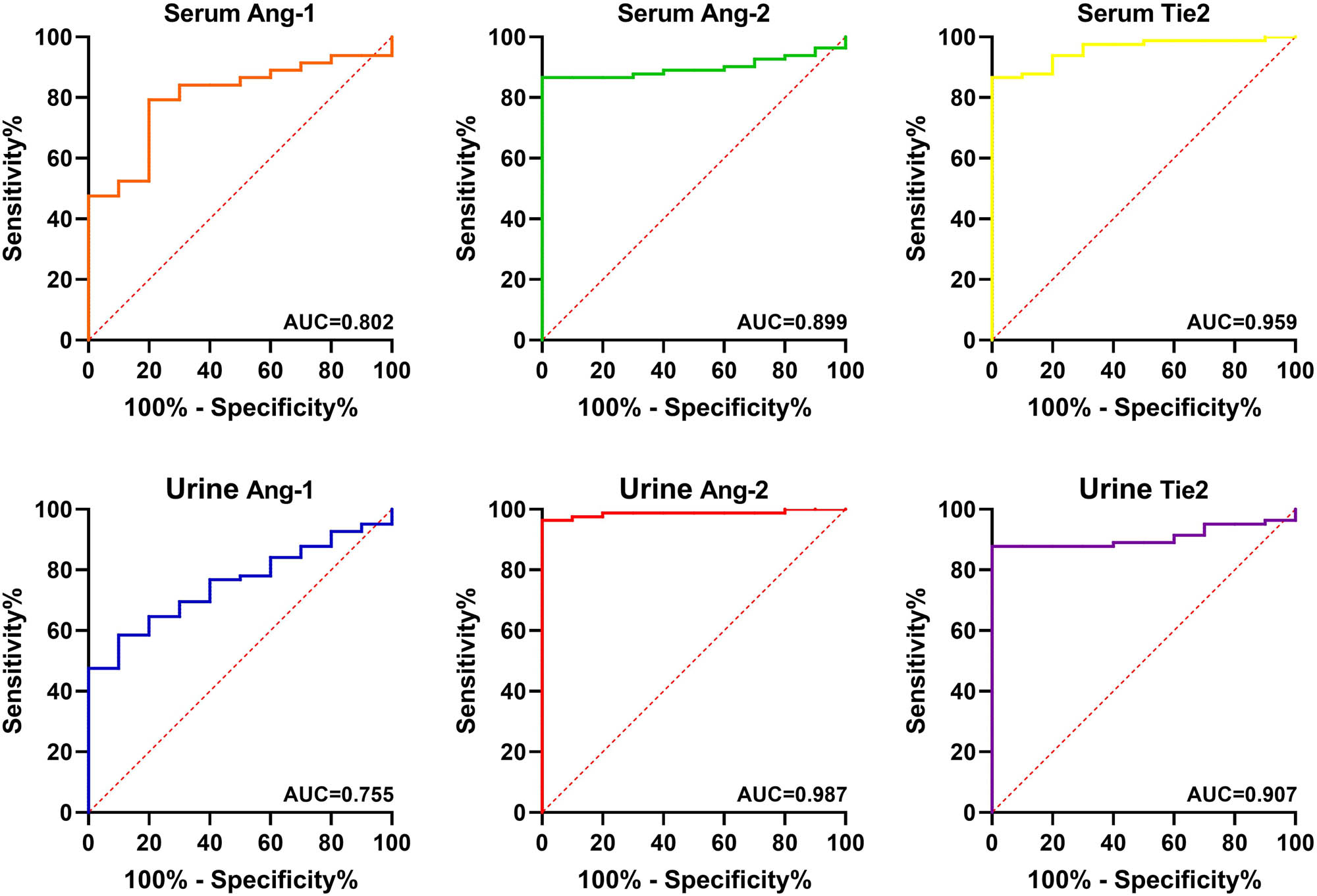
ROC curves of Ang-1, Ang-2, and Tie2 in serum and urine for the diagnosis of HSP.

ROC curves of Ang-1, Ang-2, and Tie2 in serum and urine for the diagnosis of pSLE.
4 Discussion
HSP is the most frequently occurring small-vessel leukocytoclastic vasculitis in childhood, characterized by the predominant deposition of IgA-immune complexes in and around small blood vessels [28,29]. SLE is a chronic autoimmune disease with important correlations to cardiovascular disease and inflammation, impacting critical organs, such as central nervous system vasculitis and thrombotic microangiopathy [30–32]. Children with HSP and SLE often experience extensive diffuse organ involvement more often than adults do [33,34]. Diagnosing HSP and pSLE remains challenging due to the heterogeneous presentation of symptoms and signs, as well as the absence of specific diagnostic tests [35,36]. A diagnostic biomarker is a biological marker primarily used to detect and confirm a specific disease and identify individuals with a particular subtype [37]. ROC curves have facilitated a systematic approach to evaluate biomarkers [38]. In the present study, compared to the levels in the control group, the Ang-2 and Tie2 levels were significantly elevated in both the serum and urine of HSP and pSLE patients, while the Ang-1/Ang-2 ratio was reduced. Additionally, Ang-1 was highly expressed in the serum and urine of HSP patients and in the serum of pSLE patients. Furthermore, Ang-1, Ang-2, and Tie2 exhibited differential expression patterns across various types of HSP and pSLE. Ang-1, Ang-2, and Tie2 in the serum and urine may serve as potential biomarkers for HSP and pSLE, as indicated by the ROC curve analysis. Ang-1 and Ang-2 belong to a family of vascular growth factors that exert contrasting physiological effects on endothelial activation and dysfunction when combined with Tie2. Ang-1 binding stimulates vascular growth and stability, reduces inflammation, and supports endothelial cell survival [39,40]. In addition to its important role in angiogenesis and vascular stabilization, Ang-1 also maintains vascular permeability and lymphatic integrity [41]. Ang-2, often referred to as a Tie-2 antagonist, counteracts the stabilizing effect of Ang-1 by causing the loss of intercellular contact through autocrine signaling of the Tie-2 receptor [21]. Ang-2 binding promotes vascular activation, vascular inflammation, microvascular leakage, and neoangiogenesis [25,40]. Blood vessel growth, maintenance, leakiness, and instability depend on the local balance between Ang-1 and Ang-2 as well as other angiogenic agents [25]. Furthermore, Ang-1 has multiple anti-inflammatory properties, while Ang-2 promotes inflammatory responses [42,43]. The increase in Ang-2 and Tie2, but not in Ang-1 suggest a shift in the balance toward endothelial cell activation and inflammation [44]. In our investigation, the Ang-1/Ang-2 ratio was reduced in HSP and pSLE patients. A decrease in the Ang-1/Ang-2 ratio may promote the development of HSP and pSLE diseases. Moreover, a dysregulation of the balance between Ang-1 and Ang-2 can serve as a predictive factor for multiple diseases. The Ang-1/Ang-2 ratio is significantly elevated in diabetic retinopathy, and the difference in Ang-1/Ang-2 ratio precedes measurable changes in Ang-1 and Ang-2 individually, suggesting the Ang-1/Ang-2 ratio as a potential diagnostic biomarker [45]. Plasma Ang-2 levels are associated with cytokines in patients with sepsis, and the Ang-2/Ang-1 ratio is a reliable predictor of 28-day mortality in these individuals [46]. In children with septic shock, the Ang-1/Ang-2 ratio is reduced, and is associated with an increased use of vasoactive agents, prolonged intensive care unit stay, and correlation with the severity of illness score [47]. Therefore, we speculate that the Ang-1/Ang-2 ratio may serve as a biomarker of HSP and pSLE.
HSP can be classified into various types based on clinical traits. Skin HSP is an inflammatory disorder affecting the walls of dermal blood vessels and can cause a diverse range of clinical manifestations including urticarial lesions, plaques, papules, palpable purpura, ulcers, nodules, and livedo [48]. Renal HSP can occur in a considerable proportion of HSP patients, with rates as high as 30–50%, and children (1–3%) still experience renal insufficiency that can progress to end-stage renal failure [49,50]. The clinical features of abdominal HSP include diarrhea, abdominal pain, vomiting, gastrointestinal bleeding and blood in the stool, and even intussusception and intestinal perforation, which may be followed by intestinal obstruction and intussusception in severe cases [51,52]. Patients with joint-type HSP exhibit symptoms, such as swelling and pain in the joints [53]. Mixed HSP patients present with symptoms of more than two types of HSP. In our study, we found a significant reduction in the Ang-1/Ang-2 ratio in both urine and serum of patients with renal HSP. Renal HSP more frequently affects children and is the leading cause of secondary kidney diseases in pediatric patients [54]. Additionally, renal dysfunction is the main factor contributing to mortality in children with HSP [55]. Extensive IgA deposits have been identified in the renal vessels of HSP patients, and renal damage plays a pivotal role in their prognosis [56]. A decreased Ang-1/Ang-2 ratio may contribute to the development of nephropathy in HSP. Different HSPs are regulated by different genes [57]. For example, HMGB1 has an altered distribution in the lesional skin of HSP patients and may be an important mediator of endothelial inflammation through the induction of tumor necrosis factor-alpha and interleukin-6 production [57]. Serum galactose-deficient IgA1 levels are highly heritable in pediatric patients with renal-type HSP [58]. In the present study, we found that Ang-1, Ang-2, and Tie2 were differentially expressed in various types of HSP. pSLE is classified into two types, the non-active and active pSLE is regulated by multiple genes. In patients with active pSLE, the serum concentrations of VEGF, Tie2, and thrombomodulin are markedly elevated, whereas that of serum ADAMTS13 is reduced compared to the levels in individuals with inactive diseases [8]. In the present study, we identified distinct expression patterns of Ang-1, Ang-2, and Tie2 in different types of pSLE. Consequently, we hypothesized that Ang-1, Ang-2, and Tie2 may be used as biomarkers to distinguish between different types of HSP and pSLE.
Recently, an increasing number of diagnostic biomarkers for HSP and pSLE have been identified. The MCP-1 levels in the serum and urine were significantly elevated in SLE patients compared to those in healthy individuals, irrespective of the degree of disease activity and renal involvement, and it can be used as a biomarker for SLE [59]. In adult HSP patients, elevated VEGF-A and VEGFR-1 levels and the VEGFR-1/VEGF-A ratio in serum may serve as valuable indicators of inflammatory processes and vascular endothelial injury, and VEGFR-1 appears to be a particularly important marker in disease progression [60]. VEGF, Tie2, thrombomodulin, and ADAMTS13 are potent biomarkers for pSLE activity and organ involvement [8]. Immune complexes, anti-endothelial cell antibodies, and the pro-inflammatory effects of various cytokines can mediate vasculopathy in pSLE [61]. Furthermore, genes associated with endothelial dysregulation have been identified as highly effective biomarkers for the activity of pSLE and the extent of organ involvement in affected patients [8]. Thus, endothelial cell factors play an important role in HSP and pSLE patients.
In this study, it was found that Ang-1, Ang-2, and Tie2 in the serum and urine can serve as biomarkers for HSP and pSLE.
5 Conclusion
Ang-1/Ang-2 levels are reduced in HSP and pSLE patients. Ang-1, Ang-2, and Tie2 are promising biomarkers for HSP and pSLE. These biomarkers may prove valuable in elucidating vascular pathogeneses and in monitoring disease progression.
-
Funding information: This work is supported by the Suzhou, 2020, the 29th batch of science and technology development plan project (SYS2020068).
-
Author contributions: Lishan Jia and Xiaozhong Li: conception, design and analysis of data, performed the data analyses and wrote the manuscript; Jiayun Shen, Yan Teng and Baoqin Zhang: contributed to the conception of the study; wrote the manuscript; Min Zhang, Yueqin Gu and Hong Xu: contributed significantly to analysis and manuscript preparation; wrote the manuscript; All authors have read and approved the manuscript.
-
Conflict of interest: Authors state no conflict of interest.
-
Data availability statement: The datasets generated during and/or analyzed during the current study are available from the corresponding author on reasonable request.
References
[1] Song Y, Huang X, Yu G, Qiao J, Cheng J, Wu J, et al. Pathogenesis of IgA vasculitis: an up-to-date review. Front Immunol. 2021;12:771619. Epub 2021/12/04.10.3389/fimmu.2021.771619Search in Google Scholar PubMed PubMed Central
[2] Gardner-Medwin J, Dolezalova P, Cummins C, Southwood TJL. Incidence of Henoch-Schönlein purpura, Kawasaki disease, and rare vasculitides in children of different ethnic origins. Lancet. 2002;360(9341):1197–202.10.1016/S0140-6736(02)11279-7Search in Google Scholar PubMed
[3] Penido M, Palma LMP. IgA vasculitis in children. J Bras Nefrol. 2022;44(1):3–5. Epub 2022/03/22.10.1590/2175-8239-jbn-2022-e002Search in Google Scholar PubMed PubMed Central
[4] Aslan C, Goknar N, Kelesoglu E, Uckardes D, Candan C. Long-term results in children with Henoch-Schonlein nephritis. Medeni Med J. 2022;37(2):159–64. Epub 2022/06/24.10.4274/MMJ.galenos.2022.92331Search in Google Scholar PubMed PubMed Central
[5] Ozen S, Pistorio A, Iusan SM, Bakkaloglu A, Herlin T, Brik R, et al. EULAR/PRINTO/PRES criteria for Henoch-Schonlein purpura, childhood polyarteritis nodosa, childhood Wegener granulomatosis and childhood Takayasu arteritis: Ankara 2008. Part II: final classification criteria. Ann Rheum Dis. 2010;69(5):798–806. Epub 2010/04/24.10.1136/ard.2009.116657Search in Google Scholar PubMed
[6] Ozen S, Marks SD, Brogan P, Groot N, de Graeff N, Avcin T, et al. European consensus-based recommendations for diagnosis and treatment of immunoglobulin A vasculitis-the SHARE initiative. Rheumatology (Oxf). 2019;58(9):1607–16. Epub 2019/03/18.10.1093/rheumatology/kez041Search in Google Scholar PubMed
[7] Du L, Wang P, Liu C, Li S, Yue S, Yang Y. Multisystemic manifestations of IgA vasculitis. Clin Rheumatol. 2021;40(1):43–52. Epub 2020/06/20.10.1007/s10067-020-05166-5Search in Google Scholar PubMed
[8] Lee WF, Wu CY, Yang HY, Lee WI, Chen LC, Ou LS, et al. Biomarkers associating endothelial dysregulation in pediatric-onset systemic lupus erythematous. Pediatr Rheumatol Online J. 2019;17(1):69. Epub 2019/10/28.10.1186/s12969-019-0369-7Search in Google Scholar PubMed PubMed Central
[9] Barile-Fabris L, Hernandez-Cabrera MF, Barragan-Garfias JA. Vasculitis in systemic lupus erythematosus. Curr Rheumatol Rep. 2014;16(9):440. Epub 2014/07/16.10.1007/s11926-014-0440-9Search in Google Scholar PubMed
[10] Afzali P, Isaeian A, Sadeghi P, Moazzami B, Parvaneh N, Robatjazi M, et al. Complement deficiency in pediatric-onset systemic lupus erythematosus. J Lab Physicians. 2018;10(2):232–6. Epub 2018/04/26.10.4103/JLP.JLP_171_17Search in Google Scholar PubMed PubMed Central
[11] Trindade VC, Carneiro-Sampaio M, Bonfa E, Silva CA. An update on the management of childhood-onset systemic lupus erythematosus. Paediatr Drugs. 2021;23(4):331–47. Epub 20210710.10.1007/s40272-021-00457-zSearch in Google Scholar PubMed PubMed Central
[12] Chang J, Knight A, Lawson E. Patterns of healthcare use and medication adherence among youth with systemic lupus erythematosus during transfer from pediatric to adult care. J Rheumatol. 2021;48(1):105–13.10.3899/jrheum.191029Search in Google Scholar PubMed PubMed Central
[13] Charras A, Smith E, Hedrich CM. Systemic lupus erythematosus in children and young people. Curr Rheumatol Rep. 2021;23(3):20. Epub 20210210.10.1007/s11926-021-00985-0Search in Google Scholar PubMed PubMed Central
[14] Bundhun P, Kumari A, Huang F. Differences in clinical features observed between childhood-onset versus adult-onset systemic lupus erythematosus: a systematic review and meta-analysis. Medicine. 2017;96(37):e8086.10.1097/MD.0000000000008086Search in Google Scholar PubMed PubMed Central
[15] Akca UK, Batu ED, Kisaarslan AP, Poyrazoglu H, Ayaz NA, Sozeri B, et al. Hematological involvement in pediatric systemic lupus erythematosus: a multi-center study. Lupus. 2021;30(12):1983–90. Epub 20210828.10.1177/09612033211038824Search in Google Scholar PubMed
[16] Zivkovic V, Mitic B, Stamenkovic B, Stojanovic S, Dinic BR, Stojanovic M, et al. Analysis on the risk factors for organ damage in patients with systemic lupus erythematosus: a cross-sectional single-center experience. Sao Paulo Med J. 2019;137(2):155–61. Epub 20190715.10.1590/1516-3180.2018.0258060219Search in Google Scholar PubMed PubMed Central
[17] Zhang CX, Cai L, Zhou ZY, Mao YY, Huang H, Yin L, et al. Clinical manifestations, immunological features and prognosis of Chinese pediatric systemic lupus erythematosus: a single-center study. Int J Rheum Dis. 2019;22(6):1070–6. Epub 2019/04/09.10.1111/1756-185X.13547Search in Google Scholar PubMed
[18] Hossny E, El-Ghoneimy D, Soliman DA, Ashour A. Diagnostic value of serum high-mobility group box-1 in pediatric systemic lupus erythematosus. Int J Rheum Dis. 2019;22(8):1402–9. Epub 2019/04/03.10.1111/1756-185X.13556Search in Google Scholar PubMed
[19] Trapani S, Rubino C, Simonini G, Indolfi G. Gastrointestinal and hepatic involvement in paediatric systemic lupus erythematosus. Clin Exp Rheumatol. 2021;39(4):899–906. Epub 2021/03/06.10.55563/clinexprheumatol/oebrcqSearch in Google Scholar PubMed
[20] Gale NW, Yancopoulos GD. Growth factors acting via endothelial cell-specific receptor tyrosine kinases: VEGFs, angiopoietins, and ephrins in vascular development. Genes Dev. 1999;13(9):1055–66. Epub 1999/05/14.10.1101/gad.13.9.1055Search in Google Scholar PubMed
[21] Joussen AM, Ricci F, Paris LP, Korn C, Quezada-Ruiz C, Zarbin M. Angiopoietin/Tie2 signalling and its role in retinal and choroidal vascular diseases: a review of preclinical data. Eye (Lond). 2021;35(5):1305–16. Epub 20210209.10.1038/s41433-020-01377-xSearch in Google Scholar PubMed PubMed Central
[22] Tsakogiannis D, Nikolakopoulou A, Zagouri F, Stratakos G, Syrigos K, Zografos E, et al. Update overview of the role of angiopoietins in lung cancer. Medicina (Kaunas). 2021;57(11):1191. Epub 2021/11/28.10.3390/medicina57111191Search in Google Scholar PubMed PubMed Central
[23] Yu X, Ye F. Role of angiopoietins in development of cancer and neoplasia associated with viral infection. Cells. 2020;9(2):457. Epub 20200218.10.3390/cells9020457Search in Google Scholar PubMed PubMed Central
[24] Luck R, Karakatsani A, Shah B, Schermann G, Adler H, Kupke J, et al. The angiopoietin-Tie2 pathway regulates Purkinje cell dendritic morphogenesis in a cell-autonomous manner. Cell Rep. 2021;36(7):109522.10.1016/j.celrep.2021.109522Search in Google Scholar PubMed PubMed Central
[25] Khan M, Aziz AA, Shafi NA, Abbas T, Khanani AM. Targeting angiopoietin in retinal vascular diseases: a literature review and summary of clinical trials involving faricimab. Cells. 2020;9(8):1869. Epub 20200810.10.3390/cells9081869Search in Google Scholar PubMed PubMed Central
[26] Lin Y, McClennan A, Hoffman L. Characterization of the Ang/Tie2 signaling pathway in the diaphragm muscle of DMD mice. Biomedicines. 2023;11(8):2265. Epub 20230814.10.3390/biomedicines11082265Search in Google Scholar PubMed PubMed Central
[27] Jurisic V. Multiomic analysis of cytokines in immuno-oncology. Expert Rev Proteom. 2020;17(9):663–74. Epub 2020/11/03.10.1080/14789450.2020.1845654Search in Google Scholar PubMed
[28] Gomez S, Perez M, Pellegrini M, Isern E, Quintana C, Artacho P, et al. Henoch-Schonlein purpura in pediatrics: ten years of experience at a moderate risk office of a general hospital. Arch Argent Pediatr. 2020;118(1):31–7. Epub 2020/01/28. Purpura de Schonlein-Henoch en pediatria: Diez anos de experiencia en un consultorio de moderado riesgo en un hospital general.10.5546/aap.2020.31Search in Google Scholar
[29] Liu Q, Yang Y, Ge S, Huo J, Wang D, Ma Y, et al. Serum level of advanced oxidation protein products (AOPPs) in patients with Henoch-Schonlein purpura and its relationship with aberrant glycosylation of IgA1 and Cosmc mRNA expression. Int J Dermatol. 2019;58(9):1092–7. Epub 2019/06/30.10.1111/ijd.14550Search in Google Scholar PubMed PubMed Central
[30] Yu C, Li P, Dang X, Zhang X, Mao Y, Chen X. Lupus nephritis: new progress in diagnosis and treatment. J Autoimmun. 2022;132:102871. Epub 2022/08/24.10.1016/j.jaut.2022.102871Search in Google Scholar PubMed
[31] Schwartz N, Stock AD, Putterman C. Neuropsychiatric lupus: new mechanistic insights and future treatment directions. Nat Rev Rheumatol. 2019;15(3):137–52. Epub 2019/01/20.10.1038/s41584-018-0156-8Search in Google Scholar PubMed PubMed Central
[32] Stanescu C, Andronesi AG, Jurcut C, Gherghiceanu M, Vornicu A, Burcea FA, et al. Successful treatment of catastrophic antiphospholipid syndrome using rituximab: case report and review of the literature. Medicina (Kaunas). 2021;57(9):912. Epub 2021/09/29.10.3390/medicina57090912Search in Google Scholar PubMed PubMed Central
[33] Alexander T, Hedrich CM. Systemic lupus erythematosus-are children small adults? Z Rheumatol. 2022;81(1):28–35. Epub 2021/11/09. Systemischer Lupus erythematodes - sind Kinder kleine Erwachsene?.10.1007/s00393-021-01116-xSearch in Google Scholar PubMed
[34] Ambrose N, Morgan TA, Galloway J, Ionnoau Y, Beresford MW, Isenberg DA, et al. Differences in disease phenotype and severity in SLE across age groups. Lupus. 2016;25(14):1542–50. Epub 2016/11/02.10.1177/0961203316644333Search in Google Scholar PubMed PubMed Central
[35] Barut K, Sahin S, Kasapcopur O. Pediatric vasculitis. Curr Opin Rheumatol. 2016;28(1):29–38. Epub 2015/11/12.10.1097/BOR.0000000000000236Search in Google Scholar PubMed
[36] Lewandowski LB, Watt MH, Schanberg LE, Thielman NM, Scott C. Missed opportunities for timely diagnosis of pediatric lupus in South Africa: a qualitative study. Pediatr Rheumatol Online J. 2017;15(1):14. Epub 2017/02/25.10.1186/s12969-017-0144-6Search in Google Scholar PubMed PubMed Central
[37] Califf RM. Biomarker definitions and their applications. Exp Biol Med (Maywood). 2018;243(3):213–21. Epub 2018/02/07.10.1177/1535370217750088Search in Google Scholar PubMed PubMed Central
[38] Martin M, Kleinhenz MD, Schwartzkopf-Genswein KS, Melendez D, Marti S, Pajor EA, et al. Characterizing the diagnostic sensitivity and specificity of pain biomarkers in cattle using receiver operating characteristic curves. J Dairy Sci. 2022;105(12):9853–68. Epub 2022/10/08.10.3168/jds.2021-21393Search in Google Scholar PubMed
[39] Fu X, Wang J, Cai H, Jiang H, Han S. C16 peptide and Ang-1 improve functional disability and pathological changes in an Alzheimer’s disease model associated with vascular dysfunction. Pharmaceuticals (Basel). 2022;15(4):471. Epub 2022/04/24.10.3390/ph15040471Search in Google Scholar PubMed PubMed Central
[40] Magkouta S, Kollintza A, Moschos C, Spella M, Skianis I, Pappas A, et al. Role of angiopoietins in mesothelioma progression. Cytokine. 2019;118:99–106. Epub 2018/09/12.10.1016/j.cyto.2018.08.006Search in Google Scholar PubMed
[41] Gurnik S, Devraj K, Macas J, Yamaji M, Starke J, Scholz A, et al. Angiopoietin-2-induced blood-brain barrier compromise and increased stroke size are rescued by VE-PTP-dependent restoration of Tie2 signaling. Acta Neuropathol. 2016;131(5):753–73. Epub 20160301.10.1007/s00401-016-1551-3Search in Google Scholar PubMed PubMed Central
[42] Cantero-Navarro E, Fernandez-Fernandez B, Ramos AM, Rayego-Mateos S, Rodrigues-Diez RR, Sanchez-Nino MD, et al. Renin-angiotensin system and inflammation update. Mol Cell Endocrinol. 2021;529:111254. Epub 20210330.10.1016/j.mce.2021.111254Search in Google Scholar PubMed
[43] Wu Q, Xu WD, Huang AF. Role of angiopoietin-2 in inflammatory autoimmune diseases: a comprehensive review. Int Immunopharmacol. 2020;80:106223. Epub 20200125.10.1016/j.intimp.2020.106223Search in Google Scholar PubMed
[44] Carvalheiro T, Lopes A, van der Kroef M, Malvar-Fernandez B, Rafael-Vidal C, Hinrichs A, et al. Angiopoietin-2 promotes inflammatory activation in monocytes of systemic sclerosis patients. Int J Mol Sci. 2020;21(24):9544.10.3390/ijms21249544Search in Google Scholar PubMed PubMed Central
[45] Wang Y, Fang J, Niu T, Xing X, Wang H, Shi X, et al. Serum Ang-1/Ang-2 ratio may be a promising biomarker for evaluating severity of diabetic retinopathy. Graefe’s Arch Clin Exp Ophthalmol. 2023;261(1):49–55.10.1007/s00417-022-05745-zSearch in Google Scholar PubMed
[46] Seol CH, Yong SH, Shin JH, Lee SH, Leem AY, Park MS, et al. The ratio of plasma angiopoietin-2 to angiopoietin-1 as a prognostic biomarker in patients with sepsis. Cytokine. 2020;129:155029. Epub 2020/02/15.10.1016/j.cyto.2020.155029Search in Google Scholar PubMed
[47] Melendez E, Whitney JE, Norton JS, Silverman M, Harju-Baker S, Mikacenic C, et al. Systemic angiopoietin-1/2 dysregulation in pediatric sepsis and septic shock. Int J Med Sci. 2019;16(2):318–23. Epub 2019/02/13.10.7150/ijms.27731Search in Google Scholar PubMed PubMed Central
[48] Frumholtz L, Laurent-Roussel S, Lipsker D, Terrier B. Cutaneous vasculitis: review on diagnosis and clinicopathologic correlations. Clin Rev Allergy Immunol. 2021;61(2):181–93. Epub 2020/05/08.10.1007/s12016-020-08788-4Search in Google Scholar PubMed
[49] Cao T, Zhu Y, Zhu Y, Wong K. Construction of prediction model of renal damage in children with Henoch-Schönlein purpura based on machine learning. Comput Math Methods Med. 2022;2022:1–7.10.1155/2022/6991218Search in Google Scholar PubMed PubMed Central
[50] Audemard-Verger A, Terrier B, Dechartres A, Chanal J, Amoura Z, Le Gouellec N, et al. Characteristics and management of IgA vasculitis (Henoch-Schonlein) in adults: data from 260 patients included in a French multicenter retrospective survey. Arthritis Rheumatol. 2017;69(9):1862–70. Epub 2017/06/13.10.1002/art.40178Search in Google Scholar PubMed
[51] Brogan P, Eleftheriou D, Dillon M. Small vessel vasculitis. Pediatr Nephrol. 2010;25(6):1025–35. Epub 2009/11/04.10.1007/s00467-009-1317-4Search in Google Scholar PubMed PubMed Central
[52] Choong CK, Beasley SW. Intra-abdominal manifestations of Henoch-Schonlein purpura. J Paediatr Child Health. 1998;34(5):405–9. Epub 1998/10/10.10.1046/j.1440-1754.1998.00263.xSearch in Google Scholar PubMed
[53] Short CD, Mallick NP. Renal manifestations of systemic disorders. Practitioner. 1981;225(1357):997–1005. Epub 1981/07/01.Search in Google Scholar
[54] Liu F, Wang C, Wang R, Wang W, Li M. Henoch-schonlein purpura nephritis with renal interstitial lesions. Open Med (Wars). 2018;13:597–604. Epub 2018/12/07.10.1515/med-2018-0088Search in Google Scholar PubMed PubMed Central
[55] Kawasaki Y. Mechanism of onset and exacerbation of chronic glomerulonephritis and its treatment. Pediatr Int. 2011;53(6):795–806.10.1111/j.1442-200X.2011.03469.xSearch in Google Scholar PubMed
[56] Reicy R, Jari M. Comparison of different treatment regimens for long-term improvement of renal function in patients with Henoch-Schönlein purpura: a systematic review. Curr Rheumatol Rev. 2023.10.2174/1573397119666230825163008Search in Google Scholar PubMed
[57] Chen T, Guo ZP, Wang WJ, Qin S, Cao N, Li MM. Increased serum HMGB1 levels in patients with Henoch-Schonlein purpura. Exp Dermatol. 2014;23(6):419–23. Epub 2014/04/25.10.1111/exd.12422Search in Google Scholar PubMed
[58] Kiryluk K, Moldoveanu Z, Sanders JT, Eison TM, Suzuki H, Julian BA, et al. Aberrant glycosylation of IgA1 is inherited in both pediatric IgA nephropathy and Henoch–Schönlein purpura nephritis. Kidney Int. 2011;80(1):79–87.10.1038/ki.2011.16Search in Google Scholar PubMed PubMed Central
[59] Zivkovic V, Cvetkovic T, Mitic B, Stamenkovic B, Stojanovic S, Radovanovic-Dinic B, et al. Monocyte chemoattractant protein-1 as a marker of systemic lupus erythematosus: an observational study. Rheumatol Int. 2018;38(6):1003–8. Epub 20171127.Search in Google Scholar
[60] Yucel C, Sertoglu E, Firat Oguz E, Hayran Y, Omma A, Ozgurtas T. Serum VEGF-A and VEGFR-1 levels in patients with adult immunoglobulin A vasculitis. Int J Rheum Dis. 2021;24(6):789–94. Epub 2021/04/24.10.1111/1756-185X.14115Search in Google Scholar PubMed
[61] Cieslik P, Hrycek A, Klucinski P. Vasculopathy and vasculitis in systemic lupus erythematosus. Pol Arch Med Wewn. 2008;118(1–2):57–63. Epub 2008/04/15.10.20452/pamw.306Search in Google Scholar
© 2024 the author(s), published by De Gruyter
This work is licensed under the Creative Commons Attribution 4.0 International License.
Articles in the same Issue
- Biomedical Sciences
- Constitutive and evoked release of ATP in adult mouse olfactory epithelium
- LARP1 knockdown inhibits cultured gastric carcinoma cell cycle progression and metastatic behavior
- PEGylated porcine–human recombinant uricase: A novel fusion protein with improved efficacy and safety for the treatment of hyperuricemia and renal complications
- Research progress on ocular complications caused by type 2 diabetes mellitus and the function of tears and blepharons
- The role and mechanism of esketamine in preventing and treating remifentanil-induced hyperalgesia based on the NMDA receptor–CaMKII pathway
- Brucella infection combined with Nocardia infection: A case report and literature review
- Detection of serum interleukin-18 level and neutrophil/lymphocyte ratio in patients with antineutrophil cytoplasmic antibody-associated vasculitis and its clinical significance
- Ang-1, Ang-2, and Tie2 are diagnostic biomarkers for Henoch-Schönlein purpura and pediatric-onset systemic lupus erythematous
- PTTG1 induces pancreatic cancer cell proliferation and promotes aerobic glycolysis by regulating c-myc
- Role of serum B-cell-activating factor and interleukin-17 as biomarkers in the classification of interstitial pneumonia with autoimmune features
- Effectiveness and safety of a mumps containing vaccine in preventing laboratory-confirmed mumps cases from 2002 to 2017: A meta-analysis
- Low levels of sex hormone-binding globulin predict an increased breast cancer risk and its underlying molecular mechanisms
- A case of Trousseau syndrome: Screening, detection and complication
- Application of the integrated airway humidification device enhances the humidification effect of the rabbit tracheotomy model
- Preparation of Cu2+/TA/HAP composite coating with anti-bacterial and osteogenic potential on 3D-printed porous Ti alloy scaffolds for orthopedic applications
- Aquaporin-8 promotes human dermal fibroblasts to counteract hydrogen peroxide-induced oxidative damage: A novel target for management of skin aging
- Current research and evidence gaps on placental development in iron deficiency anemia
- Single-nucleotide polymorphism rs2910829 in PDE4D is related to stroke susceptibility in Chinese populations: The results of a meta-analysis
- Pheochromocytoma-induced myocardial infarction: A case report
- Kaempferol regulates apoptosis and migration of neural stem cells to attenuate cerebral infarction by O‐GlcNAcylation of β-catenin
- Sirtuin 5 regulates acute myeloid leukemia cell viability and apoptosis by succinylation modification of glycine decarboxylase
- Apigenin 7-glucoside impedes hypoxia-induced malignant phenotypes of cervical cancer cells in a p16-dependent manner
- KAT2A changes the function of endometrial stromal cells via regulating the succinylation of ENO1
- Current state of research on copper complexes in the treatment of breast cancer
- Exploring antioxidant strategies in the pathogenesis of ALS
- Helicobacter pylori causes gastric dysbacteriosis in chronic gastritis patients
- IL-33/soluble ST2 axis is associated with radiation-induced cardiac injury
- The predictive value of serum NLR, SII, and OPNI for lymph node metastasis in breast cancer patients with internal mammary lymph nodes after thoracoscopic surgery
- Carrying SNP rs17506395 (T > G) in TP63 gene and CCR5Δ32 mutation associated with the occurrence of breast cancer in Burkina Faso
- P2X7 receptor: A receptor closely linked with sepsis-associated encephalopathy
- Probiotics for inflammatory bowel disease: Is there sufficient evidence?
- Identification of KDM4C as a gene conferring drug resistance in multiple myeloma
- Microbial perspective on the skin–gut axis and atopic dermatitis
- Thymosin α1 combined with XELOX improves immune function and reduces serum tumor markers in colorectal cancer patients after radical surgery
- Highly specific vaginal microbiome signature for gynecological cancers
- Sample size estimation for AQP4-IgG seropositive optic neuritis: Retinal damage detection by optical coherence tomography
- The effects of SDF-1 combined application with VEGF on femoral distraction osteogenesis in rats
- Fabrication and characterization of gold nanoparticles using alginate: In vitro and in vivo assessment of its administration effects with swimming exercise on diabetic rats
- Mitigating digestive disorders: Action mechanisms of Mediterranean herbal active compounds
- Distribution of CYP2D6 and CYP2C19 gene polymorphisms in Han and Uygur populations with breast cancer in Xinjiang, China
- VSP-2 attenuates secretion of inflammatory cytokines induced by LPS in BV2 cells by mediating the PPARγ/NF-κB signaling pathway
- Factors influencing spontaneous hypothermia after emergency trauma and the construction of a predictive model
- Long-term administration of morphine specifically alters the level of protein expression in different brain regions and affects the redox state
- Application of metagenomic next-generation sequencing technology in the etiological diagnosis of peritoneal dialysis-associated peritonitis
- Clinical diagnosis, prevention, and treatment of neurodyspepsia syndrome using intelligent medicine
- Case report: Successful bronchoscopic interventional treatment of endobronchial leiomyomas
- Preliminary investigation into the genetic etiology of short stature in children through whole exon sequencing of the core family
- Cystic adenomyoma of the uterus: Case report and literature review
- Mesoporous silica nanoparticles as a drug delivery mechanism
- Dynamic changes in autophagy activity in different degrees of pulmonary fibrosis in mice
- Vitamin D deficiency and inflammatory markers in type 2 diabetes: Big data insights
- Lactate-induced IGF1R protein lactylation promotes proliferation and metabolic reprogramming of lung cancer cells
- Meta-analysis on the efficacy of allogeneic hematopoietic stem cell transplantation to treat malignant lymphoma
- Mitochondrial DNA drives neuroinflammation through the cGAS-IFN signaling pathway in the spinal cord of neuropathic pain mice
- Application value of artificial intelligence algorithm-based magnetic resonance multi-sequence imaging in staging diagnosis of cervical cancer
- Embedded monitoring system and teaching of artificial intelligence online drug component recognition
- Investigation into the association of FNDC1 and ADAMTS12 gene expression with plumage coloration in Muscovy ducks
- Yak meat content in feed and its impact on the growth of rats
- A rare case of Richter transformation with breast involvement: A case report and literature review
- First report of Nocardia wallacei infection in an immunocompetent patient in Zhejiang province
- Rhodococcus equi and Brucella pulmonary mass in immunocompetent: A case report and literature review
- Downregulation of RIP3 ameliorates the left ventricular mechanics and function after myocardial infarction via modulating NF-κB/NLRP3 pathway
- Evaluation of the role of some non-enzymatic antioxidants among Iraqi patients with non-alcoholic fatty liver disease
- The role of Phafin proteins in cell signaling pathways and diseases
- Ten-year anemia as initial manifestation of Castleman disease in the abdominal cavity: A case report
- Coexistence of hereditary spherocytosis with SPTB P.Trp1150 gene variant and Gilbert syndrome: A case report and literature review
- Utilization of convolutional neural networks to analyze microscopic images for high-throughput screening of mesenchymal stem cells
- Exploratory evaluation supported by experimental and modeling approaches of Inula viscosa root extract as a potent corrosion inhibitor for mild steel in a 1 M HCl solution
- Imaging manifestations of ductal adenoma of the breast: A case report
- Gut microbiota and sleep: Interaction mechanisms and therapeutic prospects
- Isomangiferin promotes the migration and osteogenic differentiation of rat bone marrow mesenchymal stem cells
- Prognostic value and microenvironmental crosstalk of exosome-related signatures in human epidermal growth factor receptor 2 positive breast cancer
- Circular RNAs as potential biomarkers for male severe sepsis
- Knockdown of Stanniocalcin-1 inhibits growth and glycolysis in oral squamous cell carcinoma cells
- The expression and biological role of complement C1s in esophageal squamous cell carcinoma
- A novel GNAS mutation in pseudohypoparathyroidism type 1a with articular flexion deformity: A case report
- Predictive value of serum magnesium levels for prognosis in patients with non-small cell lung cancer undergoing EGFR-TKI therapy
- HSPB1 alleviates acute-on-chronic liver failure via the P53/Bax pathway
- IgG4-related disease complicated by PLA2R-associated membranous nephropathy: A case report
- Baculovirus-mediated endostatin and angiostatin activation of autophagy through the AMPK/AKT/mTOR pathway inhibits angiogenesis in hepatocellular carcinoma
- Metformin mitigates osteoarthritis progression by modulating the PI3K/AKT/mTOR signaling pathway and enhancing chondrocyte autophagy
- Evaluation of the activity of antimicrobial peptides against bacterial vaginosis
- Atypical presentation of γ/δ mycosis fungoides with an unusual phenotype and SOCS1 mutation
- Analysis of the microecological mechanism of diabetic kidney disease based on the theory of “gut–kidney axis”: A systematic review
- Omega-3 fatty acids prevent gestational diabetes mellitus via modulation of lipid metabolism
- Refractory hypertension complicated with Turner syndrome: A case report
- Interaction of ncRNAs and the PI3K/AKT/mTOR pathway: Implications for osteosarcoma
- Association of low attenuation area scores with pulmonary function and clinical prognosis in patients with chronic obstructive pulmonary disease
- Long non-coding RNAs in bone formation: Key regulators and therapeutic prospects
- The deubiquitinating enzyme USP35 regulates the stability of NRF2 protein
- Neutrophil-to-lymphocyte ratio and platelet-to-lymphocyte ratio as potential diagnostic markers for rebleeding in patients with esophagogastric variceal bleeding
- G protein-coupled receptor 1 participating in the mechanism of mediating gestational diabetes mellitus by phosphorylating the AKT pathway
- LL37-mtDNA regulates viability, apoptosis, inflammation, and autophagy in lipopolysaccharide-treated RLE-6TN cells by targeting Hsp90aa1
- The analgesic effect of paeoniflorin: A focused review
- Chemical composition’s effect on Solanum nigrum Linn.’s antioxidant capacity and erythrocyte protection: Bioactive components and molecular docking analysis
- Knockdown of HCK promotes HREC cell viability and inner blood–retinal barrier integrity by regulating the AMPK signaling pathway
- The role of rapamycin in the PINK1/Parkin signaling pathway in mitophagy in podocytes
- Laryngeal non-Hodgkin lymphoma: Report of four cases and review of the literature
- Clinical value of macrogenome next-generation sequencing on infections
- Overview of dendritic cells and related pathways in autoimmune uveitis
- TAK-242 alleviates diabetic cardiomyopathy via inhibiting pyroptosis and TLR4/CaMKII/NLRP3 pathway
- Hypomethylation in promoters of PGC-1α involved in exercise-driven skeletal muscular alterations in old age
- Profile and antimicrobial susceptibility patterns of bacteria isolated from effluents of Kolladiba and Debark hospitals
- The expression and clinical significance of syncytin-1 in serum exosomes of hepatocellular carcinoma patients
- A histomorphometric study to evaluate the therapeutic effects of biosynthesized silver nanoparticles on the kidneys infected with Plasmodium chabaudi
- PGRMC1 and PAQR4 are promising molecular targets for a rare subtype of ovarian cancer
- Analysis of MDA, SOD, TAOC, MNCV, SNCV, and TSS scores in patients with diabetes peripheral neuropathy
- SLIT3 deficiency promotes non-small cell lung cancer progression by modulating UBE2C/WNT signaling
- The relationship between TMCO1 and CALR in the pathological characteristics of prostate cancer and its effect on the metastasis of prostate cancer cells
- Heterogeneous nuclear ribonucleoprotein K is a potential target for enhancing the chemosensitivity of nasopharyngeal carcinoma
- PHB2 alleviates retinal pigment epithelium cell fibrosis by suppressing the AGE–RAGE pathway
- Anti-γ-aminobutyric acid-B receptor autoimmune encephalitis with syncope as the initial symptom: Case report and literature review
- Comparative analysis of chloroplast genome of Lonicera japonica cv. Damaohua
- Human umbilical cord mesenchymal stem cells regulate glutathione metabolism depending on the ERK–Nrf2–HO-1 signal pathway to repair phosphoramide mustard-induced ovarian cancer cells
- Electroacupuncture on GB acupoints improves osteoporosis via the estradiol–PI3K–Akt signaling pathway
- Renalase protects against podocyte injury by inhibiting oxidative stress and apoptosis in diabetic nephropathy
- Review: Dicranostigma leptopodum: A peculiar plant of Papaveraceae
- Combination effect of flavonoids attenuates lung cancer cell proliferation by inhibiting the STAT3 and FAK signaling pathway
- Renal microangiopathy and immune complex glomerulonephritis induced by anti-tumour agents: A case report
- Correlation analysis of AVPR1a and AVPR2 with abnormal water and sodium and potassium metabolism in rats
- Gastrointestinal health anti-diarrheal mixture relieves spleen deficiency-induced diarrhea through regulating gut microbiota
- Myriad factors and pathways influencing tumor radiotherapy resistance
- Exploring the effects of culture conditions on Yapsin (YPS) gene expression in Nakaseomyces glabratus
- Screening of prognostic core genes based on cell–cell interaction in the peripheral blood of patients with sepsis
- Coagulation factor II thrombin receptor as a promising biomarker in breast cancer management
- Ileocecal mucinous carcinoma misdiagnosed as incarcerated hernia: A case report
- Methyltransferase like 13 promotes malignant behaviors of bladder cancer cells through targeting PI3K/ATK signaling pathway
- The debate between electricity and heat, efficacy and safety of irreversible electroporation and radiofrequency ablation in the treatment of liver cancer: A meta-analysis
- ZAG promotes colorectal cancer cell proliferation and epithelial–mesenchymal transition by promoting lipid synthesis
- Baicalein inhibits NLRP3 inflammasome activation and mitigates placental inflammation and oxidative stress in gestational diabetes mellitus
- Impact of SWCNT-conjugated senna leaf extract on breast cancer cells: A potential apoptotic therapeutic strategy
- MFAP5 inhibits the malignant progression of endometrial cancer cells in vitro
- Major ozonated autohemotherapy promoted functional recovery following spinal cord injury in adult rats via the inhibition of oxidative stress and inflammation
- Axodendritic targeting of TAU and MAP2 and microtubule polarization in iPSC-derived versus SH-SY5Y-derived human neurons
- Differential expression of phosphoinositide 3-kinase/protein kinase B and Toll-like receptor/nuclear factor kappa B signaling pathways in experimental obesity Wistar rat model
- The therapeutic potential of targeting Oncostatin M and the interleukin-6 family in retinal diseases: A comprehensive review
- BA inhibits LPS-stimulated inflammatory response and apoptosis in human middle ear epithelial cells by regulating the Nf-Kb/Iκbα axis
- Role of circRMRP and circRPL27 in chronic obstructive pulmonary disease
- Investigating the role of hyperexpressed HCN1 in inducing myocardial infarction through activation of the NF-κB signaling pathway
- Characterization of phenolic compounds and evaluation of anti-diabetic potential in Cannabis sativa L. seeds: In vivo, in vitro, and in silico studies
- Quantitative immunohistochemistry analysis of breast Ki67 based on artificial intelligence
- Ecology and Environmental Science
- Screening of different growth conditions of Bacillus subtilis isolated from membrane-less microbial fuel cell toward antimicrobial activity profiling
- Degradation of a mixture of 13 polycyclic aromatic hydrocarbons by commercial effective microorganisms
- Evaluation of the impact of two citrus plants on the variation of Panonychus citri (Acari: Tetranychidae) and beneficial phytoseiid mites
- Prediction of present and future distribution areas of Juniperus drupacea Labill and determination of ethnobotany properties in Antalya Province, Türkiye
- Population genetics of Todarodes pacificus (Cephalopoda: Ommastrephidae) in the northwest Pacific Ocean via GBS sequencing
- A comparative analysis of dendrometric, macromorphological, and micromorphological characteristics of Pistacia atlantica subsp. atlantica and Pistacia terebinthus in the middle Atlas region of Morocco
- Macrofungal sporocarp community in the lichen Scots pine forests
- Assessing the proximate compositions of indigenous forage species in Yemen’s pastoral rangelands
- Food Science
- Gut microbiota changes associated with low-carbohydrate diet intervention for obesity
- Reexamination of Aspergillus cristatus phylogeny in dark tea: Characteristics of the mitochondrial genome
- Differences in the flavonoid composition of the leaves, fruits, and branches of mulberry are distinguished based on a plant metabolomics approach
- Investigating the impact of wet rendering (solventless method) on PUFA-rich oil from catfish (Clarias magur) viscera
- Non-linear associations between cardiovascular metabolic indices and metabolic-associated fatty liver disease: A cross-sectional study in the US population (2017–2020)
- Knockdown of USP7 alleviates atherosclerosis in ApoE-deficient mice by regulating EZH2 expression
- Utility of dairy microbiome as a tool for authentication and traceability
- Agriculture
- Enhancing faba bean (Vicia faba L.) productivity through establishing the area-specific fertilizer rate recommendation in southwest Ethiopia
- Impact of novel herbicide based on synthetic auxins and ALS inhibitor on weed control
- Perspectives of pteridophytes microbiome for bioremediation in agricultural applications
- Fertilizer application parameters for drip-irrigated peanut based on the fertilizer effect function established from a “3414” field trial
- Improving the productivity and profitability of maize (Zea mays L.) using optimum blended inorganic fertilization
- Application of leaf multispectral analyzer in comparison to hyperspectral device to assess the diversity of spectral reflectance indices in wheat genotypes
- Animal Sciences
- Knockdown of ANP32E inhibits colorectal cancer cell growth and glycolysis by regulating the AKT/mTOR pathway
- Development of a detection chip for major pathogenic drug-resistant genes and drug targets in bovine respiratory system diseases
- Exploration of the genetic influence of MYOT and MB genes on the plumage coloration of Muscovy ducks
- Transcriptome analysis of adipose tissue in grazing cattle: Identifying key regulators of fat metabolism
- Comparison of nutritional value of the wild and cultivated spiny loaches at three growth stages
- Transcriptomic analysis of liver immune response in Chinese spiny frog (Quasipaa spinosa) infected with Proteus mirabilis
- Disruption of BCAA degradation is a critical characteristic of diabetic cardiomyopathy revealed by integrated transcriptome and metabolome analysis
- Plant Sciences
- Effect of long-term in-row branch covering on soil microorganisms in pear orchards
- Photosynthetic physiological characteristics, growth performance, and element concentrations reveal the calcicole–calcifuge behaviors of three Camellia species
- Transcriptome analysis reveals the mechanism of NaHCO3 promoting tobacco leaf maturation
- Bioinformatics, expression analysis, and functional verification of allene oxide synthase gene HvnAOS1 and HvnAOS2 in qingke
- Water, nitrogen, and phosphorus coupling improves gray jujube fruit quality and yield
- Improving grape fruit quality through soil conditioner: Insights from RNA-seq analysis of Cabernet Sauvignon roots
- Role of Embinin in the reabsorption of nucleus pulposus in lumbar disc herniation: Promotion of nucleus pulposus neovascularization and apoptosis of nucleus pulposus cells
- Revealing the effects of amino acid, organic acid, and phytohormones on the germination of tomato seeds under salinity stress
- Combined effects of nitrogen fertilizer and biochar on the growth, yield, and quality of pepper
- Comprehensive phytochemical and toxicological analysis of Chenopodium ambrosioides (L.) fractions
- Impact of “3414” fertilization on the yield and quality of greenhouse tomatoes
- Exploring the coupling mode of water and fertilizer for improving growth, fruit quality, and yield of the pear in the arid region
- Metagenomic analysis of endophytic bacteria in seed potato (Solanum tuberosum)
- Antibacterial, antifungal, and phytochemical properties of Salsola kali ethanolic extract
- Exploring the hepatoprotective properties of citronellol: In vitro and in silico studies on ethanol-induced damage in HepG2 cells
- Enhanced osmotic dehydration of watermelon rind using honey–sucrose solutions: A study on pre-treatment efficacy and mass transfer kinetics
- Effects of exogenous 2,4-epibrassinolide on photosynthetic traits of 53 cowpea varieties under NaCl stress
- Comparative transcriptome analysis of maize (Zea mays L.) seedlings in response to copper stress
- An optimization method for measuring the stomata in cassava (Manihot esculenta Crantz) under multiple abiotic stresses
- Fosinopril inhibits Ang II-induced VSMC proliferation, phenotype transformation, migration, and oxidative stress through the TGF-β1/Smad signaling pathway
- Antioxidant and antimicrobial activities of Salsola imbricata methanolic extract and its phytochemical characterization
- Bioengineering and Biotechnology
- Absorbable calcium and phosphorus bioactive membranes promote bone marrow mesenchymal stem cells osteogenic differentiation for bone regeneration
- New advances in protein engineering for industrial applications: Key takeaways
- An overview of the production and use of Bacillus thuringiensis toxin
- Research progress of nanoparticles in diagnosis and treatment of hepatocellular carcinoma
- Bioelectrochemical biosensors for water quality assessment and wastewater monitoring
- PEI/MMNs@LNA-542 nanoparticles alleviate ICU-acquired weakness through targeted autophagy inhibition and mitochondrial protection
- Unleashing of cytotoxic effects of thymoquinone-bovine serum albumin nanoparticles on A549 lung cancer cells
- Erratum
- Erratum to “Investigating the association between dietary patterns and glycemic control among children and adolescents with T1DM”
- Erratum to “Activation of hypermethylated P2RY1 mitigates gastric cancer by promoting apoptosis and inhibiting proliferation”
- Retraction
- Retraction to “MiR-223-3p regulates cell viability, migration, invasion, and apoptosis of non-small cell lung cancer cells by targeting RHOB”
- Retraction to “A data mining technique for detecting malignant mesothelioma cancer using multiple regression analysis”
- Special Issue on Advances in Neurodegenerative Disease Research and Treatment
- Transplantation of human neural stem cell prevents symptomatic motor behavior disability in a rat model of Parkinson’s disease
- Special Issue on Multi-omics
- Inflammasome complex genes with clinical relevance suggest potential as therapeutic targets for anti-tumor drugs in clear cell renal cell carcinoma
- Gastroesophageal varices in primary biliary cholangitis with anti-centromere antibody positivity: Early onset?