Effect of RUNX1/FOXP3 axis on apoptosis of T and B lymphocytes and immunosuppression in sepsis
-
Yangfa Chao


Abstract
Lymphocyte apoptosis is a latent factor for immunosuppression in sepsis. Forkhead box protein P3 (FOXP3) can interact with RUNX family transcription factor 1 (RUNX1) in regulatory T cells. Our research was to probe whether RUNX1/FOXP3 axis affects immunosuppression in the process of sepsis by modulating T and B lymphocyte apoptosis. We constructed sepsis model in mice and mouse CD4+ T and CD19+ B lymphocytes. RUNX1 and FOXP3 expressions and apoptosis in cells were assessed by western blot, quantitative real-time PCR, and flow cytometer. Inflammation of serum and pathological damage was assessed by ELISA and H&E staining. Relationship between RUNX1 and FOXP3 was assessed by co-immunoprecipitation. The findings showed that RUNX1 ameliorated the survival rate, pathological damage, and decreased inflammation-related factors, and inhibited apoptosis of CD4+ T and CD19+ B cells in cecal ligation and puncture mice. Furthermore, RUNX1 up-regulated the viability and down-regulated apoptotic rate with the changed expressions of apoptosis-related molecules in lipopolysaccharide (LPS)-mediated CD4+ T and CD19+ B cells. Additionally, FOXP3 interacted with RUNX1, and its silencing decreased RUNX1 expression and reversed the inhibitory effect of RUNX1 on apoptosis of LPS-mediated CD4+ T and CD19+ B cells. In summary, the RUNX1/FOXP3 axis alleviated immunosuppression in sepsis progression by weakening T and B lymphocyte apoptosis.
1 Introduction
Sepsis is a fatal organ function damage caused by the uncontrolled immune response of the body to infection, and is a significant cause of death in critically ill patients [1]. Epidemiological studies manifested that there were six million deaths from sepsis worldwide every year, and the mortality of severe sepsis is as high as 25–30% [2]. Some studies on the prognosis of sepsis have pointed out that surviving sepsis patients have poor long-term survival, with physical and psychological dysfunction, decreased quality of life, increased risk of readmission, and become a burden on family and society [3,4].
In the early stage of sepsis, a large number of pro-inflammatory factors and oxygen free radicals are produced under the stimulation of pathogenic microorganisms and metabolites. With the continuous improvement of organ protection and alternative treatment technologies, patients with sepsis can pass through the period of excessive inflammatory response and enter the long-term immunosuppressive state [5]. Mounting studies have exhibited that immunosuppression is the main pathophysiological mechanism in the initiation and development of sepsis and severe immunosuppression is a considerable element leading to the deterioration of the patient’s condition [6]. It has been reported that lymphocyte apoptosis is tightly correlated with immunosuppression [7]. If lymphocyte apoptosis is a significant mechanism, then specific lymphocyte subsets may be more vulnerable [8].
RUNX family transcription factor 1 (RUNX1) is a gene related to immunosuppression, which can regulate the expression of a variety of transcription factors; the study unveiled that deletion of RUNX1 can cause autoimmune lung disease in mice [9]. Hsu et al. have reported that loss of RUNX1 function resulted in the loss of CD4+ T cells [10]. Some scholars have illustrated that knockdown of RUNX1 led to B cell immunodeficiency in adult zebrafish [11]. Another research has clarified that miR-338-3p mimic impedes Treg-induced immunosuppression in pemphigus vulgaris through modulating RUNX1 expression [12]. In addition, Zhang et al. has reported that exosomal miR-21-5p mitigated sepsis-triggered acute kidney damage through restraining RUNX1 level [13]. However, whether RUNX1 affects immunosuppression in the process of sepsis by modulating lymphocyte apoptosis remains to be further investigated. On the other hand, the Forkhead box protein P3 (FOXP3) is a master cell lineage regulator in CD4+ CD25+ natural regulatory T cell (Treg) development. FOXP3 can interact with RUNX1 in regulatory T cells [14,15]. Furthermore, FOXP3+ regulatory T cells are necessary for recovery from severe sepsis [16].
Based on the above research evidence, this study probes the role of RUNX1 on inflammation and pathological damage of liver, kidney, and lung tissues in cecal ligation and puncture (CLP) mice. Besides, we further explore the effect of RUNX1 on inflammation response in serum and apoptosis in CD4+ T and CD19+ B cells from CLP mice. Meanwhile, CD4+ T and CD19+ B cells induced by lipopolysaccharide (LPS) were served as the experimental subjects in vitro to analyze the role and mechanism of RUNX1 with FOXP3 in CD4+ T and CD19+ B cells.
2 Materials and methods
2.1 Animals
We bought 100 male specific pathogen-free C57BL/6 mice (8–12 week old) from Chengdu Dashuo Experimental Animal Co., Ltd (China). They were acclimatized for 7 days under the same facilities (22–24℃, 60 ± 2% humidity, 12 h light/dark cycle).
2.2 CLP model
CLP surgery was performed on 75 mice to construct a sepsis model [17]. To relieve pain, all mice received buprenorphine (0.1 mg/kg, B-044, Supelco, USA) before surgery. First, mice were anesthetized by intraperitoneal injection with 0.3% pentobarbital sodium (P3761, Sigma-Aldrich, USA). A 1 cm incision was made in the abdominal midline of the mice. Next, we ligated the cecum approximately 1 cm from the end of the cecum using a 4-0 surgical suture. Then, we used no. 21 needle to puncture the ligated cecum twice. Finally, the abdominal incision was sutured layer by layer. The remaining 25 mice belonged to the control group and underwent sham surgery. Mice in the control group were only treated with laparotomy and cecum explantation. All mice were injected subcutaneously with ceftriaxone (25 mg/kg, C304513, Aladdin, China) and metronidazole (12.5 mg/kg, M109873, Aladdin, China) after surgery and resuscitated with 1 mL of normal saline. To observe the survival of mice, we observed the animals every 12 hours within 10 days after operation. A total of 60 mice were used for survival study.
2.3 Animal treatment
The lentiviral vectors expressing the plasmids (RUNX1 or corresponding negative controls [empty vector]) were injected into mice with sepsis at a titer of 1 × 10⁹ via the tail vein. Injection was conducted 48 h after CLP.
2.4 Isolation of CD4+ T and CD19+ B lymphocytes from mouse spleen
We first harvested the spleen tissues of C57BL/6 mice, and then prepared a single cell suspension, as previously reported [17]. Then, the red blood cells were lysed and primary mouse CD19+ B and CD4+ T lymphocytes were isolated using a MagniSort™ Mouse CD19 Positive Selection Kit (8802-6847-74, Invitrogen, USA) and Dynabeads™ Untouched™ Mouse CD4 Cells Kit (11416D, Invitrogen, USA), respectively. Primary CD4+ T and CD19+ B cells were kept in RPMI-1640 medium (22400089, Gibco, USA) augmented with 10% fetal bovine serum (10099141, Gibco, USA) and 1% penicillin–streptomycin liquid (P1400, Solarbio, China) at 37℃ with 5% CO2.
2.5 ELISA
Mice were euthanized after Day 5 of CLP, and then we collected whole blood through a cardiac puncture. The blood samples were centrifuged (1,000g, 10 min), and the supernatant was gained and stored at −80℃ until further analysis. Mouse tumor necrosis factor α (TNF-α) kit (BPE20220), interleukin 6 (IL-6) kit (BPE20012), and IL-1β kit were ordered from Lengton (China). Serum samples (50 μL) and biotin antigen working solution (50 μL) were added to the sample wells, and then reacted at room temperature (RT) for 0.5 h. After thoroughly cleaning each well, 50 μL avidin-horseradish peroxidase (HRP) was added to the sample wells and reacted at RT for 0.5 h. After fully washing each well again, chromogenic agents A and B were successively added to each well. After 10 min of reaction at RT, we added stop solution to each well. Lastly, we measured the absorbance (450 nm) of each well with the aid of a microplate reader (RT-6000; Rayto, China).
2.6 Apoptosis analysis in CD4+ T cells and CD19+ B cells from mice
The whole blood (100 μL) was lysed with VersaLyse lysing solution (A09777, Beckman-Coulter, Hialeah, FL, USA) for 15 min at RT. After washing, cells were incubated with phycoerythrin-labeled antibodies directed against CD4 and CD19 (CD4+ T cells and CD19+ B cells). RIPA buffer (R0010, Solarbio, China) was taken to lyse the cells. After rinsing, the lysed samples were subjected to PE Annexin V Apoptosis Detection Kit (559763, BD Biosciences, USA) based on kit instructions. Then, the results were assessed with the help of a flow cytometer (PAS III, Partec, Germany) within 0.5 h. Regarding the caspase-3 intracellular staining, fixation/permeabilization solution kit (554715, BD Biosciences, USA) was taken to fix and permeate the cells. Subsequently, PE rabbit anti-active caspase-3 antibody (561011, BD Biosciences, USA) was added. After cells were rinsed, we utilized the flow cytometer to estimate the results. Data are presented as percentages of respective Annexin V+ or caspase-3+ cell populations. A positive threshold was established based on non-stained control cells.
2.7 H&E staining
H&E staining kit (G1120, Solarbio, China) was utilized in this research. We harvested the liver tissues, kidney tissues, and lung tissues from the mice. All tissues were fixed in 4% paraformaldehyde (G1101, Servicebio, China). The fixed tissues were dehydrated, embedded in paraffin, and cut into 3 μm slices. Next, the slices were deparaffinized in xylene, rehydrated in ethanol, and then stained utilizing hematoxylin solution. After that, the slices were subjected to differentiation. After being stained with eosin solution, the slices went through ethanol dehydration and xylene transparent. Lastly, the samples were observed with the aid of an optical microscope (×40, ×100, Ts2R-FL) after being sealed through neutral balsam (D054-1-1, Jiancheng, China).
2.8 Cell transfection and grouping
T lymphocytes (CD4+ T and CD19+ B) were transfected with lentiviral vectors expressing shRNA against FOXP3 (sh-FOXP3), RUNX1 overexpression, or corresponding negative controls (empty vector). To probe the role of RUNX1 on cells, cells were assigned to the control group, LPS group (cells were subjected to 1 μg/mL LPS for 24 h), and LPS + vector/LPS + RUNX1 group (cells transfected with overexpressed RUNX1 or vector were exposed to 1 μg/mL LPS for 24 h). Next, to analyze the functions of RUNX1 and FOXP3 on cells, cells were separated into the LPS + vector group, LPS + RUNX1 group, LPS + sh-FOXP3 group, and LPS + RUNX1 + sh-FOXP3 group.
2.9 Cell viability analysis
CD4+ T and CD19+ B cells (1 × 10⁴ per well) plated into the 96-well plates were subjected to transfection and LPS intervention. Thereafter, a further 4 h culture was done after 10 μL MTT solution (M1020, Solarbio, China) was added. Then, the OD value (490 nm) was evaluated with the aid of the microplate reader after being added to 110 μL formazan solution.
2.10 Apoptosis experiment
Apoptosis of CD4+ T and CD19+ B cells (1.2 × 10⁶) was estimated with the help of the Annexin V-FITC/PI kit (556547, BD, USA). The treated cells of each group were rinsed, digested, and then centrifuged. Cell precipitation was re-suspended in 1× binding buffer and cell concentration was adjusted to 1 × 10⁶ cells/mL. After that, 5 μL Annexin V-FITC and 10 μL PI were injected into each tube. After being reacted at 37℃ without light for 15 min, we utilized the flow cytometer to evaluate apoptosis of each group.
2.11 Co-immunoprecipitation (Co-IP)
Beyotime (China) provided the IP buffer (P0013). We collected CD4+ T and CD19+ B cells (1 × 10⁵ cells/mL). After being lysed with IP buffer, cells were subjected to ultrasonic treatment followed by centrifugation. The supernatant was then reacted with anti-rabbit IgG antibody (1:8,000, ab205718; Abcam, UK), anti-FOXP3 antibody (1:30, ab215206; Abcam, UK), anti-RUNX1 antibody (1:20, ab92336; Abcam, UK), and BeyoMag™ Protein A + G Magnetic Beads (P2173, Beyotime, China) at 4℃ overnight. Afterward, the complex was rinsed and the binding proteins were eluted with SDS sample buffer. In the end, the precipitated proteins were determined through western blot.
2.12 Western blot
CD4+ T cells and CD19+ B cells from mice were lysed through the RIPA buffer (WB038, GEFAN, China) [18]. The extracted protein was centrifuged and then quantified with the help of the BCA kit (23225, Thermo Scientific, USA). After being electrophoresed, they were transferred to the PVDF membrane before being blocked with 5% skim milk. Next, we utilized primary antibodies to treat the membrane at 4℃ all night. After that, second antibody was used to treat the membrane at 37℃ for 90 min. After rinsing, the reactive bands were reacted with the color reagent (34075, PIERCE, USA) in a gel imaging system (610020-9Q, Clinx, China). The primary antibodies of RUNX1 (1:1,000, ab240639, 48 kDa), FOXP3 (1:1,000, ab20034, 50 kDa), Bax (1:8,000, ab32503, 21 kDa), Bcl-2 (1:2,000, ab196495, 26 kDa), cleaved caspase-3 (1:5,000, ab214430, 17 kDa), caspase-3 (1:2,000, ab184787, 32 kDa), and GAPDH (1:10,000, ab181602, 36 kDa), and the second antibodies of goat anti-rabbit IgG H&L (HRP) (1:2,000, ab205718) and goat anti-mouse IgG H&L (HRP) (1:2,000, ab205719) were gained from Abcam (UK). GAPDH was exploited to the housekeeping gene. The protein level was normalized to GAPDH, and presented relative to control cells (set as 1).
2.13 Quantitative real-time PCR (qRT-PCR)
We obtained total RNA from cells with the help of the RNA extraction kit (HYY0420-50, Huayueyang, China). Subsequently, the one-step RT-qPCR system (A6020) offered by Promega (USA) was applied to conduct the qRT-PCR reaction. The reaction system was done with the aid of a PCR system (ABI7900, Applied Biosystems, USA). GAPDH was the housekeeping gene and the 2−ΔΔCt was utilized for data evaluation [19]. Primer sequences are listed in Table 1.
Primer sequences used in qRT-PCR
| Genes | Forward (5′ → 3′) | Reverse (5′ → 3′) |
|---|---|---|
| RUNX1 | ACGATGAAAACTACTCGGCAG | CTGAGGTCGTTGAATCTCGCT |
| FOXP3 | CCCATCCCCAGGAGTCTTG | ACCATGACTAGGGGCACTGTA |
| GAPDH | TGACCTCAACTACATGGTCTACA | CTTCCCATTCTCGGCCTTG |
2.14 Statistics
There were at least three independent experiments for data analysis. GraphPad Prism 8.0 (GraphPad Software Inc., USA) was exploited to process data. Quantitative data were manifested as mean ± standard deviation. Comparisons between two groups were assessed by an independent sample t-test. Comparisons among multiple groups were conducted utilizing a one-way ANOVA followed by Tukey’s post hoc test. A P-value < 0.05 was considered significant.
-
Ethics statement: All animal studies were done in compliance with the guidelines for China Animal Care and Use Committee. All animal operations were performed in Nanfang Hospital and were allowed by the Ethics Committee of Nanfang Hospital (2020041569). Every effort was made to minimize pain and discomfort to the animals.
3 Results
3.1 Down-regulation of RUNX1 in CD4+ T and CD19+ B cells from CLP mice was reversed by overexpressed RUNX1, and FOXP3 was lowly expressed in CD4+ T and CD19+ B cells
The expressions of RUNX1 and FOXP3 in CD4+ T and CD19+ B cells of CLP mice were lower than those in the control group (Figure 1a–c, P < 0.01). Next, we assessed the survival rate of mice in each group. The survival rate of CLP mice was lower than that of the control group, while RUNX1 overexpression ameliorated the survival rate of CLP mice (Figure 1d, P = 0.003). After injecting RUNX1 overexpression vectors, the mRNA and protein levels of RUNX1 in CD4+ T and CD19+ B cells from CLP mice were notably augmented (Figure 1e–j; P < 0.05). The data indicated that RUNX1 and FOXP3 was lowly expressed in CD4+ T and CD19+ B cells, and RUNX1 overexpression vectors reversed the down-regulation of RUNX1 in CD4+ T and CD19+ B cells from CLP mice.

Down-regulation of RUNX1 in CD4+ T and CD19+ B cells from CLP mice was reversed by overexpressed RUNX1, and FOXP3 was lowly expressed in CD4+ T and CD19+ B cells (a)–(c) RUNX1 and FOXP3 expressions in CD4+ T and CD19+ B cells of mice were assessed by western blot. (d) Survival rate of mice in each group. (e)–(j) Role of overexpressed RUNX1 on RUNX1 expression in CD4+ T and CD19+ B cells of CLP mice was examined by qRT-PCR and western blot, with GAPDH as the endogenous gene. ** P < 0.01, *** P < 0.001 vs control; +++ P < 0.001 vs CLP + Vector.
3.2 Up-regulation of inflammatory factor in serum and apoptosis in CD4+ T and CD19+ B cells from CLP mice was partially offset by RUNX1 overexpression
Then, we utilized ELISA to determine the content of inflammatory factors in the serum of mice in each group. TNF-α, IL-1β, and IL-6 contents in the serum of mice in the CLP group were higher than the control group, whereas RUNX1 overexpression evidently attenuated TNF-α, IL-1β, and IL-6 contents in the serum of mice (Figure 2a–c, P < 0.001). We further discovered a notable elevation of annexin-V binding and active caspase-3 in CD4+ T and CD19+ B cells of CLP mice (Figure 2d–g, P < 0.001). Importantly, overexpressed RUNX1 repressed annexin-V binding and active caspase-3 in CD4+ T and CD19+ B cells of CLP mice (Figure 2d–g, P < 0.001). The data indicated that RUNX1 overexpression exerts inhibitory effects on the content of inflammatory factors in serum and apoptosis in CD4+ T and CD19+ B cells from CLP mice.

Up-regulation of inflammatory factor and apoptosis in CD4+ T and CD19+ B cells from CLP mice was partially offset by RUNX1 overexpression. The role of overexpressed RUNX1 on inflammatory factor (a)–(c) and apoptosis (d)–(g) in CD4+ T and CD19+ B cells from CLP mice was examined by ELISA and flow cytometer. *** P < 0.001 vs control; +++ P < 0.001 vs CLP + Vector.
3.3 RUNX1 overexpression alleviated liver, kidney, and lung injury in CLP mice
In order to probe the organ damage of mice in each group, we harvested the liver, kidney, and lung tissues of mice for H&E staining. Compared with the control group, CLP mice had increased alveolar wall thickness and inflammatory cell infiltration; CLP also resulted in evident necrosis of liver tissue and exfoliation of renal tubular epithelial cells in mice (Figure 3). Application of RUNX1 overexpression ameliorated liver, kidney, and lung injury in CLP mice (Figure 3). The data indicated that RUNX1 overexpression alleviated liver, kidney, and lung injury in CLP mice.
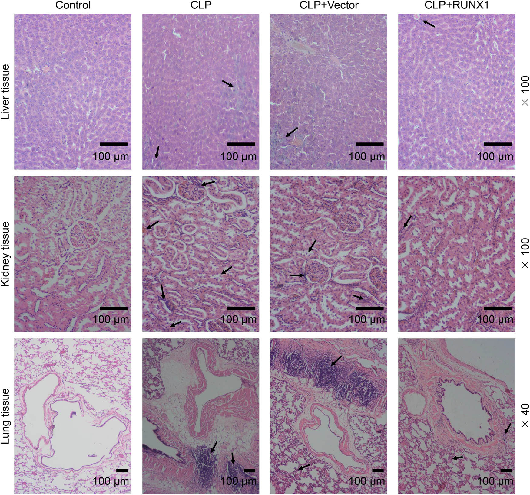
RUNX1 overexpression alleviated liver, kidney, and lung injury in CLP mice. The effect of overexpressed RUNX1 on liver, kidney, and lung injury in CLP mice was examined by H&E staining (100×, 40×, 100 μm). Infiltration of inflammatory cells and the damaged sites are indicated by arrows.
3.4 RUNX1 up-regulation intensified RUNX1 expression and cell viability in LPS-mediated CD4+ T and CD19+ B cells
To probe the role of RUNX1 on CD4+ T and CD19+ B cells, cells transfected with or without RUNX1 overexpression vector were reacted with 1 μg/mL LPS for 24 h. As displayed in Figure 4a–f, LPS caused the down-regulation of the mRNA and protein levels of RUNX1, while overexpressed RUNX1 reversed its effects (P < 0.01). Meanwhile, overexpressed RUNX1 largely elevated the cell viability of CD4+ T and CD19+ B cells induced by LPS (Figure 4g and h, P < 0.05). The data indicated that RUNX1 overexpression elevated the expression of RUNX1 and cell viability of CD4+ T and CD19+ B cells during LPS condition.

RUNX1 up-regulation intensified RUNX1 expression and cell viability in LPS-mediated CD4+ T and CD19+ B cells. (a)–(f) Effect of RUNX1 up-regulation on the mRNA and protein expressions of RUNX1 was assessed by qRT-PCR and western blot, with GAPDH as the endogenous gene. (g) and (h) Effect of RUNX1 up-regulation on cell viability in LPS-mediated CD4+ T and CD19+ B cells was estimated by MTT. ** P < 0.01, *** P < 0.001 vs control; + P < 0.05, +++ P < 0.001 vs LPS + Vector.
3.5 RUNX1 up-regulation regulated apoptosis rate, the expressions of apoptosis-related markers, and FOXP3 expressions in CD4+ T and CD19+ B cells induced by LPS
The flow cytometer analysis exhibited that RUNX1 up-regulation extremely mitigated apoptosis rate of CD4+ T and CD19+ B cells triggered by LPS (Figure 5a–c, P < 0.001). At the molecular level, we discovered that LPS resulted in the reduction of FOXP3 and Bcl-2 and the increase of Bax, cleaved caspase-3, the ratio of cleaved caspase-3/caspase-3, and Bax/Bcl-2, whereas RUNX1 up-regulation partially offset the effect of LPS (Figure 6a and b, P < 0.001). The data indicated that RUNX1 overexpression repressed apoptosis and elevated FOXP3 expression in CD4+ T and CD19+ B cells under LPS condition.
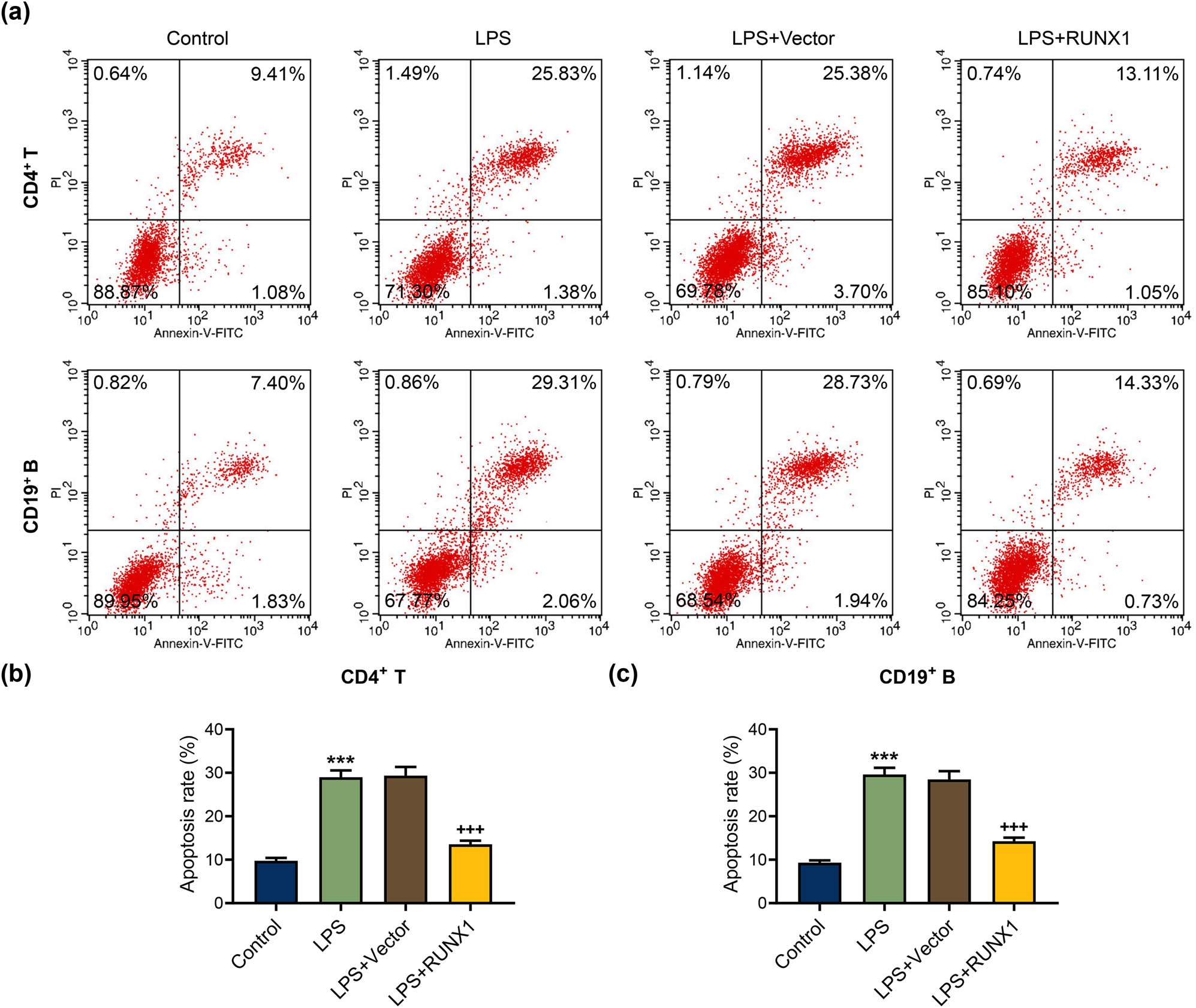
RUNX1 up-regulation repressed apoptosis rate in CD4+ T and CD19+ B cells induced by LPS. (a)–(c) The effect of RUNX1 up-regulation on apoptosis rate in CD4+ T and CD19+ B cells induced by LPS was assessed by flow cytometer. *** P < 0.001 vs control; +++ P < 0.001 vs LPS + Vector.

RUNX1 up-regulation regulated apoptosis-related markers and FOXP3 expressions in CD4+ T and CD19+ B cells induced by LPS. (a) and (b) Effect of RUNX1 up-regulation on FOXP3 expression and apoptosis-related marker expressions in CD4+ T and CD19+ B cells induced by LPS was assessed by western blot. *** P < 0.001 vs control; ++ P < 0.01, +++ P < 0.001 vs LPS + Vector.
3.6 FOXP3 interacted with RUNX1 in CD4+ T and CD19+ B cells
In the next research, we examined the relationship between RUNX1 and FOXP3 with the help of CO-IP. As exhibited in Figure 7a–d, the results clarified that RUNX1 and FOXP3 formed a complex and interacted with each other in the CD4+ T and CD19+ B cells. Moreover, we also uncovered that FOXP3 silencing greatly weakened the mRNA and protein expressions of RUNX1 and FOXP3 in CD4+ T and CD19+ B cells (Figure 7e–i, P < 0.01). The data indicated that FOXP3 interacted with RUNX1 in CD4+ T and CD19+ B cells.

FOXP3 interacted with RUNX1. (a)–(d) Relationship between RUNX1 and FOXP3 was assessed by Co-IP. (e)–(i) RUNX1 and FOXP3 expressions in CD4+ T and CD19+ B cells transfected with sh-FOXP3 were assessed by qRT-PCR and western blot, with GAPDH as the endogenous gene. ** P < 0.01, *** P < 0.001 vs Vector.
3.7 FOXP3 silencing reversed the suppression of overexpressed RUNX1 on apoptosis in LPS-mediated CD4+ T and CD19+ B cells
In Figure 8a–c, the flow cytometer assay illustrated that FOXP3 knockdown prominently facilitated apoptosis of LPS-mediated CD4+ T and CD19+ B cells and reversed the repression of overexpressed RUNX1 on apoptosis (P < 0.001). Moreover, the western blot experiment illuminated that FOXP3 silencing evidently enhanced the expressions of Bax, cleaved caspase-3, the ratio of cleaved caspase-3/caspase-3, and Bax/Bcl-2, whereas weakened Bcl-2 expression in CD4+ T and CD19+ B cells mediated by LPS (Figure 9a and b, P < 0.01). Importantly, the modulation of overexpressed RUNX1 on apoptosis-related factors was partially offset by FOXP3 silencing (Figure 9a and b, P < 0.05). The data revealed the interaction of FOXP3 and RUNX1 on apoptosis of CD4+ T and CD19+ B cells in the context of LPS.

FOXP3 silencing reversed the suppression of overexpressed RUNX1 on apoptosis in LPS-mediated CD4+ T and CD19+ B cells. (a)–(c) Roles of RUNX1 and FOXP3 on apoptosis in LPS-mediated CD4+ T and CD19+ B cells were assessed by flow cytometer. *** P < 0.001 vs LPS + Vector; +++ P < 0.001 vs LPS + short hairpin RNA targeting FOXP3 (sh-FOXP3); ^^^ P < 0.001 vs LPS + RUNX1.

Repression of overexpressed RUNX1 on apoptosis-related factors in LPS-mediated CD4+ T and CD19+ B cells was rescued by FOXP3 silencing. (a) and (b) Roles of RUNX1 and FOXP3 on apoptosis-related factors in LPS-mediated CD4+ T and CD19+ B cells were assessed by western blot. * P < 0.05, ** P < 0.01, *** P < 0.001 vs LPS + Vector; +++ P < 0.001 vs LPS + sh-FOXP3; ^^ P < 0.01, ^^^ P < 0.001 vs LPS + RUNX1.
4 Discussion
One study has reported that RUNX1 may be a new potential therapeutic target for the prevention of sepsis [20]. In our study, we uncovered that RUNX1 and FOXP3 are under-expressed in CLP mice or LPS-mediated CD4+ T and CD19+ B cells. Importantly, RUNX1 overexpression ameliorated the survival rate, pathological damage in liver, kidney and lung tissues, decreased inflammation in serum, and apoptosis of CD4+ T and CD19+ B cells in CLP mice. Furthermore, overexpression of RUNX1 up-regulated the viability and down-regulated apoptosis in LPS-mediated CD4+ T and CD19+ B cells. Additionally, FOXP3 silencing reversed the inhibitory effect of RUNX1 on apoptosis of LPS-mediated CD4+ T and CD19+ B cells by interacting with RUNX1.
In our study, RUNX1 was decreased in CD4+ T and CD19+ B cells in CLP mice, and RUNX1 overexpression increases percent survival, and alleviates pathological damage in liver, kidney, and lung tissues of CLP mice. With the deepening of the research on the pathophysiological mechanism of sepsis, modulation of immune-inflammatory disorders has become a new direction for the treatment of sepsis [21]. In sepsis, infection first causes increased secretion of core pro-inflammatory factors such as TNF-α and IL-1β [22,23]. It has been reported that IL-6 was tightly associated with sepsis mortality and can be used as a predictor of sepsis severity due to its slow metabolism and easy detection [24,25]. In this research, our data exhibited that RUNX1 overexpression evidently attenuated inflammation in the serum of mice. These present findings indicated that RUNX1 increases survival rate of CLP mice, accompanied by decreasing pathological damage in liver, kidney, and lung tissues and serum inflammation.
Depletion of T lymphocytes is one of the significant causes of immune dysfunction, mainly manifested in the reduction of CD4+ T and CD8+ T, which further affects the immune function of the body [26]. B lymphocytes play the role of humoral immunity by specifically presenting antigens, secreting antibodies, and releasing cytokines [27]. Lymphocyte apoptosis exhibits a considerable role in immunosuppression of sepsis, and inhibition of lymphocyte apoptosis can ameliorate the prognosis of sepsis. Garofalo et al. illustrated that massive apoptosis of immune cells such as T cells, dendritic cells, and B cells were observed in many organs of deceased patients with sepsis [28]. Li et al. reported that the expressions of caspase-3, caspase-8, and caspase-9 were notably elevated, while the expressions of CD4+/CD8+ T lymphocytes and CD19+ B lymphocytes were evidently decreased in a mouse model of sepsis formed by CLP [29]. Reséndiz-Martínez et al. compared the children with sepsis and healthy people and found that the apoptosis of monocytes and the expression of Fas in the peripheral blood of the children with sepsis increased, and the level of apoptosis was positively correlated with the expression of Fas [30]. Moreover, another research clarified that the expressions of cytochrome C, caspase-3, caspase-8, and caspase-9 in CLP mice were obviously up-regulated, while the expression of Bcl-2 was prominently decreased, which in turn promoted T lymphocyte apoptosis [31]. Similar to the results of previous studies, we discovered a notable elevation of annexin-V binding and active caspase-3 in CD4+ T and CD19+ B cells of CLP mice; meanwhile, in in vitro experiments, LPS resulted in the reduction of Bcl-2 and the increase of Bax and cleaved caspase-3 in CD4+ T and CD19+ B cells. More important, we discovered that overexpressed RUNX1 repressed apoptosis of CD4+ T and CD19+ B cells in CLP mice, and in LPS-mediated CD4+ T and CD19+ B cells.
Subsequently, through literature search and bioinformatics prediction, it has been found that FOXP3 could interact with RUNX1 in T cells [32–34]. Also, FOXP3+ modulatory T cells were necessary for recovery from serious sepsis [16]. In addition, RUNX1 is also a gene that activates FOXP3 expression [12]. The present results clarified that RUNX1 and FOXP3 formed a complex and interacted with each other in CD4+ T and CD19+ B cells. FOXP3 forms a head-to-head dimerization, which confers distinct DNA-binding specificity and creates a docking site for RUNX1 [35]. Importantly, FOXP3 silencing reversed the suppression of overexpressed RUNX1 on apoptosis in LPS-mediated CD4+ T and CD19+ B cells. These findings indicated that RUNX1 inhibited apoptosis of LPS-mediated CD4+ T and CD19+ B cells by interacting with FOXP3.
Collectively, these findings described that the RUNX1/FOXP3 axis alleviated immunosuppression in sepsis progression by weakening T and B lymphocyte apoptosis. In future studies, we need to further examine the expression levels of apoptosis-related markers in CD4+ T and CD19+ B cells of CLP mice.
-
Funding information: This work was supported by the Scientific Research Project of Guangdong Administration of Traditional Chinese Medicine (20171089).
-
Conflict of interest: The authors declare no conflicts of interest.
-
Data availability statement: The analyzed data sets generated during the study are available from the corresponding author on reasonable request.
References
[1] Rhodes A, Evans LE, Alhazzani W, Levy MM, Antonelli M, Ferrer R, et al. Surviving sepsis campaign: International Guidelines for Management of Sepsis and Septic Shock: 2016. Intensive Care Med. 2017;43(3):304–77.10.1007/s00134-017-4683-6Search in Google Scholar PubMed
[2] Xie J, Wang H, Kang Y, Zhou L, Liu Z, Qin B, et al. The epidemiology of sepsis in Chinese ICUs: a national cross-sectional survey. Crit Care Med. 2020;48(3):e209–e18.10.1097/CCM.0000000000004155Search in Google Scholar PubMed
[3] Yende S, Austin S, Rhodes A, Finfer S, Opal S, Thompson T, et al. Long-term quality of life among survivors of severe sepsis: analyses of two international trials. Crit Care Med. 2016;44(8):1461–7.10.1097/CCM.0000000000001658Search in Google Scholar PubMed PubMed Central
[4] Goodwin AJ, Rice DA, Simpson KN, Ford DW. Frequency, cost, and risk factors of readmissions among severe sepsis survivors. Crit Care Med. 2015;43(4):738–46.10.1097/CCM.0000000000000859Search in Google Scholar PubMed PubMed Central
[5] Venet F, Monneret G. Advances in the understanding and treatment of sepsis-induced immunosuppression. Nat Rev Nephrol. 2018;14(2):121–37.10.1038/nrneph.2017.165Search in Google Scholar PubMed
[6] Shankar-Hari M, Harrison DA, Rubenfeld GD, Rowan K. Epidemiology of sepsis and septic shock in critical care units: comparison between sepsis-2 and sepsis-3 populations using a national critical care database. Br J Anaesth. 2017;119(4):626–36.10.1093/bja/aex234Search in Google Scholar PubMed
[7] Delano MJ, Ward PA. Sepsis-induced immune dysfunction: can immune therapies reduce mortality? J Clin Invest. 2016;126(1):23–31.10.1172/JCI82224Search in Google Scholar PubMed PubMed Central
[8] Hotchkiss RS, Tinsley KW, Swanson PE, Schmieg RE Jr., Hui JJ, Chang KC, et al. Sepsis-induced apoptosis causes progressive profound depletion of B and CD4+ T lymphocytes in humans. J Immunol. 2001;166(11):6952–63.10.4049/jimmunol.166.11.6952Search in Google Scholar PubMed
[9] Wong WF, Kohu K, Nagashima T, Funayama R, Matsumoto M, Movahed E, et al. The artificial loss of Runx1 reduces the expression of quiescence-associated transcription factors in CD4( +) T lymphocytes. Mol Immunol. 2015;68(2 Pt A):223–33.10.1016/j.molimm.2015.08.012Search in Google Scholar PubMed
[10] Hsu FC, Shapiro MJ, Dash B, Chen CC, Constans MM, Chung JY, et al. An essential role for the transcription factor runx1 in T cell maturation. Sci Rep. 2016;6:23533.10.1038/srep23533Search in Google Scholar PubMed PubMed Central
[11] Chi Y, Huang Z, Chen Q, Xiong X, Chen K, Xu J, et al. Loss of Runx1 function results in B cell immunodeficiency but not T cell in adult zebrafish. Open Biol. 2018;8(7):180043.10.1098/rsob.180043Search in Google Scholar PubMed PubMed Central
[12] Xu M, Liu Q, Li S, Zhang W, Huang X, Han K, et al. Increased expression of miR-338-3p impairs Treg-mediated immunosuppression in pemphigus vulgaris by targeting RUNX1. Exp Dermatol. 2020;29(7):623–9.10.1111/exd.14111Search in Google Scholar PubMed
[13] Zhang Y, Huang H, Liu W, Liu S, Wang XY, Diao ZL, et al. Endothelial progenitor cells-derived exosomal microRNA-21-5p alleviates sepsis-induced acute kidney injury by inhibiting RUNX1 expression. Cell Death Dis. 2021;12(4):335.10.1038/s41419-021-03578-ySearch in Google Scholar PubMed PubMed Central
[14] Ono M. Control of regulatory T-cell differentiation and function by T-cell receptor signalling and Foxp3 transcription factor complexes. Immunology. 2020;160(1):24–37.10.1111/imm.13178Search in Google Scholar PubMed PubMed Central
[15] Mertowska P, Mertowski S, Grywalska E, Podgajna M. The importance of the transcription factor foxp3 in the development of primary immunodeficiencies. J Clin Med. 2022;11(4):947.10.3390/jcm11040947Search in Google Scholar PubMed PubMed Central
[16] Kühlhorn F, Rath M, Schmoeckel K, Cziupka K, Nguyen HH, Hildebrandt P, et al. Foxp3 + regulatory T cells are required for recovery from severe sepsis. PLoS One. 2013;8(5):e65109.10.1371/journal.pone.0065109Search in Google Scholar PubMed PubMed Central
[17] Chen JX, Xu X, Zhang S. Silence of long noncoding RNA NEAT1 exerts suppressive effects on immunity during sepsis by promoting microRNA-125-dependent MCEMP1 downregulation. IUBMB Life. 2019;71(7):956–68.10.1002/iub.2033Search in Google Scholar PubMed
[18] Ren Q, Guo F, Tao S, Huang R, Ma L, Fu P. Flavonoid fisetin alleviates kidney inflammation and apoptosis via inhibiting Src-mediated NF-κB p65 and MAPK signaling pathways in septic AKI mice. Biomed Pharmacother. 2020;122:109772.10.1016/j.biopha.2019.109772Search in Google Scholar PubMed
[19] Livak KJ, Schmittgen TD. Analysis of relative gene expression data using real-time quantitative PCR and the 2(-Delta Delta C(T)) method. Methods. 2001;25(4):402–8.10.1006/meth.2001.1262Search in Google Scholar PubMed
[20] Luo MC, Zhou SY, Feng DY, Xiao J, Li WY, Xu CD, et al. Runt-related transcription factor 1 (RUNX1) binds to p50 in macrophages and enhances TLR4-triggered inflammation and septic shock. J Biol Chem. 2016;291(42):22011–20.10.1074/jbc.M116.715953Search in Google Scholar PubMed PubMed Central
[21] Delano MJ, Ward PA. The immune system’s role in sepsis progression, resolution, and long-term outcome. Immunol Rev. 2016;274(1):330–53.10.1111/imr.12499Search in Google Scholar PubMed PubMed Central
[22] Kurt AN, Aygun AD, Godekmerdan A, Kurt A, Dogan Y, Yilmaz E. Serum IL-1beta, IL-6, IL-8, and TNF-alpha levels in early diagnosis and management of neonatal sepsis. Mediators Inflamm. 2007;2007:31397.10.1155/2007/31397Search in Google Scholar PubMed PubMed Central
[23] Machado JR, Soave DF, da Silva MV, de Menezes LB, Etchebehere RM, Monteiro ML, et al. Neonatal sepsis and inflammatory mediators. Mediators Inflamm. 2014;2014:269681.10.1155/2014/269681Search in Google Scholar PubMed PubMed Central
[24] Qiu X, Zhang L, Tong Y, Qu Y, Wang H, Mu D. Interleukin-6 for early diagnosis of neonatal sepsis with premature rupture of the membranes: a meta-analysis. Medicine (Baltimore). 2018;97(47):e13146.10.1097/MD.0000000000013146Search in Google Scholar PubMed PubMed Central
[25] Smok B, Domagalski K, Pawłowska M. Diagnostic and prognostic value of IL-6 and sTREM-1 in SIRS and sepsis in children. Mediators Inflamm. 2020;2020:8201585.10.1155/2020/8201585Search in Google Scholar PubMed PubMed Central
[26] Pauken KE, Wherry EJ. Snapshot: T cell exhaustion. Cell. 2015;163(4):1038-.e1.10.1016/j.cell.2015.10.054Search in Google Scholar PubMed
[27] Liu Q, Lu Y, An L, Li CS. B- and T-lymphocyte attenuator expression on regulatory T-cells in patients with severe sepsis. Chin Med J (Engl). 2018;131(21):2637–9.10.4103/0366-6999.244104Search in Google Scholar PubMed PubMed Central
[28] Garofalo AM, Lorente-Ros M, Goncalvez G, Carriedo D, Ballén-Barragán A, Villar-Fernández A, et al. Histopathological changes of organ dysfunction in sepsis. Intensive Care Med Exp. 2019;7(Suppl 1):45.10.1186/s40635-019-0236-3Search in Google Scholar PubMed PubMed Central
[29] Li S, Zhu FX, Zhao XJ, An YZ. The immunoprotective activity of interleukin-33 in mouse model of cecal ligation and puncture-induced sepsis. Immunol Lett. 2016;169:1–7.10.1016/j.imlet.2015.11.009Search in Google Scholar PubMed
[30] Reséndiz-Martínez J, Asbun-Bojalil J, Huerta-Yepez S, Vega M. Correlation of the expression of YY1 and Fas cell surface death receptor with apoptosis of peripheral blood mononuclear cells, and the development of multiple organ dysfunction in children with sepsis. Mol Med Rep. 2017;15(5):2433–42.10.3892/mmr.2017.6310Search in Google Scholar PubMed PubMed Central
[31] Luan YY, Yin CF, Qin QH, Dong N, Zhu XM, Sheng ZY, et al. Effect of regulatory T cells on promoting apoptosis of T lymphocyte and its regulatory mechanism in sepsis. J Interferon Cytokine Res. 2015;35(12):969–80.10.1089/jir.2014.0235Search in Google Scholar PubMed PubMed Central
[32] Recouvreux MS, Grasso EN, Echeverria PC, Rocha-Viegas L, Castilla LH, Schere-Levy C, et al. RUNX1 and FOXP3 interplay regulates expression of breast cancer related genes. Oncotarget. 2016;7(6):6552–65.10.18632/oncotarget.6771Search in Google Scholar PubMed PubMed Central
[33] Ono M, Yaguchi H, Ohkura N, Kitabayashi I, Nagamura Y, Nomura T, et al. Foxp3 controls regulatory T-cell function by interacting with AML1/Runx1. Nature. 2007;446(7136):685–9.10.1038/nature05673Search in Google Scholar PubMed
[34] Zhang F, Meng G, Strober W. Interactions among the transcription factors Runx1, RORgammat and Foxp3 regulate the differentiation of interleukin 17-producing T cells. Nat Immunol. 2008;9(11):1297–306.10.1038/ni.1663Search in Google Scholar PubMed PubMed Central
[35] Leng F, Zhang W, Ramirez RN, Leon J, Zhong Y, Hou L, et al. The transcription factor FoxP3 can fold into two dimerization states with divergent implications for regulatory T cell function and immune homeostasis. Immunity. 2022;55(8):1354–69, e8.10.1016/j.immuni.2022.07.002Search in Google Scholar PubMed PubMed Central
© 2023 the author(s), published by De Gruyter
This work is licensed under the Creative Commons Attribution 4.0 International License.
Articles in the same Issue
- Research Articles
- Exosomes derived from mesenchymal stem cells overexpressing miR-210 inhibits neuronal inflammation and contribute to neurite outgrowth through modulating microglia polarization
- Current situation of acute ST-segment elevation myocardial infarction in a county hospital chest pain center during an epidemic of novel coronavirus pneumonia
- circ-IARS depletion inhibits the progression of non-small-cell lung cancer by circ-IARS/miR-1252-5p/HDGF ceRNA pathway
- circRNA ITGA7 restrains growth and enhances radiosensitivity by up-regulating SMAD4 in colorectal carcinoma
- WDR79 promotes aerobic glycolysis of pancreatic ductal adenocarcinoma (PDAC) by the suppression of SIRT4
- Up-regulation of collagen type V alpha 2 (COL5A2) promotes malignant phenotypes in gastric cancer cell via inducing epithelial–mesenchymal transition (EMT)
- Inhibition of TERC inhibits neural apoptosis and inflammation in spinal cord injury through Akt activation and p-38 inhibition via the miR-34a-5p/XBP-1 axis
- 3D-printed polyether-ether-ketone/n-TiO2 composite enhances the cytocompatibility and osteogenic differentiation of MC3T3-E1 cells by downregulating miR-154-5p
- Propofol-mediated circ_0000735 downregulation restrains tumor growth by decreasing integrin-β1 expression in non-small cell lung cancer
- PVT1/miR-16/CCND1 axis regulates gastric cancer progression
- Silencing of circ_002136 sensitizes gastric cancer to paclitaxel by targeting the miR-16-5p/HMGA1 axis
- Short-term outcomes after simultaneous gastrectomy plus cholecystectomy in gastric cancer: A pooling up analysis
- SCARA5 inhibits oral squamous cell carcinoma via inactivating the STAT3 and PI3K/AKT signaling pathways
- Molecular mechanism by which the Notch signaling pathway regulates autophagy in a rat model of pulmonary fibrosis in pigeon breeder’s lung
- lncRNA TPT1-AS1 promotes cell migration and invasion in esophageal squamous-cell carcinomas by regulating the miR-26a/HMGA1 axis
- SIRT1/APE1 promotes the viability of gastric cancer cells by inhibiting p53 to suppress ferroptosis
- Glycoprotein non-metastatic melanoma B interacts with epidermal growth factor receptor to regulate neural stem cell survival and differentiation
- Treatments for brain metastases from EGFR/ALK-negative/unselected NSCLC: A network meta-analysis
- Association of osteoporosis and skeletal muscle loss with serum type I collagen carboxyl-terminal peptide β glypeptide: A cross-sectional study in elder Chinese population
- circ_0000376 knockdown suppresses non-small cell lung cancer cell tumor properties by the miR-545-3p/PDPK1 pathway
- Delivery in a vertical birth chair supported by freedom of movement during labor: A randomized control trial
- UBE2J1 knockdown promotes cell apoptosis in endometrial cancer via regulating PI3K/AKT and MDM2/p53 signaling
- Metabolic resuscitation therapy in critically ill patients with sepsis and septic shock: A pilot prospective randomized controlled trial
- Lycopene ameliorates locomotor activity and urinary frequency induced by pelvic venous congestion in rats
- UHRF1-induced connexin26 methylation is involved in hearing damage triggered by intermittent hypoxia in neonatal rats
- LINC00511 promotes melanoma progression by targeting miR-610/NUCB2
- Ultra-high-performance liquid chromatography-tandem mass spectrometry analysis of serum metabolomic characteristics in people with different vitamin D levels
- Role of Jumonji domain-containing protein D3 and its inhibitor GSK-J4 in Hashimoto’s thyroiditis
- circ_0014736 induces GPR4 to regulate the biological behaviors of human placental trophoblast cells through miR-942-5p in preeclampsia
- Monitoring of sirolimus in the whole blood samples from pediatric patients with lymphatic anomalies
- Effects of osteogenic growth peptide C-terminal pentapeptide and its analogue on bone remodeling in an osteoporosis rat model
- A novel autophagy-related long non-coding RNAs signature predicting progression-free interval and I-131 therapy benefits in papillary thyroid carcinoma
- WGCNA-based identification of potential targets and pathways in response to treatment in locally advanced breast cancer patients
- Radiomics model using preoperative computed tomography angiography images to differentiate new from old emboli of acute lower limb arterial embolism
- Dysregulated lncRNAs are involved in the progress of myocardial infarction by constructing regulatory networks
- Single-arm trial to evaluate the efficacy and safety of baclofen in treatment of intractable hiccup caused by malignant tumor chemotherapy
- Genetic polymorphisms of MRPS30-DT and NINJ2 may influence lung cancer risk
- Efficacy of immune checkpoint inhibitors in patients with KRAS-mutant advanced non-small cell lung cancer: A retrospective analysis
- Pyroptosis-based risk score predicts prognosis and drug sensitivity in lung adenocarcinoma
- Upregulation of lncRNA LANCL1-AS1 inhibits the progression of non-small-cell lung cancer via the miR-3680-3p/GMFG axis
- CircRANBP17 modulated KDM1A to regulate neuroblastoma progression by sponging miR-27b-3p
- Exosomal miR-93-5p regulated the progression of osteoarthritis by targeting ADAMTS9
- Downregulation of RBM17 enhances cisplatin sensitivity and inhibits cell invasion in human hypopharyngeal cancer cells
- HDAC5-mediated PRAME regulates the proliferation, migration, invasion, and EMT of laryngeal squamous cell carcinoma via the PI3K/AKT/mTOR signaling pathway
- The association between sleep duration, quality, and nonalcoholic fatty liver disease: A cross-sectional study
- Myostatin silencing inhibits podocyte apoptosis in membranous nephropathy through Smad3/PKA/NOX4 signaling pathway
- A novel long noncoding RNA AC125257.1 facilitates colorectal cancer progression by targeting miR-133a-3p/CASC5 axis
- Impact of omicron wave and associated control measures in Shanghai on health management and psychosocial well-being of patients with chronic conditions
- Clinicopathological characteristics and prognosis of young patients aged ≤45 years old with non-small cell lung cancer
- TMT-based comprehensive proteomic profiling identifies serum prognostic signatures of acute myeloid leukemia
- The dose limits of teeth protection for patients with nasopharyngeal carcinoma undergoing radiotherapy based on the early oral health-related quality of life
- miR-30b-5p targeting GRIN2A inhibits hippocampal damage in epilepsy
- Long non-coding RNA AL137789.1 promoted malignant biological behaviors and immune escape of pancreatic carcinoma cells
- IRF6 and FGF1 polymorphisms in non-syndromic cleft lip with or without cleft palate in the Polish population
- Comprehensive analysis of the role of SFXN family in breast cancer
- Efficacy of bronchoscopic intratumoral injection of endostar and cisplatin in lung squamous cell carcinoma patients underwent conventional chemoradiotherapy
- Silencing of long noncoding RNA MIAT inhibits the viability and proliferation of breast cancer cells by promoting miR-378a-5p expression
- AG1024, an IGF-1 receptor inhibitor, ameliorates renal injury in rats with diabetic nephropathy via the SOCS/JAK2/STAT pathway
- Downregulation of KIAA1199 alleviated the activation, proliferation, and migration of hepatic stellate cells by the inhibition of epithelial–mesenchymal transition
- Exendin-4 regulates the MAPK and WNT signaling pathways to alleviate the osteogenic inhibition of periodontal ligament stem cells in a high glucose environment
- Inhibition of glycolysis represses the growth and alleviates the endoplasmic reticulum stress of breast cancer cells by regulating TMTC3
- The function of lncRNA EMX2OS/miR-653-5p and its regulatory mechanism in lung adenocarcinoma
- Tectorigenin alleviates the apoptosis and inflammation in spinal cord injury cell model through inhibiting insulin-like growth factor-binding protein 6
- Ultrasound examination supporting CT or MRI in the evaluation of cervical lymphadenopathy in patients with irradiation-treated head and neck cancer
- F-box and WD repeat domain containing 7 inhibits the activation of hepatic stellate cells by degrading delta-like ligand 1 to block Notch signaling pathway
- Knockdown of circ_0005615 enhances the radiosensitivity of colorectal cancer by regulating the miR-665/NOTCH1 axis
- Long noncoding RNA Mhrt alleviates angiotensin II-induced cardiac hypertrophy phenotypes by mediating the miR-765/Wnt family member 7B pathway
- Effect of miR-499-5p/SOX6 axis on atrial fibrosis in rats with atrial fibrillation
- Cholesterol induces inflammation and reduces glucose utilization
- circ_0004904 regulates the trophoblast cell in preeclampsia via miR-19b-3p/ARRDC3 axis
- NECAB3 promotes the migration and invasion of liver cancer cells through HIF-1α/RIT1 signaling pathway
- The poor performance of cardiovascular risk scores in identifying patients with idiopathic inflammatory myopathies at high cardiovascular risk
- miR-2053 inhibits the growth of ovarian cancer cells by downregulating SOX4
- Nucleophosmin 1 associating with engulfment and cell motility protein 1 regulates hepatocellular carcinoma cell chemotaxis and metastasis
- α-Hederin regulates macrophage polarization to relieve sepsis-induced lung and liver injuries in mice
- Changes of microbiota level in urinary tract infections: A meta-analysis
- Identification of key enzalutamide-resistance-related genes in castration-resistant prostate cancer and verification of RAD51 functions
- Falls during oxaliplatin-based chemotherapy for gastrointestinal malignancies – (lessons learned from) a prospective study
- Outcomes of low-risk birth care during the Covid-19 pandemic: A cohort study from a tertiary care center in Lithuania
- Vitamin D protects intestines from liver cirrhosis-induced inflammation and oxidative stress by inhibiting the TLR4/MyD88/NF-κB signaling pathway
- Integrated transcriptome analysis identifies APPL1/RPS6KB2/GALK1 as immune-related metastasis factors in breast cancer
- Genomic analysis of immunogenic cell death-related subtypes for predicting prognosis and immunotherapy outcomes in glioblastoma multiforme
- Circular RNA Circ_0038467 promotes the maturation of miRNA-203 to increase lipopolysaccharide-induced apoptosis of chondrocytes
- An economic evaluation of fine-needle cytology as the primary diagnostic tool in the diagnosis of lymphadenopathy
- Midazolam impedes lung carcinoma cell proliferation and migration via EGFR/MEK/ERK signaling pathway
- Network pharmacology combined with molecular docking and experimental validation to reveal the pharmacological mechanism of naringin against renal fibrosis
- PTPN12 down-regulated by miR-146b-3p gene affects the malignant progression of laryngeal squamous cell carcinoma
- miR-141-3p accelerates ovarian cancer progression and promotes M2-like macrophage polarization by targeting the Keap1-Nrf2 pathway
- lncRNA OIP5-AS1 attenuates the osteoarthritis progression in IL-1β-stimulated chondrocytes
- Overexpression of LINC00607 inhibits cell growth and aggressiveness by regulating the miR-1289/EFNA5 axis in non-small-cell lung cancer
- Subjective well-being in informal caregivers during the COVID-19 pandemic
- Nrf2 protects against myocardial ischemia-reperfusion injury in diabetic rats by inhibiting Drp1-mediated mitochondrial fission
- Unfolded protein response inhibits KAT2B/MLKL-mediated necroptosis of hepatocytes by promoting BMI1 level to ubiquitinate KAT2B
- Bladder cancer screening: The new selection and prediction model
- circNFATC3 facilitated the progression of oral squamous cell carcinoma via the miR-520h/LDHA axis
- Prone position effect in intensive care patients with SARS-COV-2 pneumonia
- Clinical observation on the efficacy of Tongdu Tuina manipulation in the treatment of primary enuresis in children
- Dihydroartemisinin ameliorates cerebral I/R injury in rats via regulating VWF and autophagy-mediated SIRT1/FOXO1 pathway
- Knockdown of circ_0113656 assuages oxidized low-density lipoprotein-induced vascular smooth muscle cell injury through the miR-188-3p/IGF2 pathway
- Low Ang-(1–7) and high des-Arg9 bradykinin serum levels are correlated with cardiovascular risk factors in patients with COVID-19
- Effect of maternal age and body mass index on induction of labor with oral misoprostol for premature rupture of membrane at term: A retrospective cross-sectional study
- Potential protective effects of Huanglian Jiedu Decoction against COVID-19-associated acute kidney injury: A network-based pharmacological and molecular docking study
- Clinical significance of serum MBD3 detection in girls with central precocious puberty
- Clinical features of varicella-zoster virus caused neurological diseases detected by metagenomic next-generation sequencing
- Collagen treatment of complex anorectal fistula: 3 years follow-up
- LncRNA CASC15 inhibition relieves renal fibrosis in diabetic nephropathy through down-regulating SP-A by sponging to miR-424
- Efficacy analysis of empirical bismuth quadruple therapy, high-dose dual therapy, and resistance gene-based triple therapy as a first-line Helicobacter pylori eradication regimen – An open-label, randomized trial
- SMOC2 plays a role in heart failure via regulating TGF-β1/Smad3 pathway-mediated autophagy
- A prospective cohort study of the impact of chronic disease on fall injuries in middle-aged and older adults
- circRNA THBS1 silencing inhibits the malignant biological behavior of cervical cancer cells via the regulation of miR-543/HMGB2 axis
- hsa_circ_0000285 sponging miR-582-3p promotes neuroblastoma progression by regulating the Wnt/β-catenin signaling pathway
- Long non-coding RNA GNAS-AS1 knockdown inhibits proliferation and epithelial–mesenchymal transition of lung adenocarcinoma cells via the microRNA-433-3p/Rab3A axis
- lncRNA UCA1 regulates miR-132/Lrrfip1 axis to promote vascular smooth muscle cell proliferation
- Twenty-four-color full spectrum flow cytometry panel for minimal residual disease detection in acute myeloid leukemia
- Hsa-miR-223-3p participates in the process of anthracycline-induced cardiomyocyte damage by regulating NFIA gene
- Anti-inflammatory effect of ApoE23 on Salmonella typhimurium-induced sepsis in mice
- Analysis of somatic mutations and key driving factors of cervical cancer progression
- Hsa_circ_0028007 regulates the progression of nasopharyngeal carcinoma through the miR-1179/SQLE axis
- Variations in sexual function after laparoendoscopic single-site hysterectomy in women with benign gynecologic diseases
- Effects of pharmacological delay with roxadustat on multi-territory perforator flap survival in rats
- Analysis of heroin effects on calcium channels in rat cardiomyocytes based on transcriptomics and metabolomics
- Risk factors of recurrent bacterial vaginosis among women of reproductive age: A cross-sectional study
- Alkbh5 plays indispensable roles in maintaining self-renewal of hematopoietic stem cells
- Study to compare the effect of casirivimab and imdevimab, remdesivir, and favipiravir on progression and multi-organ function of hospitalized COVID-19 patients
- Correlation between microvessel maturity and ISUP grades assessed using contrast-enhanced transrectal ultrasonography in prostate cancer
- The protective effect of caffeic acid phenethyl ester in the nephrotoxicity induced by α-cypermethrin
- Norepinephrine alleviates cyclosporin A-induced nephrotoxicity by enhancing the expression of SFRP1
- Effect of RUNX1/FOXP3 axis on apoptosis of T and B lymphocytes and immunosuppression in sepsis
- The function of Foxp1 represses β-adrenergic receptor transcription in the occurrence and development of bladder cancer through STAT3 activity
- Risk model and validation of carbapenem-resistant Klebsiella pneumoniae infection in patients with cerebrovascular disease in the ICU
- Calycosin protects against chronic prostatitis in rats via inhibition of the p38MAPK/NF-κB pathway
- Pan-cancer analysis of the PDE4DIP gene with potential prognostic and immunotherapeutic values in multiple cancers including acute myeloid leukemia
- The safety and immunogenicity to inactivated COVID-19 vaccine in patients with hyperlipemia
- Circ-UBR4 regulates the proliferation, migration, inflammation, and apoptosis in ox-LDL-induced vascular smooth muscle cells via miR-515-5p/IGF2 axis
- Clinical characteristics of current COVID-19 rehabilitation outpatients in China
- Luteolin alleviates ulcerative colitis in rats via regulating immune response, oxidative stress, and metabolic profiling
- miR-199a-5p inhibits aortic valve calcification by targeting ATF6 and GRP78 in valve interstitial cells
- The application of iliac fascia space block combined with esketamine intravenous general anesthesia in PFNA surgery of the elderly: A prospective, single-center, controlled trial
- Elevated blood acetoacetate levels reduce major adverse cardiac and cerebrovascular events risk in acute myocardial infarction
- The effects of progesterone on the healing of obstetric anal sphincter damage in female rats
- Identification of cuproptosis-related genes for predicting the development of prostate cancer
- Lumican silencing ameliorates β-glycerophosphate-mediated vascular smooth muscle cell calcification by attenuating the inhibition of APOB on KIF2C activity
- Targeting PTBP1 blocks glutamine metabolism to improve the cisplatin sensitivity of hepatocarcinoma cells through modulating the mRNA stability of glutaminase
- A single center prospective study: Influences of different hip flexion angles on the measurement of lumbar spine bone mineral density by dual energy X-ray absorptiometry
- Clinical analysis of AN69ST membrane continuous venous hemofiltration in the treatment of severe sepsis
- Antibiotics therapy combined with probiotics administered intravaginally for the treatment of bacterial vaginosis: A systematic review and meta-analysis
- Construction of a ceRNA network to reveal a vascular invasion associated prognostic model in hepatocellular carcinoma
- A pan-cancer analysis of STAT3 expression and genetic alterations in human tumors
- A prognostic signature based on seven T-cell-related cell clustering genes in bladder urothelial carcinoma
- Pepsin concentration in oral lavage fluid of rabbit reflux model constructed by dilating the lower esophageal sphincter
- The antihypertensive felodipine shows synergistic activity with immune checkpoint blockade and inhibits tumor growth via NFAT1 in LUSC
- Tanshinone IIA attenuates valvular interstitial cells’ calcification induced by oxidized low density lipoprotein via reducing endoplasmic reticulum stress
- AS-IV enhances the antitumor effects of propofol in NSCLC cells by inhibiting autophagy
- Establishment of two oxaliplatin-resistant gallbladder cancer cell lines and comprehensive analysis of dysregulated genes
- Trial protocol: Feasibility of neuromodulation with connectivity-guided intermittent theta-burst stimulation for improving cognition in multiple sclerosis
- LncRNA LINC00592 mediates the promoter methylation of WIF1 to promote the development of bladder cancer
- Factors associated with gastrointestinal dysmotility in critically ill patients
- Mechanisms by which spinal cord stimulation intervenes in atrial fibrillation: The involvement of the endothelin-1 and nerve growth factor/p75NTR pathways
- Analysis of two-gene signatures and related drugs in small-cell lung cancer by bioinformatics
- Silencing USP19 alleviates cigarette smoke extract-induced mitochondrial dysfunction in BEAS-2B cells by targeting FUNDC1
- Menstrual irregularities associated with COVID-19 vaccines among women in Saudi Arabia: A survey during 2022
- Ferroptosis involves in Schwann cell death in diabetic peripheral neuropathy
- The effect of AQP4 on tau protein aggregation in neurodegeneration and persistent neuroinflammation after cerebral microinfarcts
- Activation of UBEC2 by transcription factor MYBL2 affects DNA damage and promotes gastric cancer progression and cisplatin resistance
- Analysis of clinical characteristics in proximal and distal reflux monitoring among patients with gastroesophageal reflux disease
- Exosomal circ-0020887 and circ-0009590 as novel biomarkers for the diagnosis and prediction of short-term adverse cardiovascular outcomes in STEMI patients
- Upregulated microRNA-429 confers endometrial stromal cell dysfunction by targeting HIF1AN and regulating the HIF1A/VEGF pathway
- Bibliometrics and knowledge map analysis of ultrasound-guided regional anesthesia
- Knockdown of NUPR1 inhibits angiogenesis in lung cancer through IRE1/XBP1 and PERK/eIF2α/ATF4 signaling pathways
- D-dimer trends predict COVID-19 patient’s prognosis: A retrospective chart review study
- WTAP affects intracranial aneurysm progression by regulating m6A methylation modification
- Using of endoscopic polypectomy in patients with diagnosed malignant colorectal polyp – The cross-sectional clinical study
- Anti-S100A4 antibody administration alleviates bronchial epithelial–mesenchymal transition in asthmatic mice
- Prognostic evaluation of system immune-inflammatory index and prognostic nutritional index in double expressor diffuse large B-cell lymphoma
- Prevalence and antibiogram of bacteria causing urinary tract infection among patients with chronic kidney disease
- Reactive oxygen species within the vaginal space: An additional promoter of cervical intraepithelial neoplasia and uterine cervical cancer development?
- Identification of disulfidptosis-related genes and immune infiltration in lower-grade glioma
- A new technique for uterine-preserving pelvic organ prolapse surgery: Laparoscopic rectus abdominis hysteropexy for uterine prolapse by comparing with traditional techniques
- Self-isolation of an Italian long-term care facility during COVID-19 pandemic: A comparison study on care-related infectious episodes
- A comparative study on the overlapping effects of clinically applicable therapeutic interventions in patients with central nervous system damage
- Low intensity extracorporeal shockwave therapy for chronic pelvic pain syndrome: Long-term follow-up
- The diagnostic accuracy of touch imprint cytology for sentinel lymph node metastases of breast cancer: An up-to-date meta-analysis of 4,073 patients
- Mortality associated with Sjögren’s syndrome in the United States in the 1999–2020 period: A multiple cause-of-death study
- CircMMP11 as a prognostic biomarker mediates miR-361-3p/HMGB1 axis to accelerate malignant progression of hepatocellular carcinoma
- Analysis of the clinical characteristics and prognosis of adult de novo acute myeloid leukemia (none APL) with PTPN11 mutations
- KMT2A maintains stemness of gastric cancer cells through regulating Wnt/β-catenin signaling-activated transcriptional factor KLF11
- Evaluation of placental oxygenation by near-infrared spectroscopy in relation to ultrasound maturation grade in physiological term pregnancies
- The role of ultrasonographic findings for PIK3CA-mutated, hormone receptor-positive, human epidermal growth factor receptor-2-negative breast cancer
- Construction of immunogenic cell death-related molecular subtypes and prognostic signature in colorectal cancer
- Long-term prognostic value of high-sensitivity cardiac troponin-I in patients with idiopathic dilated cardiomyopathy
- Establishing a novel Fanconi anemia signaling pathway-associated prognostic model and tumor clustering for pediatric acute myeloid leukemia patients
- Integrative bioinformatics analysis reveals STAT2 as a novel biomarker of inflammation-related cardiac dysfunction in atrial fibrillation
- Adipose-derived stem cells repair radiation-induced chronic lung injury via inhibiting TGF-β1/Smad 3 signaling pathway
- Real-world practice of idiopathic pulmonary fibrosis: Results from a 2000–2016 cohort
- lncRNA LENGA sponges miR-378 to promote myocardial fibrosis in atrial fibrillation
- Diagnostic value of urinary Tamm-Horsfall protein and 24 h urine osmolality for recurrent calcium oxalate stones of the upper urinary tract: Cross-sectional study
- The value of color Doppler ultrasonography combined with serum tumor markers in differential diagnosis of gastric stromal tumor and gastric cancer
- The spike protein of SARS-CoV-2 induces inflammation and EMT of lung epithelial cells and fibroblasts through the upregulation of GADD45A
- Mycophenolate mofetil versus cyclophosphamide plus in patients with connective tissue disease-associated interstitial lung disease: Efficacy and safety analysis
- MiR-1278 targets CALD1 and suppresses the progression of gastric cancer via the MAPK pathway
- Metabolomic analysis of serum short-chain fatty acid concentrations in a mouse of MPTP-induced Parkinson’s disease after dietary supplementation with branched-chain amino acids
- Cimifugin inhibits adipogenesis and TNF-α-induced insulin resistance in 3T3-L1 cells
- Predictors of gastrointestinal complaints in patients on metformin therapy
- Prescribing patterns in patients with chronic obstructive pulmonary disease and atrial fibrillation
- A retrospective analysis of the effect of latent tuberculosis infection on clinical pregnancy outcomes of in vitro fertilization–fresh embryo transferred in infertile women
- Appropriateness and clinical outcomes of short sustained low-efficiency dialysis: A national experience
- miR-29 regulates metabolism by inhibiting JNK-1 expression in non-obese patients with type 2 diabetes mellitus and NAFLD
- Clinical features and management of lymphoepithelial cyst
- Serum VEGF, high-sensitivity CRP, and cystatin-C assist in the diagnosis of type 2 diabetic retinopathy complicated with hyperuricemia
- ENPP1 ameliorates vascular calcification via inhibiting the osteogenic transformation of VSMCs and generating PPi
- Significance of monitoring the levels of thyroid hormone antibodies and glucose and lipid metabolism antibodies in patients suffer from type 2 diabetes
- The causal relationship between immune cells and different kidney diseases: A Mendelian randomization study
- Interleukin 33, soluble suppression of tumorigenicity 2, interleukin 27, and galectin 3 as predictors for outcome in patients admitted to intensive care units
- Identification of diagnostic immune-related gene biomarkers for predicting heart failure after acute myocardial infarction
- Long-term administration of probiotics prevents gastrointestinal mucosal barrier dysfunction in septic mice partly by upregulating the 5-HT degradation pathway
- miR-192 inhibits the activation of hepatic stellate cells by targeting Rictor
- Diagnostic and prognostic value of MR-pro ADM, procalcitonin, and copeptin in sepsis
- Review Articles
- Prenatal diagnosis of fetal defects and its implications on the delivery mode
- Electromagnetic fields exposure on fetal and childhood abnormalities: Systematic review and meta-analysis
- Characteristics of antibiotic resistance mechanisms and genes of Klebsiella pneumoniae
- Saddle pulmonary embolism in the setting of COVID-19 infection: A systematic review of case reports and case series
- Vitamin C and epigenetics: A short physiological overview
- Ebselen: A promising therapy protecting cardiomyocytes from excess iron in iron-overloaded thalassemia patients
- Aspirin versus LMWH for VTE prophylaxis after orthopedic surgery
- Mechanism of rhubarb in the treatment of hyperlipidemia: A recent review
- Surgical management and outcomes of traumatic global brachial plexus injury: A concise review and our center approach
- The progress of autoimmune hepatitis research and future challenges
- METTL16 in human diseases: What should we do next?
- New insights into the prevention of ureteral stents encrustation
- VISTA as a prospective immune checkpoint in gynecological malignant tumors: A review of the literature
- Case Reports
- Mycobacterium xenopi infection of the kidney and lymph nodes: A case report
- Genetic mutation of SLC6A20 (c.1072T > C) in a family with nephrolithiasis: A case report
- Chronic hepatitis B complicated with secondary hemochromatosis was cured clinically: A case report
- Liver abscess complicated with multiple organ invasive infection caused by hematogenous disseminated hypervirulent Klebsiella pneumoniae: A case report
- Urokinase-based lock solutions for catheter salvage: A case of an upcoming kidney transplant recipient
- Two case reports of maturity-onset diabetes of the young type 3 caused by the hepatocyte nuclear factor 1α gene mutation
- Immune checkpoint inhibitor-related pancreatitis: What is known and what is not
- Does total hip arthroplasty result in intercostal nerve injury? A case report and literature review
- Clinicopathological characteristics and diagnosis of hepatic sinusoidal obstruction syndrome caused by Tusanqi – Case report and literature review
- Synchronous triple primary gastrointestinal malignant tumors treated with laparoscopic surgery: A case report
- CT-guided percutaneous microwave ablation combined with bone cement injection for the treatment of transverse metastases: A case report
- Malignant hyperthermia: Report on a successful rescue of a case with the highest temperature of 44.2°C
- Anesthetic management of fetal pulmonary valvuloplasty: A case report
- Rapid Communication
- Impact of COVID-19 lockdown on glycemic levels during pregnancy: A retrospective analysis
- Erratum
- Erratum to “Inhibition of miR-21 improves pulmonary vascular responses in bronchopulmonary dysplasia by targeting the DDAH1/ADMA/NO pathway”
- Erratum to: “Fer exacerbates renal fibrosis and can be targeted by miR-29c-3p”
- Retraction
- Retraction of “Study to compare the effect of casirivimab and imdevimab, remdesivir, and favipiravir on progression and multi-organ function of hospitalized COVID-19 patients”
- Retraction of “circ_0062491 alleviates periodontitis via the miR-142-5p/IGF1 axis”
- Retraction of “miR-223-3p alleviates TGF-β-induced epithelial-mesenchymal transition and extracellular matrix deposition by targeting SP3 in endometrial epithelial cells”
- Retraction of “SLCO4A1-AS1 mediates pancreatic cancer development via miR-4673/KIF21B axis”
- Retraction of “circRNA_0001679/miR-338-3p/DUSP16 axis aggravates acute lung injury”
- Retraction of “lncRNA ACTA2-AS1 inhibits malignant phenotypes of gastric cancer cells”
- Special issue Linking Pathobiological Mechanisms to Clinical Application for cardiovascular diseases
- Effect of cardiac rehabilitation therapy on depressed patients with cardiac insufficiency after cardiac surgery
- Special issue The evolving saga of RNAs from bench to bedside - Part I
- FBLIM1 mRNA is a novel prognostic biomarker and is associated with immune infiltrates in glioma
- Special Issue Computational Intelligence Methodologies Meets Recurrent Cancers - Part III
- Development of a machine learning-based signature utilizing inflammatory response genes for predicting prognosis and immune microenvironment in ovarian cancer