Tectorigenin alleviates the apoptosis and inflammation in spinal cord injury cell model through inhibiting insulin-like growth factor-binding protein 6
-
 und
und

Abstract
Since tectorigenin has been reported to possess anti-inflammation, redox balance restoration, and anti-apoptosis properties, we determine to unravel whether tectorigenin has potential in alleviating spinal cord injury (SCI). Herein, PC12 cells were induced by lipopolysaccharide (LPS) to establish in vitro SCI models. The cell viability and apoptosis were detected through cell counting kit-8 and flow cytometry assays. The caspase-3/8/9 content was measured by colorimetric method. Western blot was conducted to quantify the expressions of cleaved caspse-3/8/9, IGFBP6, TLR4, IκBα, p-IκBα, RELA proto-oncogene, p65, and p-p65. Enzyme-linked immunosorbent assay and real-time quantitative polymerase chain reaction were carried out to quantitate expressions of IGFBP6, interleukin-1β (IL-1β), interleukin-6 (IL-6), and tumor necrosis factor-α (TNF-α). SwissTargetPrediction and GSE21497 database were utilized to predict the potential therapeutic targets of tectorigenin. Comparison of IGFBP6 expression in SCI tissues and normal tissues was analyzed by GEO2R. Our study found that LPS induced the declined cell viability, elevated cell apoptosis, upregulation of caspase-3/8/9, cleaved caspase-3/8/9, IL-1β, IL-6, TNF-α, IGFBP6, and TLR4, and the activation of IκBα and p65 in PC12 cells. Tectorigenin reversed the above effects of LPS. IGFBP6 was predicted to be the potential therapeutic target of tectorigenin and was overexpressed in SCI tissues. Notably, IGFBP6 overexpression offset the effects of tectorigenin on PC12 cells. In conclusion, tectorigenin could alleviate the LPS-induced apoptosis, inflammation, and activation of NF-κB signaling in SCI cell models via inhibiting IGFBP6.
1 Introduction
Traumatic spinal cord injury (SCI) is a troublesome disease that not only seriously disturbs patients physically or mentally but also brings heavy burden [1,2]. SCI could be classified into primary and secondary phases [3,4]. The secondary SCI could occur after primary events and enlarge the lesion of neural tissue injury or aggravate the neurological deficits [5,6]. In this phase, the macrophages, microglia, T cells, and neutrophils could be recruited to form inflammatory infiltration and contribute to the disruption of blood–spinal cord barrier [2]. The release of interleukin-1β (IL-1β), interleukin-6 (IL-6), and tumor necrosis factor-α (TNF-α) is triggered, and the disrupted ionic homeostasis-associated cell apoptosis also occurred in the complex complications [7,8,9,10,11]. Hence, it can be concluded that anti-inflammation and anti-apoptosis are fundamental strategies in attenuating SCI.
After first aid and diagnosis, treatment should focus on patients’ life-threatening injuries and long-term complications, depending on the possibility of severe vascular tone or bradycardia triggered by the interruption of spinal cord sympathetic fibers [12]. Apart from newly examined pharmacological, non-pharmacological, or cell therapies, traditional steroids, such as methylprednisolone, riluzole, magnesium, and minocycline, are widely adopted to exert the effects of anti-inflammation, neuroprotection, wound healing, or excitoinhibition [13,14,15,16,17]. Despite the abundant therapeutic choices, more candidates still need to be explored, due to the possible medical contraindication of patients or the shortcomings of existing drugs.
Tectorigenin (TEC) is one of the flavonoids extracted from Belamcanda chinensis, which was suggested to be an antagonism of the activation of nuclear factor kappa B subunit 1 (NF-κB) signaling pathway to exert an anti-inflammatory effect [18]. TEC also plays protective roles in many organ injuries through suppressing inflammation or oxidation [19,20,21,22,23,24,25]. Notably, TEC could alleviate the lipopolysaccharide (LPS)-induced acute lung injury through promoting superoxidase dismutase activity and inhibiting myeloperoxidase activity, NF-κB activation, and neutrophil infiltration in lung tissues [19]. Its influences on improving hepatic failure are achieved via the declined serum contents of alanine aminotransferase and aspartate aminotransferase in mice and the inhibition of toll-like receptor 4 (TLR4)/MAPK or TLR4/NF-κB in macrophages [20]. The H2O2-induced oxidative stress injury in human umbilical vein endothelial cells could also be mitigated by TEC through rebalancing redox and inhibiting apoptosis [21]. As an anti-inflammation drug, dexamethasone also exerted cytotoxicity toward human airway epithelial cells, which could be relieved by TEC through promoting cell viability and inhibiting cell apoptosis [24]. Given the above information, it is worth exploring the exact role of TEC in SCI.
Moreover, TLR4 is one of the cell membrane receptors of LPS [26]. It could transmit signaling to myeloid differentiation factor 88 (MyD88) and then activate the downstream NF-κB or MAPKs [27]. Thus, TLR4/NF-κB signaling axis is the main inflammatory cascade involved in our researches for illuminating the functions of TEC in SCI.
2 Materials and methods
2.1 Drugs
Analytical grade LPS (L3024) was obtained from Sigma-Aldrich (St. Louis, MO, USA, https://www.sigmaaldrich.cn/CN/zh/product/sigma/l3024?context=product), which was dissolved in cell culture medium and stored at 2–8℃.
Analytical grade TEC (PHL80544, purity ≥95%) was also purchased from Sigma-Aldrich (St. Louis, MO, USA, https://www.sigmaaldrich.cn/CN/zh/product/supelco/phl80544?context=product).
2.2 Cell culture
Rat pheochromocytoma cell line PC12 (CRL-1721) was ordered from American Type Culture Collection (ATCC, Manassas, VA, USA). Cells were maintained in ATCC-formulated RPMI-1640 medium (30-2001, ATCC) supplemented with 10% fetal bovine serum (FBS; 10099, Gibco, Waltham, MA, USA) and 100 U/mL of penicillin and 100 μg/mL streptomycin (1% P/S, 15140122; Gibco) and cultivated in a 5% CO2 incubator (3110; Thermo Fisher Scientific, Waltham, MA, USA) at 37℃.
PC12 cells treated with 25, 50, 100, and 200 μM TEC for 12 h was used to detect corresponding cell viability [20]. Thereafter, PC12 cells used for subsequent experiments were treated with the selected concentrations of TEC (25, 50, and 100 μM) for 12 h and then induced by 5 μg/mL LPS for 12 h in LPS + TEC25/50/100 group. As a comparison, PC12 cells in the control group were cultured at the normal condition for 24 h. PC12 cells in the LPS + negative control (NC) group were transfected with empty vector, cultured under the normal condition for 12 h, and then stimulated with 5 μg/mL LPS for 12 h. PC12 cells in the LPS + TEC + IGFBP6/NC group were transfected with IGFBP6 overexpression vector or empty vector before treatment with 100 μM TEC for 12 h and the following 5 μg/mL LPS stimulation for 12 h. As a control, PC12 cells in the NC group were transfected with empty vector and cultured under the normal condition for 24 h [28].
2.3 Cell transfection
Overexpression plasmids for IGFBP6 were constructed by inserting whole coding sequence of IGFBP6 into pcDNA 3.1 empty vector (V79020, Invitrogen, Carlsbad, CA, USA). Empty vector was used in the NC group. Based on the provider’s procedure, PC12 cells were cultured overnight in advance. The next day, fresh 1640 medium without FBS was used to dilute Lipofectamine 2000 reagent (11668027, Invitrogen) and IGFBP6 overexpression plasmid (IGFBP6 group) or NC, respectively. Then, the two dilution reagents were mixed well and then stood for 5 min. Later, the mixture was added to the cells in each well, and after 4–6 h of incubation, the culture medium was replaced with complete medium. Cells were transfected for a total of 24 h.
2.4 Cell counting kit-8 (CCK-8) assay
CCK-8 assay reagent (CK04, Dojindo, Tokyo, Japan) was used to evaluate cell viability. Cells in 96-well plates (3,000 cells/well) were treated with 25, 50, 100, and 200 μM TEC for 12 h as mentioned above, and the experiments were repeated in triplicate. After that, 10 μL of CCK-8 solution was added into each well and cultivated in a 5% CO2 incubator at 37℃ for another 4 h. Finally, the OD value was read at 450 nm by a microplate reader (Sunrise, Tecan, Austria) to measure the viability of PC12 cells.
2.5 Enzyme-linked immunosorbent assay (ELISA)
The culture media of PC12 cells under diverse treatments mentioned above were collected and centrifuged at 500g for 5 min, and the supernatant was used for ELISA detection. IL-1β, IL-6, and TNF-α levels in the supernatant were measured using corresponding ELISA kits (MM-0047R1 for IL-1β, MM-0190R2 for IL-6, MM-0180R2 for TNF-α, Meimian, Jiangsu, China, http://www.mmbio.cn/). First, standard control reagents, washing buffer, and substrate solution were prepared in advance according to the instructions. Then, the prepared standard sample was diluted through adding calibrator buffer. The serial concentrations for IL-1β, IL-6, and TNF-α were diluted for establishing standard samples. The pure calibrator diluent was used as 0 pg/mL standard control. Then, pre-coated reaction well was taken out and fixed on plates.
The serial concentrations of standard IL-1β, IL-6, or TNF-α samples and pre-diluted samples to be tested were supplemented to a whole aliquot of 100 μL. The microplates were sealed with adhesive strips and incubated at room temperature (RT) for 2 h. Each well was washed using washing buffer five times and then patted until dry. Next, 100 μL of biotinylated IL-1β, IL-6, or TNF-α antibody was first seeded into each well, and 100 μL of horseradish peroxidase (HRP)-labeled streptavidin was then added. Following both steps above, the microplates were sealed and incubated at RT for 20 min in the dark, and the plates were patted dry. Later, 100 μL of TMB chromogenic reagent was added to each well, and the microplates were sealed and incubated at RT for 20 min in dark. Finally, 50 μL of termination buffer solution was added to each well, and all solutions were mixed through slight shaking. The OD value (450 nm) was detected using xMark Microplate absorbance spectrophotometer (# 1681150; Bio-Rad, CA, USA). The concentration of IL-1β, IL-6, or TNF-α was calculated according to the established standard curve.
2.6 Flow cytometry analysis
The apoptosis rate of PC12 cells with corresponding treatment in each group was detected using Annexin V-FITC/propidium iodide (PI) Apoptosis Detection Kit (C1062L; Beyotime). Following the manufacturer’s guideline, adherent PC12 cells were digested using trypsin (C0202; Beyotime) and rinsed off. Then, cells were collected, centrifuged, and re-suspended using phosphate-buffered saline (PBS). Cell number was counted. About 5 × 10⁴ re-suspended PC12 cells were centrifuged. The precipitate was re-suspended again using 195 μL of Annexin V-FITC binding buffer. Then, the re-suspended cells were added with 5 μL of Annexin V-FITC and 10 μL of PI and incubated in the dark at RT for 15 min. Cell apoptosis was detected by flow cytometry instrument (BD Biosciences), and the data were analyzed with FlowJo software (Stanford University, San Francisco, CA, USA).
2.7 Colorimetric detection for caspase 3/8/9
The colorimetric kits for caspase-3 (KGA204), caspase-8 (KGA304), and caspase-9 (KGA404) were purchased from Nanjing KeyGen Biotech (Nanjing, China). After cells were lysed using radio immunoprecipitation assay (RIPA) lysis buffer (P0013B; Beyotime), the lysate was applied to detect the activity of caspases mentioned above according to their individual specification. Following incubation with their specific reaction buffer at 37℃ for 4 h in the dark, the results were counted through measuring absorbance (405 nm) with Sunrise microplate reader [29].
2.8 Bioinformatic analysis
SwissTargetPrediction (http://www.swisstargetprediction.ch/) and GEO2R (https://www.ncbi.nlm.nih.gov/geo/geo2r/) were applied to predict the potential effectors of TEC in disease treatment. GEO2R was also used to analyze the expression level of IGFBP6 in SCI tissues, when compared with that in normal tissues using GSE21497 database.
2.9 Real-time quantitative polymerase chain reaction (RT-qPCR) assay
Total RNA of PC12 cells treated as mentioned above was isolated using TRIzol Reagent (15596026; Invitrogen) and reversely transcribed into cDNA by reverse transcription reagents (AH401-01; TransGen, Beijing, China). Next, SYBR Green Real-Time PCR Master Mix (AQ301-01; TransGen) and primers were used to detect the gene expression using CFX Opus Real-time PCR system (CFX96; Bio-Rad). The results were calculated by 2−ΔΔCt method [30] with glyceraldehyde-3-phosphate dehydrogenase (GAPDH) as the internal reference. The primers of IL-1β, IL-6, TNF-α, IGFBP6, and GAPDH were as follows: IL-1β, 5′-GGCTGACAGACCCCAAAAGA-3′ (forward), 5′-TGTCGAGATGCTGCTGTGAG-3′ (reverse); IL-6, 5′-AGAGACTTCCAGCCAGTTGC-3′ (forward), 5′-AGTCTCCTCTCCGGACTTGT-3′ (reverse); TNF-α, 5′-ATGGGCTCCCTCTCATCAGT-3′ (forward), 5′-GCTTGGTGGTTTGCTACGAC-3′ (reverse); IGFBP6, 5′-CCAAGGAGAGCAAACCCCAT-3′ (forward), 5′-CTTGAACAGGACTGGGCCTT-3′ (reverse); and GAPDH, 5′-CACCATCTTCCAGGAGCGAG-3′ (forward), 5′-GACTCCACGACGTACTCAGC-3′ (reverse).
2.10 Western blot (WB) assay
Total protein in PC12 cells treated as mentioned above was extracted through RIPA lysis buffer supplemented with protease and phosphatase inhibitor (P1051; Beyotime) and 0.1 mM phenylmethylsulfonyl fluoride (ST506; Beyotime). BCA protein assay kit (P0010; Beyotime) was applied to detect protein concentration. All protein samples to be used in one experiment were diluted to the same concentration. Then, an equal number of proteins were subjected to sodium dodecyl sulfate-polyacrylamide gel electrophoresis (P0012A; Beyotime) and transferred to polyvinylidene fluoride (PVDF) membrane (IPSN07852; Millipore, Bedford, MA, USA). PVDF membranes were subsequently blocked with 5% nonfat milk for 1 h and then cut into strips containing different protein bands according to the protein marker.
Strips were incubated with primary antibodies for IGFBP6 (bs-4064R, rabbit polyclonal, 23 kDa, dilution 1:500; Bioss, MA, USA), cleaved caspase-3 (# 9661, rabbit antibody, 17 and 19 kDa, dilution 1:1,000; Cell signaling technology, Danvers, MA, USA), cleaved caspase-8 (#98134 rabbit antibody, 41 kDa, dilution 1:1,000; Cell signaling technology), cleaved caspase-9 (# 9507, rabbit antibody, 17 and 38 kDa, dilution 1:1,000; Cell Signaling Technology), caspase-3 (#9662, rabbit antibody, 30 kDa, dilution 1:1000, Cell signaling technology), caspase-8 (#4927, rabbit antibody, 57 kDa, dilution 1:1000, Cell signaling technology), caspase-9 (#9504, rabbit antibody, 49 kDa, dilution 1:1000, Cell signaling technology), TLR4 (ab13867, rabbit polyclonal, 90 kDa, dilution 1:500; Abcam, Cambridge, UK), p-IκBα (# 2859, rabbit monoclonal, phospho-Ser32, 40 kDa, dilution 1:1,000; Cell Signaling Technology), IκBα (# 4814, mouse monoclonal, 39 kDa, dilution 1:1,000; Cell Signaling Technology), p-p65 (# 3033, rabbit monoclonal, phospho-Ser536, 65 kDa, dilution 1:1,000; Cell Signaling Technology), p65 (# 8242, rabbit monoclonal, 65 kDa, dilution 1:1,000; Cell Signaling Technology), and GAPDH (ab181602, rabbit monoclonal, 36 kDa, dilution 1:10,000; Abcam) at 4℃ overnight. Then, the strips were washed by 0.1 M TBS containing 0.1% Tween-20 (ST673; Beyotime) three times, followed by incubation with HRP-conjugated secondary antibodies (ab205718, ab205719, dilution 1:5,000; Abcam) for 2 h at RT. Specific protein signals were tested using UltraSignal luminol-based enhanced chemiluminescence Western Blotting Substrate (4AW011-200; 4A Biotech, Beijing, China) and quantified using the ImageJ software (Wayne Rasband, NIH, USA).
2.11 Statistical analysis
All data were analyzed by Graphpad Prism 8.0 (GraphPad Software Inc, La Jolla, CA, USA), and the results were exhibited as mean ± standard deviation (SD). The significance of differences among multiple groups was analyzed using one-way analysis of variance. Tukey’s multiple comparison test was carried out for post hoc test. The data with P < 0.05 were considered statistical significant.
3 Results
3.1 LPS inhibited the viability and promoted the apoptosis of PC12 cells, while TEC could reverse the effects of LPS
The chemical structure of TEC is presented in Figure 1a. First, we detected the toxic effect of TEC on the PC12 cells. Through treating cells with 25, 50, 100, and 200 μM TEC, we found that only 200 μM TEC exerted slight cytotoxicity toward PC12 cells (Figure 1b, P < 0.01). Accordingly, 25, 50, and 100 μM TEC were adopted in the following experiments.

The concentration of TEC that was non-toxic to PC12 cells was detected. (a) The chemical structure of tectorigenin was presented. (b) CCK-8 assay was conducted to detect the viability of PC12 cells under the treatment of 0, 25, 50, 100, and 200 μM TEC. ** P < 0.01 vs 0 μM TEC group. Results were described as means ± SD of triplicate determination.
Then, PC12 cells were treated with 5 μg/mL of LPS to mimic the SCI model in vitro. The effect of TEC on the LPS-induced PC12 cells was determined. The results showed that LPS treatment inhibited the viability of PC12 cells, and TEC at 50 and 100 μM concentrations could reverse the inhibiting effect of LPS (Figure 2a, P < 0.05). In addition, LPS treatment promoted the apoptosis, the activity of caspase-3, caspase-8, and caspase-9, and the expressions of cleaved caspase-3, cleaved caspase-8, and cleaved caspase-9 (Figure 2b–j, P < 0.001). However, TEC (50 and 100 μM) could attenuate the LPS-induced elevation of PC12 cell apoptosis and the corresponding apoptosis-related factors including cleaved caspase-3, cleaved caspase-8, and cleaved caspase-9, as well as their concomitant activation forms (Figure 2b–j, P < 0.01).
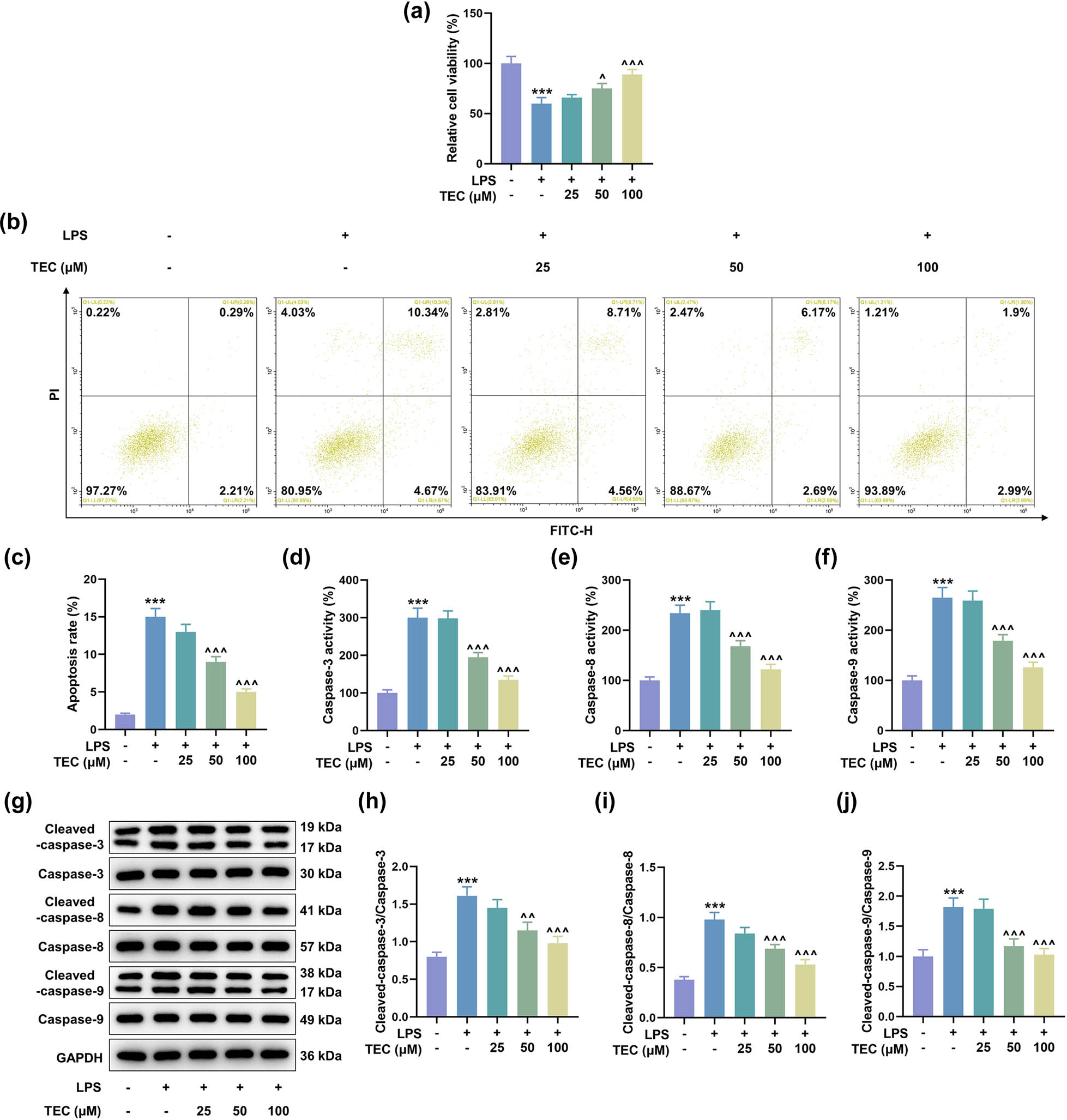
TEC alleviated the LPS-induced PC12 cellular injuries at a dose-dependent manner. (a) CCK-8 assay was applied to determine the viability of PC12 cells under the treatment of 5 μg/mL LPS coupled with or without treatment of 25, 50, or 100 μM TEC. *** P < 0.001 vs control group, ^ P < 0.05 vs LPS group, ^^^ P < 0.001 vs LPS group. (b and c) Flow cytometry was performed to detect the apoptotic rate of PC12 cells under the treatment of 5 μg/mL LPS coupled with or without treatment of 25, 50, or 100 μM TEC. *** P < 0.001 vs control group, ^^^ P < 0.001 vs LPS group. (d–f) Colorimetric assay was conducted to measure the expressions of apoptosis-related factors including caspase-3, caspase-8, and caspase-9. *** P < 0.001 vs control group, ^^^ P < 0.001 vs LPS group. (g–j) WB assay was utilized to quantitate the expressions of apoptosis-related factors including caspase-3, caspase-8, caspase-9, cleaved caspase-3, cleaved caspase-8, and cleaved caspase-9. *** P < 0.001 vs control group, ^^ P < 0.01 vs LPS group, ^^^ P < 0.001 vs LPS group. GAPDH was used as the internal reference for WB assay. All results in this figure were described as means ± SD of triplicate determination.
3.2 TEC alleviated the promoting effect of LPS on the inflammatory factors in PC12 cells
Controlling the inflammatory response is a critical step in the treatment of SCI. The expressions of pro-inflammatory factors, including IL-1β, IL-6, and TNF-α, were found to be up-regulated under LPS inducement and then were diminished by TEC (50 and 100 μM) in LPS-induced PC12 cells (Figure 3a–f, P < 0.001).
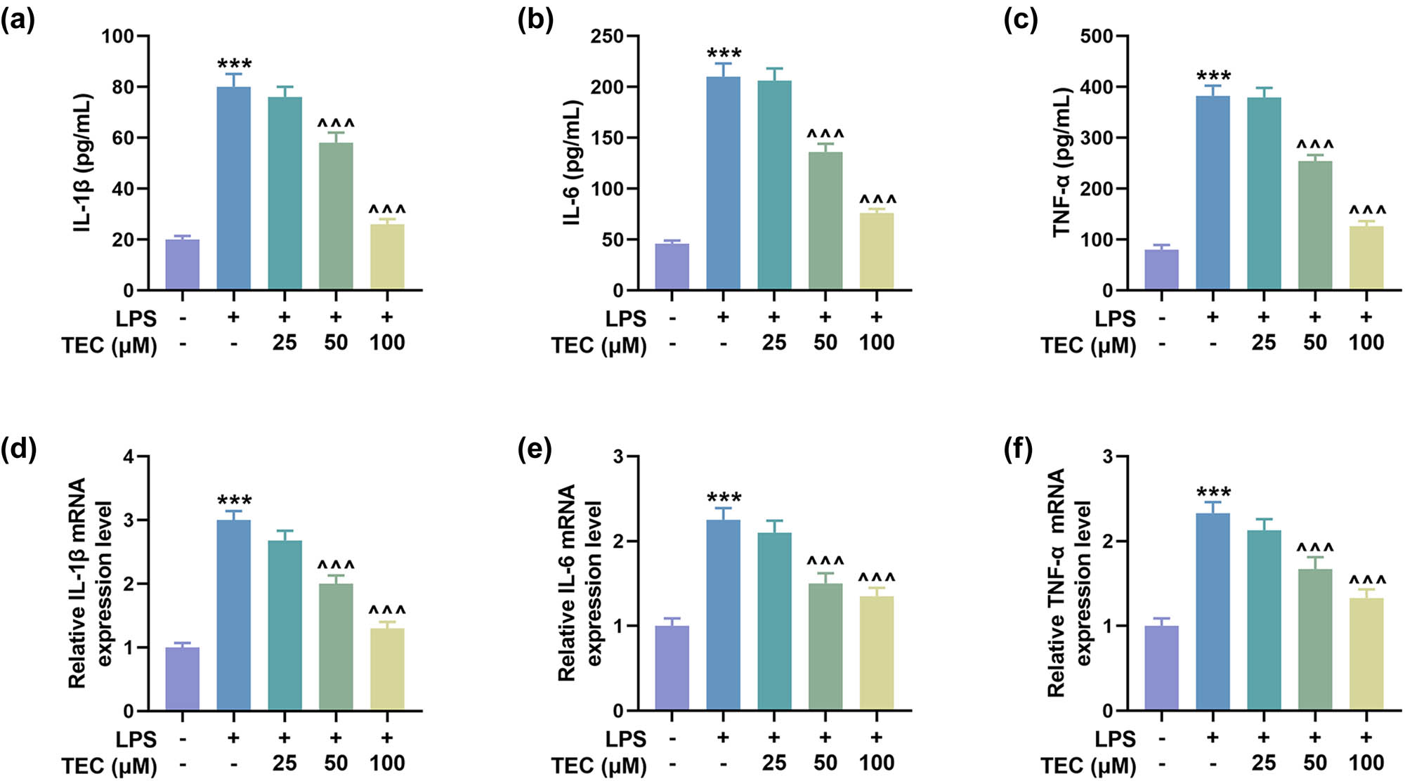
TEC abrogated the LPS-induced up-regulation of pro-inflammatory factors in PC12 cells. (a–c) ELISA was used to measure the levels of pro-inflammatory factors, including IL-1β, IL-6, and TNF-α, under LPS inducement with or without treatment of 25, 50, or 100 μM TEC. *** P < 0.001 vs control group, ^^^ P < 0.001 vs LPS group. (d–f) RT-qPCR was performed to quantitate the expressions of pro-inflammatory factors including, IL-1β, IL-6, and TNF-α, under various treatments. *** P < 0.001 vs control group, ^^^ P < 0.001 vs LPS group. GAPDH was used as the internal reference for RT-qPCR assay. All results in this figure were described as means ± SD of triplicate determination.
3.3 TEC inhibited IGFBP6 expression in LPS-induced PC12 cells
To unveil the associated downstream effector of TEC in SCI cell models, we used SwissTargetPrediction and GSE21497 to predict the potential target of TEC in disease treatment, and four targets were found, including TBXAS1, IGFBP6, OPRD1, and CA3 (Figure 4a). Since IGFBP6 was reported to aggravate SCI [31], we chose IGFBP6 as the research target in subsequent experiments. GEO2R was applied to analyze GSE21497 database, which revealed that IGFBP6 was overexpressed in SCI tissues (Figure 4b and c, P < 0.05). We then discovered that the expression of IGFBP6 was elevated in LPS-induced PC12 cells but then was dwindled by TEC treatment at a dose-dependent manner (Figure 4d and e, P < 0.05).
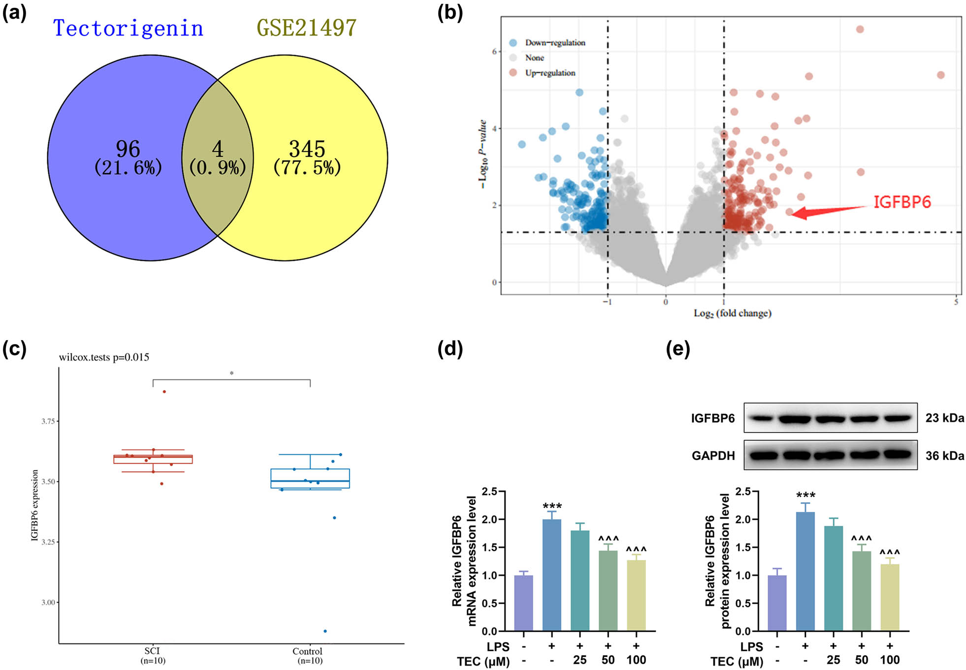
The expression of IGFBP6 was predicted and validated to be induced by LPS and inhibited by TEC. (a) SwissTargetPrediction (http://www.swisstargetprediction.ch/) and GSE21497 database were used to predict the potential target of TEC in disease treatment, and four targets were found, including TBXAS1, IGFBP6, OPRD1, and CA3. (b and c) GEO2R (https://www.ncbi.nlm.nih.gov/geo/geo2r/) was applied to analyze GSE21497 database, thereby obtaining the results that IGFBP6 was overexpressed in SCI tissues. (d and e) RT-qPCR and WB assays were carried out to measure the expression of IGFBP6 in PC12 cells under LPS inducement with or without the treatment of 25, 50, or 100 μM TEC. *** P < 0.001 vs control group, ^^^ P < 0.001 vs LPS group. GAPDH was used as the internal reference for RT-qPCR and WB assays. All results in this figure were described as means ± SD of triplicate determination.
3.4 IGFBP6 overexpression attenuated the effects of TEC on the apoptosis of LPS-induced PC12 cells
To investigate whether TEC plays a role in SCI by regulating IGFBP6, PC12 cells were transfected with IGFBP6 overexpression plasmids, and the up-regulation of IGFBP6 in PC12 cells indicated the successful transfection (Figure 5a and b, P < 0.001). As depicted in Figure 5c and d, IGFBP6 overexpression reversed the declined IGFBP6 expressions in PC12 cells under TEC treatment (P < 0.01). TEC treatment could significantly offset the effects of LPS on the apoptosis and apoptosis-related factor expressions in PC12 cells. We then discovered that IGFBP6 overexpression reversed the inhibiting effect of TEC on apoptosis, activity of caspase-3, caspase-8, and caspase-9, and expressions of cleaved caspase-3, cleaved caspase-8, and cleaved caspase-9 in LPS-induced PC12 cells (Figure 5e–m, P < 0.001).
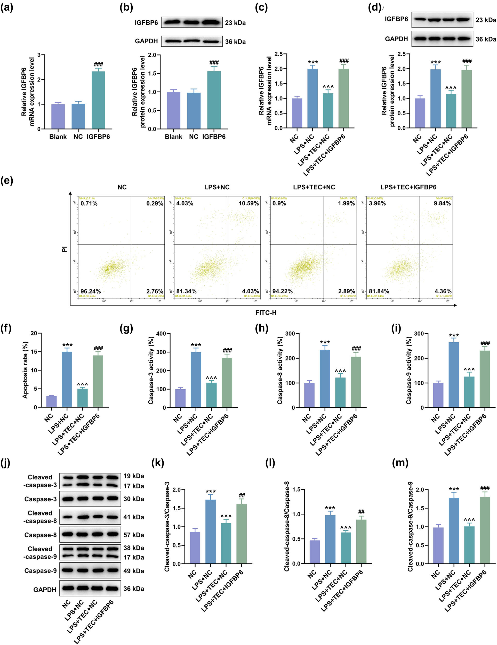
Overexpressed IGFBP6 reversed the inhibiting role of TEC in the apoptosis of LPS-induced PC12 cells. (a and b) RT-qPCR and WB assays were performed to detect the expression of IGFBP6 in PC12 cells with or without transfection of IGFBP6 overexpression plasmid (IGFBP6 group). ### P < 0.001 vs. negative control (NC) group. (c and d) RT-qPCR and WB assays were applied to quantify the expression of IGFBP6 in PC12 cells with or without LPS inducement coupled with the treatment of NC, TEC + NC, or TEC + IGFBP6 overexpression plasmid. *** P < 0.001 vs NC group, ^^^ P < 0.001 vs LPS + NC group, ### P < 0.001 vs LPS + TEC + NC group. (e and f) Flow cytometry was performed to detect the apoptotic degree of PC12 cells with or without LPS inducement coupled with the treatment of NC, TEC + NC, or TEC + IGFBP6 overexpression plasmid. *** P < 0.001 vs NC group, ^^^ P < 0.001 vs LPS + NC group, and ### P < 0.001 vs LPS + TEC + NC group. (g–i) Colorimetric method was used to quantitate the apoptosis-related factors, including caspase-3, caspase-8, and caspase-9, that were released from PC12 cells with or without LPS inducement coupled with the treatment of NC, TEC + NC, or TEC + IGFBP6 overexpression plasmid. *** P < 0.001 vs NC group, ^^^ P < 0.001 vs LPS + NC group, and ### P < 0.001 vs LPS + TEC + NC group. (j–m) WB assay was utilized to determine the expressions of apoptosis-related factors, including caspase-3, caspase-8, caspase-9, cleaved caspase-3, cleaved caspase-8, and cleaved caspase-9, in PC12 cells under various conditions. *** P < 0.001 vs NC group, ^^^ P < 0.001 vs LPS + NC group, and ### P < 0.001 vs LPS + TEC + NC group. GAPDH was used as the internal reference for RT-qPCR and WB assays. All results in this figure were described as mean ± SD of triplicate determination.
3.5 IGFBP6 overexpression counteracted the effects of TEC on the inflammation and TLR4/NF-κB pathway in LPS-induced PC12 cells
The effect of IGFBP6 on the pro-inflammatory factors was also detected. The results proved that IGFBP6 overexpression reversed the effects of TEC on down-regulating expressions of pro-inflammatory factors, including IL-1β, IL-6, and TNF-α (Figure 6a–f, P < 0.01). The regulatory effect of TEC on inflammatory response in diseases is realized by inhibiting TLR4/NF-κB signaling pathway [20], which is also related to the expression of IGFBP6 [32]. The results demonstrated that LPS induced the activation of TLR4/NF-κB signaling pathway, as evidenced by the up-regulation of TLR4, p-IκBα/IκBα, and p-p65/p-65 (Figure 6g–j, P < 0.01). TEC treatment inhibited the activation of TLR4/NF-κB signaling pathway, while the function of TEC was offset by IGFBP6 overexpression (Figure 6g–j, P < 0.05).
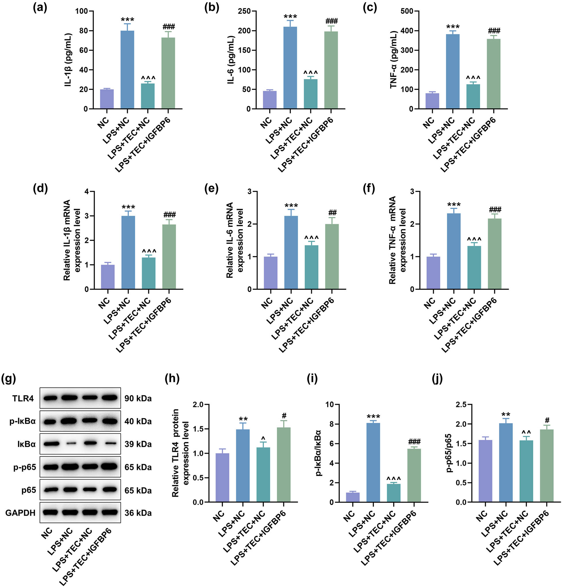
Overexpressed IGFBP6 offset the suppressing effect of TEC on inflammation in LPS-induced PC12 cells. (a–c) ELISA was used to measure the levels of pro-inflammatory factors, including IL-1β, IL-6, and TNF-α, in PC12 cells with or without LPS inducement coupled with the treatment of NC, TEC + NC, or TEC + IGFBP6 overexpression plasmid. *** P < 0.001 vs NC group, ^^^ P < 0.001 vs LPS + NC group, and ### P < 0.001 vs LPS + TEC + NC group. (d–f) RT-qPCR assay was exploited to detect the levels of pro-inflammatory factors, including IL-1β, IL-6, and TNF-α, in PC12 cells under various treatments. *** P < 0.001 vs NC group, ^^^ P < 0.001 vs LPS + NC group, ## P < 0.01 vs LPS + TEC + NC group, and ### P < 0.001 vs LPS + TEC + NC group. (g–j) WB assay was performed to determine the expressions of TLR4/NF-κB pathway-related factors, including TLR4, IκBα, p-IκBα, p65, and p-p65, in PC12 cells under various conditions. ** P < 0.01 vs NC group, *** P < 0.001 vs NC group, ^^ P < 0.01 vs LPS + NC group, # P < 0.05 vs LPS + TEC + NC group, and ## P < 0.01 vs LPS + TEC + NC group. GAPDH was used as the internal reference for RT-qPCR and WB assays. All results in this figure were described as mean ± SD of triplicate determination.
4 Discussion
Through analyzing all experimental results, it can be concluded that TEC could exert neuroprotective effects toward inflammatory infiltration of neurons and reduce the associated cellular apoptosis. Notably, all these functions were realized through inhibiting downstream apoptotic or inflammatory effector, IGFBP6.
The anti-inflammation or anti-apoptosis effect of TEC was not only validated in our experiments but also reviewed in our introduction section. Therefore, TEC is actually beneficial to mitigate SCI especially in the second phase that is full of inflammation infiltration and accompanied with various complications. Some similar or different neuroprotective effects of TEC have also been reported as follows [33,34,35]. TEC could modulate the expression of erythropoietin through inhibiting the degradation of hypoxia-inducible factor-1α (HIF-1α), therefore exerting neuroprotective functions on in vitro cultured neuron-like NT2/D1 cells and rat cortical neurons [33]. TEC could also inhibit TLR4/MyD88/NF-κB and ERK/JNK signaling pathways in microglial cells that are over-activated under the treatment of LPS [34]. The overproduction of relative inflammatory mediators including, NO synthase, cycloxygenase-2, TNF-α, and IL-6, are also attenuated through TEC treatment [34]. Accordingly, the neuroprotective role of TEC is out of question.
We then strictly carried out experiments to identify whether IGFBP6 can promote apoptosis or inflammation, since its expression was suppressed by TEC. The results confirmed that IGFBP6 could activate apoptotic cascade, pro-inflammatory reactions, and the NF-κB signaling. According to former findings, IGFBP6 has been examined in diverse situations and these results are controversial [36,37,38,39,40,41,42,43,44,45,46]. Several literatures reported that IGFBP6 is the predominant IGFBP synthesized by PC12 pheochromocytoma [36,37,38]. A review proposed that IGFBP6 plays an important role in the control of cell-specific immunologic adaptation following hyperthermia treatment [39]. A simultaneously published article elucidated that IGFBP6 could induce the oxidative burst, degranulation and chemotaxis of neutrophils [40]. A similar report also confirmed the chemotactic role of IGFBP6 in rheumatoid arthritis [41]. Moreover, the content of IGFBP6 could be lessened through the treatment of dexamethasone, an anti-inflammation drug [38,41]. IGFBP6 has been demonstrated to be positively correlated with NF-κB signaling or other inflammatory factors, including IL-6 and IL-17B [42,43].
In addition to the aforementioned report revealing IGFBP6 as a pro-inflammatory effector, some contrary findings have been published [44,45,46]. IGFBP6 has been verified to be associated with anti-inflammatory factor TGF-β1 and enhance epithelial-mesenchymal transition in fibroblasts to promote the recovery of skin injuries under the treatment of ozone oil [44]. The adipose stem cell-derived extracellular vesicles, which have been identified to suppress allergic airway inflammation, could promote the expression of IGFBP6 [45]. Furthermore, IGFBP6 has a negative relationship with IL-6 or IL-1β, since neonatal astrocytes induced by external injuries could produce IL-6 or IL-1β to facilitate neurogenesis, while IGFBP6 inhibits the differentiation of multipotent neural stem/progenitor cells to block neurogenesis [46]. Apart from pro-inflammatory effects, the pro-apoptotic role of IGFBP6 is also controversial [31,47]. IGFBP6 expression is upregulated after the establishment of SCI model, and IGFBP6 is positively correlated with the expressions of pro-apoptotic p53 and cleaved caspase-3 [31]. IGFBP6 derived from human bone marrow mesenchymal stem cells exerts neuroprotective effects through inhibiting the activation of Bax and promoting the phosphorylation of Akt [47].
5 Conclusions
Collectively, TEC might be an efficient substance that could repair the peripheral nervous system after exogenous incidence causes SCI through maintaining neural cell numbers and functions. Targeting IGFBP6 also seems to be useful in the recovery of SCI. The precision and complexity of nervous systems are well known, leading to controversial conclusions in previous reports. Hence, more precise results should be provided in future validation.
Acknowledgements
Not applicable.
-
Funding information: Not applicable.
-
Author contributions: Substantial contributions to conception and design: Liqiang Zhou, Kui Yan. Data acquisition, data analysis and interpretation: Shuxing Xing, Jun Cheng. Drafting the article or critically revising it for important intellectual content: Liqiang Zhou, Kui Yan. Final approval of the version to be published: Liqiang Zhou, Kui Yan, Shuxing Xing, Jun Cheng. Agreement to be accountable for all aspects of the work in ensuring that questions related to the accuracy or integrity of the work are appropriately investigated and resolved: Liqiang Zhou, Kui Yan, Shuxing Xing, Jun Cheng.
-
Conflict of interest: The authors declare no conflicts of interest.
-
Data availability statement: The analyzed data sets generated during the study are available from the corresponding author on reasonable request.
References
[1] National Spinal Cord Injury Statistical Center. Spinal cord injury facts and figures at a glance. J Spinal Cord Med. 2014;37(3):355–6. 10.1179/1079026814z.000000000260.Suche in Google Scholar PubMed PubMed Central
[2] Ahuja CS, Nori S, Tetreault L, Wilson J, Kwon B, Harrop J, et al. Traumatic spinal cord injury-repair and regeneration. Neurosurgery. 2017;80(3s):S9–22. 10.1093/neuros/nyw080.Suche in Google Scholar PubMed
[3] Tator CH. Update on the pathophysiology and pathology of acute spinal cord injury. Brain Pathol. 1995;5(4):407–13. 10.1111/j.1750-3639.1995.tb00619.x.Suche in Google Scholar PubMed
[4] McDonald JW, Sadowsky C. Spinal-cord injury. Lancet. 2002;359(9304):417–25. 10.1016/s0140-6736(02)07603-1.Suche in Google Scholar
[5] Yip PK, Malaspina A. Spinal cord trauma and the molecular point of no return. Mol Neurodegeneration. 2012;7:6. 10.1186/1750-1326-7-6.Suche in Google Scholar PubMed PubMed Central
[6] Norenberg MD, Smith J, Marcillo A. The pathology of human spinal cord injury: Defining the problems. J Neurotrauma. 2004;21(4):429–40. 10.1089/089771504323004575.Suche in Google Scholar PubMed
[7] Nakamura M, Houghtling RA, MacArthur L, Bayer BM, Bregman BS. Differences in cytokine gene expression profile between acute and secondary injury in adult rat spinal cord. Exp Neurol. 2003;184(1):313–25. 10.1016/s0014-4886(03)00361-3.Suche in Google Scholar PubMed
[8] Ulndreaj A, Chio JC, Ahuja CS, Fehlings MG. Modulating the immune response in spinal cord injury. Expert Rev Neurotherapeutics. 2016;16(10):1127–9. 10.1080/14737175.2016.1207532.Suche in Google Scholar PubMed
[9] Wang Y, Wang H, Tao Y, Zhang S, Wang J, Feng X. Necroptosis inhibitor necrostatin-1 promotes cell protection and physiological function in traumatic spinal cord injury. Neuroscience. 2014;266:91–101. 10.1016/j.neuroscience.2014.02.007.Suche in Google Scholar PubMed
[10] Li S, Stys PK. Mechanisms of ionotropic glutamate receptor-mediated excitotoxicity in isolated spinal cord white matter. J Neurosci: Off J Soc Neuroscience. 2000;20(3):1190–8. 10.1523/jneurosci.20-03-01190.2000.Suche in Google Scholar PubMed PubMed Central
[11] Liu M, Wu W, Li H, Li S, Huang LT, Yang YQ, et al. Necroptosis, a novel type of programmed cell death, contributes to early neural cells damage after spinal cord injury in adult mice. J Spinal Cord Med. 2015;38(6):745–53. 10.1179/2045772314y.0000000224.Suche in Google Scholar
[12] Walters BC, Hadley MN, Hurlbert RJ, Aarabi B, Dhall SS, Gelb DE, et al. Guidelines for the management of acute cervical spine and spinal cord injuries: 2013 update. Neurosurgery. 2013;60(CN_suppl_1):82–91. 10.1227/01.neu.0000430319.32247.7f.Suche in Google Scholar PubMed
[13] Bracken MB. Steroids for acute spinal cord injury. Cochrane Database Syst Rev. 2012;1(1):Cd001046. 10.1002/14651858.CD001046.pub2.Suche in Google Scholar PubMed PubMed Central
[14] Azbill RD, Mu X, Springer JE. Riluzole increases high-affinity glutamate uptake in rat spinal cord synaptosomes. Brain Res. 2000;871(2):175–80. 10.1016/s0006-8993(00)02430-6.Suche in Google Scholar PubMed
[15] Nógrádi A, Szabó A, Pintér S, Vrbová G. Delayed riluzole treatment is able to rescue injured rat spinal motoneurons. Neuroscience. 2007;144(2):431–8. 10.1016/j.neuroscience.2006.09.046.Suche in Google Scholar PubMed
[16] Kaptanoglu E, Beskonakli E, Solaroglu I, Kilinc A, Taskin Y. Magnesium sulfate treatment in experimental spinal cord injury: emphasis on vascular changes and early clinical results. Neurosurgical Rev. 2003;26(4):283–7. 10.1007/s10143-003-0272-y.Suche in Google Scholar PubMed
[17] Drabek T, Janata A, Wilson CD, Stezoski J, Janesko-Feldman K, Tisherman SA, et al. Minocycline attenuates brain tissue levels of TNF-α produced by neurons after prolonged hypothermic cardiac arrest in rats. Resuscitation. 2014;85(2):284–91. 10.1016/j.resuscitation.2013.10.015.Suche in Google Scholar PubMed PubMed Central
[18] Li QY, Chen L, Yan MM, Shi XJ, Zhong MK. Tectorigenin regulates adipogenic differentiation and adipocytokines secretion via PPARγ and IKK/NF-κB signaling. Pharm Biol. 2015;53(11):1567–75. 10.3109/13880209.2014.993038.Suche in Google Scholar PubMed
[19] Ma CH, Liu JP, Qu R, Ma SP. Tectorigenin inhibits the inflammation of LPS-induced acute lung injury in mice. Chin J Nat Med. 2014;12(11):841–6. 10.1016/s1875-5364(14)60126-6.Suche in Google Scholar PubMed
[20] Zhang L, Zhao Y, Fan L, Xu K, Ji F, Xie Z, et al. Tectorigenin protects against experimental fulminant hepatic failure by regulating the TLR4/mitogen-activated protein kinase and TLR4/nuclear factor-κB pathways and autophagy. Phytotherapy Res. 2019;33(4):1055–64. 10.1002/ptr.6299.Suche in Google Scholar PubMed PubMed Central
[21] Chen X, Zhang W, Sun L, Lian Y. Tectorigenin protect HUVECs from H(2)O(2)-induced oxidative stress injury by regulating PI3K/Akt pathway. Tissue Cell. 2021;68:101475. 10.1016/j.tice.2020.101475.Suche in Google Scholar PubMed
[22] Lee HU, Bae EA, Kim DH. Hepatoprotective effect of tectoridin and tectorigenin on tert-butyl hyperoxide-induced liver injury. J Pharmacol Sci. 2005;97(4):541–4. 10.1254/jphs.scz040467.Suche in Google Scholar PubMed
[23] Lee HW, Choo MK, Bae EA, Kim DH. Beta-glucuronidase inhibitor tectorigenin isolated from the flower of Pueraria thunbergiana protects carbon tetrachloride-induced liver injury. Liver Int: Off J Int Assoc Study Liver. 2003;23(4):221–6. 10.1034/j.1600-0676.2003.00830.x.Suche in Google Scholar PubMed
[24] Qian X, Xiao Q, Li Z. Tectorigenin regulates migration, invasion, and apoptosis in dexamethasone-induced human airway epithelial cells through up-regulating miR-222-3p. Drug Dev Res. 2021;82(7):959–68. 10.1002/ddr.21795.Suche in Google Scholar PubMed
[25] Lee SY, Kim GT, Yun HM, Kim YC, Kwon IK, Kim EC. Tectorigenin Promotes Osteoblast Differentiation and in vivo Bone Healing, but Suppresses Osteoclast Differentiation and in vivo Bone Resorption. Mol Cell. 2018;41(5):476–85. 10.14348/molcells.2018.0056.Suche in Google Scholar PubMed PubMed Central
[26] Zhang Y, Lu Y, Ma L, Cao X, Xiao J, Chen J, et al. Activation of vascular endothelial growth factor receptor-3 in macrophages restrains TLR4-NF-κB signaling and protects against endotoxin shock. Immunity. 2014;40(4):501–14. 10.1016/j.immuni.2014.01.013.Suche in Google Scholar PubMed
[27] Seong KJ, Lee HG, Kook MS, Ko HM, Jung JY, Kim WJ. Epigallocatechin-3-gallate rescues LPS-impaired adult hippocampal neurogenesis through suppressing the TLR4-NF-κB signaling pathway in mice. Korean J Physiol Pharmacol. 2016;20(1):41–51. 10.4196/kjpp.2016.20.1.41.Suche in Google Scholar PubMed PubMed Central
[28] Ma S, Zhang C, Zhang Z, Dai Y, Gu R, Jiang R. Geniposide protects PC12 cells from lipopolysaccharide-evoked inflammatory injury via up-regulation of miR-145-5p. Artif Cells, Nanomed Biotechnol. 2019;47(1):2875–81. 10.1080/21691401.2019.1626406.Suche in Google Scholar PubMed
[29] Yang H, Liu JX, Shang HX, Lin S, Zhao JY, Lin JM. Qingjie Fuzheng granules inhibit colorectal cancer cell growth by the PI3K/AKT and ERK pathways. World J Gastrointest Oncol. 2019;11(5):377–92. 10.4251/wjgo.v11.i5.377.Suche in Google Scholar PubMed PubMed Central
[30] Livak KJ, Schmittgen TD. Analysis of relative gene expression data using real-time quantitative PCR and the 2(-Delta Delta C(T)) Method. Methods. 2001;25(4):402–8. 10.1006/meth.2001.1262.Suche in Google Scholar PubMed
[31] Wang S, Liu Y, Wu C, Zhao W, Zhang J, Bao G, et al. The Expression of IGFBP6 after Spinal Cord Injury: Implications for Neuronal Apoptosis. Neurochem Res. 2017;42(2):455–67. 10.1007/s11064-016-2092-9.Suche in Google Scholar PubMed
[32] Cacalano NA, Le D, Paranjpe A, Wang MY, Fernandez A, Evazyan T, et al. Regulation of IGFBP6 gene and protein is mediated by the inverse expression and function of c-jun N-terminal kinase (JNK) and NFkappaB in a model of oral tumor cells. Apoptosis An Int J Program Cell Death. 2008;13(12):1439–49. 10.1007/s10495-008-0270-1.Suche in Google Scholar PubMed
[33] Liu EY, Zheng ZX, Zheng BZ, Xia Y, Guo MS, Dong TT, et al. Tectorigenin, an isoflavone aglycone from the rhizome of Belamcanda chinensis, induces neuronal expression of erythropoietin via accumulation of hypoxia-inducible factor-1α. Phytother Res. 2020;34(6):1329–37. 10.1002/ptr.6599.Suche in Google Scholar PubMed
[34] Lim HS, Kim YJ, Kim BY, Park G, Jeong SJ. The Anti-neuroinflammatory Activity of Tectorigenin Pretreatment via Downregulated NF-κB and ERK/JNK Pathways in BV-2 Microglial and Microglia Inactivation in Mice With Lipopolysaccharide. Front Pharmacol. 2018;9:462. 10.3389/fphar.2018.00462.Suche in Google Scholar PubMed PubMed Central
[35] Vaswani KK, Wu GS, Ledeen RW. Exogenous gangliosides stimulate breakdown of neuro-2A phosphoinositides in a manner unrelated to neurite outgrowth. J Neurochem. 1990;55(2):492–9. 10.1111/j.1471-4159.1990.tb04162.x.Suche in Google Scholar PubMed
[36] Bach LA, Tseng LY, Swartz JE, Rechler MM. Rat PC12 pheochromocytoma cells synthesize insulin-like growth factor-binding protein-6. Endocrinology. 1993;133(3):990–5. 10.1210/endo.133.3.7689963.Suche in Google Scholar PubMed
[37] Bach LA, Leeding KS, Leng SL. Cyclic AMP agonists increase levels of insulin-like growth factor (IGF) binding protein-6 in PC12 rat phaeochromocytoma cells. Growth Hormone IGF Res. 1998;8(3):265–71. 10.1016/s1096-6374(98)80119-6.Suche in Google Scholar PubMed
[38] Bach LA, Leeding KS, Leng SL. Regulation of IGF-binding protein-6 by dexamethasone and IGFs in PC12 rat phaeochromocytoma cells. J Endocrinol. 1997;155(2):225–32. 10.1677/joe.0.1550225.Suche in Google Scholar PubMed
[39] Liso A, Capitanio N, Gerli R, Conese M. From fever to immunity: A new role for IGFBP-6? J Cell Mol Med. 2018;22(10):4588–96. 10.1111/jcmm.13738.Suche in Google Scholar PubMed PubMed Central
[40] Conese M, D’Oria S, Castellani S, Trotta R, Montemurro P, Liso A. Insulin-like growth factor-6 (IGFBP-6) stimulates neutrophil oxidative burst, degranulation and chemotaxis. Inflamm Res. 2018;67(2):107–9. 10.1007/s00011-017-1107-6.Suche in Google Scholar PubMed
[41] Alunno A, Bistoni O, Manetti M, Cafaro G, Valentini V, Bartoloni E, et al. Insulin-Like Growth Factor Binding Protein 6 in Rheumatoid Arthritis: A Possible Novel Chemotactic Factor. Front Immunol. 2017;8:554. 10.3389/fimmu.2017.00554.Suche in Google Scholar PubMed PubMed Central
[42] Park JH, Lee SW, Kim IT, Shin BS, Cheong SW, Cho UH, et al. TCDD-up-regulation of IGFBP-6 and IL-5R alpha subunit genes in vivo and in vitro. Mol Cell. 2001;12(3):372–9.10.1016/S1016-8478(23)17111-1Suche in Google Scholar
[43] Ivanov VN, Ghandhi SA, Zhou H, Huang SX, Chai Y, Amundson SA, et al. Radiation response and regulation of apoptosis induced by a combination of TRAIL and CHX in cells lacking mitochondrial DNA: a role for NF-κB-STAT3-directed gene expression. Exp Cell Res. 2011;317(11):1548–66. 10.1016/j.yexcr.2011.03.012.Suche in Google Scholar PubMed PubMed Central
[44] Xiao W, Tang H, Wu M, Liao Y, Li K, Li L, et al. Ozone oil promotes wound healing by increasing the migration of fibroblasts via PI3K/Akt/mTOR signaling pathway. Biosci Rep. 2017;37(6):BSR20170658. 10.1042/bsr20170658.Suche in Google Scholar PubMed PubMed Central
[45] Kim SD, Kang SA, Kim YW, Yu HS, Cho KS, Roh HJ. Screening and Functional Pathway Analysis of Pulmonary Genes Associated with Suppression of Allergic Airway Inflammation by Adipose Stem Cell-Derived Extracellular Vesicles. Stem Cell Int. 2020;2020:5684250. 10.1155/2020/5684250.Suche in Google Scholar PubMed PubMed Central
[46] Barkho BZ, Song H, Aimone JB, Smrt RD, Kuwabara T, Nakashima K, et al. Identification of astrocyte-expressed factors that modulate neural stem/progenitor cell differentiation. Stem Cell Dev. 2006;15(3):407–21. 10.1089/scd.2006.15.407.Suche in Google Scholar PubMed PubMed Central
[47] Jeon HJ, Park J, Shin JH, Chang MS. Insulin-like growth factor binding protein-6 released from human mesenchymal stem cells confers neuronal protection through IGF-1R-mediated signaling. Int J Mol Med. 2017;40(6):1860–8. 10.3892/ijmm.2017.3173.Suche in Google Scholar PubMed PubMed Central
© 2023 the author(s), published by De Gruyter
This work is licensed under the Creative Commons Attribution 4.0 International License.
Artikel in diesem Heft
- Research Articles
- Exosomes derived from mesenchymal stem cells overexpressing miR-210 inhibits neuronal inflammation and contribute to neurite outgrowth through modulating microglia polarization
- Current situation of acute ST-segment elevation myocardial infarction in a county hospital chest pain center during an epidemic of novel coronavirus pneumonia
- circ-IARS depletion inhibits the progression of non-small-cell lung cancer by circ-IARS/miR-1252-5p/HDGF ceRNA pathway
- circRNA ITGA7 restrains growth and enhances radiosensitivity by up-regulating SMAD4 in colorectal carcinoma
- WDR79 promotes aerobic glycolysis of pancreatic ductal adenocarcinoma (PDAC) by the suppression of SIRT4
- Up-regulation of collagen type V alpha 2 (COL5A2) promotes malignant phenotypes in gastric cancer cell via inducing epithelial–mesenchymal transition (EMT)
- Inhibition of TERC inhibits neural apoptosis and inflammation in spinal cord injury through Akt activation and p-38 inhibition via the miR-34a-5p/XBP-1 axis
- 3D-printed polyether-ether-ketone/n-TiO2 composite enhances the cytocompatibility and osteogenic differentiation of MC3T3-E1 cells by downregulating miR-154-5p
- Propofol-mediated circ_0000735 downregulation restrains tumor growth by decreasing integrin-β1 expression in non-small cell lung cancer
- PVT1/miR-16/CCND1 axis regulates gastric cancer progression
- Silencing of circ_002136 sensitizes gastric cancer to paclitaxel by targeting the miR-16-5p/HMGA1 axis
- Short-term outcomes after simultaneous gastrectomy plus cholecystectomy in gastric cancer: A pooling up analysis
- SCARA5 inhibits oral squamous cell carcinoma via inactivating the STAT3 and PI3K/AKT signaling pathways
- Molecular mechanism by which the Notch signaling pathway regulates autophagy in a rat model of pulmonary fibrosis in pigeon breeder’s lung
- lncRNA TPT1-AS1 promotes cell migration and invasion in esophageal squamous-cell carcinomas by regulating the miR-26a/HMGA1 axis
- SIRT1/APE1 promotes the viability of gastric cancer cells by inhibiting p53 to suppress ferroptosis
- Glycoprotein non-metastatic melanoma B interacts with epidermal growth factor receptor to regulate neural stem cell survival and differentiation
- Treatments for brain metastases from EGFR/ALK-negative/unselected NSCLC: A network meta-analysis
- Association of osteoporosis and skeletal muscle loss with serum type I collagen carboxyl-terminal peptide β glypeptide: A cross-sectional study in elder Chinese population
- circ_0000376 knockdown suppresses non-small cell lung cancer cell tumor properties by the miR-545-3p/PDPK1 pathway
- Delivery in a vertical birth chair supported by freedom of movement during labor: A randomized control trial
- UBE2J1 knockdown promotes cell apoptosis in endometrial cancer via regulating PI3K/AKT and MDM2/p53 signaling
- Metabolic resuscitation therapy in critically ill patients with sepsis and septic shock: A pilot prospective randomized controlled trial
- Lycopene ameliorates locomotor activity and urinary frequency induced by pelvic venous congestion in rats
- UHRF1-induced connexin26 methylation is involved in hearing damage triggered by intermittent hypoxia in neonatal rats
- LINC00511 promotes melanoma progression by targeting miR-610/NUCB2
- Ultra-high-performance liquid chromatography-tandem mass spectrometry analysis of serum metabolomic characteristics in people with different vitamin D levels
- Role of Jumonji domain-containing protein D3 and its inhibitor GSK-J4 in Hashimoto’s thyroiditis
- circ_0014736 induces GPR4 to regulate the biological behaviors of human placental trophoblast cells through miR-942-5p in preeclampsia
- Monitoring of sirolimus in the whole blood samples from pediatric patients with lymphatic anomalies
- Effects of osteogenic growth peptide C-terminal pentapeptide and its analogue on bone remodeling in an osteoporosis rat model
- A novel autophagy-related long non-coding RNAs signature predicting progression-free interval and I-131 therapy benefits in papillary thyroid carcinoma
- WGCNA-based identification of potential targets and pathways in response to treatment in locally advanced breast cancer patients
- Radiomics model using preoperative computed tomography angiography images to differentiate new from old emboli of acute lower limb arterial embolism
- Dysregulated lncRNAs are involved in the progress of myocardial infarction by constructing regulatory networks
- Single-arm trial to evaluate the efficacy and safety of baclofen in treatment of intractable hiccup caused by malignant tumor chemotherapy
- Genetic polymorphisms of MRPS30-DT and NINJ2 may influence lung cancer risk
- Efficacy of immune checkpoint inhibitors in patients with KRAS-mutant advanced non-small cell lung cancer: A retrospective analysis
- Pyroptosis-based risk score predicts prognosis and drug sensitivity in lung adenocarcinoma
- Upregulation of lncRNA LANCL1-AS1 inhibits the progression of non-small-cell lung cancer via the miR-3680-3p/GMFG axis
- CircRANBP17 modulated KDM1A to regulate neuroblastoma progression by sponging miR-27b-3p
- Exosomal miR-93-5p regulated the progression of osteoarthritis by targeting ADAMTS9
- Downregulation of RBM17 enhances cisplatin sensitivity and inhibits cell invasion in human hypopharyngeal cancer cells
- HDAC5-mediated PRAME regulates the proliferation, migration, invasion, and EMT of laryngeal squamous cell carcinoma via the PI3K/AKT/mTOR signaling pathway
- The association between sleep duration, quality, and nonalcoholic fatty liver disease: A cross-sectional study
- Myostatin silencing inhibits podocyte apoptosis in membranous nephropathy through Smad3/PKA/NOX4 signaling pathway
- A novel long noncoding RNA AC125257.1 facilitates colorectal cancer progression by targeting miR-133a-3p/CASC5 axis
- Impact of omicron wave and associated control measures in Shanghai on health management and psychosocial well-being of patients with chronic conditions
- Clinicopathological characteristics and prognosis of young patients aged ≤45 years old with non-small cell lung cancer
- TMT-based comprehensive proteomic profiling identifies serum prognostic signatures of acute myeloid leukemia
- The dose limits of teeth protection for patients with nasopharyngeal carcinoma undergoing radiotherapy based on the early oral health-related quality of life
- miR-30b-5p targeting GRIN2A inhibits hippocampal damage in epilepsy
- Long non-coding RNA AL137789.1 promoted malignant biological behaviors and immune escape of pancreatic carcinoma cells
- IRF6 and FGF1 polymorphisms in non-syndromic cleft lip with or without cleft palate in the Polish population
- Comprehensive analysis of the role of SFXN family in breast cancer
- Efficacy of bronchoscopic intratumoral injection of endostar and cisplatin in lung squamous cell carcinoma patients underwent conventional chemoradiotherapy
- Silencing of long noncoding RNA MIAT inhibits the viability and proliferation of breast cancer cells by promoting miR-378a-5p expression
- AG1024, an IGF-1 receptor inhibitor, ameliorates renal injury in rats with diabetic nephropathy via the SOCS/JAK2/STAT pathway
- Downregulation of KIAA1199 alleviated the activation, proliferation, and migration of hepatic stellate cells by the inhibition of epithelial–mesenchymal transition
- Exendin-4 regulates the MAPK and WNT signaling pathways to alleviate the osteogenic inhibition of periodontal ligament stem cells in a high glucose environment
- Inhibition of glycolysis represses the growth and alleviates the endoplasmic reticulum stress of breast cancer cells by regulating TMTC3
- The function of lncRNA EMX2OS/miR-653-5p and its regulatory mechanism in lung adenocarcinoma
- Tectorigenin alleviates the apoptosis and inflammation in spinal cord injury cell model through inhibiting insulin-like growth factor-binding protein 6
- Ultrasound examination supporting CT or MRI in the evaluation of cervical lymphadenopathy in patients with irradiation-treated head and neck cancer
- F-box and WD repeat domain containing 7 inhibits the activation of hepatic stellate cells by degrading delta-like ligand 1 to block Notch signaling pathway
- Knockdown of circ_0005615 enhances the radiosensitivity of colorectal cancer by regulating the miR-665/NOTCH1 axis
- Long noncoding RNA Mhrt alleviates angiotensin II-induced cardiac hypertrophy phenotypes by mediating the miR-765/Wnt family member 7B pathway
- Effect of miR-499-5p/SOX6 axis on atrial fibrosis in rats with atrial fibrillation
- Cholesterol induces inflammation and reduces glucose utilization
- circ_0004904 regulates the trophoblast cell in preeclampsia via miR-19b-3p/ARRDC3 axis
- NECAB3 promotes the migration and invasion of liver cancer cells through HIF-1α/RIT1 signaling pathway
- The poor performance of cardiovascular risk scores in identifying patients with idiopathic inflammatory myopathies at high cardiovascular risk
- miR-2053 inhibits the growth of ovarian cancer cells by downregulating SOX4
- Nucleophosmin 1 associating with engulfment and cell motility protein 1 regulates hepatocellular carcinoma cell chemotaxis and metastasis
- α-Hederin regulates macrophage polarization to relieve sepsis-induced lung and liver injuries in mice
- Changes of microbiota level in urinary tract infections: A meta-analysis
- Identification of key enzalutamide-resistance-related genes in castration-resistant prostate cancer and verification of RAD51 functions
- Falls during oxaliplatin-based chemotherapy for gastrointestinal malignancies – (lessons learned from) a prospective study
- Outcomes of low-risk birth care during the Covid-19 pandemic: A cohort study from a tertiary care center in Lithuania
- Vitamin D protects intestines from liver cirrhosis-induced inflammation and oxidative stress by inhibiting the TLR4/MyD88/NF-κB signaling pathway
- Integrated transcriptome analysis identifies APPL1/RPS6KB2/GALK1 as immune-related metastasis factors in breast cancer
- Genomic analysis of immunogenic cell death-related subtypes for predicting prognosis and immunotherapy outcomes in glioblastoma multiforme
- Circular RNA Circ_0038467 promotes the maturation of miRNA-203 to increase lipopolysaccharide-induced apoptosis of chondrocytes
- An economic evaluation of fine-needle cytology as the primary diagnostic tool in the diagnosis of lymphadenopathy
- Midazolam impedes lung carcinoma cell proliferation and migration via EGFR/MEK/ERK signaling pathway
- Network pharmacology combined with molecular docking and experimental validation to reveal the pharmacological mechanism of naringin against renal fibrosis
- PTPN12 down-regulated by miR-146b-3p gene affects the malignant progression of laryngeal squamous cell carcinoma
- miR-141-3p accelerates ovarian cancer progression and promotes M2-like macrophage polarization by targeting the Keap1-Nrf2 pathway
- lncRNA OIP5-AS1 attenuates the osteoarthritis progression in IL-1β-stimulated chondrocytes
- Overexpression of LINC00607 inhibits cell growth and aggressiveness by regulating the miR-1289/EFNA5 axis in non-small-cell lung cancer
- Subjective well-being in informal caregivers during the COVID-19 pandemic
- Nrf2 protects against myocardial ischemia-reperfusion injury in diabetic rats by inhibiting Drp1-mediated mitochondrial fission
- Unfolded protein response inhibits KAT2B/MLKL-mediated necroptosis of hepatocytes by promoting BMI1 level to ubiquitinate KAT2B
- Bladder cancer screening: The new selection and prediction model
- circNFATC3 facilitated the progression of oral squamous cell carcinoma via the miR-520h/LDHA axis
- Prone position effect in intensive care patients with SARS-COV-2 pneumonia
- Clinical observation on the efficacy of Tongdu Tuina manipulation in the treatment of primary enuresis in children
- Dihydroartemisinin ameliorates cerebral I/R injury in rats via regulating VWF and autophagy-mediated SIRT1/FOXO1 pathway
- Knockdown of circ_0113656 assuages oxidized low-density lipoprotein-induced vascular smooth muscle cell injury through the miR-188-3p/IGF2 pathway
- Low Ang-(1–7) and high des-Arg9 bradykinin serum levels are correlated with cardiovascular risk factors in patients with COVID-19
- Effect of maternal age and body mass index on induction of labor with oral misoprostol for premature rupture of membrane at term: A retrospective cross-sectional study
- Potential protective effects of Huanglian Jiedu Decoction against COVID-19-associated acute kidney injury: A network-based pharmacological and molecular docking study
- Clinical significance of serum MBD3 detection in girls with central precocious puberty
- Clinical features of varicella-zoster virus caused neurological diseases detected by metagenomic next-generation sequencing
- Collagen treatment of complex anorectal fistula: 3 years follow-up
- LncRNA CASC15 inhibition relieves renal fibrosis in diabetic nephropathy through down-regulating SP-A by sponging to miR-424
- Efficacy analysis of empirical bismuth quadruple therapy, high-dose dual therapy, and resistance gene-based triple therapy as a first-line Helicobacter pylori eradication regimen – An open-label, randomized trial
- SMOC2 plays a role in heart failure via regulating TGF-β1/Smad3 pathway-mediated autophagy
- A prospective cohort study of the impact of chronic disease on fall injuries in middle-aged and older adults
- circRNA THBS1 silencing inhibits the malignant biological behavior of cervical cancer cells via the regulation of miR-543/HMGB2 axis
- hsa_circ_0000285 sponging miR-582-3p promotes neuroblastoma progression by regulating the Wnt/β-catenin signaling pathway
- Long non-coding RNA GNAS-AS1 knockdown inhibits proliferation and epithelial–mesenchymal transition of lung adenocarcinoma cells via the microRNA-433-3p/Rab3A axis
- lncRNA UCA1 regulates miR-132/Lrrfip1 axis to promote vascular smooth muscle cell proliferation
- Twenty-four-color full spectrum flow cytometry panel for minimal residual disease detection in acute myeloid leukemia
- Hsa-miR-223-3p participates in the process of anthracycline-induced cardiomyocyte damage by regulating NFIA gene
- Anti-inflammatory effect of ApoE23 on Salmonella typhimurium-induced sepsis in mice
- Analysis of somatic mutations and key driving factors of cervical cancer progression
- Hsa_circ_0028007 regulates the progression of nasopharyngeal carcinoma through the miR-1179/SQLE axis
- Variations in sexual function after laparoendoscopic single-site hysterectomy in women with benign gynecologic diseases
- Effects of pharmacological delay with roxadustat on multi-territory perforator flap survival in rats
- Analysis of heroin effects on calcium channels in rat cardiomyocytes based on transcriptomics and metabolomics
- Risk factors of recurrent bacterial vaginosis among women of reproductive age: A cross-sectional study
- Alkbh5 plays indispensable roles in maintaining self-renewal of hematopoietic stem cells
- Study to compare the effect of casirivimab and imdevimab, remdesivir, and favipiravir on progression and multi-organ function of hospitalized COVID-19 patients
- Correlation between microvessel maturity and ISUP grades assessed using contrast-enhanced transrectal ultrasonography in prostate cancer
- The protective effect of caffeic acid phenethyl ester in the nephrotoxicity induced by α-cypermethrin
- Norepinephrine alleviates cyclosporin A-induced nephrotoxicity by enhancing the expression of SFRP1
- Effect of RUNX1/FOXP3 axis on apoptosis of T and B lymphocytes and immunosuppression in sepsis
- The function of Foxp1 represses β-adrenergic receptor transcription in the occurrence and development of bladder cancer through STAT3 activity
- Risk model and validation of carbapenem-resistant Klebsiella pneumoniae infection in patients with cerebrovascular disease in the ICU
- Calycosin protects against chronic prostatitis in rats via inhibition of the p38MAPK/NF-κB pathway
- Pan-cancer analysis of the PDE4DIP gene with potential prognostic and immunotherapeutic values in multiple cancers including acute myeloid leukemia
- The safety and immunogenicity to inactivated COVID-19 vaccine in patients with hyperlipemia
- Circ-UBR4 regulates the proliferation, migration, inflammation, and apoptosis in ox-LDL-induced vascular smooth muscle cells via miR-515-5p/IGF2 axis
- Clinical characteristics of current COVID-19 rehabilitation outpatients in China
- Luteolin alleviates ulcerative colitis in rats via regulating immune response, oxidative stress, and metabolic profiling
- miR-199a-5p inhibits aortic valve calcification by targeting ATF6 and GRP78 in valve interstitial cells
- The application of iliac fascia space block combined with esketamine intravenous general anesthesia in PFNA surgery of the elderly: A prospective, single-center, controlled trial
- Elevated blood acetoacetate levels reduce major adverse cardiac and cerebrovascular events risk in acute myocardial infarction
- The effects of progesterone on the healing of obstetric anal sphincter damage in female rats
- Identification of cuproptosis-related genes for predicting the development of prostate cancer
- Lumican silencing ameliorates β-glycerophosphate-mediated vascular smooth muscle cell calcification by attenuating the inhibition of APOB on KIF2C activity
- Targeting PTBP1 blocks glutamine metabolism to improve the cisplatin sensitivity of hepatocarcinoma cells through modulating the mRNA stability of glutaminase
- A single center prospective study: Influences of different hip flexion angles on the measurement of lumbar spine bone mineral density by dual energy X-ray absorptiometry
- Clinical analysis of AN69ST membrane continuous venous hemofiltration in the treatment of severe sepsis
- Antibiotics therapy combined with probiotics administered intravaginally for the treatment of bacterial vaginosis: A systematic review and meta-analysis
- Construction of a ceRNA network to reveal a vascular invasion associated prognostic model in hepatocellular carcinoma
- A pan-cancer analysis of STAT3 expression and genetic alterations in human tumors
- A prognostic signature based on seven T-cell-related cell clustering genes in bladder urothelial carcinoma
- Pepsin concentration in oral lavage fluid of rabbit reflux model constructed by dilating the lower esophageal sphincter
- The antihypertensive felodipine shows synergistic activity with immune checkpoint blockade and inhibits tumor growth via NFAT1 in LUSC
- Tanshinone IIA attenuates valvular interstitial cells’ calcification induced by oxidized low density lipoprotein via reducing endoplasmic reticulum stress
- AS-IV enhances the antitumor effects of propofol in NSCLC cells by inhibiting autophagy
- Establishment of two oxaliplatin-resistant gallbladder cancer cell lines and comprehensive analysis of dysregulated genes
- Trial protocol: Feasibility of neuromodulation with connectivity-guided intermittent theta-burst stimulation for improving cognition in multiple sclerosis
- LncRNA LINC00592 mediates the promoter methylation of WIF1 to promote the development of bladder cancer
- Factors associated with gastrointestinal dysmotility in critically ill patients
- Mechanisms by which spinal cord stimulation intervenes in atrial fibrillation: The involvement of the endothelin-1 and nerve growth factor/p75NTR pathways
- Analysis of two-gene signatures and related drugs in small-cell lung cancer by bioinformatics
- Silencing USP19 alleviates cigarette smoke extract-induced mitochondrial dysfunction in BEAS-2B cells by targeting FUNDC1
- Menstrual irregularities associated with COVID-19 vaccines among women in Saudi Arabia: A survey during 2022
- Ferroptosis involves in Schwann cell death in diabetic peripheral neuropathy
- The effect of AQP4 on tau protein aggregation in neurodegeneration and persistent neuroinflammation after cerebral microinfarcts
- Activation of UBEC2 by transcription factor MYBL2 affects DNA damage and promotes gastric cancer progression and cisplatin resistance
- Analysis of clinical characteristics in proximal and distal reflux monitoring among patients with gastroesophageal reflux disease
- Exosomal circ-0020887 and circ-0009590 as novel biomarkers for the diagnosis and prediction of short-term adverse cardiovascular outcomes in STEMI patients
- Upregulated microRNA-429 confers endometrial stromal cell dysfunction by targeting HIF1AN and regulating the HIF1A/VEGF pathway
- Bibliometrics and knowledge map analysis of ultrasound-guided regional anesthesia
- Knockdown of NUPR1 inhibits angiogenesis in lung cancer through IRE1/XBP1 and PERK/eIF2α/ATF4 signaling pathways
- D-dimer trends predict COVID-19 patient’s prognosis: A retrospective chart review study
- WTAP affects intracranial aneurysm progression by regulating m6A methylation modification
- Using of endoscopic polypectomy in patients with diagnosed malignant colorectal polyp – The cross-sectional clinical study
- Anti-S100A4 antibody administration alleviates bronchial epithelial–mesenchymal transition in asthmatic mice
- Prognostic evaluation of system immune-inflammatory index and prognostic nutritional index in double expressor diffuse large B-cell lymphoma
- Prevalence and antibiogram of bacteria causing urinary tract infection among patients with chronic kidney disease
- Reactive oxygen species within the vaginal space: An additional promoter of cervical intraepithelial neoplasia and uterine cervical cancer development?
- Identification of disulfidptosis-related genes and immune infiltration in lower-grade glioma
- A new technique for uterine-preserving pelvic organ prolapse surgery: Laparoscopic rectus abdominis hysteropexy for uterine prolapse by comparing with traditional techniques
- Self-isolation of an Italian long-term care facility during COVID-19 pandemic: A comparison study on care-related infectious episodes
- A comparative study on the overlapping effects of clinically applicable therapeutic interventions in patients with central nervous system damage
- Low intensity extracorporeal shockwave therapy for chronic pelvic pain syndrome: Long-term follow-up
- The diagnostic accuracy of touch imprint cytology for sentinel lymph node metastases of breast cancer: An up-to-date meta-analysis of 4,073 patients
- Mortality associated with Sjögren’s syndrome in the United States in the 1999–2020 period: A multiple cause-of-death study
- CircMMP11 as a prognostic biomarker mediates miR-361-3p/HMGB1 axis to accelerate malignant progression of hepatocellular carcinoma
- Analysis of the clinical characteristics and prognosis of adult de novo acute myeloid leukemia (none APL) with PTPN11 mutations
- KMT2A maintains stemness of gastric cancer cells through regulating Wnt/β-catenin signaling-activated transcriptional factor KLF11
- Evaluation of placental oxygenation by near-infrared spectroscopy in relation to ultrasound maturation grade in physiological term pregnancies
- The role of ultrasonographic findings for PIK3CA-mutated, hormone receptor-positive, human epidermal growth factor receptor-2-negative breast cancer
- Construction of immunogenic cell death-related molecular subtypes and prognostic signature in colorectal cancer
- Long-term prognostic value of high-sensitivity cardiac troponin-I in patients with idiopathic dilated cardiomyopathy
- Establishing a novel Fanconi anemia signaling pathway-associated prognostic model and tumor clustering for pediatric acute myeloid leukemia patients
- Integrative bioinformatics analysis reveals STAT2 as a novel biomarker of inflammation-related cardiac dysfunction in atrial fibrillation
- Adipose-derived stem cells repair radiation-induced chronic lung injury via inhibiting TGF-β1/Smad 3 signaling pathway
- Real-world practice of idiopathic pulmonary fibrosis: Results from a 2000–2016 cohort
- lncRNA LENGA sponges miR-378 to promote myocardial fibrosis in atrial fibrillation
- Diagnostic value of urinary Tamm-Horsfall protein and 24 h urine osmolality for recurrent calcium oxalate stones of the upper urinary tract: Cross-sectional study
- The value of color Doppler ultrasonography combined with serum tumor markers in differential diagnosis of gastric stromal tumor and gastric cancer
- The spike protein of SARS-CoV-2 induces inflammation and EMT of lung epithelial cells and fibroblasts through the upregulation of GADD45A
- Mycophenolate mofetil versus cyclophosphamide plus in patients with connective tissue disease-associated interstitial lung disease: Efficacy and safety analysis
- MiR-1278 targets CALD1 and suppresses the progression of gastric cancer via the MAPK pathway
- Metabolomic analysis of serum short-chain fatty acid concentrations in a mouse of MPTP-induced Parkinson’s disease after dietary supplementation with branched-chain amino acids
- Cimifugin inhibits adipogenesis and TNF-α-induced insulin resistance in 3T3-L1 cells
- Predictors of gastrointestinal complaints in patients on metformin therapy
- Prescribing patterns in patients with chronic obstructive pulmonary disease and atrial fibrillation
- A retrospective analysis of the effect of latent tuberculosis infection on clinical pregnancy outcomes of in vitro fertilization–fresh embryo transferred in infertile women
- Appropriateness and clinical outcomes of short sustained low-efficiency dialysis: A national experience
- miR-29 regulates metabolism by inhibiting JNK-1 expression in non-obese patients with type 2 diabetes mellitus and NAFLD
- Clinical features and management of lymphoepithelial cyst
- Serum VEGF, high-sensitivity CRP, and cystatin-C assist in the diagnosis of type 2 diabetic retinopathy complicated with hyperuricemia
- ENPP1 ameliorates vascular calcification via inhibiting the osteogenic transformation of VSMCs and generating PPi
- Significance of monitoring the levels of thyroid hormone antibodies and glucose and lipid metabolism antibodies in patients suffer from type 2 diabetes
- The causal relationship between immune cells and different kidney diseases: A Mendelian randomization study
- Interleukin 33, soluble suppression of tumorigenicity 2, interleukin 27, and galectin 3 as predictors for outcome in patients admitted to intensive care units
- Identification of diagnostic immune-related gene biomarkers for predicting heart failure after acute myocardial infarction
- Long-term administration of probiotics prevents gastrointestinal mucosal barrier dysfunction in septic mice partly by upregulating the 5-HT degradation pathway
- miR-192 inhibits the activation of hepatic stellate cells by targeting Rictor
- Diagnostic and prognostic value of MR-pro ADM, procalcitonin, and copeptin in sepsis
- Review Articles
- Prenatal diagnosis of fetal defects and its implications on the delivery mode
- Electromagnetic fields exposure on fetal and childhood abnormalities: Systematic review and meta-analysis
- Characteristics of antibiotic resistance mechanisms and genes of Klebsiella pneumoniae
- Saddle pulmonary embolism in the setting of COVID-19 infection: A systematic review of case reports and case series
- Vitamin C and epigenetics: A short physiological overview
- Ebselen: A promising therapy protecting cardiomyocytes from excess iron in iron-overloaded thalassemia patients
- Aspirin versus LMWH for VTE prophylaxis after orthopedic surgery
- Mechanism of rhubarb in the treatment of hyperlipidemia: A recent review
- Surgical management and outcomes of traumatic global brachial plexus injury: A concise review and our center approach
- The progress of autoimmune hepatitis research and future challenges
- METTL16 in human diseases: What should we do next?
- New insights into the prevention of ureteral stents encrustation
- VISTA as a prospective immune checkpoint in gynecological malignant tumors: A review of the literature
- Case Reports
- Mycobacterium xenopi infection of the kidney and lymph nodes: A case report
- Genetic mutation of SLC6A20 (c.1072T > C) in a family with nephrolithiasis: A case report
- Chronic hepatitis B complicated with secondary hemochromatosis was cured clinically: A case report
- Liver abscess complicated with multiple organ invasive infection caused by hematogenous disseminated hypervirulent Klebsiella pneumoniae: A case report
- Urokinase-based lock solutions for catheter salvage: A case of an upcoming kidney transplant recipient
- Two case reports of maturity-onset diabetes of the young type 3 caused by the hepatocyte nuclear factor 1α gene mutation
- Immune checkpoint inhibitor-related pancreatitis: What is known and what is not
- Does total hip arthroplasty result in intercostal nerve injury? A case report and literature review
- Clinicopathological characteristics and diagnosis of hepatic sinusoidal obstruction syndrome caused by Tusanqi – Case report and literature review
- Synchronous triple primary gastrointestinal malignant tumors treated with laparoscopic surgery: A case report
- CT-guided percutaneous microwave ablation combined with bone cement injection for the treatment of transverse metastases: A case report
- Malignant hyperthermia: Report on a successful rescue of a case with the highest temperature of 44.2°C
- Anesthetic management of fetal pulmonary valvuloplasty: A case report
- Rapid Communication
- Impact of COVID-19 lockdown on glycemic levels during pregnancy: A retrospective analysis
- Erratum
- Erratum to “Inhibition of miR-21 improves pulmonary vascular responses in bronchopulmonary dysplasia by targeting the DDAH1/ADMA/NO pathway”
- Erratum to: “Fer exacerbates renal fibrosis and can be targeted by miR-29c-3p”
- Retraction
- Retraction of “Study to compare the effect of casirivimab and imdevimab, remdesivir, and favipiravir on progression and multi-organ function of hospitalized COVID-19 patients”
- Retraction of “circ_0062491 alleviates periodontitis via the miR-142-5p/IGF1 axis”
- Retraction of “miR-223-3p alleviates TGF-β-induced epithelial-mesenchymal transition and extracellular matrix deposition by targeting SP3 in endometrial epithelial cells”
- Retraction of “SLCO4A1-AS1 mediates pancreatic cancer development via miR-4673/KIF21B axis”
- Retraction of “circRNA_0001679/miR-338-3p/DUSP16 axis aggravates acute lung injury”
- Retraction of “lncRNA ACTA2-AS1 inhibits malignant phenotypes of gastric cancer cells”
- Special issue Linking Pathobiological Mechanisms to Clinical Application for cardiovascular diseases
- Effect of cardiac rehabilitation therapy on depressed patients with cardiac insufficiency after cardiac surgery
- Special issue The evolving saga of RNAs from bench to bedside - Part I
- FBLIM1 mRNA is a novel prognostic biomarker and is associated with immune infiltrates in glioma
- Special Issue Computational Intelligence Methodologies Meets Recurrent Cancers - Part III
- Development of a machine learning-based signature utilizing inflammatory response genes for predicting prognosis and immune microenvironment in ovarian cancer