Automated high-throughput analysis of B cell spreading on immobilized antibodies with whole slide imaging
-
Veit Wiesmann
 ,
Dorothea Reimer
,
Dorothea Reimer

Abstract
Automated image processing methods enable objective, reproducible and high quality analysis of fluorescent cell images in a reasonable amount of time. Therefore, we propose the application of image processing pipelines based on established segmentation algorithms which can handle massive amounts of whole slide imaging data of multiple fluorescent labeled cells. After automated parameter adaption the segmentation pipelines provide high quality cell delineations revealing significant differences in the spreading of B cells: LPS-activated B cells spread significantly less on anti CD19 mAb than on anti BCR mAb and both processes could be inhibited by the F-actin destabilizing drug Cytochalasin D. Moreover, anti CD19 mAb induce a more symmetrical spreading than anti BCR mAb as reflected by the higher cell circularity.
1 Introduction
In the life sciences, various light microscopy techniques are currently employed for the visual assessment, observation and quantification of changes in cell morphology during cell spreading experiments. Typically, such experiments include the acquisition of images at different points in time (time lapse experiments) or the analysis and comparison of different experiment.
For such high-throughput analysis experiments, the whole slide scanning technologies enable experimental-ists to acquire massive amounts of multiple-stainded fluorescence images with a high quality. Manual assessment of these amounts of micrographs is a tedious and time consuming task. Therefore, the application of automated image processing and analysis algorithms is strongly required to solve tasks like cell detection and and nuclei and plasma segmentation in adequate amount of time. It simultaneously increases quality, objectiveness and reproducibility of these experiments.
We combine fluorescence, whole slide imaging with automated image analysis to investigate B cell spreading during the interaction with antigen presenting cells (APC). B cells are lymphocytes of the adaptive immune system.
In the past, various methods for cells and nuclei segmentation in fluorescence microscopy images have been proposed. Most commonly, these methods incorporate thresholding methods for figure-ground separation as well as the watershed transform [12] for object spitting [1, 11, 13]. Alternative approaches apply level sets [2] or are based on the graph cut algorithm [8].
Nevertheless, for each new image analysis application a new set of methods and an adequate processing pipeline has to be established and the corresponding parameters of each part of the pipeline have to be fine-tuned and adapted to the experiments. In contrast to a manual parameter tuning, we adjust parameters with an automated adatpion scheme, which is based on small but representative set of manually labeled cells [7].
2 Materials and methods
Our line of action consists of three steps. First step is the preparation of the B cells on a well slide (cf. Section 2.1). The slides are then captured with a fluorescence whole slide scanner (cf. Section 2.2). Finally, the acquired fluorescence micrographs are automatically analyzed with adequate image processing methods (cf. Section 2.3).
2.1 Samples
Naive murine B cells from C57Bl/6 mice were isolated from the spleen by negative selection and activated with lipopolysaccharide (LPS, 10 µg/ml) for 72h in RPMI1640 medium supplemented with fetal calf serum (FCS)(10%), L-glutamine (2 mM), Pyruvate (1 mM), Penicillin (50 U/ml), Streptomycin (50 μg/ml) and β-Mercaptoethanol (50 μM). Teflon-coated microscope slides with 8 wells each and a thickness of 6 mm were prepared for coating with αBCR (10μg/ml rat anti BCR monoclonal) [3] or αCD19 (rat anti CD19 monoclonal) [10]. Per well, 2x10⁴ B cells (in 25 μl volume) were seeded on the coated slides and incubated for 45min in a humidified incubator (5% CO2 atmosphere in RPMI1640 supplemented as described above but without FCS). As a control, B cells were treated with Cytochalasin D [5], a mycotoxin that inhibits actin polymerization. Cell spreading was stopped by fixating the cells in phosphate buffered saline (PBS) containing 4% para-formaldehyde. Fixed cells were washed and permeabilized in PBS with 0.1% Triton X-100. F-actin was specifically stained intracellularly with Phalloidin-Rhodamin (Molecular Probes) and nuclei were stained with DAPI (Roth). Slides were mounted in MOWIOL(Roth).
2.2 Imaging
Each well was automatically recorded with around 25 visual fields and two fluorescent dyes with an Axio Scan.Z1 whole slide scanning fluorescence microscope (Zeiss). The DAPI stained nuclei were used to generate the focus map for each individual well. Then all images were acquired with two fluorescent channels, namely in DAPI for the cell nuclei and Phalloidin-Rhodamin for the cell F-Actin cytoskeleton. Each micrograph has a spatial resolution of 1388 pixels × 1040 pixels, where the physical pixel size is 0.163 μm × 0.163 μm. Wells with exceedingly high cell density were neglected and excluded for further analysis. In total, we acquired 7 slides and used 48 wells and 2418 images for further analysis. Fig. 1 shows exemplary parts of recorded images from different conditions.
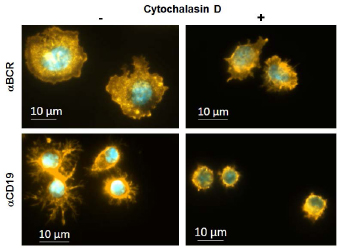
Different cell spreading behavior of LPS activated B cells treated with (right) or without (left) Cytochalasin D on immobilized antibodies BCR or CD19. )). Immunofluorescent staining was performed for F-Actin (Phalloidin-Rhodamin) and DNA (DAPI).
2.3 Image analysis
The key issue during an automated cell image analysis is an appropriate segmentation of the cell regions. Usually this includes preprocessing the image (preprocessing), separating foreground pixels from background pixels (figure-ground separation), and, if necessary, separating cells from each other (cell splitting). An additional measurement step for e.g. the assessment of cell area or cell circularity follows cell segmentation. Segmentation and measurements are integrated in the experimental analysis tool (CaeT). CaeT allows to combine various methods with each other to design cell image processing pipelines of arbitrary length. This is a similar approach like in the CellProfiler [4].
In this study, we have applied a combination of various algorithms to nuclei and to cell segmentation. Fig. 2 depicts the segmentation pipeline for nuclei segmentation. Gaussian smoothing filters an image with a Gaussian kernel with the standard deviation σn and produces a smoothed version of the original image (preprocessing). The smoothed image is used for figure-ground separation with a method that is based on k-means clustering with one parameter κn, the number of clusters [7]. Nuclei splitting is performed with a watershed approach. After smoothing the distance transformed result of the figure-ground separation with Gaussian smoothing with standard deviation σnd, it applies the watershed transfrom and neglects segments with areas smaller than αn to prevent oversegmentation.

Image processing pipeline for nuclei segmentation.
Fig. 3 shows the image processing pipeline for cell segmentation. Preprocessing is performed with a Difference of Gaussians filter (DoG). This filter generates two smoothed versions of the original image with the Gaussian filter with the standard deviations σc and σcb. The foreground of the micrograph is estimated by subtracting these smoothed images from each other. Figure-ground separation is performed with k-means clustering with the number of clusters κc on the result of the DoG. Now, the seeded watershed algorithm is used for cell splitting and uses the previously detected nuclei regions as starting point for the watershed transform. Parameters are σcd for smoothing the distance transformed image and αc determining the minimum cell size.

Image processing pipeline for cell segmentation.
In order to obtain the best result fro the image analysis challenge, all parameters p=( σn, κn, σnd, αn, σc, σcb, κc, σcd, αc) of all methods applied within the image processing pipeline have to be adapted to the experimental image data. For human experts, this is an elaborate and time-consuming task. Held et al. [9] have presented a method to automatically optimize parameters of a three-step image processing pipeline with respect to a small set of hand labeled reference image data. In this contribution this approach has been extended to adapt the parameters of image processing pipelines with arbitrary length. The optimization algorithm makes use of coordinate descend approach in order to find the best fitting parameter set p* using the combined Jaccard metric presented in [9] as optimization measure. To provide the ground truth data for the parameter optimization, a subset of 15 representative images with 149 cells and 165 nuclei, respectively, were manually labeled by an human expert using a Wacom Board [6]. Incomplete cells and nuclei witch contact to image tile borders were excluded.
3 Results
In order to process the described whole slide image data, an appropriate parameter set p* has to be found for each image processing pipelines. These parameters are separately adapted for the nuclei segmentation pipeline (see fig. 2) and for the F-actin cytoskeleton segmentation pipeline (see fig. 3) with respect to the hand labeled reference image data. As segmented nuclei regions are input into the F-actin cytoskeleton pipeline, the parameter set for the nuclei segmentation is the first to be adapted. For nuclei, the performance measured with two-fold cross validation and the combined Jaccard metric ispn=0.73 with a hit quality ofhn=0.91. Fig. 4 shows an exemplary segmentation result. For F-actin cytoskeleton, the performance waspc=0.64 with a hit qualityhc=0.84. Fig. 5 depicts an exemplary segmentation.
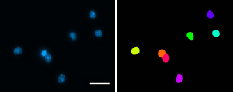
Exemplary segmentation result for nuclei: part of original DAPI image (left); segmentation result (right); scale bar corresponds to 20 μm.
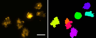
Exemplary segmentation result for F-actin cytoskeleton: brightness adapted part of original Phalloidin-Rhodamin image (left); segmentation result (right); scale bar corresponds to 20 μm.
The quality measures based on the combined Jaccard index are affected by differences between human labeling and automated image processing. The automated process uses the DAPI channel as a reference channel and tries to delineate exactly one cell region for each detected nucleus region. In contrast, a human expert’s decision depends more on the channel currently depected. This means if the staining works fine in the DAPI channel the human expert might have delineated a nucleus although there is not a visible cell in the F-actin cytoskeleton channel and vice versa. Also, the human experts were able to recognize dividing nuclei based on the F-actin cytoskeleton channel, and have labeled the nucleus as a single one. However, the algorithm separates the nucleus into two entities if the division process is sufficiently advanced and two distinct nuclei ca be detected. There also exist differences in the handling of debris and dirt in the micrograph. Human experts are able to distinguish between dirt, debris and cells based on morphology and texture, while the algorithm is only able to sort out too small regions.
With the parameters obtained during the optimization process the complete experimental image data was processed. This resulted in a total number of 16.715 detected and segmented cells and nuclei, respectively. Based on the segmented cell regions, the cell area and the cell circularity were calculated to describe the morphology of each cell. The results are visualized in fig. 6. The analysis shows that LPS-activated primary murine B cells, attached on glass slides coated either with anti BCR mAb, or coated with antibodies against the BCR co-receptor CD19, in the absence (-) or in the presence of the F-actin destabilizing drug Cytochalasin D, spread significantly less on anti CD19 mAb than on anti BCR mAb. The F-Actin dependency of both processes could be shown via specific inhibition by Cytochalasin D. Furthermore, anti CD19 mAb induce a more symmetrical spreading of B lymphocytes than anti BCR mAb as reflected by the significant higher cell circularity.
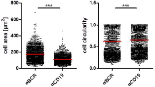
Measurements depict highly significant results in cell area and circularity between B cells spreading on immobilized BCR or CD19 antibodies. Significance according to Mann-Whitney-U test (*** p= 0.0001 - 0.001). Red line represents the mean.
4 Conclusion
In summary, we have established a protocol and an algorithm that allows the quantification of large numbers of fluorescent labeled and fixed B cells attached to glass slides. This method allows rapid screening of cytoskeletal effector molecules and of mouse mutants suspected to effect cytoskeletal rearrangement.
Acknowledgment
This work has been supported by the Collaborative Research Center 796 Project A4 and transregional collaborative research center 130 TP03. Both research centers are funded by the German Research Foundation.
Author's Statement
Conflict of interest: Authors state no conflict of interest. Material and Methods: Informed consent: Informed consent has been obtained from all individuals included in this study. Ethical approval: The research related to human use has been complied with all the relevant national regulations, institutional policies and in accordance the tenets of the Helsinki Declaration, and has been approved by the authors’ institutional review board or equivalent committee.
References
[1] Bengtsson EC, Wählby C. Robust cell image segmentation methods. Pattern Recognition and Image Analysis. 2004; 14(2):157-167.Suche in Google Scholar
[2] Bunyak F, Palaniappan K, Nath SK, Baskin TI, Dong G. Quantitative cell motility for in vitro wound healing using level set-based active contour tracking. Proc IEEE Int Symp Biomed Imaging. 2006; 6:1040-1043.10.1109/ISBI.2006.1625099Suche in Google Scholar
[3] Cambier JC, Heusser CH, Julius MH. Abortive activation of B lymphocytes by monoclonal anti-immunoglobulin antibodies. J Immunol. 1986;136(9):3140-6.10.4049/jimmunol.136.9.3140Suche in Google Scholar
[4] Carpenter AE, Jones TR, Lamprecht MR, et al. CellProfiler: Image Analysis Software for Identifying and Quantifying Cell Pheno-types. Genome Biology. 2007; 7:R10010.1186/gb-2006-7-10-r100Suche in Google Scholar
[5] Cooper, JA (1987). Effects of cytochalasin and phalloidin on actin. J Cell Biol. 1987; 105(4): 1473–1478.10.1083/jcb.105.4.1473Suche in Google Scholar
[6] Dach C, Held C, Palmisano R, Wittenberg T, Friedl S. Evaluation of input modalities for the interactive image segmentation of fluorescent micrographs. Biomed Tech. 2011; 56 (Suppl. 1).Suche in Google Scholar
[7] Held C. Towards Increased Efficiency and Automation in Fluorescence Micrograph Analysis Based on Hand-Labeled Data. Universität Bielefeld. 2013.Suche in Google Scholar
[8] Held C, Wenzel J, Wiesmann V, Palmisano R, Lang R, Wittenberg T. Enhancing automated micrograph-based evaluation of LPS-stimulated macrophage spreading. Cytometry A. 2013; 83(4):409-18.10.1002/cyto.a.22248Suche in Google Scholar
[9] Held C, Nattkemper T, Palmisano R, Wittenberg T. Approaches to automatic parameter fitting in a microscopy image segmentation pipeline: An exploratory parameter space analysis. J Pathol Inform. 2013; 4(Suppl): S5.10.4103/2153-3539.109831Suche in Google Scholar
[10] Krop I, Shaffer AL, Fearon DT, Schlissel MS. The signaling activity of murine CD19 is regulated during cell development. J Immunol. 1996;157(1):48-56.10.4049/jimmunol.157.1.48Suche in Google Scholar
[11] Malpica N, Ortiz de Solorzano C, Vaquero JJ, et al. Applying watershed algorithms to the segmentation of clustered nuclei. Cytometry. 1997; 28(4):289-297.10.1002/(SICI)1097-0320(19970801)28:4<289::AID-CYTO3>3.0.CO;2-7Suche in Google Scholar
[12] Roerdink JB, Meijster A. The watershed transform: Definitions, algorithms and parallelization strategies. Fundamenta informaticae. 2000; 41. Jg., Nr. 1:187-228.10.3233/FI-2000-411207Suche in Google Scholar
[13] Wählby C, Lindblad J, Vondrus M, Bengtsson E, Björkesten L. Algorithms for cytoplasm segmentation of fluorescence labelled cells. Anal Cell Pathol. 2002;24(2-3):101-11.10.1155/2002/821782Suche in Google Scholar
© 2015 by Walter de Gruyter GmbH, Berlin/Boston
This article is distributed under the terms of the Creative Commons Attribution Non-Commercial License, which permits unrestricted non-commercial use, distribution, and reproduction in any medium, provided the original work is properly cited.
Artikel in diesem Heft
- Research Article
- Development and characterization of superparamagnetic coatings
- Research Article
- The development of an experimental setup to measure acousto-electric interaction signal
- Research Article
- Stability analysis of ferrofluids
- Research Article
- Investigation of endothelial growth using a sensors-integrated microfluidic system to simulate physiological barriers
- Research Article
- Energy harvesting for active implants: powering a ruminal pH-monitoring system
- Research Article
- New type of fluxgate magnetometer for the heart’s magnetic fields detection
- Research Article
- Field mapping of ballistic pressure pulse sources
- Research Article
- Development of a new homecare sleep monitor using body sounds and motion tracking
- Research Article
- Noise properties of textile, capacitive EEG electrodes
- Research Article
- Detecting phase singularities and rotor center trajectories based on the Hilbert transform of intraatrial electrograms in an atrial voxel model
- Research Article
- Spike sorting: the overlapping spikes challenge
- Research Article
- Separating the effect of respiration from the heart rate variability for cases of constant harmonic breathing
- Research Article
- Locating regions of arrhythmogenic substrate by analyzing the duration of triggered atrial activities
- Research Article
- Combining different ECG derived respiration tracking methods to create an optimal reconstruction of the breathing pattern
- Research Article
- Atrial and ventricular signal averaging electrocardiography in pacemaker and cardiac resynchronization therapy
- Research Article
- Estimation of a respiratory signal from a single-lead ECG using the 4th order central moments
- Research Article
- Compressed sensing of multi-lead ECG signals by compressive multiplexing
- Research Article
- Heart rate monitoring in ultra-high-field MRI using frequency information obtained from video signals of the human skin compared to electrocardiography and pulse oximetry
- Research Article
- Synchronization in wireless biomedical-sensor networks with Bluetooth Low Energy
- Research Article
- Automated classification of stages of anaesthesia by populations of evolutionary optimized fuzzy rules
- Research Article
- Effects of sampling rate on automated fatigue recognition in surface EMG signals
- Research Article
- Closed-loop transcranial alternating current stimulation of slow oscillations
- Research Article
- Cardiac index in atrio- and interventricular delay optimized cardiac resynchronization therapy and cardiac contractility modulation
- Research Article
- The role of expert evaluation for microsleep detection
- Research Article
- The impact of baseline wander removal techniques on the ST segment in simulated ischemic 12-lead ECGs
- Research Article
- Metal artifact reduction by projection replacements and non-local prior image integration
- Research Article
- A novel coaxial nozzle for in-process adjustment of electrospun scaffolds’ fiber diameter
- Research Article
- Processing of membranes for oxygenation using the Bellhouse-effect
- Research Article
- Inkjet printing of viable human dental follicle stem cells
- Research Article
- The use of an icebindingprotein out of the snowflea Hypogastrura harveyi as a cryoprotectant in the cryopreservation of mesenchymal stem cells
- Research Article
- New NIR spectroscopy based method to determine ischemia in vivo in liver – a first study on rats
- Research Article
- QRS and QT ventricular conduction times and permanent pacemaker therapy after transcatheter aortic valve implantation
- Research Article
- Adopting oculopressure tonometry as a transient in vivo rabbit glaucoma model
- Research Article
- Next-generation vision testing: the quick CSF
- Research Article
- Improving tactile sensation in laparoscopic surgery by overcoming size restrictions
- Research Article
- Design and control of a 3-DOF hydraulic driven surgical instrument
- Research Article
- Evaluation of endourological tools to improve the diagnosis and therapy of ureteral tumors – from model development to clinical application
- Research Article
- Frequency based assessment of surgical activities
- Research Article
- “Hands free for intervention”, a new approach for transoral endoscopic surgery
- Research Article
- Pseudo-haptic feedback in medical teleoperation
- Research Article
- Feasibility of interactive gesture control of a robotic microscope
- Research Article
- Towards structuring contextual information for workflow-driven surgical assistance functionalities
- Research Article
- Towards a framework for standardized semantic workflow modeling and management in the surgical domain
- Research Article
- Closed-loop approach for situation awareness of medical devices and operating room infrastructure
- Research Article
- Kinect based physiotherapy system for home use
- Research Article
- Evaluating the microsoft kinect skeleton joint tracking as a tool for home-based physiotherapy
- Research Article
- Integrating multimodal information for intraoperative assistance in neurosurgery
- Research Article
- Respiratory motion tracking using Microsoft’s Kinect v2 camera
- Research Article
- Using smart glasses for ultrasound diagnostics
- Research Article
- Measurement of needle susceptibility artifacts in magnetic resonance images
- Research Article
- Dimensionality reduction of medical image descriptors for multimodal image registration
- Research Article
- Experimental evaluation of different weighting schemes in magnetic particle imaging reconstruction
- Research Article
- Evaluation of CT capability for the detection of thin bone structures
- Research Article
- Towards contactless optical coherence elastography with acoustic tissue excitation
- Research Article
- Development and implementation of algorithms for automatic and robust measurement of the 2D:4D digit ratio using image data
- Research Article
- Automated high-throughput analysis of B cell spreading on immobilized antibodies with whole slide imaging
- Research Article
- Tissue segmentation from head MRI: a ground truth validation for feature-enhanced tracking
- Research Article
- Video tracking of swimming rodents on a reflective water surface
- Research Article
- MR imaging of model drug distribution in simulated vitreous
- Research Article
- Studying the extracellular contribution to the double wave vector diffusion-weighted signal
- Research Article
- Artifacts in field free line magnetic particle imaging in the presence of inhomogeneous and nonlinear magnetic fields
- Research Article
- Introducing a frequency-tunable magnetic particle spectrometer
- Research Article
- Imaging of aortic valve dynamics in 4D OCT
- Research Article
- Intravascular optical coherence tomography (OCT) as an additional tool for the assessment of stent structures
- Research Article
- Simple concept for a wide-field lensless digital holographic microscope using a laser diode
- Research Article
- Intraoperative identification of somato-sensory brain areas using optical imaging and standard RGB camera equipment – a feasibility study
- Research Article
- Respiratory surface motion measurement by Microsoft Kinect
- Research Article
- Improving image quality in EIT imaging by measurement of thorax excursion
- Research Article
- A clustering based dual model framework for EIT imaging: first experimental results
- Research Article
- Three-dimensional anisotropic regularization for limited angle tomography
- Research Article
- GPU-based real-time generation of large ultrasound volumes from freehand 3D sweeps
- Research Article
- Experimental computer tomograph
- Research Article
- US-tracked steered FUS in a respiratory ex vivo ovine liver phantom
- Research Article
- Contribution of brownian rotation and particle assembly polarisation to the particle response in magnetic particle spectrometry
- Research Article
- Preliminary investigations of magnetic modulated nanoparticles for microwave breast cancer detection
- Research Article
- Construction of a device for magnetic separation of superparamagnetic iron oxide nanoparticles
- Research Article
- An IHE-conform telecooperation platform supporting the treatment of dementia patients
- Research Article
- Automated respiratory therapy system based on the ARDSNet protocol with systemic perfusion control
- Research Article
- Identification of surgical instruments using UHF-RFID technology
- Research Article
- A generic concept for the development of model-guided clinical decision support systems
- Research Article
- Evaluation of local alterations in femoral bone mineral density measured via quantitative CT
- Research Article
- Creating 3D gelatin phantoms for experimental evaluation in biomedicine
- Research Article
- Influence of short-term fixation with mixed formalin or ethanol solution on the mechanical properties of human cortical bone
- Research Article
- Analysis of the release kinetics of surface-bound proteins via laser-induced fluorescence
- Research Article
- Tomographic particle image velocimetry of a water-jet for low volume harvesting of fat tissue for regenerative medicine
- Research Article
- Wireless medical sensors – context, robustness and safety
- Research Article
- Sequences for real-time magnetic particle imaging
- Research Article
- Speckle-based off-axis holographic detection for non-contact photoacoustic tomography
- Research Article
- A machine learning approach for planning valve-sparing aortic root reconstruction
- Research Article
- An in-ear pulse wave velocity measurement system using heart sounds as time reference
- Research Article
- Measuring different oxygenation levels in a blood perfusion model simulating the human head using NIRS
- Research Article
- Multisegmental fusion of the lumbar spine a curse or a blessing?
- Research Article
- Numerical analysis of the biomechanical complications accompanying the total hip replacement with NANOS-Prosthetic: bone remodelling and prosthesis migration
- Research Article
- A muscle model for hybrid muscle activation
- Research Article
- Mathematical, numerical and in-vitro investigation of cooling performance of an intra-carotid catheter for selective brain hypothermia
- Research Article
- An ideally parameterized unscented Kalman filter for the inverse problem of electrocardiography
- Research Article
- Interactive visualization of cardiac anatomy and atrial excitation for medical diagnosis and research
- Research Article
- Virtualizing clinical cases of atrial flutter in a fast marching simulation including conduction velocity and ablation scars
- Research Article
- Mesh structure-independent modeling of patient-specific atrial fiber orientation
- Research Article
- Accelerating mono-domain cardiac electrophysiology simulations using OpenCL
- Research Article
- Understanding the cellular mode of action of vernakalant using a computational model: answers and new questions
- Research Article
- A java based simulator with user interface to simulate ventilated patients
- Research Article
- Evaluation of an algorithm to choose between competing models of respiratory mechanics
- Research Article
- Numerical simulation of low-pulsation gerotor pumps for use in the pharmaceutical industry and in biomedicine
- Research Article
- Numerical and experimental flow analysis in centifluidic systems for rapid allergy screening tests
- Research Article
- Biomechanical parameter determination of scaffold-free cartilage constructs (SFCCs) with the hyperelastic material models Yeoh, Ogden and Demiray
- Research Article
- FPGA controlled artificial vascular system
- Research Article
- Simulation based investigation of source-detector configurations for non-invasive fetal pulse oximetry
- Research Article
- Test setup for characterizing the efficacy of embolic protection devices
- Research Article
- Impact of electrode geometry on force generation during functional electrical stimulation
- Research Article
- 3D-based visual physical activity assessment of children
- Research Article
- Realtime assessment of foot orientation by Accelerometers and Gyroscopes
- Research Article
- Image based reconstruction for cystoscopy
- Research Article
- Image guided surgery innovation with graduate students - a new lecture format
- Research Article
- Multichannel FES parameterization for controlling foot motion in paretic gait
- Research Article
- Smartphone supported upper limb prosthesis
- Research Article
- Use of quantitative tremor evaluation to enhance target selection during deep brain stimulation surgery for essential tremor
- Research Article
- Evaluation of adhesion promoters for Parylene C on gold metallization
- Research Article
- The influence of metallic ions from CoCr28Mo6 on the osteogenic differentiation and cytokine release of human osteoblasts
- Research Article
- Increasing the visibility of thin NITINOL vascular implants
- Research Article
- Possible reasons for early artificial bone failure in biomechanical tests of ankle arthrodesis systems
- Research Article
- Development of a bending test procedure for the characterization of flexible ECoG electrode arrays
- Research Article
- Tubular manipulators: a new concept for intracochlear positioning of an auditory prosthesis
- Research Article
- Investigation of the dynamic diameter deformation of vascular stents during fatigue testing with radial loading
- Research Article
- Electrospun vascular grafts with anti-kinking properties
- Research Article
- Integration of temperature sensors in polyimide-based thin-film electrode arrays
- Research Article
- Use cases and usability challenges for head-mounted displays in healthcare
- Research Article
- Device- and service profiles for integrated or systems based on open standards
- Research Article
- Risk management for medical devices in research projects
- Research Article
- Simulation of varying femoral attachment sites of medial patellofemoral ligament using a musculoskeletal multi-body model
- Research Article
- Does enhancing consciousness for strategic planning processes support the effectiveness of problem-based learning concepts in biomedical education?
- Research Article
- SPIO processing in macrophages for MPI: The breast cancer MPI-SNLB-concept
- Research Article
- Numerical simulations of airflow in the human pharynx of OSAHS patients