Use of quantitative tremor evaluation to enhance target selection during deep brain stimulation surgery for essential tremor
-
A. Shah


Abstract
Deep brain stimulation (DBS), an effective surgical treatment for Essential Tremor (ET), requires test stimulations in the thalamus to find the optimum site for permanent electrode implantation. During these test stimulations, the changes in tremor are only visually evaluated. This, along with other parameters, increases the subjectivity when comparing the efficacy of different thalamic nuclei. We developed a method to quantitatively evaluate tremor during the test stimulations of DBS surgery and applied to 6 ET patients undergoing this treatment. From the quantitative data collected, we identified effective stimulation amplitudes for every test stimulation position and compared it with the ones identified visually during the surgery. We also classified the data based on the thalamic nuclei in which the center of the stimulating contact was present during test stimulations. Results indicate that, to achieve the same reduction in tremor, on average, the stimulation amplitude identified by our method was 0.6 mA lower than those identified by visual evaluation. The comparison of the different thalamic nuclei showed that stimulations in the Ventro-oral and the Intermediolateral nuclei of the thalamus result in higher reduction in tremor for similar stimulation amplitudes as the frequently targeted Ventrointermediate nucleus. We conclude that our quantitative tremor evaluation method is more sensitive than the widely used visual evaluation. Using such quantitative methods will aid in identifying the optimum target structure for patients undergoing DBS.
1 Background
Deep Brain Stimulation (DBS) is now a routinely used surgical treatment for movement disorders like Essential Tremor (ET) [1]. Electrical stimulation of certain brain structures is performed by implanting electrodes and connecting them to a subcutaneously implanted neurostimulator. Although the use of DBS is increasing, the optimum target structure is still debated. The limited knowledge of the mechanisms of action of DBS being one of the reasons, we believe the under-utilization of intraoperatively obtained data is another.
A typical DBS surgery is preceded by a planning session during which a trajectory is planned from an entry point in the skull to the target structure on the patient’s anatomical images using stereotactic planning software. With the aid of stereotactic equipment, this trajectory is then explored during the surgery. In most of the surgeries, neuronal activity is recorded and test stimulations are performed at pre-planned positions on this trajectory while the patient is awake. The final implant position is decided after comparing the reduction in tremor, needed stimulation current for inducing those reductions and the side effects observed during the test stimulations.
In the routing clinical practice, the symptom evaluation methods are subjective [2]. Tremor is evaluated by a visual rating of the change compared to its level observed before stimulation. We have previously described the method of evaluating tremor during DBS quantitatively by using accelerometers [3]. We used this method to intraoperatively evaluate tremor from 6 ET patients who participated in a clinical study in the University Hospital in Clermont-Ferrand, France. In the current paper, we describe the result of the comparison between visual and quantitative evaluations. Further, we classified the results based on the anatomical location of the electrode and compared them to identify the target structure which is the most effective in reducing tremor.
2 Method
2.1 Surgical protocol
The primary goal of the DBS surgery remains the same for all surgical centres: to determine the optimal implantation position. However, the actual procedure may differ significantly. Lemaire et al have described, in detail, the routine surgical procedure at the University Hospital in Clermont-Ferrand [4]. For treating ET patients using DBS, the primary target structure is the Ventrointermedius nucleus (VIM) of the thalamus. At the University Hospital in Clermont-Ferrand, during the planning session on patient’s images using a commercial stereotactic surgery planning software (iPlan Stereotaxy, Brainlab, Germany), the surgical team identifies and outlines different nuclei in the thalamus using MRI images of the patient [5]. After the definition of target and entry point, test stimulation positions are planned on the resulting trajectory. After performing micro-electrode recording (MER) at these positions, test stimulations are performed. For each test stimulation, the stimulation current is varied from 0 to 3 mA in steps of 0.2 mA and the changes in tremor are observed visually. The highest reduction in tremor (Visually identified Change, VC) and the corresponding stimulation amplitude (Visually identified Amplitude, VA) at which it was observed are noted for every test stimulation location. Along with these, the occurrence of side-effects, their type and their corresponding amplitudes are also noted. On completion of all test stimulations, these data are discussed upon by the surgical team and a position is determined as the “final implant location” for the chronic DBS electrode.
2.2 Acceleration data recording
Tremor was quantified during the surgery using a commercial acceleration sensor evaluation board (STEVALMKI022VI, ST Micro, Switzerland). Time-stamped acceleration values from 3 axes were obtained at a frequency of 400 Hz and a range of 8 g. The evaluation board was fixed in an in-house developed non-conductive printed plastic case which was attached to the patient’s wrist using a Velcro strap. The acceleration data were recorded by in-house developed software installed on a laptop that was interfaced with the sensor via an USB cable. Furthermore, the recording software was also interfaced with the electro-physiology system to synchronize the acceleration data recording with the test stimulation data.
For the simplification of acceleration data recording and analysis, a data recording protocol was defined for all the surgeries. Acceleration data recording is started before the beginning of the test stimulation to record the data representing the baseline tremor for that test stimulation, and is continued till all the stimulation amplitudes are tested. The acceleration data and the related information about the test stimulation position and amplitude were saved in data files for offline analysis [3].
2.3 Post-operative data analysis
Acceleration data were analysed post-operatively using Matlab (Mathwork, USA). In brief, the resultant acceleration was calculated from the three axes and filtered to remove low frequency (2 Hz) movements and high frequency noise (10 Hz). Statistical features were extracted from these filtered acceleration data and the features from different stimulation amplitudes were normalized (%) using the features from the baseline data. These normalized features were used to identify a dataset of 2 values for each test stimulation: 1.) The change in acceleration features (Quantitatively identified Change, QC) at the visually identified best stimulation amplitude VA and 2.) The stimulation amplitude (Quantitatively equivalent Amplitude, QA) at which the quantitative change QC was similar to VC. These data were then statistically compared to the corresponding values from the visual evaluation (VC vs QC and VA vs QA) using the Wilcoxon two-sided signed rank test.
In order to analyse the correlation with the anatomical position of the electrode, the anatomical structure present at the centre of the stimulating electrode was determined. Data were then grouped based on the thalamic nucleus attributed to the test stimulation position. Furthermore, from the acceleration data of every test stimulation, we also calculated the minimum stimulation amplitude required to obtain a 75% reduction in the statistical features compared to baseline (Amp75). These data along with the occurrence of side-effects were used to compare the efficiency of different structures in reducing the patient’s tremor by a fixed amount.
2.4 Patients
The above method was applied to 6 ET patients under a clinical study at the University Hospital in Clermont-Ferrand (2011-A00774-37 / AU905). Out of the 167 test stimulations that were performed, 148 positions were found where both evaluation methods identified a reduction in tremor.
3 Results
The Wilcoxon signed rank test revealed a statistically significant difference between VA and QA (W=4256, p< 0.001, alpha = 0.001) which can also be observed in the box plot of the two data sets (Figure 1). On the other hand, the difference between VC and QC at the visually identified amplitude VA was not statistically significant (W = 3004, p=0.72, alpha = 0.001).
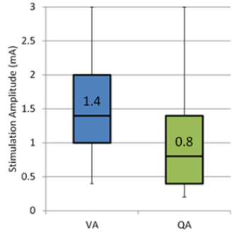
Box plot comparing visually identified effective stimulation amplitude VA to quantitatively identified effective stimulation amplitude QA for the same clinical improvement.
From the analysis of the electrode position during test stimulation, it was found that the positions were distributed in 7 thalamic nuclei: Center Median (CM), Intermedio-Lateral (InL), Ventrointermediate (VIM), Ventrocaudal lateral (VCL), Ventrocaudal medial (VCM), Ventro-Oral (VO) and also in Preleminiscal Radiations (PreR). Table 1 shows the average values of the different parameters extracted from the visual and the quantitative evaluations. The number of evaluations in the CM region (3) of the thalamus is small compared to the others and thus the significance of the results is very low. The values of QA are lower than VA for all the structures, the difference between them being the largest for InL, VO and the VIM.
Figure 2A shows the average minimum stimulation amplitude required to see 75% reduction in tremor quantitatively while 2B shows the number of test stimulations with and without side-effects (red and blue respectively). The VCL and the PreR achieve similar reductions in tremor at lower amplitudes. But these structures also have higher occurrence of side effects. The VCM require higher stimulation amplitudes than VCL and PreR, but also has high occurrence of side-effects. The InL and the VO require similar stimulation amplitudes as the VIM but have much lower occurrence of side effects. The VIM requires the highest stimulation amplitude to achieve 75% reduction, but also has high occurrence of side-effects.
4 Discussion
Various methods have been used previously to quantitatively evaluate tremor [6], but only a handful have been reported to be used during DBS surgery. Shamir et al. [7] used goniometers for Parkinson’s patients undergoing DBS, but only during MER to identify borders of sub-thalamic nucleus and not to quantitatively evaluate tremor during test stimulations. Other quantitative evaluation techniques have been used to optimize the stimulation parameters of DBS but only after the implant position of the electrode was already decided [8, 9]. To the best of our knowledge, no other group has previously used quantitative tremor evaluation to compare the efficacy of different thalamic nuclei.
One of the observations of the application of our method to the current set of patients was that the use of quantitative evaluation during DBS surgery did not increase the duration of the surgery or cause any discomfort to the patient. Moreover, our results suggest that this quantitative method is more sensitive than the current visual evaluation methods used during the surgery. The significant difference (0.6 mA) between average values of VA and QA shows that stimulation parameters may be further optimized by using quantitative tremor evaluation.
The anatomical analysis of the data obtained using quantitative tremor evaluation suggests that the conventional target, VIM, may not be the most efficient target for ET patients undergoing DBS surgery. All other structures required stimulation amplitude equal to or lower than the VIM to reduce tremor by 75%. However, after looking at the number of positions with side effects, the VO and InL seem to be more efficient in reducing tremor and limiting side-effects of the therapy. This seems to confirm published data [5] suggesting that parts of the ventro-oral nucleus (VO) could be appropriate targets as well.
Average visual and quantitative parameters for different thalamic nuclei
| CM | InL | VO | VIM | VCL | VCM | PreR | |
|---|---|---|---|---|---|---|---|
| No. of Stimulations | 3 | 19 | 18 | 52 | 13 | 24 | 15 |
| No. of side-effect occurrence | 0 | 1 | 1 | 10 | 2 | 6 | 2 |
| Average VC (%) | 50 | 45 | 63 | 60 | 65 | 72 | 67 |
| Average QC (%) | 76 | 66 | 65 | 57 | 76 | 66 | 57 |
| Average VA (mA) | 1.1 | 2.2 | 1.9 | 1.8 | 1.4 | 1.6 | 1 |
| Average QA (mA) | 0.4 | 1.3 | 1.1 | 1.3 | 1 | 1.5 | 0.7 |
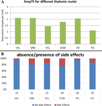
Comparison of efficacy of different thalamic nuclei. A) Average stimulation amplitude required to achieve 75% reduction in tremor. B) Stacked column plot of number of test stimulations with and without side-effects.
The results have also indicated some limitations of the current method. One of the main factors that influences the proper functioning of the method is the recording of baseline data. A minimum baseline recording of 5 seconds is necessary to extract proper statistical features. However, in case a baseline recording is not available for the current position, the baseline data of the previous recording can be used to perform the analysis. Furthermore, the current analysis method is post-operative. The next step for this method will to perform real-time data analysis so that the results are available to the surgical team when they identify the final implant site.
5 Conclusion
The current paper presents the results of quantitative tremor evaluation during DBS surgery for ET patients. The results indicate that our method is more sensitive to changes in tremor than the current visual evaluation methods. Furthermore, the results of the anatomical analysis suggest that the thalamic sub-structures InL and VO are more efficient targets for DBS than the conventional and targeted VIM.
Funding: This work was supported by the Swiss National Science Foundation (www.snf.ch).
Author's Statement
Conflict of interest: Authors state no conflict of interest. Material and Methods: Informed consent: Informed consent has been obtained from all individuals included in this study. Ethical approval: The research related to human use has been complied with all the relevant national regulations, institutional policies and in accordance the tenets of the Helsinki Declaration, and has been approved by the authors’ institutional review board or equivalent committee.
References
[1] Sarem-Aslani A, Mullett K. Industrial Perspective on Deep Brain Stimulation: History, Current State, and Future Developments. Front. Integr. Neurosci. 2011;5.10.3389/fnint.2011.00046Search in Google Scholar PubMed PubMed Central
[2] Hemm S, Wĺrdell K. Stereotactic implantation of deep brain stimulation electrodes: a review of technical systems, methods and emerging tools. Med Biol Eng Comput 2010;48:611–24.10.1007/s11517-010-0633-ySearch in Google Scholar PubMed
[3] Shah A, Coste J, Schkommodau E, Lemaire JJ, Hemm-Ode S. Using acceleration sensors to quantify symptoms during deep brain stimulation surgery. Biomedical Engineering / Biomedizinische Technik 2013.10.1515/bmt-2013-4007Search in Google Scholar PubMed
[4] Lemaire J, Coste J, Ouchchane L, Hemm S, Derost P, Ulla M, Siadoux S et al. MRI anatomical mapping and direct stereotactic targeting in the subthalamic region: functional and anatomical correspondence in Parkinson’s disease. Int J CARS 2007;2:75–85.10.1007/s11548-007-0124-2Search in Google Scholar
[5] Vassal F, Coste J, Derost P, Mendes V, Gabrillargues J, Nuti C, Durif F et al. Direct stereotactic targeting of the ventrointermediate nucleus of the thalamus based on anatomic 1.5-T MRI mapping with a white matter attenuated inversion recovery (WAIR) sequence. Brain Stimul. 2012;5:625–3310.1016/j.brs.2011.10.007Search in Google Scholar PubMed
[6] Mansur PHG, Cury LKP, Andrade AO, Pereira AA, Miotto GAA, Soares AB, Naves ELM. A review on techniques for tremor recording and quantification. Crit Rev Biomed Eng 2007;35:343–62.10.1615/CritRevBiomedEng.v35.i5.10Search in Google Scholar
[7] Shamir RR, Eitan R, Sheffer S, Marmor-Levin O, Valsky D, Moshel S, Zaidel A et al. Intra-operative Identification of the Subthalamic Nucleus Motor Zone Using Goniometers. Information Processing in Computer-Assisted Interventions. Berlin, Heidelberg: Springer Berlin Heidelberg; 2013. p. 21–29.10.1007/978-3-642-38568-1_3Search in Google Scholar
[8] Papapetropoulos S, Jagid JR, Sengun C, Singer C, Gallo BV. Objective monitoring of tremor and bradykinesia during DBS surgery for Parkinson disease. Neurology 2008;70:1244–9.10.1212/01.wnl.0000308936.27780.94Search in Google Scholar PubMed
[9] Journee HL, Postma AA, Staal MJ. Intraoperative neurophysiological assessment of disabling symptoms in DBS surgery. Neurophysiol Clin 2007;37:467–710.1016/j.neucli.2007.10.006Search in Google Scholar PubMed
© 2015 by Walter de Gruyter GmbH, Berlin/Boston
This article is distributed under the terms of the Creative Commons Attribution Non-Commercial License, which permits unrestricted non-commercial use, distribution, and reproduction in any medium, provided the original work is properly cited.
Articles in the same Issue
- Research Article
- Development and characterization of superparamagnetic coatings
- Research Article
- The development of an experimental setup to measure acousto-electric interaction signal
- Research Article
- Stability analysis of ferrofluids
- Research Article
- Investigation of endothelial growth using a sensors-integrated microfluidic system to simulate physiological barriers
- Research Article
- Energy harvesting for active implants: powering a ruminal pH-monitoring system
- Research Article
- New type of fluxgate magnetometer for the heart’s magnetic fields detection
- Research Article
- Field mapping of ballistic pressure pulse sources
- Research Article
- Development of a new homecare sleep monitor using body sounds and motion tracking
- Research Article
- Noise properties of textile, capacitive EEG electrodes
- Research Article
- Detecting phase singularities and rotor center trajectories based on the Hilbert transform of intraatrial electrograms in an atrial voxel model
- Research Article
- Spike sorting: the overlapping spikes challenge
- Research Article
- Separating the effect of respiration from the heart rate variability for cases of constant harmonic breathing
- Research Article
- Locating regions of arrhythmogenic substrate by analyzing the duration of triggered atrial activities
- Research Article
- Combining different ECG derived respiration tracking methods to create an optimal reconstruction of the breathing pattern
- Research Article
- Atrial and ventricular signal averaging electrocardiography in pacemaker and cardiac resynchronization therapy
- Research Article
- Estimation of a respiratory signal from a single-lead ECG using the 4th order central moments
- Research Article
- Compressed sensing of multi-lead ECG signals by compressive multiplexing
- Research Article
- Heart rate monitoring in ultra-high-field MRI using frequency information obtained from video signals of the human skin compared to electrocardiography and pulse oximetry
- Research Article
- Synchronization in wireless biomedical-sensor networks with Bluetooth Low Energy
- Research Article
- Automated classification of stages of anaesthesia by populations of evolutionary optimized fuzzy rules
- Research Article
- Effects of sampling rate on automated fatigue recognition in surface EMG signals
- Research Article
- Closed-loop transcranial alternating current stimulation of slow oscillations
- Research Article
- Cardiac index in atrio- and interventricular delay optimized cardiac resynchronization therapy and cardiac contractility modulation
- Research Article
- The role of expert evaluation for microsleep detection
- Research Article
- The impact of baseline wander removal techniques on the ST segment in simulated ischemic 12-lead ECGs
- Research Article
- Metal artifact reduction by projection replacements and non-local prior image integration
- Research Article
- A novel coaxial nozzle for in-process adjustment of electrospun scaffolds’ fiber diameter
- Research Article
- Processing of membranes for oxygenation using the Bellhouse-effect
- Research Article
- Inkjet printing of viable human dental follicle stem cells
- Research Article
- The use of an icebindingprotein out of the snowflea Hypogastrura harveyi as a cryoprotectant in the cryopreservation of mesenchymal stem cells
- Research Article
- New NIR spectroscopy based method to determine ischemia in vivo in liver – a first study on rats
- Research Article
- QRS and QT ventricular conduction times and permanent pacemaker therapy after transcatheter aortic valve implantation
- Research Article
- Adopting oculopressure tonometry as a transient in vivo rabbit glaucoma model
- Research Article
- Next-generation vision testing: the quick CSF
- Research Article
- Improving tactile sensation in laparoscopic surgery by overcoming size restrictions
- Research Article
- Design and control of a 3-DOF hydraulic driven surgical instrument
- Research Article
- Evaluation of endourological tools to improve the diagnosis and therapy of ureteral tumors – from model development to clinical application
- Research Article
- Frequency based assessment of surgical activities
- Research Article
- “Hands free for intervention”, a new approach for transoral endoscopic surgery
- Research Article
- Pseudo-haptic feedback in medical teleoperation
- Research Article
- Feasibility of interactive gesture control of a robotic microscope
- Research Article
- Towards structuring contextual information for workflow-driven surgical assistance functionalities
- Research Article
- Towards a framework for standardized semantic workflow modeling and management in the surgical domain
- Research Article
- Closed-loop approach for situation awareness of medical devices and operating room infrastructure
- Research Article
- Kinect based physiotherapy system for home use
- Research Article
- Evaluating the microsoft kinect skeleton joint tracking as a tool for home-based physiotherapy
- Research Article
- Integrating multimodal information for intraoperative assistance in neurosurgery
- Research Article
- Respiratory motion tracking using Microsoft’s Kinect v2 camera
- Research Article
- Using smart glasses for ultrasound diagnostics
- Research Article
- Measurement of needle susceptibility artifacts in magnetic resonance images
- Research Article
- Dimensionality reduction of medical image descriptors for multimodal image registration
- Research Article
- Experimental evaluation of different weighting schemes in magnetic particle imaging reconstruction
- Research Article
- Evaluation of CT capability for the detection of thin bone structures
- Research Article
- Towards contactless optical coherence elastography with acoustic tissue excitation
- Research Article
- Development and implementation of algorithms for automatic and robust measurement of the 2D:4D digit ratio using image data
- Research Article
- Automated high-throughput analysis of B cell spreading on immobilized antibodies with whole slide imaging
- Research Article
- Tissue segmentation from head MRI: a ground truth validation for feature-enhanced tracking
- Research Article
- Video tracking of swimming rodents on a reflective water surface
- Research Article
- MR imaging of model drug distribution in simulated vitreous
- Research Article
- Studying the extracellular contribution to the double wave vector diffusion-weighted signal
- Research Article
- Artifacts in field free line magnetic particle imaging in the presence of inhomogeneous and nonlinear magnetic fields
- Research Article
- Introducing a frequency-tunable magnetic particle spectrometer
- Research Article
- Imaging of aortic valve dynamics in 4D OCT
- Research Article
- Intravascular optical coherence tomography (OCT) as an additional tool for the assessment of stent structures
- Research Article
- Simple concept for a wide-field lensless digital holographic microscope using a laser diode
- Research Article
- Intraoperative identification of somato-sensory brain areas using optical imaging and standard RGB camera equipment – a feasibility study
- Research Article
- Respiratory surface motion measurement by Microsoft Kinect
- Research Article
- Improving image quality in EIT imaging by measurement of thorax excursion
- Research Article
- A clustering based dual model framework for EIT imaging: first experimental results
- Research Article
- Three-dimensional anisotropic regularization for limited angle tomography
- Research Article
- GPU-based real-time generation of large ultrasound volumes from freehand 3D sweeps
- Research Article
- Experimental computer tomograph
- Research Article
- US-tracked steered FUS in a respiratory ex vivo ovine liver phantom
- Research Article
- Contribution of brownian rotation and particle assembly polarisation to the particle response in magnetic particle spectrometry
- Research Article
- Preliminary investigations of magnetic modulated nanoparticles for microwave breast cancer detection
- Research Article
- Construction of a device for magnetic separation of superparamagnetic iron oxide nanoparticles
- Research Article
- An IHE-conform telecooperation platform supporting the treatment of dementia patients
- Research Article
- Automated respiratory therapy system based on the ARDSNet protocol with systemic perfusion control
- Research Article
- Identification of surgical instruments using UHF-RFID technology
- Research Article
- A generic concept for the development of model-guided clinical decision support systems
- Research Article
- Evaluation of local alterations in femoral bone mineral density measured via quantitative CT
- Research Article
- Creating 3D gelatin phantoms for experimental evaluation in biomedicine
- Research Article
- Influence of short-term fixation with mixed formalin or ethanol solution on the mechanical properties of human cortical bone
- Research Article
- Analysis of the release kinetics of surface-bound proteins via laser-induced fluorescence
- Research Article
- Tomographic particle image velocimetry of a water-jet for low volume harvesting of fat tissue for regenerative medicine
- Research Article
- Wireless medical sensors – context, robustness and safety
- Research Article
- Sequences for real-time magnetic particle imaging
- Research Article
- Speckle-based off-axis holographic detection for non-contact photoacoustic tomography
- Research Article
- A machine learning approach for planning valve-sparing aortic root reconstruction
- Research Article
- An in-ear pulse wave velocity measurement system using heart sounds as time reference
- Research Article
- Measuring different oxygenation levels in a blood perfusion model simulating the human head using NIRS
- Research Article
- Multisegmental fusion of the lumbar spine a curse or a blessing?
- Research Article
- Numerical analysis of the biomechanical complications accompanying the total hip replacement with NANOS-Prosthetic: bone remodelling and prosthesis migration
- Research Article
- A muscle model for hybrid muscle activation
- Research Article
- Mathematical, numerical and in-vitro investigation of cooling performance of an intra-carotid catheter for selective brain hypothermia
- Research Article
- An ideally parameterized unscented Kalman filter for the inverse problem of electrocardiography
- Research Article
- Interactive visualization of cardiac anatomy and atrial excitation for medical diagnosis and research
- Research Article
- Virtualizing clinical cases of atrial flutter in a fast marching simulation including conduction velocity and ablation scars
- Research Article
- Mesh structure-independent modeling of patient-specific atrial fiber orientation
- Research Article
- Accelerating mono-domain cardiac electrophysiology simulations using OpenCL
- Research Article
- Understanding the cellular mode of action of vernakalant using a computational model: answers and new questions
- Research Article
- A java based simulator with user interface to simulate ventilated patients
- Research Article
- Evaluation of an algorithm to choose between competing models of respiratory mechanics
- Research Article
- Numerical simulation of low-pulsation gerotor pumps for use in the pharmaceutical industry and in biomedicine
- Research Article
- Numerical and experimental flow analysis in centifluidic systems for rapid allergy screening tests
- Research Article
- Biomechanical parameter determination of scaffold-free cartilage constructs (SFCCs) with the hyperelastic material models Yeoh, Ogden and Demiray
- Research Article
- FPGA controlled artificial vascular system
- Research Article
- Simulation based investigation of source-detector configurations for non-invasive fetal pulse oximetry
- Research Article
- Test setup for characterizing the efficacy of embolic protection devices
- Research Article
- Impact of electrode geometry on force generation during functional electrical stimulation
- Research Article
- 3D-based visual physical activity assessment of children
- Research Article
- Realtime assessment of foot orientation by Accelerometers and Gyroscopes
- Research Article
- Image based reconstruction for cystoscopy
- Research Article
- Image guided surgery innovation with graduate students - a new lecture format
- Research Article
- Multichannel FES parameterization for controlling foot motion in paretic gait
- Research Article
- Smartphone supported upper limb prosthesis
- Research Article
- Use of quantitative tremor evaluation to enhance target selection during deep brain stimulation surgery for essential tremor
- Research Article
- Evaluation of adhesion promoters for Parylene C on gold metallization
- Research Article
- The influence of metallic ions from CoCr28Mo6 on the osteogenic differentiation and cytokine release of human osteoblasts
- Research Article
- Increasing the visibility of thin NITINOL vascular implants
- Research Article
- Possible reasons for early artificial bone failure in biomechanical tests of ankle arthrodesis systems
- Research Article
- Development of a bending test procedure for the characterization of flexible ECoG electrode arrays
- Research Article
- Tubular manipulators: a new concept for intracochlear positioning of an auditory prosthesis
- Research Article
- Investigation of the dynamic diameter deformation of vascular stents during fatigue testing with radial loading
- Research Article
- Electrospun vascular grafts with anti-kinking properties
- Research Article
- Integration of temperature sensors in polyimide-based thin-film electrode arrays
- Research Article
- Use cases and usability challenges for head-mounted displays in healthcare
- Research Article
- Device- and service profiles for integrated or systems based on open standards
- Research Article
- Risk management for medical devices in research projects
- Research Article
- Simulation of varying femoral attachment sites of medial patellofemoral ligament using a musculoskeletal multi-body model
- Research Article
- Does enhancing consciousness for strategic planning processes support the effectiveness of problem-based learning concepts in biomedical education?
- Research Article
- SPIO processing in macrophages for MPI: The breast cancer MPI-SNLB-concept
- Research Article
- Numerical simulations of airflow in the human pharynx of OSAHS patients