Towards contactless optical coherence elastography with acoustic tissue excitation
-
 ,
,

Abstract
Elastography presents an interesting approach to complement image data with mechanical tissue properties. Typically, the tissue is excited by direct contact to a probe. We study contactless elastography based on optical coherence tomography (OCT) and dynamic acoustic tissue excitation with airborne sound. We illustrate the principle and an implementation using sound waves of 135 Hz to excite the tissue. The displacement is measured and results of several tests indicate the feasibility to obtain a qualitative measure of the mechanical tissue properties. The approach is interesting for optical palpation, e.g., to enhance navigation and tissue characterization in minimally invasive and robot-assisted surgery.
1 Introduction
Measuring the mechanical properties of tissue can aid the diagnosis and localization of lesions, e.g., during surgery. For instance, tumor tissue is often stiffer than healthy tissue, and hence palpation is used to detect soft tissue lesion, e.g., in breast or prostate. In principle, the different response of the tissue with respect to an external load is measured. This measurement can be done by imaging and is called elastography [1]. Two widely used elastography modalities are ultrasound and MRI elastography, which each have different merits and drawbacks, e.g., related to the temporal and spatial resolution. Optical coherence elastography is another method to measure and display tissue elastic properties based on optical coherence tomography [2]. OCE allows for a high temporal and spatial resolution and can be implemented in endoscopes and catheter probes. One approach for an OCT based tactile sensor has been shown by Kennedy et al. The authors propose a setup were a translucent layer of compliant silicone is placed on the tissue surface. Applying a defined load through the compliant sensor, the tissue mechanical properties can be displayed in an en face image [3, 4]. However, the proposed method requires the presence of an additional sensor element and is therefore difficult to realize in minimal invasive scenarios. We belive that a non-contact measurement of the local tissue mechanical properties would be advantageous. Air puff devices and focused ultrasound have been proposed to realize non-contact excitation of tissue on a micrometer scale, but usually require a rather sophisticated calibration of the imaging position and excitation hot spot [5, 6]. As an alternative acoustic waves can be transmitted through narrow tubes, thus could be implemented in endoscopic applications [7]. Subhash et al used such an approach to realize a OCT based middle ear vibrometry [8]. We study whether contactless acoustic tissue excitation and OCE can be used to identify differences in tissue elasticity. Our results illustrate that this is feasible and even subsurface variations in mechanical tissue properties can be detected.
2 Methods
Contactless tissue excitation is achieved with a miniature fullrange speaker with 28 mm diaphragm (VISATON GmbH & Co KG, Germany). An acoustic waveguide of 100 mm length is mounted in front of the speaker and tightly sealed. The duct inside the waveguide features a gradually tapering cross section and terminates in a circular port of 3 mm diameter, compare Figure 1. The objective of this waveguide is to generate a point-like acoustic source in the very proximity of the tissue, yet in a controllable and reproducible site. Taking advantage of the inverse-square law governing the distance dependency of sound intensity, this reduces excitation of other system components while maintaining a sufficient intensity to induce measurable tissue vibrations. The speaker is driven with an electrical sine wave of 135 Hz produced by a function generator and reinforced to 1.4 VRMS by an audio amplifier. This excitation frequency was chosen as a multiple of the image rate of the OCT device, which in turn was measured to be 2.7 s−1 at the imaging parameters described below.
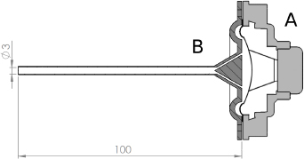
Cross section of the acoustic excitation device consisting of speaker (A) and waveguide (B).
Spatially resolved phase data is acquired with a TELESTO fourier domain OCT device (Thorlabs Inc.) utilizing a super luminescent diode with 1325 nm central wavelength and 150 nm bandwith as the source of low coherence light. The OCT system is characterized by an axial resolution of 6.5 µm in air over a depth range of 2.5 mm and a lateral resolution of 15 µm over a field of view of 10 mm × 10 mm. To obtain a time and depth-resolved phase image (M-scan), 2000 subsequent line scans, each consisting of 512 raw data points distributed over the depth range are acquired and further reduced to an array of 999 by 255 relative phase values, representing the phase shift in adjacent data points due to tissue motion. To reduce phase noise originating from the spectrometer, five subsequent M-scans are averaged and exported to Matlab (The Math-Works, Inc.). The depth-resolved amplitude of phase differences at the excitation frequency, being the representation of acoustically induced tissue motion is then extracted by row wise application of the FFT algorithm.
The experimental set-up can be subdivided into three functional blocks, which are depicted in Figure 2. Four different tissue mimicking gelatin phantoms have been used to perform the experiments. Phantom A and B are equally shaped discs with a diameter of 55 mm, a height of 10 mm and a weight of 40 g. Both phantoms differ in their gelatin concentrations to archive different elastic properties. While the hard phantom contains 6 g of gelatin, phantom B contains only 2 g of gelatin. Phantom C is made from the same low-modulus gelatin concentration as phantom B but contains a spherical inclusion of high-modulus silicone rubber with a diameter of 20 mm centrally located inside the phantom with its highest point resting approx. 3 mm under the upper surface. Phantom D is a cube of 1 cm and consists of three upright layers, with the outer ones being made from the low-modulus gelatin and the inner one of the high-modulus gelatin. The gelatin material of all three phantoms is mixed with a small amount of TiO2 to ensure sufficient incident light scattering inside the phantom for reliable phase data acquisition.

Photograph of the set-up. A: tissue-mimicking phantom, B: waveguide, C: OCT probe.
A total of 40 individual measurements from different sites on hard phantom A (N=20) and soft phantom B (N=20) were taken and processed to yield the same amount of data sets of depth resolved vibrational displacement amplitudes at the excitation frequency. A mean value for the first 100 data points lying below the surface was then calculated for each measurement to further reduce each data set to a single value.
Additionally, measurements of the inclusion phantom C and layered phantom D were taken while laterally scanning the low coherent light beam over a 1 cm long line on the surface, with help of the integrated galvanometer driven mirror scanner. Thus, a spatially resolved phase image (B-scan) is acquired. B-Scans of phantom D were collected from lines directly located over the silicone sphere, as well as from lines not over the inclusion for reference. Multiple B-Scans from adjacent lines on phantom D were acquired to obtain an en face phase image.
To retain the transverse spatial resolution in the discrete frequency transform, a different approach for frequency extraction is chosen in the form of a two-dimensional cross-correlation of the phase image with a (24x48)-convolution kernel, where all rows of the kernel contain a sine wave of the excitation frequency. The resulting cross-correlated signal is then smoothed with a rectangular average filter to account for its periodicity, compare Figure 3.
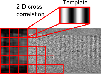
Principle of vibration detection via cross-correlation. The kernel represents the expected excitation frequency and in used to uncover the spatio-temporal motion pattern when scanning.
3 Results
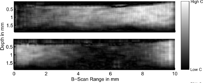
Figure 4 shows the relation between speaker voltage and the displacement. From 0 V to 1.5 V the characteristic appears partially linear with an increasing slope at approximately 1 V. For voltages higher than 1.5 V the characteristic becomes non-linear and the slope decreases to nearly zero. The two groups of mean vibrational amplitudes from the two homogenous gelatin phantoms were confirmed to both have normal distributed values. The overall mean of vibrational displacement for all points below the surface averaged are 16.1 µm (s=5.3 µm) for the hard phantom A and 36.7 µm (s=6.6 µm) for the soft phantom B. A two-sample t-test assuming unequal variances between groups was performed and revealed a highly significant difference between groups (P<0.001). The depth resolved mean vibrational displacements of both groups are depicted in Figure 5. As for the measurements of the phantom C, which features a spatially varying modulus, the cross-correlation map was plotted directly and inspected visually. Two of these are shown in Figure 6. The attenuation of the acoustically induced vibration due to the lower elasticity of the underlying inclusion in the upper image can be clearly distinguished in comparison to the reference image below.
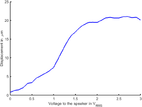
Mean displacement of a hard gelatin Phantom with increasing speaker voltage.
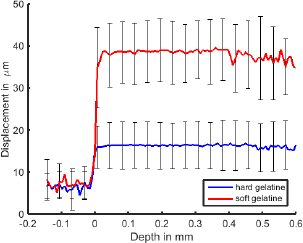
Mean displacement over depth profiles with standard deviation for 20 measurements each of hard and soft gelatin phantom A and B
The en face displacement maps of the layered phantom D were to be gained by averaging depth resolved correlation coefficients and thereby reducing each of the 25 B-scans to a two-dimensional measure of transverse spatial resolved, acoustically induced tissue displacement. Those were then plotted side by side in a log scaled, color coded en face image, depicted in Figure 7.
Spatial distribution of 2-D cross correlation coefficient between the phase distribution and a kernel consisting of the reference signal.

En face image of layered phantom D
4 Discussion
The results indicate that an OCT based elastography with acoustic excitation can help to distinguish tissues with different elastic properties. The relation between the axial displacement and the amplitude of the input signal appears partially linear, but shows a non-linear behavior for higher signal amplitudes. This is probably due to a saturation of the amplifier or a collision of the speaker membrane with the adapter for the waveguide. The detection limit due to phase wrapping or fringe washout of the OCT signal is not reached in this scenario [9]. The spherical inclusion, which is located beneath the imaging range of the OCT has a high inffiuence on the displacement of the tissue on the surface. An analog result was shown in [3, 4] and emphasizes the qualitative nature of the elasticity measurement. Currently the tissue was excited with a single frequency of 135 Hz. However, it has been shown that a frequency depended measurement could provide additional information [10]. Furthermore investigations regarding the optimal application of the acoustic radiation are planned.
Funding
This work was partially supported by DFG grant SCHL 1844/2-1.
Author's Statement
Conffiict of interest: Authors state no conffiict of interest. Material and Methods: Informed consent: Informed consent has been obtained from all individuals included in this study. Ethical approval: The research related to human use has been complied with all the relevant national regulations, institutional policies and in accordance the tenets of the Helsinki Declaration, and has been approved by the authors’ institutional review board or equivalent committee.
References
[1] Parker KJ, Doyley MM, Rubens DJ. Imaging the elastic properties of tissue: the 20 year perspective. Phys. Med. Biol.2011; 56: R1-R2910.1088/0031-9155/56/1/R01Search in Google Scholar PubMed
[2] Chin L, Kennedy BF, Kennedy KM, et al. Three-dimensional optical coherence micro-elastography of skeletal muscle tissue. Biomed Opt Express. 2014; 5(9): 3090–102.10.1364/BOE.5.003090Search in Google Scholar PubMed PubMed Central
[3] Kennedy KM, Es’haghian S, Chin L, McLaughlin RA, Sampson DD, Kennedy BF. Optical palpation: optical coherence tomography-based tactile imaging using a compliant sensor. Opt Lett. 2014; 39(10): 3014–7.10.1364/OL.39.003014Search in Google Scholar PubMed
[4] Es’haghian S, Kennedy KM, Gong P, Sampson DD, McLaughlin RA, Kennedy BF. Optical palpation in vivo: imaging human skin lesions using mechanical contrast. J Biomed Opt. 2015; 20(1): 16013.10.1117/1.JBO.20.1.016013Search in Google Scholar PubMed
[5] Qi W, Li R, Ma T, et al. Resonant acoustic radiation force optical coherence elastography. Appl Phys Lett. 2013; 103(10): 103704.10.1063/1.4820252Search in Google Scholar PubMed PubMed Central
[6] Wang S, Larin KV. Noncontact depth-resolved micro-scale optical coherence elastography of the cornea. Biomed Opt Express. 2014; 5(11): 3807–21.10.1364/BOE.5.003807Search in Google Scholar PubMed PubMed Central
[7] Subhash HM, Nguyen-Huynh A, Wang RK, Jacques SL, Choudhury N, Nuttall AL. Feasibility of spectral-domain phase-sensitive optical coherence tomography for middle ear vibrometry. J Biomed Opt. 2012; 17(6): 060505.10.1117/1.JBO.17.6.060505Search in Google Scholar PubMed PubMed Central
[8] Yazaki T, Tashiro Y, Biwa T. Measurements of sound propagation in narrow tubes. Proc. R. Soc. London, Ser. A. 2007; 463: 2855– 2862.10.1098/rspa.2007.1897Search in Google Scholar
[9] Hendargo HC, McNabb RP, Dhalla AH, Shepherd N, Izatt JA. Doppler velocity detection limitations in spectrometer-based versus swept-source optical coherence tomography. Biomed Opt Express. 2011; 2(8): 2175–88.10.1364/BOE.2.002175Search in Google Scholar PubMed PubMed Central
[10] Adie SG, Liang X, Kennedy BF, John R, Sampson DD, Boppart SA. Spectroscopic Optical Coherence Elastography. Opt Express. 2010; 18(25): 25519-25534.10.1364/OE.18.025519Search in Google Scholar PubMed PubMed Central
© 2015 by Walter de Gruyter GmbH, Berlin/Boston
This article is distributed under the terms of the Creative Commons Attribution Non-Commercial License, which permits unrestricted non-commercial use, distribution, and reproduction in any medium, provided the original work is properly cited.
Articles in the same Issue
- Research Article
- Development and characterization of superparamagnetic coatings
- Research Article
- The development of an experimental setup to measure acousto-electric interaction signal
- Research Article
- Stability analysis of ferrofluids
- Research Article
- Investigation of endothelial growth using a sensors-integrated microfluidic system to simulate physiological barriers
- Research Article
- Energy harvesting for active implants: powering a ruminal pH-monitoring system
- Research Article
- New type of fluxgate magnetometer for the heart’s magnetic fields detection
- Research Article
- Field mapping of ballistic pressure pulse sources
- Research Article
- Development of a new homecare sleep monitor using body sounds and motion tracking
- Research Article
- Noise properties of textile, capacitive EEG electrodes
- Research Article
- Detecting phase singularities and rotor center trajectories based on the Hilbert transform of intraatrial electrograms in an atrial voxel model
- Research Article
- Spike sorting: the overlapping spikes challenge
- Research Article
- Separating the effect of respiration from the heart rate variability for cases of constant harmonic breathing
- Research Article
- Locating regions of arrhythmogenic substrate by analyzing the duration of triggered atrial activities
- Research Article
- Combining different ECG derived respiration tracking methods to create an optimal reconstruction of the breathing pattern
- Research Article
- Atrial and ventricular signal averaging electrocardiography in pacemaker and cardiac resynchronization therapy
- Research Article
- Estimation of a respiratory signal from a single-lead ECG using the 4th order central moments
- Research Article
- Compressed sensing of multi-lead ECG signals by compressive multiplexing
- Research Article
- Heart rate monitoring in ultra-high-field MRI using frequency information obtained from video signals of the human skin compared to electrocardiography and pulse oximetry
- Research Article
- Synchronization in wireless biomedical-sensor networks with Bluetooth Low Energy
- Research Article
- Automated classification of stages of anaesthesia by populations of evolutionary optimized fuzzy rules
- Research Article
- Effects of sampling rate on automated fatigue recognition in surface EMG signals
- Research Article
- Closed-loop transcranial alternating current stimulation of slow oscillations
- Research Article
- Cardiac index in atrio- and interventricular delay optimized cardiac resynchronization therapy and cardiac contractility modulation
- Research Article
- The role of expert evaluation for microsleep detection
- Research Article
- The impact of baseline wander removal techniques on the ST segment in simulated ischemic 12-lead ECGs
- Research Article
- Metal artifact reduction by projection replacements and non-local prior image integration
- Research Article
- A novel coaxial nozzle for in-process adjustment of electrospun scaffolds’ fiber diameter
- Research Article
- Processing of membranes for oxygenation using the Bellhouse-effect
- Research Article
- Inkjet printing of viable human dental follicle stem cells
- Research Article
- The use of an icebindingprotein out of the snowflea Hypogastrura harveyi as a cryoprotectant in the cryopreservation of mesenchymal stem cells
- Research Article
- New NIR spectroscopy based method to determine ischemia in vivo in liver – a first study on rats
- Research Article
- QRS and QT ventricular conduction times and permanent pacemaker therapy after transcatheter aortic valve implantation
- Research Article
- Adopting oculopressure tonometry as a transient in vivo rabbit glaucoma model
- Research Article
- Next-generation vision testing: the quick CSF
- Research Article
- Improving tactile sensation in laparoscopic surgery by overcoming size restrictions
- Research Article
- Design and control of a 3-DOF hydraulic driven surgical instrument
- Research Article
- Evaluation of endourological tools to improve the diagnosis and therapy of ureteral tumors – from model development to clinical application
- Research Article
- Frequency based assessment of surgical activities
- Research Article
- “Hands free for intervention”, a new approach for transoral endoscopic surgery
- Research Article
- Pseudo-haptic feedback in medical teleoperation
- Research Article
- Feasibility of interactive gesture control of a robotic microscope
- Research Article
- Towards structuring contextual information for workflow-driven surgical assistance functionalities
- Research Article
- Towards a framework for standardized semantic workflow modeling and management in the surgical domain
- Research Article
- Closed-loop approach for situation awareness of medical devices and operating room infrastructure
- Research Article
- Kinect based physiotherapy system for home use
- Research Article
- Evaluating the microsoft kinect skeleton joint tracking as a tool for home-based physiotherapy
- Research Article
- Integrating multimodal information for intraoperative assistance in neurosurgery
- Research Article
- Respiratory motion tracking using Microsoft’s Kinect v2 camera
- Research Article
- Using smart glasses for ultrasound diagnostics
- Research Article
- Measurement of needle susceptibility artifacts in magnetic resonance images
- Research Article
- Dimensionality reduction of medical image descriptors for multimodal image registration
- Research Article
- Experimental evaluation of different weighting schemes in magnetic particle imaging reconstruction
- Research Article
- Evaluation of CT capability for the detection of thin bone structures
- Research Article
- Towards contactless optical coherence elastography with acoustic tissue excitation
- Research Article
- Development and implementation of algorithms for automatic and robust measurement of the 2D:4D digit ratio using image data
- Research Article
- Automated high-throughput analysis of B cell spreading on immobilized antibodies with whole slide imaging
- Research Article
- Tissue segmentation from head MRI: a ground truth validation for feature-enhanced tracking
- Research Article
- Video tracking of swimming rodents on a reflective water surface
- Research Article
- MR imaging of model drug distribution in simulated vitreous
- Research Article
- Studying the extracellular contribution to the double wave vector diffusion-weighted signal
- Research Article
- Artifacts in field free line magnetic particle imaging in the presence of inhomogeneous and nonlinear magnetic fields
- Research Article
- Introducing a frequency-tunable magnetic particle spectrometer
- Research Article
- Imaging of aortic valve dynamics in 4D OCT
- Research Article
- Intravascular optical coherence tomography (OCT) as an additional tool for the assessment of stent structures
- Research Article
- Simple concept for a wide-field lensless digital holographic microscope using a laser diode
- Research Article
- Intraoperative identification of somato-sensory brain areas using optical imaging and standard RGB camera equipment – a feasibility study
- Research Article
- Respiratory surface motion measurement by Microsoft Kinect
- Research Article
- Improving image quality in EIT imaging by measurement of thorax excursion
- Research Article
- A clustering based dual model framework for EIT imaging: first experimental results
- Research Article
- Three-dimensional anisotropic regularization for limited angle tomography
- Research Article
- GPU-based real-time generation of large ultrasound volumes from freehand 3D sweeps
- Research Article
- Experimental computer tomograph
- Research Article
- US-tracked steered FUS in a respiratory ex vivo ovine liver phantom
- Research Article
- Contribution of brownian rotation and particle assembly polarisation to the particle response in magnetic particle spectrometry
- Research Article
- Preliminary investigations of magnetic modulated nanoparticles for microwave breast cancer detection
- Research Article
- Construction of a device for magnetic separation of superparamagnetic iron oxide nanoparticles
- Research Article
- An IHE-conform telecooperation platform supporting the treatment of dementia patients
- Research Article
- Automated respiratory therapy system based on the ARDSNet protocol with systemic perfusion control
- Research Article
- Identification of surgical instruments using UHF-RFID technology
- Research Article
- A generic concept for the development of model-guided clinical decision support systems
- Research Article
- Evaluation of local alterations in femoral bone mineral density measured via quantitative CT
- Research Article
- Creating 3D gelatin phantoms for experimental evaluation in biomedicine
- Research Article
- Influence of short-term fixation with mixed formalin or ethanol solution on the mechanical properties of human cortical bone
- Research Article
- Analysis of the release kinetics of surface-bound proteins via laser-induced fluorescence
- Research Article
- Tomographic particle image velocimetry of a water-jet for low volume harvesting of fat tissue for regenerative medicine
- Research Article
- Wireless medical sensors – context, robustness and safety
- Research Article
- Sequences for real-time magnetic particle imaging
- Research Article
- Speckle-based off-axis holographic detection for non-contact photoacoustic tomography
- Research Article
- A machine learning approach for planning valve-sparing aortic root reconstruction
- Research Article
- An in-ear pulse wave velocity measurement system using heart sounds as time reference
- Research Article
- Measuring different oxygenation levels in a blood perfusion model simulating the human head using NIRS
- Research Article
- Multisegmental fusion of the lumbar spine a curse or a blessing?
- Research Article
- Numerical analysis of the biomechanical complications accompanying the total hip replacement with NANOS-Prosthetic: bone remodelling and prosthesis migration
- Research Article
- A muscle model for hybrid muscle activation
- Research Article
- Mathematical, numerical and in-vitro investigation of cooling performance of an intra-carotid catheter for selective brain hypothermia
- Research Article
- An ideally parameterized unscented Kalman filter for the inverse problem of electrocardiography
- Research Article
- Interactive visualization of cardiac anatomy and atrial excitation for medical diagnosis and research
- Research Article
- Virtualizing clinical cases of atrial flutter in a fast marching simulation including conduction velocity and ablation scars
- Research Article
- Mesh structure-independent modeling of patient-specific atrial fiber orientation
- Research Article
- Accelerating mono-domain cardiac electrophysiology simulations using OpenCL
- Research Article
- Understanding the cellular mode of action of vernakalant using a computational model: answers and new questions
- Research Article
- A java based simulator with user interface to simulate ventilated patients
- Research Article
- Evaluation of an algorithm to choose between competing models of respiratory mechanics
- Research Article
- Numerical simulation of low-pulsation gerotor pumps for use in the pharmaceutical industry and in biomedicine
- Research Article
- Numerical and experimental flow analysis in centifluidic systems for rapid allergy screening tests
- Research Article
- Biomechanical parameter determination of scaffold-free cartilage constructs (SFCCs) with the hyperelastic material models Yeoh, Ogden and Demiray
- Research Article
- FPGA controlled artificial vascular system
- Research Article
- Simulation based investigation of source-detector configurations for non-invasive fetal pulse oximetry
- Research Article
- Test setup for characterizing the efficacy of embolic protection devices
- Research Article
- Impact of electrode geometry on force generation during functional electrical stimulation
- Research Article
- 3D-based visual physical activity assessment of children
- Research Article
- Realtime assessment of foot orientation by Accelerometers and Gyroscopes
- Research Article
- Image based reconstruction for cystoscopy
- Research Article
- Image guided surgery innovation with graduate students - a new lecture format
- Research Article
- Multichannel FES parameterization for controlling foot motion in paretic gait
- Research Article
- Smartphone supported upper limb prosthesis
- Research Article
- Use of quantitative tremor evaluation to enhance target selection during deep brain stimulation surgery for essential tremor
- Research Article
- Evaluation of adhesion promoters for Parylene C on gold metallization
- Research Article
- The influence of metallic ions from CoCr28Mo6 on the osteogenic differentiation and cytokine release of human osteoblasts
- Research Article
- Increasing the visibility of thin NITINOL vascular implants
- Research Article
- Possible reasons for early artificial bone failure in biomechanical tests of ankle arthrodesis systems
- Research Article
- Development of a bending test procedure for the characterization of flexible ECoG electrode arrays
- Research Article
- Tubular manipulators: a new concept for intracochlear positioning of an auditory prosthesis
- Research Article
- Investigation of the dynamic diameter deformation of vascular stents during fatigue testing with radial loading
- Research Article
- Electrospun vascular grafts with anti-kinking properties
- Research Article
- Integration of temperature sensors in polyimide-based thin-film electrode arrays
- Research Article
- Use cases and usability challenges for head-mounted displays in healthcare
- Research Article
- Device- and service profiles for integrated or systems based on open standards
- Research Article
- Risk management for medical devices in research projects
- Research Article
- Simulation of varying femoral attachment sites of medial patellofemoral ligament using a musculoskeletal multi-body model
- Research Article
- Does enhancing consciousness for strategic planning processes support the effectiveness of problem-based learning concepts in biomedical education?
- Research Article
- SPIO processing in macrophages for MPI: The breast cancer MPI-SNLB-concept
- Research Article
- Numerical simulations of airflow in the human pharynx of OSAHS patients