Tissue segmentation from head MRI: a ground truth validation for feature-enhanced tracking
-
 ,
,

Abstract
Accuracy is essential for optical head-tracking in cranial radiotherapy. Recently, the exploitation of local patterns of tissue information was proposed to achieve a more robust registration. Here, we validate a ground truth for this information obtained from high resolution MRI scans. In five subjects we compared the segmentation accuracy of a semi-automatic algorithm with five human experts. While the algorithm segments the skin and bone surface with an average accuracy of less than 0.1 mm and 0.2 mm, respectively, the mean error on the tissue thickness was 0.17 mm. We conclude that this accuracy is a reasonable basis for extracting reliable cutaneous structures to support surface registration.
1 Introduction
Precise localization of the cranium is vital for effective head radiotherapy. Thus, patient immobilization is currently achieved by inconvenient thermoplastic masks with repositioning accuracies in the mm range [1]. A fast alternative for more comfortable monitoring during treatment is given by marker-less optical head-tracking. To achieve the desired sub-millimeter accuracy and robustness of skin surface registration for continuous scanning over time, feature-supported tracking was introduced recently [2]. Using machine learning models, optical backscatter features across the forehead are converted into a tissue thickness measure, which is joined with the 3D spatial information and then used for enhanced 4D surface registration.
These models are generated in a supervised manner based on a high resolution ground truth for tissue thickness across the forehead. Here, we validate the ground truth accuracy and show that the segmentation procedure used in [2] suffices to provide smooth subcutaneous structures across the forehead.
2 Material and methods
2.1 Data acquisition
High resolution MR images (0.1025 mm × 0.1025 mm 1 mm) of the forehead region were acquired on an Ingenia 3.0T scanner (Philips Healthcare) from five subjects (3 male, 2 female, aged 25-64). Fig. 1 marks the region of interest in a lower resolution T1 scan of the entire head.

Clinical T1 contrast scan of a subject’s head. The volume-of-interest (VOI) is marked in green. It contains the forehead region and MR-visible marker spheres for registering the volume into other coordinate spaces.
The imaging sequence was a gradient echo (FFE-T1) with a flip angle of 15° to rapidly acquire a volume of 210 mm × 210 mm × 70 mm. Echo time was kept as low as possible (TE/TR = 5/17 ms) to minimize susceptibility artifacts at tissue-air/bone interfaces [3]. The volume was aligned in parallel to the AC-PC line and approximately orthogonal to the forehead surface.
2.2 Tissue segmentation
The segmentation pipeline consists of nine major steps and was performed slice-wise. First, the initial segmentation of five components (grey matter, white matter, cerebral spinal fluid (CSF), meninges and skull/skin) was done using SPM8 [4]. Based on probability maps, the output indicates the likelihood for each voxel of belonging to a certain type of anatomy. Second, the first four components were merged into a negative and the last component (extra-cranial tissue and skull) into a positive mask. By masking the original image with both of them, the interior of the skull was cut out and only voxels were kept that had a probability of more than 25% of belonging to component five and more than 68% of not belonging to one of the others (cf. Fig 2A-D). The volume-of-interest (VOI) was then restricted to the forehead (Fig 2E).

Illustration of the segmentation pipeline. A: raw volume slice, B: negative mask of intra-cranial sites, C: positive mask of likely extra-cranial tissue sites, D: raw slice after masking, E: forehead region from D, F: region growing output, G: active bone contour detection using snakes (blue: initial contour, red: output contour), H: snake output segment, I: output from F and H after Canny edge detection
In a third step manual inspection allowed to correct for rare drops in local contrast or large vessels. In rare cases these may lead to holes in the tissue region which cannot be closed using a previously applied morphological closing operator and median low-pass filter. Fourth, a 2D region growing algorithm was used to extract the largest, connected tissue segment (Fig 2F). In order to obtain a smooth edge of the tissue-bone boundary, 2D snakes were applied [5] (200 support points, 150 gradient descent steps, weights for tension and stiffness were 0.03 and 0.005, respectively). The active contour also made use of a balloon force and gradient vector flow as an external force. Fig 2G shows the initial contour and the optimized result for one slice. Fig 2H plots the segment contained within this optimized contour. Canny edge detection then yielded the air-tissue boundary (cf. Fig 2I). In the eighth step agglomerative clustering rejected minor contours (e.g. surrounding vessels) and identified the point clouds for the skin and bone surface.
From these clouds the actual tissue thickness was obtained as the distance between each point on the skin surface and the penetration point of the corresponding normal vector at the bone surface (cf. Fig 3).

3D point clouds for the skin (green) and bone (red) surface. Tissue thickness is computed by normal vector (black) penetration through both surfaces. The orthogonality condition holds for the skin surface.
2.3 Expert segmentation and validation
The algorithmic segmentation as described above was validated based on the segmentation results of five skilled human experts. Each of them manually segmented the skin and bone surfaces for one slice per subject. Each slice was selected from the middle of the volume to be representative for the forehead region possibly scanned by an optical tracking system. To evaluate the segmentation error propagation to the skin thickness measure, the experts were also asked to segment five slices for subject three. Five slices were sufficient to apply the normal vector penetration procedure and to compare with the tissue thickness output by the segmentation algorithm. The deviation of the algorithmic from the expert segmentation was computed as a 3D vector using point-to-point correspondences obtained via Euclidean nearest-neighbor search. Finally, expert five segmented one slice of subject three five times, in order to get an impression of the intra-operator reproducibility.
3 Results
Fig. 4 shows the algorithmic segmentation result for subject three. The top part shows the 3D skin surface over-laid with color-coded tissue thickness. The surface shows smooth subcutaneous and cutaneous structures. The histogram reveals that large areas on the forehead had a tissue thickness between 3.4 mm and 5.6 mm. This corresponds to 80 % of the histogram area. Corresponding intervals for the other subjects (not shown) had a range of 3.35 mm (S1, 70 %), 3.87 mm (S2, 70 %), 2.60 mm (S4, 80 %), and 1.98 mm (S5, 80 %).
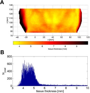
Segmentation result for subject 3. A: 3D skin surface over-laid with color-coded tissue thickness. B: Histogram of the tissue thickness across the patch shown in A.
The deviation of the algorithmic from the expert segmentation is provided in Table 1. As illustrated in Fig. 5, the tissue boundary was always identified with an average accuracy of less than 0.1 mm and the bone with less than 0.2 mm. The standard deviations shown in Table 1 contain on average 0.019 mm inter-operator variability for the skin and 0.031 for the bone surface (root-mean-square across all subjects). Expert five was able to reproduce his results with mean deviations of 0.084 mm for the skin and 0.086 mm for the bone.

Deviations of the algorithmic from the expert segmentation for all subjects averaged across all experts.
Mean and standard deviation of the absolute differences between expert and algorithmic segmentation for skin and bone contours (in mm).
| Skin [mm] | Bone [mm] | |
|---|---|---|
| Subject 1 | 0.093 ± 0.081 | 0.168 ± 0.098 |
| Subject 2 | 0.082 ± 0.072 | 0.199 ± 0.128 |
| Subject 3 | 0.101 ± 0.082 | 0.154 ± 0.126 |
| Subject 4 | 0.102 ± 0.071 | 0.155 ± 0.095 |
| Subject 5 | 0.095 ± 0.078 | 0.185 ± 0.118 |
All experts segmented five slices for subject three to evaluate how the segmentation errors on the skin and bone boundaries translate to the tissue thickness measure. The results are show in Table 2. On average, the error on the tissue thickness in that region was found to be 0.173 mm and for all experts less than 0.21 mm.
Mean and standard deviation of the absolute differences on the computed tissue thickness (in mm).
| Expert 1 | Expert 2 | Expert 3 | Expert 4 | Expert 5 | |
|---|---|---|---|---|---|
| S3 | 0.161 | 0.183 | 0.162 | 0.154 | 0.205 |
| ±0.123 | ±0.151 | ±0.145 | ±0.128 | ±0.159 |
4 Discussion and conclusion
A qualitative evaluation of the skin patches reveals that cutaneous structures were smoothly visible after segmentation. These variations in tissue thickness originate from subcutaneous vessels, facial muscles or changes in the subcutaneous fat layer. The segmentation error for the air-tissue and tissue-bone interface were on average less than twice the in-plane voxel size. With respect to the theoretical limit of half the voxel size and the main thickness interval of tissue across the forehead (here the structures were varying in ranges of 1.98 mm or more), this is acceptable. Moreover, repeated manual segmentation for one expert suggests that significant parts of this error may be due to intra-operator variability. The accuracy across subjects may vary due to motion during the acquisition process, which lasted approximately 16 min (as it was the case for subjects two and five). This motion leads to much noisier images and blurred boundaries. Large motion results in halo-like noise patterns around the head.
In general, the segmentation of the bone was more prone to errors due to a poorer contrast with respect to the adjacent cranium or meninges structures. Nevertheless, it was found that the mean boundary deviations do not additively translate to the skin thickness measure. One possible contribution to this is that the thickness was extracted along the forehead normal direction, while the slice orientation was not precisely orthogonal.
In conclusion, we have shown, that the accuracy of tissue thickness segmentation is acceptable and provides a reasonable basis for revealing smooth and prominent cutaneous structures across the forehead. These can add valuable information to a surface registration process and fix poorly defined degrees of freedom.
Acknowledgment
The authors acknowledge the support of Dr. Uwe Melchert and Christian Erdmann from the clinic of neuroradiology, University Hospital Schleswig-Holstein, respectively.
Funding
This work was supported by Varian Medical Systems, Inc. and partially funded by the Graduate School for Computing in Medicine and Life Sciences, German Excellence Initiative [DFG GSC 235/1]
Author’s Statement
Conflict of interest Authors state no conflict of interest. Material and Methods: Informed consent: Informed consent has been obtained from all individuals included in this study. Ethical approval: The research related to human use has been complied with all the relevant national regulations, institutional policies and in accordance the tenets of the Helsinki Declaration, and has been approved by the authors’ institutional review board or equivalent committee.
References
[1] Robar, JL., Clark, BG., Schella, JW., and Kim, CS. Analysis of patient repositioning accuracy in precision radiation therapy using automated image fusion. Journal of Applied Clinical Medical Physics, 2005: 6 (1): 71–83.10.1120/jacmp.v6i1.1998Search in Google Scholar
[2] Wissel, T., Stüber, P., Wagner, B., Bruder, R., Schweikard, A., and Ernst, F. Tissue Thickness Estimation for High Precision Head-Tracking using a Galvanometric Laser Scanner - A Case Study. 36th Annual International Conference of the IEEE Engineering in Medicine and Biology Society (EMBC ‘14), Chicago, USA, 2014, 3106–310910.1109/EMBC.2014.6944280Search in Google Scholar
[3] Farahani, K., Sinha, U., Sinha, S., Chiu, LCL., and Lufkin, R., Effect of field strength on susceptibility artifacts in magnetic resonance imaging. Computerized Medical Imaging and Graphics, 1990: 14 (6): 409–413.10.1016/0895-6111(90)90040-ISearch in Google Scholar
[4] Friston, KJ., Ashburner, JT., Kiebel, SJ., Nichols, TE., and Penny, WD. Statistical Parametric Mapping: The Analysis of Functional Brain Images. 2006: Elsevier, London.Search in Google Scholar
[5] Kaas, M., Witkin, A., and Terzopoulos, D. Snakes: active contour models. International Journal of Computer Vision, 1988: 1 (4): 321–331.10.1007/BF00133570Search in Google Scholar
[6] Canny, J. A computational approach to edge detection. IEEE Transaction on Pattern Analysis and Machine Intelligence. 1986: PAMI-8 (6), 679–698.10.1016/B978-0-08-051581-6.50024-6Search in Google Scholar
© 2015 by Walter de Gruyter GmbH, Berlin/Boston
This article is distributed under the terms of the Creative Commons Attribution Non-Commercial License, which permits unrestricted non-commercial use, distribution, and reproduction in any medium, provided the original work is properly cited.
Articles in the same Issue
- Research Article
- Development and characterization of superparamagnetic coatings
- Research Article
- The development of an experimental setup to measure acousto-electric interaction signal
- Research Article
- Stability analysis of ferrofluids
- Research Article
- Investigation of endothelial growth using a sensors-integrated microfluidic system to simulate physiological barriers
- Research Article
- Energy harvesting for active implants: powering a ruminal pH-monitoring system
- Research Article
- New type of fluxgate magnetometer for the heart’s magnetic fields detection
- Research Article
- Field mapping of ballistic pressure pulse sources
- Research Article
- Development of a new homecare sleep monitor using body sounds and motion tracking
- Research Article
- Noise properties of textile, capacitive EEG electrodes
- Research Article
- Detecting phase singularities and rotor center trajectories based on the Hilbert transform of intraatrial electrograms in an atrial voxel model
- Research Article
- Spike sorting: the overlapping spikes challenge
- Research Article
- Separating the effect of respiration from the heart rate variability for cases of constant harmonic breathing
- Research Article
- Locating regions of arrhythmogenic substrate by analyzing the duration of triggered atrial activities
- Research Article
- Combining different ECG derived respiration tracking methods to create an optimal reconstruction of the breathing pattern
- Research Article
- Atrial and ventricular signal averaging electrocardiography in pacemaker and cardiac resynchronization therapy
- Research Article
- Estimation of a respiratory signal from a single-lead ECG using the 4th order central moments
- Research Article
- Compressed sensing of multi-lead ECG signals by compressive multiplexing
- Research Article
- Heart rate monitoring in ultra-high-field MRI using frequency information obtained from video signals of the human skin compared to electrocardiography and pulse oximetry
- Research Article
- Synchronization in wireless biomedical-sensor networks with Bluetooth Low Energy
- Research Article
- Automated classification of stages of anaesthesia by populations of evolutionary optimized fuzzy rules
- Research Article
- Effects of sampling rate on automated fatigue recognition in surface EMG signals
- Research Article
- Closed-loop transcranial alternating current stimulation of slow oscillations
- Research Article
- Cardiac index in atrio- and interventricular delay optimized cardiac resynchronization therapy and cardiac contractility modulation
- Research Article
- The role of expert evaluation for microsleep detection
- Research Article
- The impact of baseline wander removal techniques on the ST segment in simulated ischemic 12-lead ECGs
- Research Article
- Metal artifact reduction by projection replacements and non-local prior image integration
- Research Article
- A novel coaxial nozzle for in-process adjustment of electrospun scaffolds’ fiber diameter
- Research Article
- Processing of membranes for oxygenation using the Bellhouse-effect
- Research Article
- Inkjet printing of viable human dental follicle stem cells
- Research Article
- The use of an icebindingprotein out of the snowflea Hypogastrura harveyi as a cryoprotectant in the cryopreservation of mesenchymal stem cells
- Research Article
- New NIR spectroscopy based method to determine ischemia in vivo in liver – a first study on rats
- Research Article
- QRS and QT ventricular conduction times and permanent pacemaker therapy after transcatheter aortic valve implantation
- Research Article
- Adopting oculopressure tonometry as a transient in vivo rabbit glaucoma model
- Research Article
- Next-generation vision testing: the quick CSF
- Research Article
- Improving tactile sensation in laparoscopic surgery by overcoming size restrictions
- Research Article
- Design and control of a 3-DOF hydraulic driven surgical instrument
- Research Article
- Evaluation of endourological tools to improve the diagnosis and therapy of ureteral tumors – from model development to clinical application
- Research Article
- Frequency based assessment of surgical activities
- Research Article
- “Hands free for intervention”, a new approach for transoral endoscopic surgery
- Research Article
- Pseudo-haptic feedback in medical teleoperation
- Research Article
- Feasibility of interactive gesture control of a robotic microscope
- Research Article
- Towards structuring contextual information for workflow-driven surgical assistance functionalities
- Research Article
- Towards a framework for standardized semantic workflow modeling and management in the surgical domain
- Research Article
- Closed-loop approach for situation awareness of medical devices and operating room infrastructure
- Research Article
- Kinect based physiotherapy system for home use
- Research Article
- Evaluating the microsoft kinect skeleton joint tracking as a tool for home-based physiotherapy
- Research Article
- Integrating multimodal information for intraoperative assistance in neurosurgery
- Research Article
- Respiratory motion tracking using Microsoft’s Kinect v2 camera
- Research Article
- Using smart glasses for ultrasound diagnostics
- Research Article
- Measurement of needle susceptibility artifacts in magnetic resonance images
- Research Article
- Dimensionality reduction of medical image descriptors for multimodal image registration
- Research Article
- Experimental evaluation of different weighting schemes in magnetic particle imaging reconstruction
- Research Article
- Evaluation of CT capability for the detection of thin bone structures
- Research Article
- Towards contactless optical coherence elastography with acoustic tissue excitation
- Research Article
- Development and implementation of algorithms for automatic and robust measurement of the 2D:4D digit ratio using image data
- Research Article
- Automated high-throughput analysis of B cell spreading on immobilized antibodies with whole slide imaging
- Research Article
- Tissue segmentation from head MRI: a ground truth validation for feature-enhanced tracking
- Research Article
- Video tracking of swimming rodents on a reflective water surface
- Research Article
- MR imaging of model drug distribution in simulated vitreous
- Research Article
- Studying the extracellular contribution to the double wave vector diffusion-weighted signal
- Research Article
- Artifacts in field free line magnetic particle imaging in the presence of inhomogeneous and nonlinear magnetic fields
- Research Article
- Introducing a frequency-tunable magnetic particle spectrometer
- Research Article
- Imaging of aortic valve dynamics in 4D OCT
- Research Article
- Intravascular optical coherence tomography (OCT) as an additional tool for the assessment of stent structures
- Research Article
- Simple concept for a wide-field lensless digital holographic microscope using a laser diode
- Research Article
- Intraoperative identification of somato-sensory brain areas using optical imaging and standard RGB camera equipment – a feasibility study
- Research Article
- Respiratory surface motion measurement by Microsoft Kinect
- Research Article
- Improving image quality in EIT imaging by measurement of thorax excursion
- Research Article
- A clustering based dual model framework for EIT imaging: first experimental results
- Research Article
- Three-dimensional anisotropic regularization for limited angle tomography
- Research Article
- GPU-based real-time generation of large ultrasound volumes from freehand 3D sweeps
- Research Article
- Experimental computer tomograph
- Research Article
- US-tracked steered FUS in a respiratory ex vivo ovine liver phantom
- Research Article
- Contribution of brownian rotation and particle assembly polarisation to the particle response in magnetic particle spectrometry
- Research Article
- Preliminary investigations of magnetic modulated nanoparticles for microwave breast cancer detection
- Research Article
- Construction of a device for magnetic separation of superparamagnetic iron oxide nanoparticles
- Research Article
- An IHE-conform telecooperation platform supporting the treatment of dementia patients
- Research Article
- Automated respiratory therapy system based on the ARDSNet protocol with systemic perfusion control
- Research Article
- Identification of surgical instruments using UHF-RFID technology
- Research Article
- A generic concept for the development of model-guided clinical decision support systems
- Research Article
- Evaluation of local alterations in femoral bone mineral density measured via quantitative CT
- Research Article
- Creating 3D gelatin phantoms for experimental evaluation in biomedicine
- Research Article
- Influence of short-term fixation with mixed formalin or ethanol solution on the mechanical properties of human cortical bone
- Research Article
- Analysis of the release kinetics of surface-bound proteins via laser-induced fluorescence
- Research Article
- Tomographic particle image velocimetry of a water-jet for low volume harvesting of fat tissue for regenerative medicine
- Research Article
- Wireless medical sensors – context, robustness and safety
- Research Article
- Sequences for real-time magnetic particle imaging
- Research Article
- Speckle-based off-axis holographic detection for non-contact photoacoustic tomography
- Research Article
- A machine learning approach for planning valve-sparing aortic root reconstruction
- Research Article
- An in-ear pulse wave velocity measurement system using heart sounds as time reference
- Research Article
- Measuring different oxygenation levels in a blood perfusion model simulating the human head using NIRS
- Research Article
- Multisegmental fusion of the lumbar spine a curse or a blessing?
- Research Article
- Numerical analysis of the biomechanical complications accompanying the total hip replacement with NANOS-Prosthetic: bone remodelling and prosthesis migration
- Research Article
- A muscle model for hybrid muscle activation
- Research Article
- Mathematical, numerical and in-vitro investigation of cooling performance of an intra-carotid catheter for selective brain hypothermia
- Research Article
- An ideally parameterized unscented Kalman filter for the inverse problem of electrocardiography
- Research Article
- Interactive visualization of cardiac anatomy and atrial excitation for medical diagnosis and research
- Research Article
- Virtualizing clinical cases of atrial flutter in a fast marching simulation including conduction velocity and ablation scars
- Research Article
- Mesh structure-independent modeling of patient-specific atrial fiber orientation
- Research Article
- Accelerating mono-domain cardiac electrophysiology simulations using OpenCL
- Research Article
- Understanding the cellular mode of action of vernakalant using a computational model: answers and new questions
- Research Article
- A java based simulator with user interface to simulate ventilated patients
- Research Article
- Evaluation of an algorithm to choose between competing models of respiratory mechanics
- Research Article
- Numerical simulation of low-pulsation gerotor pumps for use in the pharmaceutical industry and in biomedicine
- Research Article
- Numerical and experimental flow analysis in centifluidic systems for rapid allergy screening tests
- Research Article
- Biomechanical parameter determination of scaffold-free cartilage constructs (SFCCs) with the hyperelastic material models Yeoh, Ogden and Demiray
- Research Article
- FPGA controlled artificial vascular system
- Research Article
- Simulation based investigation of source-detector configurations for non-invasive fetal pulse oximetry
- Research Article
- Test setup for characterizing the efficacy of embolic protection devices
- Research Article
- Impact of electrode geometry on force generation during functional electrical stimulation
- Research Article
- 3D-based visual physical activity assessment of children
- Research Article
- Realtime assessment of foot orientation by Accelerometers and Gyroscopes
- Research Article
- Image based reconstruction for cystoscopy
- Research Article
- Image guided surgery innovation with graduate students - a new lecture format
- Research Article
- Multichannel FES parameterization for controlling foot motion in paretic gait
- Research Article
- Smartphone supported upper limb prosthesis
- Research Article
- Use of quantitative tremor evaluation to enhance target selection during deep brain stimulation surgery for essential tremor
- Research Article
- Evaluation of adhesion promoters for Parylene C on gold metallization
- Research Article
- The influence of metallic ions from CoCr28Mo6 on the osteogenic differentiation and cytokine release of human osteoblasts
- Research Article
- Increasing the visibility of thin NITINOL vascular implants
- Research Article
- Possible reasons for early artificial bone failure in biomechanical tests of ankle arthrodesis systems
- Research Article
- Development of a bending test procedure for the characterization of flexible ECoG electrode arrays
- Research Article
- Tubular manipulators: a new concept for intracochlear positioning of an auditory prosthesis
- Research Article
- Investigation of the dynamic diameter deformation of vascular stents during fatigue testing with radial loading
- Research Article
- Electrospun vascular grafts with anti-kinking properties
- Research Article
- Integration of temperature sensors in polyimide-based thin-film electrode arrays
- Research Article
- Use cases and usability challenges for head-mounted displays in healthcare
- Research Article
- Device- and service profiles for integrated or systems based on open standards
- Research Article
- Risk management for medical devices in research projects
- Research Article
- Simulation of varying femoral attachment sites of medial patellofemoral ligament using a musculoskeletal multi-body model
- Research Article
- Does enhancing consciousness for strategic planning processes support the effectiveness of problem-based learning concepts in biomedical education?
- Research Article
- SPIO processing in macrophages for MPI: The breast cancer MPI-SNLB-concept
- Research Article
- Numerical simulations of airflow in the human pharynx of OSAHS patients