Evaluation of CT capability for the detection of thin bone structures
-
 und
und

Abstract
Evaluation of CT capability for the detection of thin bone structures by means of modeling small bone structures and comparing with quantitative measurements. Knowledge of how thin bone structures can be or of what low density to be detectable in clinical CT images can increase the diagnostic competence to diagnose various diseases. Correlation between size and density of detectable bone structures is the key for many diagnosis purposes. This paper describes the use of clinical 64 detector scanner to image the skeletons of codfish and salmon species. Fish skeletons, such as codfish and salmon, have petite structures and lower bone density than humans. Bone structures were segmented out of the image data and 3D models of their skeletons developed. Evaluation was done by means of comparing quantitative measurements of selected bones to parameters observed from the model. Results show the limits where thin bones and of low density disappear from the clinical CT. It shows ability to reconstruct closely the diameter of the codfish bones but to a less extent the small bones of the salmon.
1 Introduction
Knowing and understanding the correlation between size and density of detectable bone structures in clinical computed tomography (CT) is the key for many diagnosis purposes. To be able to distinguish at what sizes bone structures are detectable with a clinical CT scanner, parameters from the image model have to be compared to quantitative size parameters of the bone structures.
Hangartner et al. was able to show that bone width can be measured down to a width of less than 1 mm, by using algorithm based on iteratively varying CT thresholds for narrow structures and fixed thresholds for wide structures [1]. However, they did not specify the bone density of their specimens, i.e. if it is relatively low or not.
CT is an excellent source of creating geometrically accurate 3D models by means of good segmentation methods and it is possible to convert the entire range of Hounsfield units to bone density [2]. However it should be noted that images of very thin structures show artificially decreased density values due to the well known partial-volume effects [1].
In this work a standard clinical CT scanner is evaluated for detecting small bone structures in codfish and salmon. A 3D model of the fish skeletons are developed and few specific bones are analyzed to study differences in size parameters. This is done to see if parts of the bones are not detectable by the use of a CT scanner or not included in the model due to its small thickness or low density. It is hypothesized that parts of extremely thin bone structures or of low density will be absent in the 3D model. Helgason et al. did similar research on detecting thin bones and modeling for the development of automated bone detection methods for the food processing industry [3]. Results are promising for future studies in this field.
2 Material and methods
Fish bones from a codfish and salmon were used as specimens. The two fishes were scanned separately in a standard clinical CT scanner, same parameters used for both scans. The CT data were then processed so that the skeleton was separated from the tissue and a 3D model developed. Quantitative measurements of selected bones were then compared to parameters observed from the model.
2.1 Codfish and salmon
The codfish was scanned the same day as it was caught off the coast of Iceland. We got the salmon from a fish farmer and it was kept in a freezer overnight before imaging. After imaging, the fishes were kept in a freezer until specific bones of interest were cut out for further analysis.
2.2 CT imaging system
The CT imaging system used was a scanner designed for human clinical use, Philips Brilliance 64. The scan parameters were set at the lowest energy of 80 KV with 0.67 mm slice thickness with 50% redundancy, i.e. each slice covers half of the volume as the previous slice covers. Field of view is 344 mm and each slice has an image matrix of 512 × 512 pixels.
2.3 Image processing – segmentation
Image processing was done using Mimics® (v15.0, Materialise NV, Leuven, Belgium) the specially developed software for medical image processing. Mimics® allows for easy and quick creation of accurate 3D models and segmentation from the CT imaging data.
CT image values are linearly transformed to Hounsfield units (HU) by relating the CT values to nearby water and air values calculated from the following equation [2].
where µw and µA are the CT image values of water and air. Bone density is defined as mean value expressed in HU in each pixel [2] and in a 3D image, the picture elements are referred to as voxels, volume elements.
The CT slice images obtained along the length of the bodies of the fishes can be stacked to form a 3D representation of the bodies. Each voxel in the 3D image is characterized by its HU value and specific tissue types can be segmented from the rest of the body by sorting out the voxels with their values of HU in a detailed interval. Resulting in a geometrical 3D model of the specified tissue. The method of bone segmentation used in Mimics® is described briefly in the following steps:
– A mask is created by the means of thresholding, the first action performed to create a segmentation mask. Region of interest is selected by defining an interval of gray values, interval of 226 to 3071 HU was chosen, i.e. the mask consists only of voxels containing HU from this interval.
– Region growing is used to separate masks into different parts and eliminate noise by getting rid of floating pixels. The region-growing tool separates a structure from a mask by growing out from a starting voxel within the region of interest and finding the voxels that are connected to it.
– Additional voxels that have yet failed to connect are manually added to the mask.
– Erode region growing is used to eliminate voxels that do not belong to the anatomical structure of interest. The erode tool takes away the number of pixels selected. Then using the region-growing tool again creates a new mask.
– Dilate region growing is used if too many voxels were deleted from the mask. The dilate tool adds the number of pixels selected to the boundary of the mask. Performing the function erode followed by dilation is useful for breaking small connections. Here we have obtained the geometrical structure of interest, the skeleton of the fish.
– Boolean operations are used to separate the structure into smaller structures of interest.
2.4 Bone specimens
Small structures of interest were chosen from the models of the fish skeletons for further analysis. From the codfish two sets of bones from the headmost joints from the left side were chosen (see Figure 1). From the salmon a whole vertebra was chosen with six bones attached (see Figure 2).
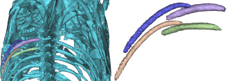
Codfish bone specimens. Annotations: S1-Short (lavender), S1-Long (blue), S2-Short (green), S2-Long (crčme).
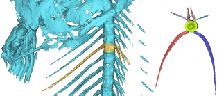
Salmon bone specimens. Annotations: Left rib (red), Right rib (blue), Left side bone (lavender), Right side bone (green), Left top bone (light pink), Right top bone (crčme).
The specific bone structures were harvested from the fish bodies. Disposable clinical scalpels were used for cutting. Afterwards the bones were soaked in 80 °C water to extract remaining flesh from the bones.
2.5 Measurements
All measurements on the model were performed using measurement tools in Mimics®. Quantitative measurements of the selected bones consist of:
Mass: all bones were weighted on the same scale with d = 0.1 mg/mg.
Length: Optical measurements by photographing the bones lying on a millimeter paper for accurate scaling in ImageJ, imaging processing software (see Figure 3). Estimated accuracy of 0.25 mm. The model length measurements are estimated to have less accuracy due to the rough surface of the modelled bones making it difficult to measure in a straight line over the surface. Estimated accuracy of 2.50 mm.
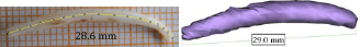
Length measurements.
Diameter: The diameters of the bones were measured with a micrometer screw gauge with a rated accuracy of 0.01 mm. The diameter was measured at three positions, at the bones fixation end, their middle and far end. Since the bones are not completely round the diameter was measured in two perpendicular directions, i.e. one in the direction of the length axis of the fish and other perpendicular to the length axis. It should be noted that the irregular shape of the bones slightly affects the accuracy of the diameter measurements.
3 Results
The 3D models of the skeletons are shown in Figure 4 and Figure 5. The specific bone structures selected for further analyzing are highlighted.

3D Model of codfish skeleton.

3D Model of the Salmon.
Mass and length: Results from mass and length measurements of the selected bones of the codfish are shown in Table 1 and selected bones of the salmon in Table 2.
Mass & Length Results for Codfish *(d=0.1mg/mg)
| Annotation | Mass* | Bone length | Model length | Difference | Difference |
|---|---|---|---|---|---|
| (g) | (mm) | (mm) | (mm) | (%) | |
| S1-Short | 0.1012 | 28.6 | 29.0 | 0.4 | 1.4% |
| S1-Long | 0.0420 | 36 | 43.98 | 7.98 | 22.2% |
| S2-Short | 0.1422 | 29.7 | 30.84 | 1.14 | 3.8% |
| S2-Long | 0.0531 | 38.7 | 43.06 | 4.36 | 11.3% |
Mass & Length Results for Salmon *(d=0.1mg/mg)
| Annotation | Mass* (g) | Bone length (mm) | Model length (mm) | Difference (mm) | Difference (%) |
|---|---|---|---|---|---|
| Left rib | 0.0197 | 67.9 | 53.35 | 14.55 | 21.4% |
| Right rib | 0.0172 | 66.2 | 54.31 | 11.89 | 18.0% |
| Left side | 0.0076 | 29.2 | 15.37 | 13.83 | 47.4% |
| Right side | 0.0067 | 32.8 | 14.33 | 18.47 | 56.3% |
| Left top | 0.0473 | 27.7 | 15.79 | 11.91 | 43.0% |
| Right top | 0.0428 | 28.5 | 15.61 | 12.89 | 45.2% |
Diameter: Comparisons of measured diameter with the millimeter screw to diameter measurements of the model are shown in the Table 3 and Table 4. The tables show results at three positions measured both in the direction of the length axis of the fish and perpendicular to the length axis.
Diameter Comparisons of Selected Bones in Codfish
| Measured over | Annotation | Fixation end (mm) | Middle (mm) | Far end (mm) |
|---|---|---|---|---|
| Sides | S1-Short | 1.41 | 1.38 | 0.58 |
| Sides | S1-Long | 1.68 | 1.41 | 1.18 |
| Sides | S2-Short | 0.85 | 0.08 | 0.29 |
| Sides | S2-Long | 1.26 | 0.69 | 1.06 |
| Arch | S1-Short | 0.8 | 0.68 | 0.45 |
| Arch | S1-Long | 1 | 1.43 | 1.04 |
| Arch | S2-Short | 0.52 | 0.99 | 0.48 |
| Arch | S2-Long | 0.22 | 1.1 | 0.57 |
Diameter Comparisons of Selected Bones in Salmon
| Measured over | Annotation | Fixation end (mm) | Middle (mm) | Far end (mm) |
|---|---|---|---|---|
| Sides | Left rib | 0.7 | 0.61 | 0.04 |
| Sides | Right rib | 0.34 | 0.6 | −0.04 |
| Sides | Left side bone | 0.24 | 0.34 | 0.28 |
| Sides | Right side bone | 0.26 | 0.51 | 0.09 |
| Sides | Left top bone | 1.2 | 0.79 | 0.42 |
| Sides | Right top bone | 0.96 | 0.55 | −0.16 |
| Arch | Left rib | −0.22 | 0.33 | −0.16 |
| Arch | Right rib | −0.67 | 0.15 | 0.06 |
| Arch | Left side bone | 0.14 | 0.51 | 0.17 |
| Arch | Right side bone | −0.18 | 0.47 | 0.12 |
| Arch | Left top bone | 0.75 | 0.00 | 0.24 |
| Arch | Right top bone | 0.20 | 0.27 | −0.05 |
4 Discussion and conclusion
The thin bone structures of the codfish and salmon are detected with a clinical CT and can be modeled. This is useful for detection of small bone fractures or thin bone structures.
It is interesting to see that the specimen bones of the codfish are thicker than of the salmon. The result show significantly larger difference in modeled parameters and measured ones for the salmon than the codfish specimen. Our results show ability to reconstruct closely the diameter of the codfish bones but to a less extent the small bones of the salmon.
Our results indicate that very thin bone structures are missing from the models due to the small thickness or low density resulting from the partial-volume-effect. Furthermore it is likely that edges of thin bone structures that are close together have merged and the structures have been modeled as a single piece. This can be improved by using a scanner with higher spatial resolution since it influences the representation of edges between objects of different densities, decreasing the field of view would also contribute to the improvement. Additionally the selection of appropriate threshold is essential to the accuracy of the geometrically segmented model.
In terms of accuracy of our models we can see that the codfish model is very close to measured values of the specimen bones (9.7 ± 9.3%), however the level of accuracy for the salmon model decreases significantly (38.5 ± 15.3%). Again this indicates that very thin bones structures or of very low densities are not completely detectable with a clinical CT. To further analyze our model it would be interesting to determine the levels of densities of the bones and compare it to the observed HU in the model. Further research on the subject is ongoing.
Acknowledgment
Special thanks to Haraldur Auđunsson for the codfish and to the company Rifós hf. for the salmon.
Author’s Statement
Conflict of interest: Authors state no conflict of interest. Material and Methods: Informed consent: Informed consent has been obtained from all individuals included in this study. Ethical approval: The research related to human use has been complied with all the relevant national regulations, institutional policies and in accordance the tenets of the Helsinki Declaration, and has been approved by the authors’ institutional review board or equivalent committee.
References
[1] Hangartner TN., Short DF. Accurate Quantification of Width and Density of Bone Structures by Computed Tomography. In: Med Phys. 2007;34(10):3777-3784.10.1118/1.2769102Suche in Google Scholar
[2] Rho JY, Hobatho MC, Ashman RB. Relations of Mechanical Properties to Density and CT Numbers in Human Bone. In: Med Eng Phys. 1995;17(5):347-355.10.1016/1350-4533(95)97314-FSuche in Google Scholar
[3] T. Helgason, R.A. Gudmundsdottir, K.L. Valtysdottir, K. Andersen. Detecting Thin Bones and Modeling Cod Skeleton. In: Proceedings of the European Modeling and Simulation Symposium, 2012; 163-168.Suche in Google Scholar
[4] T. Helgason, P. Gargiulo, S. Knútsdóttir, V. Gudmundsdottir, H. Kern, U. Carraro, P. Ingvarsson, S. Yngvason, 2011. “Monitoring Muscle Growth and Tissue Changes Induced by Electrical Stimulation of Denervated Degenerated Muscles with CT and Stereolithographic 3D Modelling.” In: Narayan, R, Calvert, P, eds. Computer Aided Biomanufacturing. Weinheim Germany, Wiley-VCH, 130-146Suche in Google Scholar
[5] P. Gargiulo, U. Carraro, T. Mandl, H. Kern, S. Zampieri, W. Mayr, T. Helgason, 2012. Anthropometry of Human Muscle Using Segmentation Techniques and 3D Modelling: Applications to Lower Motor Neuron Denervated Muscle in Spinal Cord Injury. In: Preedy, V.R., ed. Handbook af Anthropometry. Physical Measures of Human Form in Health and Disease. US:Springer, 323-35410.1007/978-1-4419-1788-1_18Suche in Google Scholar
[6] Johannesdottir F., 2006. Bone: use it or lose it. MS thesis. University of Iceland.Suche in Google Scholar
© 2015 by Walter de Gruyter GmbH, Berlin/Boston
This article is distributed under the terms of the Creative Commons Attribution Non-Commercial License, which permits unrestricted non-commercial use, distribution, and reproduction in any medium, provided the original work is properly cited.
Artikel in diesem Heft
- Research Article
- Development and characterization of superparamagnetic coatings
- Research Article
- The development of an experimental setup to measure acousto-electric interaction signal
- Research Article
- Stability analysis of ferrofluids
- Research Article
- Investigation of endothelial growth using a sensors-integrated microfluidic system to simulate physiological barriers
- Research Article
- Energy harvesting for active implants: powering a ruminal pH-monitoring system
- Research Article
- New type of fluxgate magnetometer for the heart’s magnetic fields detection
- Research Article
- Field mapping of ballistic pressure pulse sources
- Research Article
- Development of a new homecare sleep monitor using body sounds and motion tracking
- Research Article
- Noise properties of textile, capacitive EEG electrodes
- Research Article
- Detecting phase singularities and rotor center trajectories based on the Hilbert transform of intraatrial electrograms in an atrial voxel model
- Research Article
- Spike sorting: the overlapping spikes challenge
- Research Article
- Separating the effect of respiration from the heart rate variability for cases of constant harmonic breathing
- Research Article
- Locating regions of arrhythmogenic substrate by analyzing the duration of triggered atrial activities
- Research Article
- Combining different ECG derived respiration tracking methods to create an optimal reconstruction of the breathing pattern
- Research Article
- Atrial and ventricular signal averaging electrocardiography in pacemaker and cardiac resynchronization therapy
- Research Article
- Estimation of a respiratory signal from a single-lead ECG using the 4th order central moments
- Research Article
- Compressed sensing of multi-lead ECG signals by compressive multiplexing
- Research Article
- Heart rate monitoring in ultra-high-field MRI using frequency information obtained from video signals of the human skin compared to electrocardiography and pulse oximetry
- Research Article
- Synchronization in wireless biomedical-sensor networks with Bluetooth Low Energy
- Research Article
- Automated classification of stages of anaesthesia by populations of evolutionary optimized fuzzy rules
- Research Article
- Effects of sampling rate on automated fatigue recognition in surface EMG signals
- Research Article
- Closed-loop transcranial alternating current stimulation of slow oscillations
- Research Article
- Cardiac index in atrio- and interventricular delay optimized cardiac resynchronization therapy and cardiac contractility modulation
- Research Article
- The role of expert evaluation for microsleep detection
- Research Article
- The impact of baseline wander removal techniques on the ST segment in simulated ischemic 12-lead ECGs
- Research Article
- Metal artifact reduction by projection replacements and non-local prior image integration
- Research Article
- A novel coaxial nozzle for in-process adjustment of electrospun scaffolds’ fiber diameter
- Research Article
- Processing of membranes for oxygenation using the Bellhouse-effect
- Research Article
- Inkjet printing of viable human dental follicle stem cells
- Research Article
- The use of an icebindingprotein out of the snowflea Hypogastrura harveyi as a cryoprotectant in the cryopreservation of mesenchymal stem cells
- Research Article
- New NIR spectroscopy based method to determine ischemia in vivo in liver – a first study on rats
- Research Article
- QRS and QT ventricular conduction times and permanent pacemaker therapy after transcatheter aortic valve implantation
- Research Article
- Adopting oculopressure tonometry as a transient in vivo rabbit glaucoma model
- Research Article
- Next-generation vision testing: the quick CSF
- Research Article
- Improving tactile sensation in laparoscopic surgery by overcoming size restrictions
- Research Article
- Design and control of a 3-DOF hydraulic driven surgical instrument
- Research Article
- Evaluation of endourological tools to improve the diagnosis and therapy of ureteral tumors – from model development to clinical application
- Research Article
- Frequency based assessment of surgical activities
- Research Article
- “Hands free for intervention”, a new approach for transoral endoscopic surgery
- Research Article
- Pseudo-haptic feedback in medical teleoperation
- Research Article
- Feasibility of interactive gesture control of a robotic microscope
- Research Article
- Towards structuring contextual information for workflow-driven surgical assistance functionalities
- Research Article
- Towards a framework for standardized semantic workflow modeling and management in the surgical domain
- Research Article
- Closed-loop approach for situation awareness of medical devices and operating room infrastructure
- Research Article
- Kinect based physiotherapy system for home use
- Research Article
- Evaluating the microsoft kinect skeleton joint tracking as a tool for home-based physiotherapy
- Research Article
- Integrating multimodal information for intraoperative assistance in neurosurgery
- Research Article
- Respiratory motion tracking using Microsoft’s Kinect v2 camera
- Research Article
- Using smart glasses for ultrasound diagnostics
- Research Article
- Measurement of needle susceptibility artifacts in magnetic resonance images
- Research Article
- Dimensionality reduction of medical image descriptors for multimodal image registration
- Research Article
- Experimental evaluation of different weighting schemes in magnetic particle imaging reconstruction
- Research Article
- Evaluation of CT capability for the detection of thin bone structures
- Research Article
- Towards contactless optical coherence elastography with acoustic tissue excitation
- Research Article
- Development and implementation of algorithms for automatic and robust measurement of the 2D:4D digit ratio using image data
- Research Article
- Automated high-throughput analysis of B cell spreading on immobilized antibodies with whole slide imaging
- Research Article
- Tissue segmentation from head MRI: a ground truth validation for feature-enhanced tracking
- Research Article
- Video tracking of swimming rodents on a reflective water surface
- Research Article
- MR imaging of model drug distribution in simulated vitreous
- Research Article
- Studying the extracellular contribution to the double wave vector diffusion-weighted signal
- Research Article
- Artifacts in field free line magnetic particle imaging in the presence of inhomogeneous and nonlinear magnetic fields
- Research Article
- Introducing a frequency-tunable magnetic particle spectrometer
- Research Article
- Imaging of aortic valve dynamics in 4D OCT
- Research Article
- Intravascular optical coherence tomography (OCT) as an additional tool for the assessment of stent structures
- Research Article
- Simple concept for a wide-field lensless digital holographic microscope using a laser diode
- Research Article
- Intraoperative identification of somato-sensory brain areas using optical imaging and standard RGB camera equipment – a feasibility study
- Research Article
- Respiratory surface motion measurement by Microsoft Kinect
- Research Article
- Improving image quality in EIT imaging by measurement of thorax excursion
- Research Article
- A clustering based dual model framework for EIT imaging: first experimental results
- Research Article
- Three-dimensional anisotropic regularization for limited angle tomography
- Research Article
- GPU-based real-time generation of large ultrasound volumes from freehand 3D sweeps
- Research Article
- Experimental computer tomograph
- Research Article
- US-tracked steered FUS in a respiratory ex vivo ovine liver phantom
- Research Article
- Contribution of brownian rotation and particle assembly polarisation to the particle response in magnetic particle spectrometry
- Research Article
- Preliminary investigations of magnetic modulated nanoparticles for microwave breast cancer detection
- Research Article
- Construction of a device for magnetic separation of superparamagnetic iron oxide nanoparticles
- Research Article
- An IHE-conform telecooperation platform supporting the treatment of dementia patients
- Research Article
- Automated respiratory therapy system based on the ARDSNet protocol with systemic perfusion control
- Research Article
- Identification of surgical instruments using UHF-RFID technology
- Research Article
- A generic concept for the development of model-guided clinical decision support systems
- Research Article
- Evaluation of local alterations in femoral bone mineral density measured via quantitative CT
- Research Article
- Creating 3D gelatin phantoms for experimental evaluation in biomedicine
- Research Article
- Influence of short-term fixation with mixed formalin or ethanol solution on the mechanical properties of human cortical bone
- Research Article
- Analysis of the release kinetics of surface-bound proteins via laser-induced fluorescence
- Research Article
- Tomographic particle image velocimetry of a water-jet for low volume harvesting of fat tissue for regenerative medicine
- Research Article
- Wireless medical sensors – context, robustness and safety
- Research Article
- Sequences for real-time magnetic particle imaging
- Research Article
- Speckle-based off-axis holographic detection for non-contact photoacoustic tomography
- Research Article
- A machine learning approach for planning valve-sparing aortic root reconstruction
- Research Article
- An in-ear pulse wave velocity measurement system using heart sounds as time reference
- Research Article
- Measuring different oxygenation levels in a blood perfusion model simulating the human head using NIRS
- Research Article
- Multisegmental fusion of the lumbar spine a curse or a blessing?
- Research Article
- Numerical analysis of the biomechanical complications accompanying the total hip replacement with NANOS-Prosthetic: bone remodelling and prosthesis migration
- Research Article
- A muscle model for hybrid muscle activation
- Research Article
- Mathematical, numerical and in-vitro investigation of cooling performance of an intra-carotid catheter for selective brain hypothermia
- Research Article
- An ideally parameterized unscented Kalman filter for the inverse problem of electrocardiography
- Research Article
- Interactive visualization of cardiac anatomy and atrial excitation for medical diagnosis and research
- Research Article
- Virtualizing clinical cases of atrial flutter in a fast marching simulation including conduction velocity and ablation scars
- Research Article
- Mesh structure-independent modeling of patient-specific atrial fiber orientation
- Research Article
- Accelerating mono-domain cardiac electrophysiology simulations using OpenCL
- Research Article
- Understanding the cellular mode of action of vernakalant using a computational model: answers and new questions
- Research Article
- A java based simulator with user interface to simulate ventilated patients
- Research Article
- Evaluation of an algorithm to choose between competing models of respiratory mechanics
- Research Article
- Numerical simulation of low-pulsation gerotor pumps for use in the pharmaceutical industry and in biomedicine
- Research Article
- Numerical and experimental flow analysis in centifluidic systems for rapid allergy screening tests
- Research Article
- Biomechanical parameter determination of scaffold-free cartilage constructs (SFCCs) with the hyperelastic material models Yeoh, Ogden and Demiray
- Research Article
- FPGA controlled artificial vascular system
- Research Article
- Simulation based investigation of source-detector configurations for non-invasive fetal pulse oximetry
- Research Article
- Test setup for characterizing the efficacy of embolic protection devices
- Research Article
- Impact of electrode geometry on force generation during functional electrical stimulation
- Research Article
- 3D-based visual physical activity assessment of children
- Research Article
- Realtime assessment of foot orientation by Accelerometers and Gyroscopes
- Research Article
- Image based reconstruction for cystoscopy
- Research Article
- Image guided surgery innovation with graduate students - a new lecture format
- Research Article
- Multichannel FES parameterization for controlling foot motion in paretic gait
- Research Article
- Smartphone supported upper limb prosthesis
- Research Article
- Use of quantitative tremor evaluation to enhance target selection during deep brain stimulation surgery for essential tremor
- Research Article
- Evaluation of adhesion promoters for Parylene C on gold metallization
- Research Article
- The influence of metallic ions from CoCr28Mo6 on the osteogenic differentiation and cytokine release of human osteoblasts
- Research Article
- Increasing the visibility of thin NITINOL vascular implants
- Research Article
- Possible reasons for early artificial bone failure in biomechanical tests of ankle arthrodesis systems
- Research Article
- Development of a bending test procedure for the characterization of flexible ECoG electrode arrays
- Research Article
- Tubular manipulators: a new concept for intracochlear positioning of an auditory prosthesis
- Research Article
- Investigation of the dynamic diameter deformation of vascular stents during fatigue testing with radial loading
- Research Article
- Electrospun vascular grafts with anti-kinking properties
- Research Article
- Integration of temperature sensors in polyimide-based thin-film electrode arrays
- Research Article
- Use cases and usability challenges for head-mounted displays in healthcare
- Research Article
- Device- and service profiles for integrated or systems based on open standards
- Research Article
- Risk management for medical devices in research projects
- Research Article
- Simulation of varying femoral attachment sites of medial patellofemoral ligament using a musculoskeletal multi-body model
- Research Article
- Does enhancing consciousness for strategic planning processes support the effectiveness of problem-based learning concepts in biomedical education?
- Research Article
- SPIO processing in macrophages for MPI: The breast cancer MPI-SNLB-concept
- Research Article
- Numerical simulations of airflow in the human pharynx of OSAHS patients