SPIO processing in macrophages for MPI: The breast cancer MPI-SNLB-concept
-
Dominique Finas
 ,
Stegmann-Frehse Janine
,
Stegmann-Frehse Janine

Abstract
Introduction
Breast cancer (BC) is the most common cancer in women worldwide. We aim to develop a new sentinel lymph node biopsy (SLNB) method with superparamagnetic iron oxide nanoparticles (SPIOs) and magnetic particle imaging (MPI) in BC to avoid tissue damaging while axillary surgery. As we know from i.v. SPIO application in magnetic resonance imaging (MRI), macrophages (MP) are key role player in processing of SPIOs (e.g. in liver) causing a drop of signal intensity. But, knowledge lacks concerning enrichment processes of SPIOs after injection in breast tissue, the adjacent lymphatic tissues and associated cells, especially in BC and metastatic lymph nodes. We already evaluated the distribution of SPIOs in an in vivo healthy and tumor mouse model. Based on these studies we investigate the processing of the SPIOs in MP.
Material and Methods
To evaluate SPIO processing, a mouse MP cell line J774A.1 was incubated either by Resovist in culture medium (RPMI, FBS), or culture medium only as control. MP were than analyzed by transmission electron microscopy (TEM). Additionally, this process was observed in vivo by multiphoton microscopy. Detection of SPIOs was realized by excitation at 1200 nm.
Results
Resovist had no toxic effects on cells.MP showed activity in phagocytosis of Resovist after incubation in TEM as well as in multiphoton microscopy. SPIOs were detectable within intracellular vesicles by TEM and 3-photon process. The first cell associated SPIO signal was detected after 1,5 min of incubation by in vivo imaging.
Conclusion
To our knowledge this is the first time a 3-photon device was used to image SPIOs in a bio-medical context. System wide scanning is known (MRI, MPI), but nowwe are also able to identify the link to subcellular processing and localization of SPIOs. Further processing of SPIOs in MP is under development.
1 Introduction
Breast cancer (BC) is the most common cancer in women in the world in western industrialized countries [1]. BC Tumors are draining from the breast tissue to the axillary lymph nodes. Exploration of the axillary region, therefore is part of the surgical staging in breast cancer [2]. If tumor cells are histopathologically present in the extracted axillary lymph nodes, individual cancer prognosis decreases dramatically. The conventional radical axillary lymph node extraction (ALNE) for surgical staging in breast cancer is associated with high morbidity and significant loss of quality of life (QoL). These adverse e_ects were strongly reduced by introducing the concept of the so called sentinel lymph node biopsy (SLNB). Tracer substances like dyes and radio nuclides are used to mark the sentinel lymph nodes (SLNs) within this SLNB method, and are therefore injected into the breast. After a predefined waiting time, enrichment of the tracer is completed in the SLN which can then be localized intra operative as radioactive hot spots by a mobile gamma hand probe and then extracted surgically [3].
The use of dyes and radio nuclides together for detection of SLNs increases safety in the diagnosis of lymph node involvement through less false negative results [4]. Before starting the surgical procedure a static lymph scintigraphy is demanded for intra operative correlation of the detectable SNLs [2]. Unfortunately, the procedure with combination of dyes and radio nuclides has a longer learning curve than radioactive procedure alone, because of the additional intra operative use of dye [5]. Moreover, this procedure is time consuming within the surgical procedure, as scanning of the axillary region for SLNs by hand probe for radioactivity and visually scanning for blue traces (lymph vessels to lymph nodes) and blue lymph nodes takes time. Axillary morbidity increases particularly when multiple lymph nodes are labeled by radioactivity and/or dye. Moreover, esthetic problems may occur, because blue dyes can generate permanent tattooing skin effects.
We aim to develop a new sentinel lymph node biopsy concept by the use of superparamagnetic iron oxide nanoparticles (SPIOs) and a three-dimensional imaging and distinct localization of SPIOs by magnetic particle imaging (MPI) [6] in breast cancer to decrease tissue damage while diagnostic surgery of the axilla and to increase patients QoL and safety.
There are SPIOs approved specifically for MR imaging (e.g. ferucarbotran, Resovist®). After intravenous bolus administration uptake is observed in lymph nodes [7]. Nano-sized particles with superparamagnetic iron oxide, coated by carboxydextran could replace the marker substances previously described above within the new SLN procedure. So far, few local and systemic adverse effects were noted when SPIOs like Resovist® were used within MRI procedures [7, 8].
As we know from i.v. application of SPIOs in magnetic resonance imaging (MRI), macrophages are playing a key role in processing of SPIOs (e.g. in the liver) causing a drop of signal intensity [9–12]. But, knowledge lacks concerning enrichment processes of SPIOs after injection in breast tissue, the adjacent lymphatic tissues and associated cells, especially in breast cancer and metastatic lymph nodes. Therefore, tissue and cell associated processing of SPIOs is of major interest for this SLNB concept. Based on our own preliminary studies in healthy and tumor mouse model [13] we investigate the processing of SPIOs by and in macrophages for the first time [14].
To investigate those SPIO associated processes in active macrophages (MPs), life imaging by multiphoton laser microscopy seems to be a feasible method. Due to the development Ti.sapphire fs lasers in the last 10 years, multiphoton-microscopy has become a popular method in life sciences. Its ability to excite a broad variety of intrinsic fluorescent proteins such as FAD [15, 16], NADH [17] or serotonin [18] without additional staining and without extensive tissue damage is ideal for in vivo applications. Also the combined use with genetical engineered fluorescent proteins has been shown as well as the applicability to standard dyes as for example DAPI in histology or Fluo 4 for calcium measurements. It was also shown that multiphoton microscopy is able to track quantum dots in the colon to investigate the uptake of nanoparticles by the intestines.
Transmission electron microscopy (TEM) is a methodological standard for imaging of subcellular processes after fixation of biological materials [19, 20]. Therefore, it seems to be feasible for approval of in vivo results of multiphoton laser microscopy.
2 Material and Methods
2.1 Macrophages
A commercially available mouse monocyte macrophage cell line J774A.1 (BALB/c) was attached to thermanox™ coverslips to growth, than incubated in culture medium (RPMI 1640 (Roswell Park Memorial Institute), 20% fetal bovine serum (FBS), 1% Penicillin/Streptomycin) and further processed, as described follows.
2.1.1 Preparation of MPs for TEM
Pre-incubatad MPs were washed and further processed over 24 hrs either by 100 μl/mg Resovist-Fe (0.5 mmol/l) in culture medium (RPMI 1640, 20% FBS, 1% Penicillin/ Streptomycin), or culture medium only as control.
After incubation, the cells were washed with phosphate buffered saline (PBS) and fixed in paraformaldehyd (Monti Graziadei fixative: 156 ml natrium-cacodylat-buffer 0.12 M; 25 ml glutaraldehyd 25%; 19 ml paraformaldehyd 10%; 3 ml CaCl2, 3%; pH 7.35 accumulated to 312 ml with Aqua bidest [21]). Macrophages were than analyzed by TEM and phagocytosis of SPIOs was evaluated.
2.1.2 Preparation of MPs for multiphoton microscopy
Pre-incubatad MPs were washed and further processed in heated 6 well plates with 1 × 10⁵ cells per well for immediate in vivo laser procedure. Cells were analyzed directly after incubation either by 100 μl/mg Resovist-Fe (0.5 mmol/l) in culture medium (RPMI 1640, 20% FBS, 1% Penicillin/Streptomycin), or culture medium only as control with live cell imaging.
2.2 Microscope and Lasers
2.2.1 Transmission electron microscope
For TEM imaging, we used a standard transmission electron microscope: JEOL 1011 TEM, Jeol Germany GmbH, Eching. nominal acceleration voltage 80–120 kV.
2.2.2 Multiphoton laser scanning microscope
The multiphoton microscope used is an upright TriM Scope 2 (LaVision Biotec, Bielefeld Germany), with an adapter for an Olympus XLPLN25XWMP2 objective 25× NA 1.05 (Olympus, Tokyo, Japan). The detection is realised with H7422-40 photo multiplier tubes (Hamamatsu, Hamamatsu, Japan) with GaAsP-photocathodes, implemented for optimum quantum effciency in the spectral range from 435 nm to 560 nm. For the detection of the third harmonic below 435 nm a Hamamatsu PMT with an alkali cathode is used. The collected signal from the sample is separated from the excitation light by a 700nmbeam splitter. Afterwards three different dichroic mirrors divide the signal into 4 spectral channels. Below 435 nm, 435 nm to 495 nm, 495 nm to 560 nm and light above 560 nm.
Excitation is realised with two Mai Tai tuneable Ti:sapphire lasers (SpectraPhysics) with a repetition rate of 80 MHz and pulse lengths of about 100 fs. The MaiTai are combined with a 850 nm beam splitter before entering a beam shaper to provide a pre compensation for the dispersion of the fs pulses in the optics of the microscope.
As a third laser source a Insight Deep See (Spectra Physics) broad bandwidth tuneable laser is used. The Insight DS is coupled onto the same beam path as the MaiTais insight the scan head of the TriM 2 with a 1045 nm dichroic mirror. The DS supplies laser pulses at a rate of 80 MHz with about 100 fs length up to 1300 nm.
Three pockels cells control laser power at the sample. Exact confocality for all lasers is checked with fluorescent poly styrole beads of 50 nm size.
2.2.3 Fluorescence Lifetime Imaging Microscopy
The fluorescence lifetime imaging microscopy (FLIM) setup used in our lab is an simple tau 150 with a HPM-100-40 detector (Becker und Hickl GmbH, Berlin, Germany). The software for data acquisition is the proprietary Becker and Hickl SPC Software. The detector module can be easily combined with the filter box module of the TriM Scope 2. Synchronisation with the excitation lasers is done with the MaiTais sync output or a fast photo diode for the Insight DS respectively.
FLIM is a method that allows distinguishing of different fluorescent species by their fluorescence lifetime. Every excited fluorescent molecules excited state has a characteristic halftime. This time can be modified by ambient conditions like pH or calcium concentration or presence of another excitable molecule. This usually is implemented to measure these conditions. On the other hand FLIM can distinguish fluorescence from coherent processes like second harmonic generation (SHG) or third harmonic generation (THG). The limiting factor for this in time correlated single photon counting is the instrument response function (IRF), which is the response of the electronics to the detection of a photon.
The FLIM data was recorded with 100 frames (at 0.6 frames per second with 1.6 μs pixel dwell time) summed up, to get the necessary threshold of photons for fluorescence lifetime fitting. The whole collected fluorescence was directed to the detectors. A 50/50 beam splitter allowed simultaneous image acquisition with the GaAsP PMT of the TriM Scope 2.
2.3 Data collection and software
The cells are imaged under epi-illumination while, submerged in Dulbeccos modified eagles medium. The microscope objective dips into the medium.
The scanning is done at a speed of 1000 lines per second with a pixel dwell time depending on the chosen pixel size of the images taken. PMT amplification is usually set to 90% for the GaAsP detectors and to 95% for the alkali detector.
The laser power for the Insight DS during measurements is about 50mWat the sample for 1200 nm. For most measurements one MaiTai tuned to 740 nm is used to excited intrinsic fluorescent proteins. Laser power at 740 nm is kept below 10mWat all times to limit photo damage and production of photo toxic by products.
The software to collect the data and control the microscope as well as the MaiTai lasers is the Inspector application (32bit version for Windows XP) provided by LaVision Biotec. The Insight DS is controlled by a separate Laptop with Spectra Physics control software.
3 Results
We are presenting our first results of processing of SPIOs by MPs, which showed activity in phagocytosis of Resovist after incubation in TEM as well as in multiphoton microscopy.
SPIOs were detectable within intracellular vesicles by TEM. Macrophages were incorporating the SPIOs after incubation with Resovist. First, SPIOs are attaching at the cellular membrane, than a phagocytic vesicle was formed and SPIOs were incorporated actively by the cells subsequently. These intracellular phagocytic vesicles are clearly shown by TEM as phagolysosomes (Figure 1 and 2).
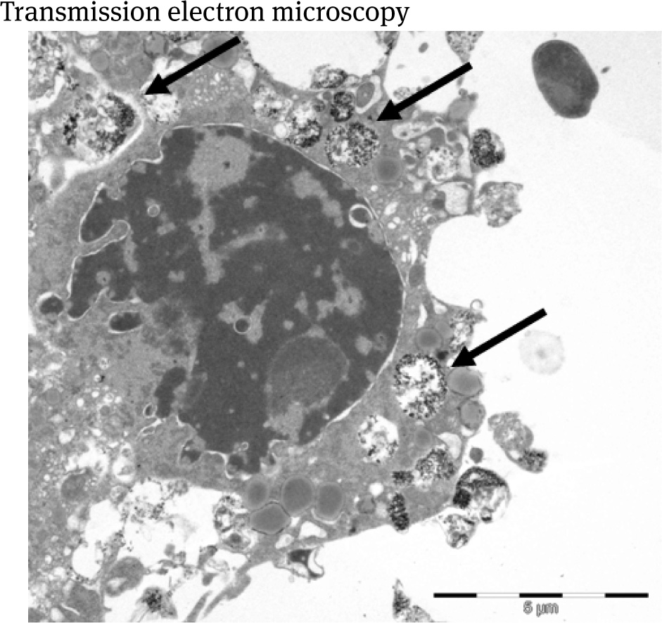
Macrophage presenting SPIOs within phagolysosomes (arrows) after incubation by 100 μl/mg Resovist-Fe over 24 hrs. The nucleus appears to be structurally normal. Bar = 5 μm.
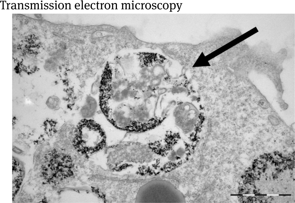
Magni_cation of an intra cellular MP phagolysosome (arrow) presenting SPIOs after incubation by 100 μl/mg Resovist-Fe over 24 hrs. Bar = 1 μm
Live cell imaging showed that Resovist had no toxic effects on MPs, even after 24 hrs of incubation within the TEM experiment.
Thus, in vivo analysis of living cells by multiphoton microscopy showed at an excitation of 1200 nm, that Resovist was clearly visualized inside the MPs in the channel below 435 nm. The excitation was changed in a range from 1100 nm to 1300 nm to check out optimal excitation wavelength. It peaked at a wavelength of about 1200 nm with a small bandwidth. Experiments with naive cells for control revealed no detectable signal in the spectral range below 435 nm by multiphoton microscopy (Figure 3).
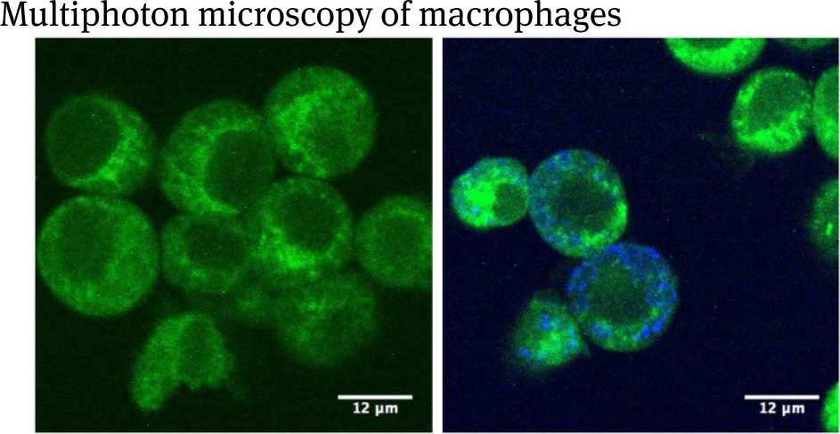
Left side: SPIO nadve macrophages; right side: macrophages incubated with Resovist-Fe. Bar = 12 μm. Cells are detected with by signal within the spectral range of 435– 495 nm in epi-direction with an excitation at 740 nm (depicted in green). Intra cellular SPIOs are shown by a signal with a detector for <435 nm, excited at 1200 nm (depicted in blue).
The incubation medium of the cells was changed to SPIO containing medium immediately before the start of continuous multiphoton microscopy scanning with 7.7 s frame intervall to avoid uptake artefacts.
Recordings were done for 760 nm and 1200 nm excitation separately because the laser pulses from MaiTai and Insight DS are not synchronous. The first signal of Resovist was detected after a minimum of 1.5 minutes after adding the iron nanoparticles containing medium. Figure 4 shows the colour-coded image for 760 nm.Mean fluorescence lifetime was determined to 1.36 ns.
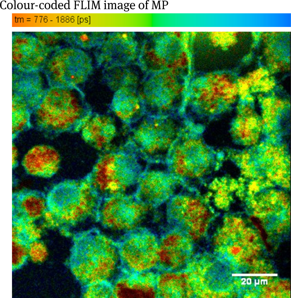
Colour-coded FLIM image of MP loaded with Resovist-Fe, excited at 760 nm
Figure 5 shows the FLIM image of the same cells with 1200 nm excitation. The mean fluorescence lifetime was determined to 0.112 ns.
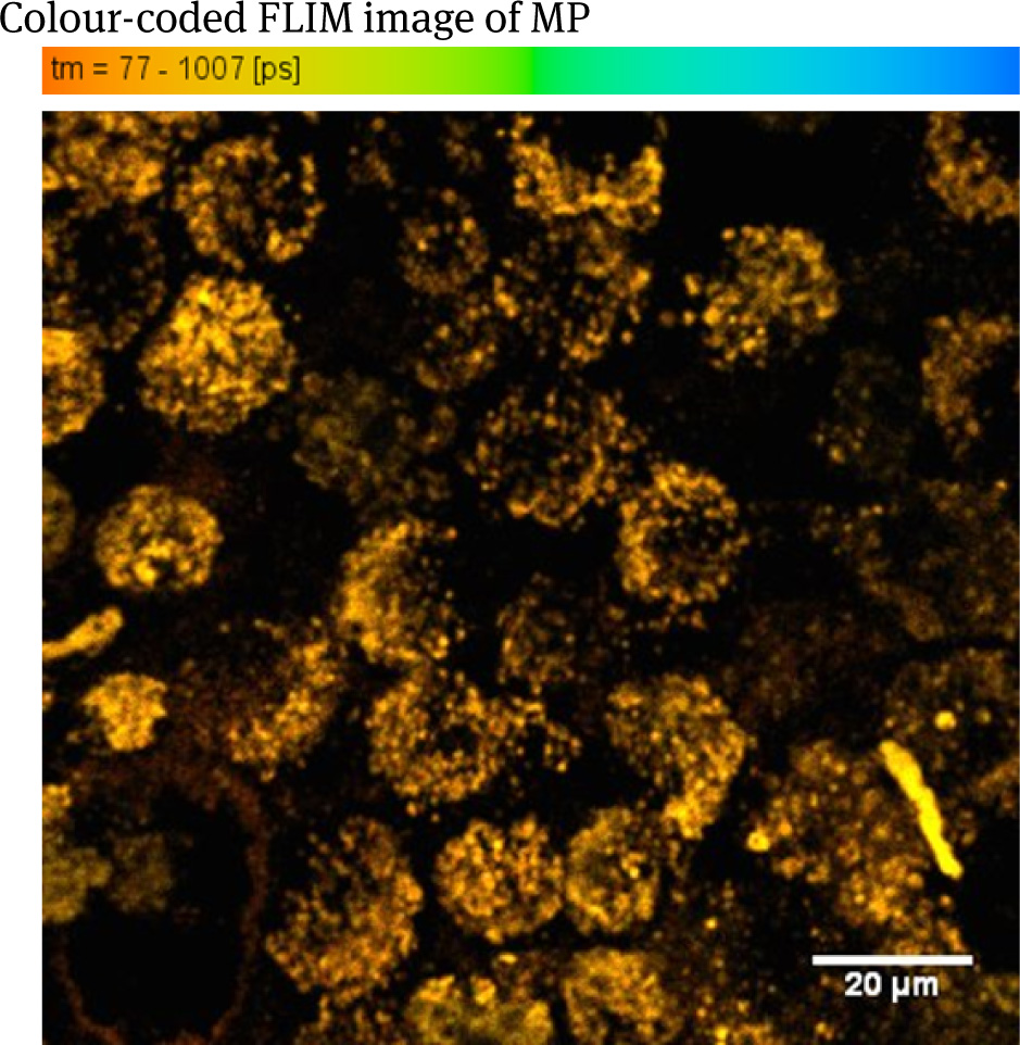
Colour-coded FLIM image MP loaded with Resovist, excited at 1200 nm.
4 Discussion
Axillary lymph node detection and extraction are still part of the standard staging procedure in breast cancer diagnosis. To leave the primary radical axillary lymph node extraction procedure the sentinel concept was established to avoid severe side effects. But, even for conventional SLNB the axilla has to be widely explored to identify the SLN. To avoid further surgically caused damage of the axillary region, an intra operative tracer targeted imaging and surgically extraction of the SNL is needed. This would be a relevant improvement in QoL for the patient and could be achieved by the use of SPIO as tracer and MPI as finder [22, 23]. Our own previous findings showed that SPIO detection within tissue is feasible and may be performed withMPI in future scenario [14]. Through the avoidance of intensive surgical exploration of the axilla, may be by using a MPI hand probe later on [24], the morbidity could be dramatically reduced. The tracer forMPI is easy to obtain. This would make the method accessible to all patients [25]. Furthermore, the SLNB-MPI concept could be applied in principle in all solid tumors if the method has proved its effectiveness under real life conditions.
Nanoparticles are widely discussed as environmental toxins.Unfortunately, knowledge lacks concerning enrichment processes of SPIOs after injection in breast tissue, the adjacent lymphatic tissues and associated cells, especially in BC and metastatic lymph nodes. We already evaluated the distribution of SPIOs in an in vivo healthy and tumor mouse model [26]. Based on these studies we further investigate the processing of the SPIOs in MP.
To our knowledge this is the first time three photon process was used to image superparamagnetic iron oxide nanoparticles in a bio-medical context. The small excitation spectrum as well as the wavelength range of the signal suggests third harmonic generation at the nanoparticle clusters, which can also be seen in the TEM images. Since single nano-particles do not generate a detectable signal, clustering size seems to be directly connected to the conversion effciency.
More experiments are necessary to confirm THG at the nanoparticles. A strong indication might be the fluorescence lifetime of the signal. A frequency tripling would have no detectable lifetime different from the instrument response function of the lifetime measurement device.
The lifetime of the Resovist signal recorded at 1200nm excitation is as short as the IRF of the setup recorded with SHG measurements with Urea cristals excited with 920 nm excitation. This is a strong indication that Resovist particle clusters are a source of THG. Similarly short lifetimes can be recorded with the fluorescence of gold nanoparticles. In contrast to those the excitation and emission spectra of the SPIOs have a much smaller FWHM.Gold nanoparticles can be excited with 740 nm to 1200 nm laser light and detected in the whole spectrum from 435 nm to above 560 nm. The SPIOs used can only be excited around 1200 nm and the spectrum of the signal can only be detected in the spectral channel below 435 nm.
5 Conclusion
This project is part of a comprehensive test program to develop a new SLNB technique. This might be less complex and incriminating for the patient and the staff, especially if the new MPI hand probe with unilateral solenoid arrangement will be ready for use in the operating theater [22, 27, 28].
The multiphoton based imaging of the SPIO clusters enables us to detect the path of the nanoparticles in histological slices as well as in vivo with a micro-meter resolution. In combination with the system wide scanning methods asMRT orMPI we are now able not only to identify hot spots of the surface modified particles but also to identify the linkage to subcellular handling and localisation of the iron nano-particles. In combination with the broad spectrum of other molecular biological methods the method of multiphoton based imaging will help to further investigate SPIO processing in cells and in the whole body. Since, SPIOs seems not to be toxic to our cells, we conclude that there will not be unexpected toxic effects in the whole organism. But, we are now encouraged to bring the proof for this hypothesis in further experiments.
Author’s Statement
Conflict of interest: Authors state no conflict of interest. Material and Methods: Informed consent: Informed consent is not applicable. Ethical approval: The conducted research is not related to either human or animals use.
Acknowledgment
This work was supported by the German Federal Ministry of Education and Research (BMBF) under Grant 01EZ0912 and by the University Research Program "Imaging of Disease Processes", University of Lübeck.
References
[1] Jemal, A., Siegel, R., Ward, E., Hao, Y., Xu, J., Murray, T., and Thun, M.J.: ‘Cancer statistics, 2008’, CA: a cancer journal for clinicians, 2008, 58, (2), pp. 71-9610.3322/CA.2007.0010Suche in Google Scholar
[2] Kuehn, T., Bembenek, A., Decker, T., Munz, D.L., Sautter-Bihl, M.L., Untch, M., and Wallwiener, D.: ‘A concept for the clinical implementation of sentinel lymph node biopsy in patients with breast carcinoma with special regard to quality assurance’, Cancer, 2005, 103, (3), pp. 451-46110.1002/cncr.20786Suche in Google Scholar
[3] Kühn, T., Bembenek, A., Büchels, H., Decker, T., Dunst, J., Müllerleile, U., Munz, D.L., Ostertag, H., Sautter-Bihl, M.L., Schirrmeister, H., Tulusan, A.H., Untch, M., Winzer, K.J., and Wittekind, C.: ‘Sentinel-Node-Biopsie beim Mammakarzinom: Interdisziplinär abgestimmter Konsensus der Deutschen Gesellschaft für Senologie für eine qualitätsgesicherte Anwendung in der klinischen Routine’, Geburtsh Frauenheilk, 2003, 63, pp. 835-840.10.1055/s-2003-42576Suche in Google Scholar
[4] Heuser, T., Rink, T., Weller, E., Fitz, H., Zippel, H.H., Kreienberg, R., and Kuhn, T.: ‘Impact of the axillary nodal status on sentinel node mapping in breast cancer and its relevance for technical proceeding’, Breast Cancer Res Treat, 2001, 67, (2), pp. 125-13210.1023/A:1010619223296Suche in Google Scholar
[5] Morrow, M., Rademaker, A.W., Bethke, K.P., Talamonti, M.S., Dawes, L.G., Clauson, J., and Hansen, N.: ‘Learning sentinel node biopsy: results of a prospective randomized trial of two techniques’, Surgery, 1999, 126, (4), pp. 714-720; discussion 720-71210.1016/S0039-6060(99)70127-3Suche in Google Scholar
[6] Gleich, B., and Weizenecker, J.: ‘Tomographic imaging using the nonlinear response of magnetic particles’, Nature, 2005, 435, (7046), pp. 1214-121710.1038/nature03808Suche in Google Scholar PubMed
[7] Wang, Y.X.: ‘Superparamagnetic iron oxide based MRI contrast agents: Current status of clinical application’, Quantitative imaging in medicine and surgery, 2011, 1, (1), pp. 35-40Suche in Google Scholar
[8] BayerSheringPharma: ‘Resovist (Fachinformation)’, 2007, pp. 1-3Suche in Google Scholar
[9] Saksena, M., Harisinghani, M., Hahn, P., Kim, J., Saokar, A., King, B., and Weissleder, R.: ‘Comparison of lymphotropic nanoparticle-enhanced MRI sequences in patients with various primary cancers’, AJR Am J Roentgenol, 2006, 187, (6), pp. W582-58810.2214/AJR.05.0873Suche in Google Scholar PubMed
[10] Harisinghani, M.G., Barentsz, J., Hahn, P.F., Deserno, W.M., Tabatabaei, S., van de Kaa, C.H., de la Rosette, J., and Weissleder, R.: ‘Noninvasive detection of clinically occult lymph-node metastases in prostate cancer’, N Engl J Med, 2003, 348, (25), pp. 2491-249910.1056/NEJMoa022749Suche in Google Scholar PubMed
[11] Anzai, Y., McLachlan, S., Morris, M., Saxton, R., and Lufkin, R.B.: ‘Dextran-coated superparamagnetic iron oxide, an MR contrast agent for assessing lymph nodes in the head and neck’, AJNR. American journal of neuroradiology, 1994, 15, (1), pp. 87-94Suche in Google Scholar
[12] Anzai, Y., Piccoli, C.W., Outwater, E.K., Stanford, W., Bluemke, D.A., Nurenberg, P., Saini, S., Maravilla, K.R., Feldman, D.E., Schmiedl, U.P., Brunberg, J.A., Francis, I.R., Harms, S.E., Som, P.M., Tempany, C.M., and Group: ‘Evaluation of neck and body metastases to nodes with ferumoxtran 10-enhanced MR imaging: phase III safety and e_cacy study’, Radiology, 2003, 228, (3), pp. 777-78810.1148/radiol.2283020872Suche in Google Scholar PubMed
[13] Finas, D., Baumann, K., Sydow, L., Heinrich, K., Grafe, K., Rody, A., Ludtke-Buzug, K., and Buzug, T.: ‘Lymphatic Tissue and Superparamagnetic Nanoparticles - Magnetic Particle Imaging for Detection and Distribution in a Breast Cancer Model’, Biomedizinische Technik. Biomedical engineering, 2013, 5810.1109/IWMPI.2013.6528390Suche in Google Scholar
[14] Finas, D., Stegmann-Frehse, J., Sauer, B., Hüttmann, G., Rody, A., Buzug, T., and Lüdtke-Buzug, K.: ‘Role of macrophages in SPIO processing in lymphatic tissue - further development of the breast cancer SNLB-concept using MPI’, in Editor (Ed.)^(Eds.): ‘Book Role of macrophages in SPIO processing in lymphatic tissue - further development of the breast cancer SNLB-concept using MPI’ (IEEE, 2015, edn.), pp. 11010.1109/IWMPI.2015.7107041Suche in Google Scholar
[15] Xu, C., Williams, R.M., Zipfel, W., and Webb, W.W.: ‘Multiphoton excitation cross-sections of molecular fluorophores’, Bioimaging, 1996, 4, pp. 198-20710.1002/1361-6374(199609)4:3<198::AID-BIO10>3.0.CO;2-XSuche in Google Scholar
[16] Xu, C., Zipfel, W., Shear, J.B., Williams, R.M., and Webb, W.W.: ‘Multiphoton fluorescence excitation: new spectral windows for biological nonlinear microscopy’, Proc Natl Acad Sci U S A, 1996, 93, (20), pp. 10763-1076810.1073/pnas.93.20.10763Suche in Google Scholar
[17] Huang, S., Heikal, A.A., and Webb, W.W.: ‘Two-photon fluorescence spectroscopy and microscopy of NAD(P)H and flavoprotein’, Biophysical journal, 2002, 82, (5), pp. 2811-282510.1016/S0006-3495(02)75621-XSuche in Google Scholar
[18] Maiti, S., Shear, J.B., Williams, R.M., Zipfel, W.R., and Webb, W.W.: ‘Measuring serotonin distribution in live cells with three-photon excitation’, Science, 1997, 275, (5299), pp. 530-53210.1126/science.275.5299.530Suche in Google Scholar
[19] Chen, C.L., Zhang, H., Ye, Q., Hsieh, W.Y., Hitchens, T.K., Shen, H.H., Liu, L., Wu, Y.J., Foley, L.M., Wang, S.J., and Ho, C.: ‘A new nano-sized iron oxide particle with high sensitivity for cellular magnetic resonance imaging’, Molecular imaging and biology : MIB : the offcial publication of the Academy of Molecular Imaging, 2011, 13, (5), pp. 825-83910.1007/s11307-010-0430-xSuche in Google Scholar
[20] Fasske, E., and Morgenroth, K.: ‘Functional morphology of phagocytosing alveolar macrophages. Long-term electron microscopic and X-ray microanalytical investigations on the rat model’, Virchows Archiv. B, Cell pathology including molecular pathology, 1985, 49, (2), pp. 195-20810.1007/BF02912097Suche in Google Scholar
[21] Graziadei, P.P., and Monti Graziadei, G.A.: ‘Neurogenesis and neuron regeneration in the olfactory system of mammals. III. Dea_erentation and reinnervation of the olfactory bulb following section of the fila olfactoria in rat’, Journal of neurocytology, 1980, 9, (2), pp. 145-16210.1007/BF01205155Suche in Google Scholar
[22] Lüdtke-Buzug, K.: ‘From synthesis to clinical application – magnetic nanoparticles’, Chem Unserer Zeit, 2012, 46, (1), pp. 32-3910.1002/ciuz.201200558Suche in Google Scholar
[23] Finas, D., Baumann, K., Sydow, L., Heinrich, K., Grafe, K., Buzug, T., and Ludtke-Buzug, K.: ‘Detection and distribution of superparamagnetic nanoparticles in lymphatic tissue in a breast cancer model for magnetic particle imaging’, Biomedizinische Technik. Biomedical engineering, 2012, 57, (Si-1), pp. 81-8310.1109/IWMPI.2013.6528390Suche in Google Scholar
[24] Haegele, J., Sattel, T., Erbe, M., Luedtke-Buzug, K., Taupitz, M., Borgert, J., Buzug, T.M., Barkhausen, J., and Vogt, F.M.: ‘[Magnetic particle imaging (MPI)]’, RoFo : Fortschritte auf dem Gebiete der Rontgenstrahlen und der Nuklearmedizin, 2012, 184, (5), pp. 420-42610.1055/s-0031-1281981Suche in Google Scholar
[25] Finas, D., Baumann, K., Ruhland, B., Heinrich, K., Lüdtke-Buzug, K., and Buzug, T.: ‘Sentinel Lymph node detection in breast cancer through superparamagnetic nanoparticles for magnetic particle imaging’, Biomed Tech, 2011, 56, (s1), pp. 1-710.1142/9789814324687_0029Suche in Google Scholar
[26] Finas, D., Baumann, K., Lotta, S., Heinrich, K., Rody, A., Gräfe, K., Buzug, T., and Lüdtke-Buzug, K.: ‘SPIO detection and distribution in biological tissue - a murine MPI-SLNB breast cancer model’, IEEE Trans Med Imaging, 2015, in press10.1109/TMAG.2014.2358272Suche in Google Scholar
[27] Grafe, K., Sattel, T.F., Ludtke-Buzug, K., Finas, D., Borgert, J., and Buzug, T.M.: ‘An Application Scenario for Single-Sided Magnetic Particle Imaging (Abstract)’, Biomedizinische Technik. Biomedical engineering, 2012, 57, (Si-1), pp. 51410.1515/bmt-2012-4343Suche in Google Scholar
[28] Sattel, T., Knopp, T., Biederer, S., Gleich, B., Weizenecker, J., Borgert, J., and Buzug, T.: ‘Single-sided device for magnetic particle imaging’, J Phys D: Appl Phys, 2009, 42, pp. 1-510.1088/0022-3727/42/2/022001Suche in Google Scholar
© 2015 Walter de Gruyter GmbH, Berlin/Boston
This article is distributed under the terms of the Creative Commons Attribution Non-Commercial License, which permits unrestricted non-commercial use, distribution, and reproduction in any medium, provided the original work is properly cited.
Artikel in diesem Heft
- Research Article
- Development and characterization of superparamagnetic coatings
- Research Article
- The development of an experimental setup to measure acousto-electric interaction signal
- Research Article
- Stability analysis of ferrofluids
- Research Article
- Investigation of endothelial growth using a sensors-integrated microfluidic system to simulate physiological barriers
- Research Article
- Energy harvesting for active implants: powering a ruminal pH-monitoring system
- Research Article
- New type of fluxgate magnetometer for the heart’s magnetic fields detection
- Research Article
- Field mapping of ballistic pressure pulse sources
- Research Article
- Development of a new homecare sleep monitor using body sounds and motion tracking
- Research Article
- Noise properties of textile, capacitive EEG electrodes
- Research Article
- Detecting phase singularities and rotor center trajectories based on the Hilbert transform of intraatrial electrograms in an atrial voxel model
- Research Article
- Spike sorting: the overlapping spikes challenge
- Research Article
- Separating the effect of respiration from the heart rate variability for cases of constant harmonic breathing
- Research Article
- Locating regions of arrhythmogenic substrate by analyzing the duration of triggered atrial activities
- Research Article
- Combining different ECG derived respiration tracking methods to create an optimal reconstruction of the breathing pattern
- Research Article
- Atrial and ventricular signal averaging electrocardiography in pacemaker and cardiac resynchronization therapy
- Research Article
- Estimation of a respiratory signal from a single-lead ECG using the 4th order central moments
- Research Article
- Compressed sensing of multi-lead ECG signals by compressive multiplexing
- Research Article
- Heart rate monitoring in ultra-high-field MRI using frequency information obtained from video signals of the human skin compared to electrocardiography and pulse oximetry
- Research Article
- Synchronization in wireless biomedical-sensor networks with Bluetooth Low Energy
- Research Article
- Automated classification of stages of anaesthesia by populations of evolutionary optimized fuzzy rules
- Research Article
- Effects of sampling rate on automated fatigue recognition in surface EMG signals
- Research Article
- Closed-loop transcranial alternating current stimulation of slow oscillations
- Research Article
- Cardiac index in atrio- and interventricular delay optimized cardiac resynchronization therapy and cardiac contractility modulation
- Research Article
- The role of expert evaluation for microsleep detection
- Research Article
- The impact of baseline wander removal techniques on the ST segment in simulated ischemic 12-lead ECGs
- Research Article
- Metal artifact reduction by projection replacements and non-local prior image integration
- Research Article
- A novel coaxial nozzle for in-process adjustment of electrospun scaffolds’ fiber diameter
- Research Article
- Processing of membranes for oxygenation using the Bellhouse-effect
- Research Article
- Inkjet printing of viable human dental follicle stem cells
- Research Article
- The use of an icebindingprotein out of the snowflea Hypogastrura harveyi as a cryoprotectant in the cryopreservation of mesenchymal stem cells
- Research Article
- New NIR spectroscopy based method to determine ischemia in vivo in liver – a first study on rats
- Research Article
- QRS and QT ventricular conduction times and permanent pacemaker therapy after transcatheter aortic valve implantation
- Research Article
- Adopting oculopressure tonometry as a transient in vivo rabbit glaucoma model
- Research Article
- Next-generation vision testing: the quick CSF
- Research Article
- Improving tactile sensation in laparoscopic surgery by overcoming size restrictions
- Research Article
- Design and control of a 3-DOF hydraulic driven surgical instrument
- Research Article
- Evaluation of endourological tools to improve the diagnosis and therapy of ureteral tumors – from model development to clinical application
- Research Article
- Frequency based assessment of surgical activities
- Research Article
- “Hands free for intervention”, a new approach for transoral endoscopic surgery
- Research Article
- Pseudo-haptic feedback in medical teleoperation
- Research Article
- Feasibility of interactive gesture control of a robotic microscope
- Research Article
- Towards structuring contextual information for workflow-driven surgical assistance functionalities
- Research Article
- Towards a framework for standardized semantic workflow modeling and management in the surgical domain
- Research Article
- Closed-loop approach for situation awareness of medical devices and operating room infrastructure
- Research Article
- Kinect based physiotherapy system for home use
- Research Article
- Evaluating the microsoft kinect skeleton joint tracking as a tool for home-based physiotherapy
- Research Article
- Integrating multimodal information for intraoperative assistance in neurosurgery
- Research Article
- Respiratory motion tracking using Microsoft’s Kinect v2 camera
- Research Article
- Using smart glasses for ultrasound diagnostics
- Research Article
- Measurement of needle susceptibility artifacts in magnetic resonance images
- Research Article
- Dimensionality reduction of medical image descriptors for multimodal image registration
- Research Article
- Experimental evaluation of different weighting schemes in magnetic particle imaging reconstruction
- Research Article
- Evaluation of CT capability for the detection of thin bone structures
- Research Article
- Towards contactless optical coherence elastography with acoustic tissue excitation
- Research Article
- Development and implementation of algorithms for automatic and robust measurement of the 2D:4D digit ratio using image data
- Research Article
- Automated high-throughput analysis of B cell spreading on immobilized antibodies with whole slide imaging
- Research Article
- Tissue segmentation from head MRI: a ground truth validation for feature-enhanced tracking
- Research Article
- Video tracking of swimming rodents on a reflective water surface
- Research Article
- MR imaging of model drug distribution in simulated vitreous
- Research Article
- Studying the extracellular contribution to the double wave vector diffusion-weighted signal
- Research Article
- Artifacts in field free line magnetic particle imaging in the presence of inhomogeneous and nonlinear magnetic fields
- Research Article
- Introducing a frequency-tunable magnetic particle spectrometer
- Research Article
- Imaging of aortic valve dynamics in 4D OCT
- Research Article
- Intravascular optical coherence tomography (OCT) as an additional tool for the assessment of stent structures
- Research Article
- Simple concept for a wide-field lensless digital holographic microscope using a laser diode
- Research Article
- Intraoperative identification of somato-sensory brain areas using optical imaging and standard RGB camera equipment – a feasibility study
- Research Article
- Respiratory surface motion measurement by Microsoft Kinect
- Research Article
- Improving image quality in EIT imaging by measurement of thorax excursion
- Research Article
- A clustering based dual model framework for EIT imaging: first experimental results
- Research Article
- Three-dimensional anisotropic regularization for limited angle tomography
- Research Article
- GPU-based real-time generation of large ultrasound volumes from freehand 3D sweeps
- Research Article
- Experimental computer tomograph
- Research Article
- US-tracked steered FUS in a respiratory ex vivo ovine liver phantom
- Research Article
- Contribution of brownian rotation and particle assembly polarisation to the particle response in magnetic particle spectrometry
- Research Article
- Preliminary investigations of magnetic modulated nanoparticles for microwave breast cancer detection
- Research Article
- Construction of a device for magnetic separation of superparamagnetic iron oxide nanoparticles
- Research Article
- An IHE-conform telecooperation platform supporting the treatment of dementia patients
- Research Article
- Automated respiratory therapy system based on the ARDSNet protocol with systemic perfusion control
- Research Article
- Identification of surgical instruments using UHF-RFID technology
- Research Article
- A generic concept for the development of model-guided clinical decision support systems
- Research Article
- Evaluation of local alterations in femoral bone mineral density measured via quantitative CT
- Research Article
- Creating 3D gelatin phantoms for experimental evaluation in biomedicine
- Research Article
- Influence of short-term fixation with mixed formalin or ethanol solution on the mechanical properties of human cortical bone
- Research Article
- Analysis of the release kinetics of surface-bound proteins via laser-induced fluorescence
- Research Article
- Tomographic particle image velocimetry of a water-jet for low volume harvesting of fat tissue for regenerative medicine
- Research Article
- Wireless medical sensors – context, robustness and safety
- Research Article
- Sequences for real-time magnetic particle imaging
- Research Article
- Speckle-based off-axis holographic detection for non-contact photoacoustic tomography
- Research Article
- A machine learning approach for planning valve-sparing aortic root reconstruction
- Research Article
- An in-ear pulse wave velocity measurement system using heart sounds as time reference
- Research Article
- Measuring different oxygenation levels in a blood perfusion model simulating the human head using NIRS
- Research Article
- Multisegmental fusion of the lumbar spine a curse or a blessing?
- Research Article
- Numerical analysis of the biomechanical complications accompanying the total hip replacement with NANOS-Prosthetic: bone remodelling and prosthesis migration
- Research Article
- A muscle model for hybrid muscle activation
- Research Article
- Mathematical, numerical and in-vitro investigation of cooling performance of an intra-carotid catheter for selective brain hypothermia
- Research Article
- An ideally parameterized unscented Kalman filter for the inverse problem of electrocardiography
- Research Article
- Interactive visualization of cardiac anatomy and atrial excitation for medical diagnosis and research
- Research Article
- Virtualizing clinical cases of atrial flutter in a fast marching simulation including conduction velocity and ablation scars
- Research Article
- Mesh structure-independent modeling of patient-specific atrial fiber orientation
- Research Article
- Accelerating mono-domain cardiac electrophysiology simulations using OpenCL
- Research Article
- Understanding the cellular mode of action of vernakalant using a computational model: answers and new questions
- Research Article
- A java based simulator with user interface to simulate ventilated patients
- Research Article
- Evaluation of an algorithm to choose between competing models of respiratory mechanics
- Research Article
- Numerical simulation of low-pulsation gerotor pumps for use in the pharmaceutical industry and in biomedicine
- Research Article
- Numerical and experimental flow analysis in centifluidic systems for rapid allergy screening tests
- Research Article
- Biomechanical parameter determination of scaffold-free cartilage constructs (SFCCs) with the hyperelastic material models Yeoh, Ogden and Demiray
- Research Article
- FPGA controlled artificial vascular system
- Research Article
- Simulation based investigation of source-detector configurations for non-invasive fetal pulse oximetry
- Research Article
- Test setup for characterizing the efficacy of embolic protection devices
- Research Article
- Impact of electrode geometry on force generation during functional electrical stimulation
- Research Article
- 3D-based visual physical activity assessment of children
- Research Article
- Realtime assessment of foot orientation by Accelerometers and Gyroscopes
- Research Article
- Image based reconstruction for cystoscopy
- Research Article
- Image guided surgery innovation with graduate students - a new lecture format
- Research Article
- Multichannel FES parameterization for controlling foot motion in paretic gait
- Research Article
- Smartphone supported upper limb prosthesis
- Research Article
- Use of quantitative tremor evaluation to enhance target selection during deep brain stimulation surgery for essential tremor
- Research Article
- Evaluation of adhesion promoters for Parylene C on gold metallization
- Research Article
- The influence of metallic ions from CoCr28Mo6 on the osteogenic differentiation and cytokine release of human osteoblasts
- Research Article
- Increasing the visibility of thin NITINOL vascular implants
- Research Article
- Possible reasons for early artificial bone failure in biomechanical tests of ankle arthrodesis systems
- Research Article
- Development of a bending test procedure for the characterization of flexible ECoG electrode arrays
- Research Article
- Tubular manipulators: a new concept for intracochlear positioning of an auditory prosthesis
- Research Article
- Investigation of the dynamic diameter deformation of vascular stents during fatigue testing with radial loading
- Research Article
- Electrospun vascular grafts with anti-kinking properties
- Research Article
- Integration of temperature sensors in polyimide-based thin-film electrode arrays
- Research Article
- Use cases and usability challenges for head-mounted displays in healthcare
- Research Article
- Device- and service profiles for integrated or systems based on open standards
- Research Article
- Risk management for medical devices in research projects
- Research Article
- Simulation of varying femoral attachment sites of medial patellofemoral ligament using a musculoskeletal multi-body model
- Research Article
- Does enhancing consciousness for strategic planning processes support the effectiveness of problem-based learning concepts in biomedical education?
- Research Article
- SPIO processing in macrophages for MPI: The breast cancer MPI-SNLB-concept
- Research Article
- Numerical simulations of airflow in the human pharynx of OSAHS patients