A machine learning approach for planning valve-sparing aortic root reconstruction
-
 ,
,

Abstract
Choosing the optimal prosthesis size and shape is a difficult task during surgical valve-sparing aortic root reconstruction. Hence, there is a need for surgery planning tools. Common surgery planning approaches try to model the mechanical behaviour of the aortic valve and its leaflets. However, these approaches suffer from inaccuracies due to unknown biomechanical properties and from a high computational complexity. In this paper, we present a new approach based on machine learning that avoids these problems. The valve geometry is described by geometrical features obtained from ultrasound images. We interpret the surgery planning as a learning problem, in which the features of the healthy valve are predicted from these of the dilated valve using support vector regression (SVR). Our first results indicate that a machine learning based surgery planning can be possible.
1 Introduction
The aortic valve is an important structure located in the aortic root to provide the right direction of the blood flow in the human body. However, the functionality of the valve can be restricted, which can finally lead to a heart insufficiency. One reason for such a restriction is the dilatation of the aortic root wall. In this case, the geometry of the valve is distorted while the shape of the valve leaflets stays unchanged [1]. This distortion causes a valve insufficiency, which happens especially in patients with the Marfan-syndrome [2].
For these patients, the valve-sparing aortic root reconstruction is an alternative to valve replacement. The basic idea of this technique is to remodel the healthy shape of the aortic root using a prosthesis [4]. The patients leaflets are attached to this prosthesis. The advantages of this method are the natural valve leaflets much better mechanical properties and the longer durability compared to artificial valve prosthesis’. Fig. 1 shows the aortic root in the healthy, the dilated and the reconstructed state.
![Figure 1 The aortic root. a) healthy state b) dilated state c) reconstructed state [3].](/document/doi/10.1515/cdbme-2015-0089/asset/graphic/j_cdbme-2015-0089_fig_001.jpg)
The aortic root. a) healthy state b) dilated state c) reconstructed state [3].
However, the estimation of the actual valve geometry and the determination of the optimal prosthesis size and shape are difficult tasks. During surgery, the heart is not pressurized, which limits the possibilities of detecting the actual root geometry. One further problem is that one can only obtain the root geometry in the dilated state, so the optimal prosthesis shape can only be estimated based on this distorted geometry. Performing this estimation before the treatment could improve the method significantly.
Previous approaches are aiming at finite element based simulation to evaluate the individual valves mechanics as well as the bloodflow through the valve with different pros-thesis sizes virtually [5]. One problem of these approaches are the complex, non-isotropic biomechanical properties of the valve leaflets, which are not yet completely understood [6], making it very hard to find biologically reasonable constraints for the model parameters. Furthermore, three-dimensional image acquisition of the valve leaflets can be quite intricate [7] and the models are computational expensive. The simulation of the blood flow raises additionally raises the complexity.
In this paper, we present a new machine learning based approach to solve this problem avoiding biomechanical uncertainties.
2 Material and methods
The basic idea of the approach is to use a geometric description (so-called features) of the pathologically dilated valve in order to estimate the geometry of the optimal pros-thesis.
In our experiments, we obtained healthy and dilated features of porcine aortic roots, which results in a (dilated features, healthy features) tupel for each examined valve. Accordingly, the problem of finding the optimal prosthesis can be described as a learning problem: the aim is to calculate the features in the healthy state based on the individual features in the dilated state. This problem can be solved using Support Vector Regression (SVR) where the obtained tupels (dilated features, healthy features) serve as training data.
The presented method can be separated into four steps: the experimental data acquisition, the feature extraction, the model training and the feature prediction. These steps are visualized in Fig. 2 and are described in the following paragraphs.
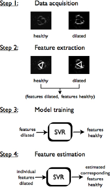
The main steps of the presented method. At first, ultra-sound images are acquired. Then, geometrical features are extracted to get tupels of features in the healthy and dilated state. With these tupels, an SVR-model is trained. This model allows feature estimation.
2.1 Data acquisition
To obtain features of an aortic root, a suitable imaging modality is needed. Due to its fine structure and its fast movement during the cardiac cycle, the detailed leaflet geometry is hard to extract from typical volumetric images like CT or MRI. Because of this reason, we previously proposed the use of transesophageal ultrasound (TEE) [8], given its high temporal resolution. Further benefits of this modality are its low examination costs (compared to CT and MRI) and its availability in clinical practice.
We designed a setup to conduct a TEE-examination on exvivo porcine aortic roots [8]. In this setup, a porcine aortic root is pressurized by a constant diastolic pressure created by a water head. The ultrasound images are taken sequentially while the imaging plane is rotating. After that, a three-dimensional reconstruction is done by transforming the image data to a Cartesian coordinate representation. The result is a volumetric image frame of the aortic root. As mentioned above, we examined ex-vivo porcine aortic roots. To get information about the root geometry before and after the pathological dilatation, we studied each aortic root in two different states: the healthy state and the dilated state. To examine the healthy state, we took volumetric ultrasound images of the unchanged root. To simulate the dilated state, the root wall was manually enlarged by adding additional aortic tissue into cuts in the root wall. After that, we acquired three-dimensional image data of the dilated root. The result are three-dimensional images of the root in both states.
2.2 Feature extraction
The aim of this step is to generate tupels of features that describe the geometry of the valve in the healthy and the dilated state. The extracted features were the three commissure distances k1, k2 and k3, the effective height heff and the leaflet-specific characteristic parameters F and, described in more detail below. The effective height is defined as the height difference between the commissure plane, where the leaflets are attached to the root wall, and the coaptation plane, where the three leaflets meet each other. It has a high influence on the functionality of the valve [5]. The leaflet-specific parameter F describes the area of the triangle formed by the two commissure points of the leaflet and the coaptation point (cf. Fig. 3). Hence, F measures the size of the leaflet. is one of the angles of the same triangle and describes the leaflets sheering. Both F and affect the performance of the valve [9]. The commis-sure distances are defined as the distances between pairs of the three commissure points, respectively (cf. Fig. 3). The features were manually obtained from the ultrasound images using the open-source image analysis tool 3dSlicer (Harvard Medical School, Boston, USA).
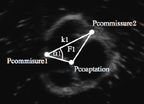
Ultrasound image of a porcine aortic root. For one leaflet, the commissure points Pcommissure1 and Pcommissure2, the coaptation point Pcoaptation and the geometrical features k1, F1 and α1 are shown.
At the moment, the prostheses used in clinical practice are shaped as a tube, so we aimed to estimate the optimal prosthesis diameter. This diameter can be calculated based on the three commissure distances as the diameter of the circumcircle of the triangle formed by the commis-sure points. Hence, the commissure distances k1, k2 and k3 are very important and the focus of this paper lies on a good estimation of them.
2.3 Model training
The surgery planning method aims at estimating the healthy features only based on the knowledge of the dilated features. In the previous steps we have obtained tupels (dilated features, healthy features). Using this data set we train a support vector regression (SVR) algorithm to learn the mapping between the dilated and the healthy states. The SVR is a machine learning method that performs regression without initial knowledge of the underlying model [10]. In this paper, we used the so called ∊-SVR with a Gaussian radial basis function kernel (RBF) using the MATLAB -implementation of the open-source library libsvm [11].
As mentioned above, the focus of this paper lies on a good estimation of the commissure distances. To find the best parameter combination for reaching this goal, we implemented a nonlinear simplex optimization method that minimizes the sum of absolute differences between the healthy commissure distances and the estimated commissure distances.
With this parameter set, we can train our SVR-model with the (dilated features, healthy features) tupels.
2.4 Feature estimation
After the training, the model is ready to reconstruct the healthy features of a valve from the individual dilated features. We obtained tupels of the healthy and dilated features of six aortic roots using the method previously described. We trained the SVM-model with five of the tupels and estimated the healthy features of the sixth valve with it. To evaluate the accuracy we compared all estimated features to the reference by calculating relative distances. Additionally, we calculated the root diameter based on the estimated features and the reference.
This evaluation was performed six times, each time estimating the healthy features of another valve while the other five valves where used for training (leave-one-out).
3 Results
We examined six porcine aortic valves using the method described in chapter 2. Accordingly, we estimated the healthy features of each of the six valves using an SVR-model trained with the other five valves’ features, respectively. Table 1 shows the relative distances between the estimated features and the reference as well as the mean difference for each feature. As mentioned in section 2.2, our aim is to calculate the optimal prosthesis diameter based on the commissure distances. For this purpose, this diameter was calculated for the estimated healthy features as well as the reference features. The resulting prosthesis sizes are presented in Table 2. As the commonly used pros-theses are produced with even-numbered diameters, we rounded our results appropriately.
4 Discussion
Table 1 shows particular differences between the estimated and the reference features. Some parameters for some valves are estimated good, others depict a high discrepancy. This is mainly due to the fact that the regression was performed with only five training samples. This is obviously not enough information to produce reliable feature estimations.
Relative differences in percentage between the estimated features and the reference for the six examined valves.
| Feature | Valve 1 | Valve 2 | Valve 3 | Valve 4 |
|---|---|---|---|---|
| k1 | 23,07 | 14,10 | 3,76 | 1,38 |
| k2 | 21,00 | 16,89 | 1,81 | 9,02 |
| k3 | 10,96 | 24,68 | 77,64 | 27,63 |
| heff | 50,901 | 69,77 | 9,30 | 38,65 |
| α1 | 8,81 | 47,18 | 54,85 | 6,31 |
| α2 | 16,24 | 26,18 | 4,48 | 2,18 |
| α3 | 0,87 | 3,41 | 1,24 | 16,76 |
| F1 | 38,22 | 50,70 | 17,64 | 27,40 |
| F2 | 26,64 | 85,92 | 110,83 | 33,21 |
| F3 | 38,77 | 39,02 | 100,82 | 18,00 |
| Feature | Valve 5 | Valve 6 | Mean |
|---|---|---|---|
| k1 | 18,61 | 2,73 | 10,61 ± 9.23 |
| k2 | 7,42 | 17,18 | 12,22 ± 7.28 |
| k3 | 4,52 | 4,94 | 25,06 ± 27.56 |
| heff | 29,93 | 10,13 | 34,78 ± 23.58 |
| α1 | 45,82 | 5,25 | 28,04 ± 23.51 |
| α2 | 3,04 | 12,20 | 10,72 ± 9.41 |
| α3 | 41,00 | 48,68 | 18,60 ± 21.24 |
| F1 | 51,06 | 18,06 | 33,85 ± 15.18 |
| F2 | 27,44 | 23,81 | 51,31 ± 37.42 |
| F3 | 80,71 | 27,72 | 50,84 ± 32.52 |
However, Table 2 depicts that in two cases, the calculated prosthesis diameter fits to the diameter of the healthy valve. In three other cases, the difference is only one pros-thesis size step. Hence, even with this small data set, an estimation of the best prosthesis diameter is possible. Further work should aim for the enlargement of the training data set. This would likely enhance the accuracy of the SVR-model. Additionally, a greater number of training samples would allow for a higher dimensional regression, i.e. the estimation of one feature could be based on all other features.
Another interesting point is the further processing of the estimated features. If the features could be translated to a geometric model of the aortic root, it would be possible to produce patient individual prosthesis’ with the specific shape of that patients aortic root in the healthy state. Estimating the prosthesis diameter could be just the beginning of individualization of cardiovascular implants.
Estimated and reference prosthesis diameters in mm.
| Valve | Reference | Estimated |
|---|---|---|
| 1 | 24 | 26 |
| 2 | 18 | 20 |
| 3 | 16 | 20 |
| 4 | 28 | 28 |
| 5 | 20 | 20 |
| 6 | 22 | 24 |
Funding
This publication is a result of the ongoing research within the LUMEN research group, which is funded by the German Bundesministerium für Bildung und Forschung (BMBF) (FKZ 13EZ1140A/B). LUMEN is a joint research project of Lübeck University of Applied Sciences and University of Lübeck and represents an own branch of the Graduate School for Computing in Medicine and Life Sciences of University of Lübeck.
Author's Statement
Conflict of interest: Authors state no conflict of interest. Material and Methods: Informed consent: Informed consent has been obtained from all individuals included in this study. Ethical approval: The research related to human use has been complied with all the relevant national regulations, institutional policies and in accordance the tenets of the Helsinki Declaration, and has been approved by the authors’ institutional review board or equivalent committee.
References
[1] M. Scharfschwerdt, HH. Sievers, A. Hussein, ED. Kraatz and M. Misfeld, Impact of progressive sinotubular junction dilatation on valve competence of the 3F Aortic and Sorin Solo stentless bioprosthetic heart valves. Eur J Cardiothorac Surg, 37:631-634, 2010.10.1016/j.ejcts.2009.09.010Search in Google Scholar PubMed
[2] A. Schuerhaus, Aortale Eingriffe bei Patienten mit Marfan-Syndrom. PhD thesis, Universität zu Lübeck, 2008.Search in Google Scholar
[3] P. Nataf and E. Lansac, Dilation thoracic aorta: medical surgical management. Heart 92(9), pp. 1345–1352, 2006.10.1136/hrt.2005.074781Search in Google Scholar
[4] J. Bechtel, A. Erasmi, M. Misfeld and HH. Sievers, Rekonstruktive Aortenklappenchirurgie: Ross-, David- und Yacoubverfahren. Herz 31(5), pp. 413-422, 2006.10.1007/s00059-006-2836-4Search in Google Scholar PubMed
[5] G. Marom, R. Haj-Ali, M. Rosenfeld, HJ. Schäfers and E. Raanani, Aortic root numeric model: annulus diameter prediction of effective height and coaptation in post-aortic valve repair. J Thorac Cardiovasc Surg 145(2),406-411, 2013.10.1016/j.jtcvs.2012.01.080Search in Google Scholar PubMed
[6] P. Hammer, C. Pacak, R. Howe and P. Nido, Collagen bundle orientation explains aortic valve leaflet coaptation. Functional Imaging and Modeling of the Heart, pp. 409-415, 2013.10.1007/978-3-642-38899-6_48Search in Google Scholar
[7] J. Hagenah, M. Scharfschwerdt, C. Metzner, A. Schlaefer, HH. Sievers, and A. Schweikard, An approach for patient specific modeling of the aortic valve leaflets. Biomedizinische Technik (BMT 2014), 2014.Search in Google Scholar
[8] J. Hagenah, M. Scharfschwerdt, B. Stender, S. Ott, R. Friedl, HH. Sievers and A. Schlaefer, A setup for ultrasound based assessment of the aortic root geometry. Biomedizinische Technik (BMT 2013), 2013.10.1515/bmt-2013-4379Search in Google Scholar PubMed
[9] J. Hagenah, Erstellung eines patientenindividuellen Modells der Aortenklappe, Bachelor thesis, Universität zu Lübeck, 2013.Search in Google Scholar
[10] AJ. Smola and B. Schöllkopf, A tutorial on support vector regression, Statistics and Computing 14(3), pp. 199-222, 2004.10.1023/B:STCO.0000035301.49549.88Search in Google Scholar
[11] CC. Chang and CJ. Lin, LIBSVM : A library for support vector machines, ACM Transactions on Intelligent Systems and Technology 2(27), pp. 1-27, 2011.10.1145/1961189.1961199Search in Google Scholar
© 2015 by Walter de Gruyter GmbH, Berlin/Boston
This article is distributed under the terms of the Creative Commons Attribution Non-Commercial License, which permits unrestricted non-commercial use, distribution, and reproduction in any medium, provided the original work is properly cited.
Articles in the same Issue
- Research Article
- Development and characterization of superparamagnetic coatings
- Research Article
- The development of an experimental setup to measure acousto-electric interaction signal
- Research Article
- Stability analysis of ferrofluids
- Research Article
- Investigation of endothelial growth using a sensors-integrated microfluidic system to simulate physiological barriers
- Research Article
- Energy harvesting for active implants: powering a ruminal pH-monitoring system
- Research Article
- New type of fluxgate magnetometer for the heart’s magnetic fields detection
- Research Article
- Field mapping of ballistic pressure pulse sources
- Research Article
- Development of a new homecare sleep monitor using body sounds and motion tracking
- Research Article
- Noise properties of textile, capacitive EEG electrodes
- Research Article
- Detecting phase singularities and rotor center trajectories based on the Hilbert transform of intraatrial electrograms in an atrial voxel model
- Research Article
- Spike sorting: the overlapping spikes challenge
- Research Article
- Separating the effect of respiration from the heart rate variability for cases of constant harmonic breathing
- Research Article
- Locating regions of arrhythmogenic substrate by analyzing the duration of triggered atrial activities
- Research Article
- Combining different ECG derived respiration tracking methods to create an optimal reconstruction of the breathing pattern
- Research Article
- Atrial and ventricular signal averaging electrocardiography in pacemaker and cardiac resynchronization therapy
- Research Article
- Estimation of a respiratory signal from a single-lead ECG using the 4th order central moments
- Research Article
- Compressed sensing of multi-lead ECG signals by compressive multiplexing
- Research Article
- Heart rate monitoring in ultra-high-field MRI using frequency information obtained from video signals of the human skin compared to electrocardiography and pulse oximetry
- Research Article
- Synchronization in wireless biomedical-sensor networks with Bluetooth Low Energy
- Research Article
- Automated classification of stages of anaesthesia by populations of evolutionary optimized fuzzy rules
- Research Article
- Effects of sampling rate on automated fatigue recognition in surface EMG signals
- Research Article
- Closed-loop transcranial alternating current stimulation of slow oscillations
- Research Article
- Cardiac index in atrio- and interventricular delay optimized cardiac resynchronization therapy and cardiac contractility modulation
- Research Article
- The role of expert evaluation for microsleep detection
- Research Article
- The impact of baseline wander removal techniques on the ST segment in simulated ischemic 12-lead ECGs
- Research Article
- Metal artifact reduction by projection replacements and non-local prior image integration
- Research Article
- A novel coaxial nozzle for in-process adjustment of electrospun scaffolds’ fiber diameter
- Research Article
- Processing of membranes for oxygenation using the Bellhouse-effect
- Research Article
- Inkjet printing of viable human dental follicle stem cells
- Research Article
- The use of an icebindingprotein out of the snowflea Hypogastrura harveyi as a cryoprotectant in the cryopreservation of mesenchymal stem cells
- Research Article
- New NIR spectroscopy based method to determine ischemia in vivo in liver – a first study on rats
- Research Article
- QRS and QT ventricular conduction times and permanent pacemaker therapy after transcatheter aortic valve implantation
- Research Article
- Adopting oculopressure tonometry as a transient in vivo rabbit glaucoma model
- Research Article
- Next-generation vision testing: the quick CSF
- Research Article
- Improving tactile sensation in laparoscopic surgery by overcoming size restrictions
- Research Article
- Design and control of a 3-DOF hydraulic driven surgical instrument
- Research Article
- Evaluation of endourological tools to improve the diagnosis and therapy of ureteral tumors – from model development to clinical application
- Research Article
- Frequency based assessment of surgical activities
- Research Article
- “Hands free for intervention”, a new approach for transoral endoscopic surgery
- Research Article
- Pseudo-haptic feedback in medical teleoperation
- Research Article
- Feasibility of interactive gesture control of a robotic microscope
- Research Article
- Towards structuring contextual information for workflow-driven surgical assistance functionalities
- Research Article
- Towards a framework for standardized semantic workflow modeling and management in the surgical domain
- Research Article
- Closed-loop approach for situation awareness of medical devices and operating room infrastructure
- Research Article
- Kinect based physiotherapy system for home use
- Research Article
- Evaluating the microsoft kinect skeleton joint tracking as a tool for home-based physiotherapy
- Research Article
- Integrating multimodal information for intraoperative assistance in neurosurgery
- Research Article
- Respiratory motion tracking using Microsoft’s Kinect v2 camera
- Research Article
- Using smart glasses for ultrasound diagnostics
- Research Article
- Measurement of needle susceptibility artifacts in magnetic resonance images
- Research Article
- Dimensionality reduction of medical image descriptors for multimodal image registration
- Research Article
- Experimental evaluation of different weighting schemes in magnetic particle imaging reconstruction
- Research Article
- Evaluation of CT capability for the detection of thin bone structures
- Research Article
- Towards contactless optical coherence elastography with acoustic tissue excitation
- Research Article
- Development and implementation of algorithms for automatic and robust measurement of the 2D:4D digit ratio using image data
- Research Article
- Automated high-throughput analysis of B cell spreading on immobilized antibodies with whole slide imaging
- Research Article
- Tissue segmentation from head MRI: a ground truth validation for feature-enhanced tracking
- Research Article
- Video tracking of swimming rodents on a reflective water surface
- Research Article
- MR imaging of model drug distribution in simulated vitreous
- Research Article
- Studying the extracellular contribution to the double wave vector diffusion-weighted signal
- Research Article
- Artifacts in field free line magnetic particle imaging in the presence of inhomogeneous and nonlinear magnetic fields
- Research Article
- Introducing a frequency-tunable magnetic particle spectrometer
- Research Article
- Imaging of aortic valve dynamics in 4D OCT
- Research Article
- Intravascular optical coherence tomography (OCT) as an additional tool for the assessment of stent structures
- Research Article
- Simple concept for a wide-field lensless digital holographic microscope using a laser diode
- Research Article
- Intraoperative identification of somato-sensory brain areas using optical imaging and standard RGB camera equipment – a feasibility study
- Research Article
- Respiratory surface motion measurement by Microsoft Kinect
- Research Article
- Improving image quality in EIT imaging by measurement of thorax excursion
- Research Article
- A clustering based dual model framework for EIT imaging: first experimental results
- Research Article
- Three-dimensional anisotropic regularization for limited angle tomography
- Research Article
- GPU-based real-time generation of large ultrasound volumes from freehand 3D sweeps
- Research Article
- Experimental computer tomograph
- Research Article
- US-tracked steered FUS in a respiratory ex vivo ovine liver phantom
- Research Article
- Contribution of brownian rotation and particle assembly polarisation to the particle response in magnetic particle spectrometry
- Research Article
- Preliminary investigations of magnetic modulated nanoparticles for microwave breast cancer detection
- Research Article
- Construction of a device for magnetic separation of superparamagnetic iron oxide nanoparticles
- Research Article
- An IHE-conform telecooperation platform supporting the treatment of dementia patients
- Research Article
- Automated respiratory therapy system based on the ARDSNet protocol with systemic perfusion control
- Research Article
- Identification of surgical instruments using UHF-RFID technology
- Research Article
- A generic concept for the development of model-guided clinical decision support systems
- Research Article
- Evaluation of local alterations in femoral bone mineral density measured via quantitative CT
- Research Article
- Creating 3D gelatin phantoms for experimental evaluation in biomedicine
- Research Article
- Influence of short-term fixation with mixed formalin or ethanol solution on the mechanical properties of human cortical bone
- Research Article
- Analysis of the release kinetics of surface-bound proteins via laser-induced fluorescence
- Research Article
- Tomographic particle image velocimetry of a water-jet for low volume harvesting of fat tissue for regenerative medicine
- Research Article
- Wireless medical sensors – context, robustness and safety
- Research Article
- Sequences for real-time magnetic particle imaging
- Research Article
- Speckle-based off-axis holographic detection for non-contact photoacoustic tomography
- Research Article
- A machine learning approach for planning valve-sparing aortic root reconstruction
- Research Article
- An in-ear pulse wave velocity measurement system using heart sounds as time reference
- Research Article
- Measuring different oxygenation levels in a blood perfusion model simulating the human head using NIRS
- Research Article
- Multisegmental fusion of the lumbar spine a curse or a blessing?
- Research Article
- Numerical analysis of the biomechanical complications accompanying the total hip replacement with NANOS-Prosthetic: bone remodelling and prosthesis migration
- Research Article
- A muscle model for hybrid muscle activation
- Research Article
- Mathematical, numerical and in-vitro investigation of cooling performance of an intra-carotid catheter for selective brain hypothermia
- Research Article
- An ideally parameterized unscented Kalman filter for the inverse problem of electrocardiography
- Research Article
- Interactive visualization of cardiac anatomy and atrial excitation for medical diagnosis and research
- Research Article
- Virtualizing clinical cases of atrial flutter in a fast marching simulation including conduction velocity and ablation scars
- Research Article
- Mesh structure-independent modeling of patient-specific atrial fiber orientation
- Research Article
- Accelerating mono-domain cardiac electrophysiology simulations using OpenCL
- Research Article
- Understanding the cellular mode of action of vernakalant using a computational model: answers and new questions
- Research Article
- A java based simulator with user interface to simulate ventilated patients
- Research Article
- Evaluation of an algorithm to choose between competing models of respiratory mechanics
- Research Article
- Numerical simulation of low-pulsation gerotor pumps for use in the pharmaceutical industry and in biomedicine
- Research Article
- Numerical and experimental flow analysis in centifluidic systems for rapid allergy screening tests
- Research Article
- Biomechanical parameter determination of scaffold-free cartilage constructs (SFCCs) with the hyperelastic material models Yeoh, Ogden and Demiray
- Research Article
- FPGA controlled artificial vascular system
- Research Article
- Simulation based investigation of source-detector configurations for non-invasive fetal pulse oximetry
- Research Article
- Test setup for characterizing the efficacy of embolic protection devices
- Research Article
- Impact of electrode geometry on force generation during functional electrical stimulation
- Research Article
- 3D-based visual physical activity assessment of children
- Research Article
- Realtime assessment of foot orientation by Accelerometers and Gyroscopes
- Research Article
- Image based reconstruction for cystoscopy
- Research Article
- Image guided surgery innovation with graduate students - a new lecture format
- Research Article
- Multichannel FES parameterization for controlling foot motion in paretic gait
- Research Article
- Smartphone supported upper limb prosthesis
- Research Article
- Use of quantitative tremor evaluation to enhance target selection during deep brain stimulation surgery for essential tremor
- Research Article
- Evaluation of adhesion promoters for Parylene C on gold metallization
- Research Article
- The influence of metallic ions from CoCr28Mo6 on the osteogenic differentiation and cytokine release of human osteoblasts
- Research Article
- Increasing the visibility of thin NITINOL vascular implants
- Research Article
- Possible reasons for early artificial bone failure in biomechanical tests of ankle arthrodesis systems
- Research Article
- Development of a bending test procedure for the characterization of flexible ECoG electrode arrays
- Research Article
- Tubular manipulators: a new concept for intracochlear positioning of an auditory prosthesis
- Research Article
- Investigation of the dynamic diameter deformation of vascular stents during fatigue testing with radial loading
- Research Article
- Electrospun vascular grafts with anti-kinking properties
- Research Article
- Integration of temperature sensors in polyimide-based thin-film electrode arrays
- Research Article
- Use cases and usability challenges for head-mounted displays in healthcare
- Research Article
- Device- and service profiles for integrated or systems based on open standards
- Research Article
- Risk management for medical devices in research projects
- Research Article
- Simulation of varying femoral attachment sites of medial patellofemoral ligament using a musculoskeletal multi-body model
- Research Article
- Does enhancing consciousness for strategic planning processes support the effectiveness of problem-based learning concepts in biomedical education?
- Research Article
- SPIO processing in macrophages for MPI: The breast cancer MPI-SNLB-concept
- Research Article
- Numerical simulations of airflow in the human pharynx of OSAHS patients