Respiratory motion tracking using Microsoft’s Kinect v2 camera
-
Floris Ernst
 and
Philipp Saß
and
Philipp Saß

Abstract
In image-guided radiotherapy, monitoring and compensating for respiratory motion is of high importance. We have analysed the possibility to use Microsoft’s Kinect v2 sensor as a low-cost tracking camera. In our experiment, eleven circular markers were printed onto a Lycra shirt and were tracked in the camera’s color image using cross correlation-based template matching. The 3D position of the marker was determined using this information and the mean distance of all template pixels from the sensor. In an experiment with four volunteers (male and female) we could demonstrate that real time position tracking is possible in 3D. By averaging over the depth values inside the template, it was possible to increase the Kinect’s depth resolution from 1 mm to 0.1 mm. The noise level was reduced to a standard deviation of 0.4 mm. Temperature sensitivity of the measured depth values was observed for about 10-15 minutes after system start.
1 Introduction
In many clinical applications, detecting and tracking of respiratory motion is required. As an example, image-guided radiotherapy (IGRT) of the chest and abdomen relies heavily on this principle: some kind of marker is placed on or attached to the patient’s chest and is monitored using a non-invasive localisation device. Subsequently, trajectory of the marker is then analysed and used to either dynamically activate the treatment beam (called gating [3]) or to guide the radiation source [6]. Especially in the second scenario, tracking one marker may not be sufficient: the actual target of the treatment beam – the tumour – is typically not observed directly. Although this could be done (either using continuous X-ray localisation [7] or 3D ultrasound tracking [1]), the current method in clinical use relies on a mathematical model linking the motion on the patient’s chest to the motion of the actual target.
It has been shown that the accuracy of these correlation algorithms can be improved by incorporating multiple markers [2]. In this work, we demonstrate how consumer hardware (Microsoft’s Kinect v2 depth sensor) can be used to accurately track the 3D position of multiple markers using a special marker shirt.
2 Methods and materials
To acquire respiratory motion traces, a special marker shirt has been developed. Eleven marker templates were printed onto a Lycra shirt, ensuring tight fit on the volunteers, while position of the markers correspond to areas relevant for the measurement. Each marker consists of a black circle surrounded by a black ring. Details and numbering of the markers are shown in Figure 1.
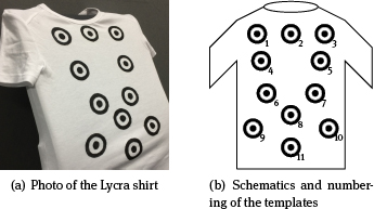
Marker shirt for motion tracking
Tracking the position of the markers is done using Microsoft’s Kinect v2 camera (see Figure 2) and the corresponding software development kit (SDK) [5]. The camera is able to simultaneously capture three different types of images at a frame rate of up to 30 Hz: a color image (1920 × 1280 pixels), an infrared-illuminated grayscale image (512 × 424 pixels), and a depth image (512 × 424 pixels, depth resolution of 1 mm). Typical images are shown in Figure 3. Details about the technology behind the sensor is given in [4].

Kinect v2 Sensor. Photograph courtesy of Microsoft Corp.
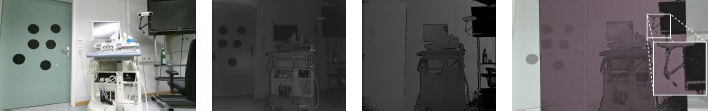
Typical frames acquired with the Kinect v2 sensor. (A): color image, (B): IR illuminated scene, (C): depth image, (D): overlay of color and depth images.
Using these images, and the known intrinsics and extrinsics of the color- and IR-cameras inside the Kinect sensor, it is possible to determine the 3D position for each pixel in the depth image. We have developed an application that allows selecting and tracking up to 15 markers in real time. The general process is as follows:
During setup, the user is shown a camera image of the subject and is asked to select the initial position of the markers and the template to use for tracking.
The position of the template in the given regions of interest (ROI) is determined
The distance of the center point of the template found is determined using template matching
The matching ROIs are centered around the position of the last match
Template matching is done using cross correlation. It is implemented in C# using a wrapper library (EmguCV) around the OpenCV computer vision library.
2.1 Volunteer study
The Kinect sensor was attached to an industrial robot (Adept Viper s850) to allow accurate and stable placement. The setup is shown schematically in Figure 5. In a small volunteer study (four participants, one female, three male), we evaluated the possibility of feature tracking. Our volunteers were asked to lie down in supine position and breathe normally for three to four minutes.

Process of region-of-interest-based template matching. (A) – template found inside ROI (black). (B) – template moved. (C) – template found in old ROI. (D) – Old ROI (gray) and new ROI (black).

Schematic setup of the experiment.
2.2 Accuracy measurements
Finally, the stability and accuracy of the Kinect sensor was evaluated in another experiment. First, the robot as shown in Figure 5 was programmed to follow a sinusoidal motion (therefore similar to respiratory motion) along the z- axis while the distance to the patient couch was computed for each camera frame. Second, the distance to the patient couch was measured repeatedly for about twelve minutes to determine the amount of noise and possible drift.
3 Results
Using our multi-threaded implementation in C#, tracking eleven markers in the color camera image – using ROIs of twice the size of the marker template – was possible in real time using multi-threading on a MacBook Pro Retina (2.3 GHz Core i7, four cores, 16 GiB RAM, SSD). In general, the runtime of one template matching iteration was around 80 ms.
3.1 Volunteer study
Recording motion traces of the markers worked for all four volunteers (three male, one female), although markers one and three were difficult to track due to stretching of the fabric. Figure 6 shows the distances measured for all eleven templates. Note the large differences in amplitude between the individual sensors.
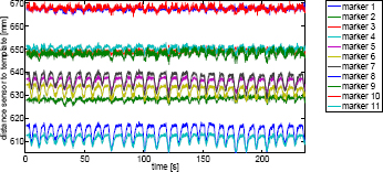
Anterior/posterior motion traces of the markers for subject one (female).
The depth motion trace of a second volunteer (subject four) is shown in Figure 7. Note the much larger amplitude for markers 5–8 and 11 and the sudden motion around t = 95 s due to the volunteer sneezing. Additionally, the values from markers one and three (red and blue, respectively) show that tracking them is difficult due to deformation.

Anterior/posterior motion traces of the markers for subject four (male). Note the sudden peak around 95 s, which is due to the volunteer sneezing and the low quality of markers one and three.
Additionally, the in-image motion of the template was also evaluated. It is exemplarily shown for one marker (marker eight of subject one) in Figure 8. Here, it is clear that there is very little motion in the left/right direction, as would be expected. In the superior/inferior-direction, however, some motion is present (one pixel corresponds to approximately 1-1.5 mm in our setup, depending on the exact distance from the sensor), albeit not as strong as in the anterior/posterior-direction.
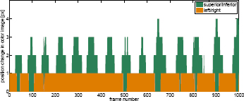
Inferior/superior and left/right motion traces for marker eight of subject one.
3.2 Accuracy measurements
Using the same setup as described before, we determined the absolute accuracy of the depth measurements. The trajectory of the robot – overlaid with the measured distance to the template – is given in Figure 9. Clearly, the distance measured by the Kinect sensor deviates substantially from the true motion of the robot, maximum is 3.7 mm and the root mean square error (RMSE) is 2.0 mm with a working distance on the order of 50 cm.

Sinusoidal motion trace performed by the robot (red) and as measured from template matching and averaging the depth values (blue).
The results of the static measurement evaluation are shown in Figure 10. The measurement was taken directly after turning on the Kinect sensor and some kind of time-dependent drift is visible. We believe that this is due to the changing temperature of the sensor PCB. The depth value is determined – as outlined above – from averaging all pixels in the template, resulting in sub-millimeter resolution. The noise level, however, is still considerable: we observe a standard deviation of 0.4 mm.

Measurement noise from a static target, recorded over twelve minutes (blue) and running average (red).
4 Discussion
We have demonstrated that the Kinect v2 sensor’s data streams – color image and depth image – can be used to track multiple markers on the human chest in 3D and in real time using standard hardware. Additionally, by averaging the depth values inside the marker template, it is possible to substantially reduce the measurement noise to a standard deviation of 0.4 mm. On the other hand, however, we observed that the depth values measured using the robotic setup and the sinusoidal motion pattern deviate strongly from the actual data: the motion amplitude of the sine was 20 mm and the amplitude of the template matching was more than 25 mm – 25 % more. We believe that this is caused by multiple factors:
Inaccurate alignment of the depth axis of the Kinect sensor with the robot’s z-axis and the template center
Errors in sensor’s calibration (the Kinect sensor stores its intrinsics and extrinsics in firmware and we did not perform camera calibration)
As next steps, we plan to perform sub-pixel template matching to increase the resolution along the L/R- and S/I-axes and to further analyze the accuracy of the setup by tracking the marker with a dedicated tracking device (like NDI’s Polaris Spectra system). Also the operating speed of the system (now about 15 fps) could be increased due to massive code parallelization, so that every frame from the Kinect v2 is used. We need to make sure, however, that the light emitted by the Kinect v2 sensor does not interfere with the IR light used by the Spectra system. Both operate in the near-infrared range around 850 to 860 nm.
Author's Statement
Conflict of interest: Authors state no conflict of interest. Material and Methods: Informed consent: Informed consent has been obtained from all individuals included in this study. Ethical approval: The research related to human use has been complied with all the relevant national regulations, institutional policies and in accordance the tenets of the Helsinki Declaration, and has been approved by the authors’ institutional review board or equivalent committee.
References
[1] O. Blanck, P. Jauer, F. Ernst, R. Bruder, and A. Schweikard. Pilot-Phantomtest zur ultraschall-geführten robotergestützten Radiochirurgie. In H. Treuer, editor,44. Jahrestagung der DGMP, Cologne, Germany, 2013. DGMP, pages 122–123.Search in Google Scholar
[2] R. Dürichen, M. A. F. Pimentel, L. Clifton, A. Schweikard, and D. A. Clifton. Multi-task gaussian processes for multivariate physiological time-series analysis. IEEE Transactions on Biomedical Engineering, 62(1):314–322, 2014. 10.1109/TBME.2014.2351376.Search in Google Scholar
[3] J. Hanley, M. M. Debois, D. Mah, G. S. Mageras, A. Raben, K. Rosenzweig, B. Mychalczak, L. H. Schwartz, P. J. Gloeggler, W. Lutz, C. C. Ling, S. A. Leibel, Z. Fuks, and G. J. Kutcher. Deep inspiration breath-hold technique for lung tumors: the potential value of target immobilization and reduced lung density in dose escalation. International Journal of Radiation Oncology, Biology, Physics, 45(3):603–611, 1999. 10.1016/s0360-3016(99)00154-6.Search in Google Scholar
[4] D. Lau. The science behind Kinects or Kinect 1.0 versus 2.0. http://www.gamasutra.com/blogs/DanielLau/20131127/205820/The_Science_Behind_Kinects_or_Kinect_10_versus_20.php, November, 2013. Online, last visited 2015-03-24.Search in Google Scholar
[5] Microsoft Corporation. Kinect for Windows SDK 2.0. http://www.microsoft.com/en-us/download/details.aspx?id=44561, October, 2014. Online, last visited 2015-03-24.Search in Google Scholar
[6] A. Schweikard, H. Shiomi, and J. R. Adler, Jr. Respiration tracking in radiosurgery. Medical Physics, 31(10):2738–2741, 2004. 10.1118/1.1774132.Search in Google Scholar
[7] H. Shirato, S. Shimizu, K. Kitamura, T. Nishioka, K. Kagei, S. Hashimoto, H. Aoyama, T. Kunieda, N. Shinohara, H. DosakaAkita, and K. Miyasaka. Four-dimensional treatment planning and fluoroscopic real-time tumor tracking radiotherapy for moving tumor. International Journal of Radiation Oncology, Biology, Physics, 48(2):435–442, 2000. 10.1016/s0360-3016(00)00625-8.Search in Google Scholar
© 2015 by Walter de Gruyter GmbH, Berlin/Boston
This article is distributed under the terms of the Creative Commons Attribution Non-Commercial License, which permits unrestricted non-commercial use, distribution, and reproduction in any medium, provided the original work is properly cited.
Articles in the same Issue
- Research Article
- Development and characterization of superparamagnetic coatings
- Research Article
- The development of an experimental setup to measure acousto-electric interaction signal
- Research Article
- Stability analysis of ferrofluids
- Research Article
- Investigation of endothelial growth using a sensors-integrated microfluidic system to simulate physiological barriers
- Research Article
- Energy harvesting for active implants: powering a ruminal pH-monitoring system
- Research Article
- New type of fluxgate magnetometer for the heart’s magnetic fields detection
- Research Article
- Field mapping of ballistic pressure pulse sources
- Research Article
- Development of a new homecare sleep monitor using body sounds and motion tracking
- Research Article
- Noise properties of textile, capacitive EEG electrodes
- Research Article
- Detecting phase singularities and rotor center trajectories based on the Hilbert transform of intraatrial electrograms in an atrial voxel model
- Research Article
- Spike sorting: the overlapping spikes challenge
- Research Article
- Separating the effect of respiration from the heart rate variability for cases of constant harmonic breathing
- Research Article
- Locating regions of arrhythmogenic substrate by analyzing the duration of triggered atrial activities
- Research Article
- Combining different ECG derived respiration tracking methods to create an optimal reconstruction of the breathing pattern
- Research Article
- Atrial and ventricular signal averaging electrocardiography in pacemaker and cardiac resynchronization therapy
- Research Article
- Estimation of a respiratory signal from a single-lead ECG using the 4th order central moments
- Research Article
- Compressed sensing of multi-lead ECG signals by compressive multiplexing
- Research Article
- Heart rate monitoring in ultra-high-field MRI using frequency information obtained from video signals of the human skin compared to electrocardiography and pulse oximetry
- Research Article
- Synchronization in wireless biomedical-sensor networks with Bluetooth Low Energy
- Research Article
- Automated classification of stages of anaesthesia by populations of evolutionary optimized fuzzy rules
- Research Article
- Effects of sampling rate on automated fatigue recognition in surface EMG signals
- Research Article
- Closed-loop transcranial alternating current stimulation of slow oscillations
- Research Article
- Cardiac index in atrio- and interventricular delay optimized cardiac resynchronization therapy and cardiac contractility modulation
- Research Article
- The role of expert evaluation for microsleep detection
- Research Article
- The impact of baseline wander removal techniques on the ST segment in simulated ischemic 12-lead ECGs
- Research Article
- Metal artifact reduction by projection replacements and non-local prior image integration
- Research Article
- A novel coaxial nozzle for in-process adjustment of electrospun scaffolds’ fiber diameter
- Research Article
- Processing of membranes for oxygenation using the Bellhouse-effect
- Research Article
- Inkjet printing of viable human dental follicle stem cells
- Research Article
- The use of an icebindingprotein out of the snowflea Hypogastrura harveyi as a cryoprotectant in the cryopreservation of mesenchymal stem cells
- Research Article
- New NIR spectroscopy based method to determine ischemia in vivo in liver – a first study on rats
- Research Article
- QRS and QT ventricular conduction times and permanent pacemaker therapy after transcatheter aortic valve implantation
- Research Article
- Adopting oculopressure tonometry as a transient in vivo rabbit glaucoma model
- Research Article
- Next-generation vision testing: the quick CSF
- Research Article
- Improving tactile sensation in laparoscopic surgery by overcoming size restrictions
- Research Article
- Design and control of a 3-DOF hydraulic driven surgical instrument
- Research Article
- Evaluation of endourological tools to improve the diagnosis and therapy of ureteral tumors – from model development to clinical application
- Research Article
- Frequency based assessment of surgical activities
- Research Article
- “Hands free for intervention”, a new approach for transoral endoscopic surgery
- Research Article
- Pseudo-haptic feedback in medical teleoperation
- Research Article
- Feasibility of interactive gesture control of a robotic microscope
- Research Article
- Towards structuring contextual information for workflow-driven surgical assistance functionalities
- Research Article
- Towards a framework for standardized semantic workflow modeling and management in the surgical domain
- Research Article
- Closed-loop approach for situation awareness of medical devices and operating room infrastructure
- Research Article
- Kinect based physiotherapy system for home use
- Research Article
- Evaluating the microsoft kinect skeleton joint tracking as a tool for home-based physiotherapy
- Research Article
- Integrating multimodal information for intraoperative assistance in neurosurgery
- Research Article
- Respiratory motion tracking using Microsoft’s Kinect v2 camera
- Research Article
- Using smart glasses for ultrasound diagnostics
- Research Article
- Measurement of needle susceptibility artifacts in magnetic resonance images
- Research Article
- Dimensionality reduction of medical image descriptors for multimodal image registration
- Research Article
- Experimental evaluation of different weighting schemes in magnetic particle imaging reconstruction
- Research Article
- Evaluation of CT capability for the detection of thin bone structures
- Research Article
- Towards contactless optical coherence elastography with acoustic tissue excitation
- Research Article
- Development and implementation of algorithms for automatic and robust measurement of the 2D:4D digit ratio using image data
- Research Article
- Automated high-throughput analysis of B cell spreading on immobilized antibodies with whole slide imaging
- Research Article
- Tissue segmentation from head MRI: a ground truth validation for feature-enhanced tracking
- Research Article
- Video tracking of swimming rodents on a reflective water surface
- Research Article
- MR imaging of model drug distribution in simulated vitreous
- Research Article
- Studying the extracellular contribution to the double wave vector diffusion-weighted signal
- Research Article
- Artifacts in field free line magnetic particle imaging in the presence of inhomogeneous and nonlinear magnetic fields
- Research Article
- Introducing a frequency-tunable magnetic particle spectrometer
- Research Article
- Imaging of aortic valve dynamics in 4D OCT
- Research Article
- Intravascular optical coherence tomography (OCT) as an additional tool for the assessment of stent structures
- Research Article
- Simple concept for a wide-field lensless digital holographic microscope using a laser diode
- Research Article
- Intraoperative identification of somato-sensory brain areas using optical imaging and standard RGB camera equipment – a feasibility study
- Research Article
- Respiratory surface motion measurement by Microsoft Kinect
- Research Article
- Improving image quality in EIT imaging by measurement of thorax excursion
- Research Article
- A clustering based dual model framework for EIT imaging: first experimental results
- Research Article
- Three-dimensional anisotropic regularization for limited angle tomography
- Research Article
- GPU-based real-time generation of large ultrasound volumes from freehand 3D sweeps
- Research Article
- Experimental computer tomograph
- Research Article
- US-tracked steered FUS in a respiratory ex vivo ovine liver phantom
- Research Article
- Contribution of brownian rotation and particle assembly polarisation to the particle response in magnetic particle spectrometry
- Research Article
- Preliminary investigations of magnetic modulated nanoparticles for microwave breast cancer detection
- Research Article
- Construction of a device for magnetic separation of superparamagnetic iron oxide nanoparticles
- Research Article
- An IHE-conform telecooperation platform supporting the treatment of dementia patients
- Research Article
- Automated respiratory therapy system based on the ARDSNet protocol with systemic perfusion control
- Research Article
- Identification of surgical instruments using UHF-RFID technology
- Research Article
- A generic concept for the development of model-guided clinical decision support systems
- Research Article
- Evaluation of local alterations in femoral bone mineral density measured via quantitative CT
- Research Article
- Creating 3D gelatin phantoms for experimental evaluation in biomedicine
- Research Article
- Influence of short-term fixation with mixed formalin or ethanol solution on the mechanical properties of human cortical bone
- Research Article
- Analysis of the release kinetics of surface-bound proteins via laser-induced fluorescence
- Research Article
- Tomographic particle image velocimetry of a water-jet for low volume harvesting of fat tissue for regenerative medicine
- Research Article
- Wireless medical sensors – context, robustness and safety
- Research Article
- Sequences for real-time magnetic particle imaging
- Research Article
- Speckle-based off-axis holographic detection for non-contact photoacoustic tomography
- Research Article
- A machine learning approach for planning valve-sparing aortic root reconstruction
- Research Article
- An in-ear pulse wave velocity measurement system using heart sounds as time reference
- Research Article
- Measuring different oxygenation levels in a blood perfusion model simulating the human head using NIRS
- Research Article
- Multisegmental fusion of the lumbar spine a curse or a blessing?
- Research Article
- Numerical analysis of the biomechanical complications accompanying the total hip replacement with NANOS-Prosthetic: bone remodelling and prosthesis migration
- Research Article
- A muscle model for hybrid muscle activation
- Research Article
- Mathematical, numerical and in-vitro investigation of cooling performance of an intra-carotid catheter for selective brain hypothermia
- Research Article
- An ideally parameterized unscented Kalman filter for the inverse problem of electrocardiography
- Research Article
- Interactive visualization of cardiac anatomy and atrial excitation for medical diagnosis and research
- Research Article
- Virtualizing clinical cases of atrial flutter in a fast marching simulation including conduction velocity and ablation scars
- Research Article
- Mesh structure-independent modeling of patient-specific atrial fiber orientation
- Research Article
- Accelerating mono-domain cardiac electrophysiology simulations using OpenCL
- Research Article
- Understanding the cellular mode of action of vernakalant using a computational model: answers and new questions
- Research Article
- A java based simulator with user interface to simulate ventilated patients
- Research Article
- Evaluation of an algorithm to choose between competing models of respiratory mechanics
- Research Article
- Numerical simulation of low-pulsation gerotor pumps for use in the pharmaceutical industry and in biomedicine
- Research Article
- Numerical and experimental flow analysis in centifluidic systems for rapid allergy screening tests
- Research Article
- Biomechanical parameter determination of scaffold-free cartilage constructs (SFCCs) with the hyperelastic material models Yeoh, Ogden and Demiray
- Research Article
- FPGA controlled artificial vascular system
- Research Article
- Simulation based investigation of source-detector configurations for non-invasive fetal pulse oximetry
- Research Article
- Test setup for characterizing the efficacy of embolic protection devices
- Research Article
- Impact of electrode geometry on force generation during functional electrical stimulation
- Research Article
- 3D-based visual physical activity assessment of children
- Research Article
- Realtime assessment of foot orientation by Accelerometers and Gyroscopes
- Research Article
- Image based reconstruction for cystoscopy
- Research Article
- Image guided surgery innovation with graduate students - a new lecture format
- Research Article
- Multichannel FES parameterization for controlling foot motion in paretic gait
- Research Article
- Smartphone supported upper limb prosthesis
- Research Article
- Use of quantitative tremor evaluation to enhance target selection during deep brain stimulation surgery for essential tremor
- Research Article
- Evaluation of adhesion promoters for Parylene C on gold metallization
- Research Article
- The influence of metallic ions from CoCr28Mo6 on the osteogenic differentiation and cytokine release of human osteoblasts
- Research Article
- Increasing the visibility of thin NITINOL vascular implants
- Research Article
- Possible reasons for early artificial bone failure in biomechanical tests of ankle arthrodesis systems
- Research Article
- Development of a bending test procedure for the characterization of flexible ECoG electrode arrays
- Research Article
- Tubular manipulators: a new concept for intracochlear positioning of an auditory prosthesis
- Research Article
- Investigation of the dynamic diameter deformation of vascular stents during fatigue testing with radial loading
- Research Article
- Electrospun vascular grafts with anti-kinking properties
- Research Article
- Integration of temperature sensors in polyimide-based thin-film electrode arrays
- Research Article
- Use cases and usability challenges for head-mounted displays in healthcare
- Research Article
- Device- and service profiles for integrated or systems based on open standards
- Research Article
- Risk management for medical devices in research projects
- Research Article
- Simulation of varying femoral attachment sites of medial patellofemoral ligament using a musculoskeletal multi-body model
- Research Article
- Does enhancing consciousness for strategic planning processes support the effectiveness of problem-based learning concepts in biomedical education?
- Research Article
- SPIO processing in macrophages for MPI: The breast cancer MPI-SNLB-concept
- Research Article
- Numerical simulations of airflow in the human pharynx of OSAHS patients