Suppression of cathepsin B attenuates myocardial injury via limiting cardiomyocyte apoptosis
-
Ruilin Su
 und
Li Xu
und
Li Xu

Abstract
Background
Inflammation plays a pivotal role in modulating the pathophysiological progression of myocardial injury and the subsequent repair and remodeling of the infarcted myocardium. Cathepsin B, a member of the cysteine protease family, has been recognized for its ability to initiate various signaling cascades essential to inflammatory processes. This study aims to investigate whether cathepsin B influences cardiomyocyte survival under inflammatory conditions.
Methods
Mice were randomly divided into four groups (n = 6 per group) based on whether they received an intraperitoneal injection of Ca-074 Me (50 mg/kg) and whether myocardial ischemia/reperfusion (I/R) surgery was performed.
Results
The cathepsin B-specific inhibitor Ca-074 Me significantly attenuated myocardial infarction caused by I/R in vivo and reduced hypoxia-induced cardiomyocyte apoptosis in vitro. Mechanistically, Ca-074 Me appeared to inhibit caspase-3 signaling, thereby mitigating cardiomyocyte apoptosis under chemical hypoxia induced by cobalt chloride or physical oxygen deprivation.
Conclusion
Targeted inhibition of cathepsin B may represent an innovative strategy for the amelioration of myocardial injury.
1 Introduction
Ischemic heart disease (IHD) is a prevalent cardiovascular condition associated with significant morbidity and mortality worldwide [1]. It is characterized by diminished myocardial perfusion that precipitates ischemic damage and subsequent injury to the cardiac muscle [1,2]. The underlying pathophysiology of IHD is complex and multifactorial, with a range of risk factors, including long-term high blood pressure, hyperlipidemia, smoking, and physical activity. The mechanistic basis of myocardial injury stemming from ischemia–reperfusion (I/R) events is relatively complex, encompassing aberrant production of reactive oxygen species, calcium dyshomeostasis, amplification of proinflammatory cytokines, and depletion of high-energy phosphate compounds [3]. Cardiomyocyte apoptosis has been acknowledged as a principal contributor to I/R injury. Accordingly, the exploration of anti-apoptotic modalities harbors therapeutic promise for IHD. Accumulating evidence indicates the inflammatory response triggered by I/R injury plays a significant role in the development and progression of IHD [3,4]. Numerous studies have demonstrated elevated levels of inflammatory mediators (i.e., C-reactive protein, IL-6, and TNF-α) in patients with IHD [4–6]. Clinical trials have exhibited that anti-inflammatory agents, such as monoclonal antibodies against IL-1β, can reduce the risk of cardiovascular events in IHD patients [7]. However, the relationship between inflammation and IHD is complex, and studies elucidating the role and function of inflammatory mediators in cardiomyocyte apoptosis might provide new insights into further directions for IHD.
Cathepsins are proteases with serine, cysteine, or aspartic acid residues as the nucleophiles, which are essential for a range of functions, including digestion, coagulation, immune response, adipogenesis, hormone release, and peptide synthesis [8]. Several studies have shown that cathepsins are involved in the regulation of inflammation [9,10]. For example, cathepsin B has been shown to activate the NLRP3 inflammasome, a key mediator of inflammation, by cleaving pro-IL-1β and pro-IL-18 [11]. During acute pancreatitis, excessive release of cathepsin B into the cytoplasm can lead to cell necrosis through a mechanism dependent on active trypsin [12]. Deletion of cathepsin B alleviated hepatic pathology and enhanced survival in mice with acute liver injury [13]. Cathepsin B seems to degrade a cysteine protease that could eliminate apoptosis, supporting Bax in the cells [14]. Notably, cathepsin B participates in hypoxia-induced disease, and administering cathepsin B inhibitors before ischemia induction can reduce post-ischemic apoptosis of hepatocytes, thereby minimizing liver injury [15]. The compound Ca-074 methyl ester (Ca-074 Me), a selective inhibitor of cathepsin B, has been demonstrated to exert protective effects within a guinea pig model of polymyositis precipitated by infection with Coxsackie virus B1 (CVB1) [16]. However, it remains unknown whether cathepsin B may influence cardiomyocyte apoptosis and contribute to the pathogenesis of myocardial injury. Further investigation is required to elucidate the potential mechanistic role of cathepsin B in the regulation of cardiac cell death and the subsequent development of myocardial damage.
2 Materials and methods
2.1 Mice
Male C57BL/6J mice (12 weeks, 22–25 g) were purchased from the Laboratory Animal Center of Southern Medical University (Guangzhou, China). The mice were housed under a specific pathogen-free condition, on a 12-h light-dark cycle, and with food and water ad libitum.
2.2 Hypoxia-induced myocardial I/R
Mice were randomly assigned to four groups (n = 6 per group) according to whether Ca-074 Me (50 mg/kg) was injected intraperitoneally simultaneously and whether myocardial I/R surgery was performed or not. At the beginning of the procedure, the mice were given an intraperitoneal injection of 50 mg/kg chloral hydrate. Then, an incision of about 1.2 cm was made over the left chest, and a purse suture was made. The fourth intercostal space was exposed after dissection and retraction of the pectoral major and minor muscles. A small hole was made at the fourth intercostal space with a mosquito clamp to open the pleural membrane and pericardium. With the clamp open, the heart was popped out through the incision. Locating the left anterior descending (LAD) branch, then using a slipknot tied around the LAD 2–3 mm from its origin with 8–0 silk suture. The heart is then quickly placed back into the thoracic space, followed by manual evacuation of air and the skin closing. The internal needle end of the slipknot suture is cut as short as possible, and the other end of the suture is about 0.8 cm long and remains outside of the chest. After 30 min of ischemia, the slipknot was released by pulling the long end of the slipknot suture smoothly and gently until a feeling of release was sensed; meanwhile, the myocardium began reperfusion. Blood and heart samples were collected after 24 h of reperfusion. Submandibular blood samples (about 200 µl) were obtained by incising the right submandibular vein of mice (22–25 g) with a sterile 4-mm lancet. The animals were euthanized with 4% isoflurane inhalation followed by cervical vertebra dislocation. Animal death was confirmed by respiratory and cardiac arrest and no righting reflex.
2.3 Cell culture and stimulation conditions
AC16 cells were cultured in DMEM/F12 (Thermo Fisher Scientific, Carlsbad, CA, USA, A4192001) supplemented with 100 U/ml penicillin, 100 μg/ml streptomycin, and 10% fetal bovine serum at 37°C in a 5% CO2 incubator. The cells were treated with different concentrations of CoCl2 (0, 10, 50, 100, 200, and 500 µmol/l) or cultured in a hypoxia environment (1% oxygen, 5% carbon dioxide, and 94% nitrogen) for 0, 1, 3, 6, 12, and 24 h to determine the suitable hypoxia stimulation condition. Next, the cells were treated with CoCl2 (200 μM, Sigma-Aldrich, 232696) or 1% O2 in the presence or absence of Ca-074 Me (10 μM, Merck, S7420). AC16 cells were also treated with Ca-074 Me (10 μM) in combination with or without Z-DEVD-FMK (20 μM, Selleck, S7312) for 24 h under hypoxia.
2.4 Generation of gene knockout cells with CRISPR/Cas9
According to the manufacturer’s instructions, AC16 cells were transfected with Cathepsin B CRISPR/Cas9 KO plasmid (Santa Cruz Biotechnology) using UltraCruz® transfection reagent (Santa Cruz Biotechnology). Twenty-four hours after transfection, the expression level of cathepsin B in the cells was assessed by immunoblotting analysis.
2.5 Plasmid overexpression
Plasmid was synthesized by the Beijing Genomics Institution (Shenzhen, China) and was used to overexpress cathepsin B expression in HK-2 cells. Before other indicated experiments, the AC16 cells were seeded into six-well plates and were transfected with 10 μg of plasmid with Lipofectamine 3000 reagent (Thermo Fisher Scientific) following the standard protocol provided by the manufacturer.
2.6 ELISA assay
Mice’s blood samples were obtained from the carotid artery and centrifuged at 4,000 × g for 15 min, and then, the supernatant was collected and set aside at −80°C for serum cytokine analysis. Cytokine levels in the sera were assessed using commercial ELISA kits purchased from eBioscience (San Diego, CA, USA).
2.7 Histological evaluation
Heart samples were collected from mice, then fixed in 4% paraformaldehyde, embedded in paraffin, and stained with hematoxylin and eosin (H&E) according to the manufacturer’s instructions. The quantification analysis of H&E staining was established based on the methods of a previous study [17]. In brief, vascular congestion, interstitial capillary damage, vascular dilatation, myocytolysis, and myocyte vacuolization were assessed and rated as minimum (1), mild (2), moderate (3), or severe (4) on a scale of 1–4, with papillary muscle involvement serving as the criterion. Histopathologic changes were graded as minimal (1) if less than 30% of the heart muscles (left and right ventricles, and interventricular septum) were affected without papillary muscle involvement; mild (2) if more than 30% of the heart muscles were affected without papillary muscle involvement; moderate–mild (3) if papillary muscle involvement was focal; and severe (4) if the heart muscle and papillary muscle involvement was diffuse. A scale from 1 to 3 was used to grade myocardial hypertrophy. Mild myocardial hypertrophy was defined as minimal reactive cellular hypertrophy of the left ventricle (1), moderate myocardial hypertrophy as reactive cellular hypertrophy of the left and right ventricles, as well as the interventricular septum (2), and severe myocardial hypertrophy as reactive cellular hypertrophy involving all heart muscles, including papillary muscles with obvious luminal narrowing (3). Scores of 1 and 2 indicated focal or diffuse intravascular inflammation, respectively. Focal intravascular inflammation was defined as dispersed inflammation within interstitial vessels without papillary muscle involvement. A pathologist carried out all scoring in a blinded fashion. For immunohistochemical staining, antigen retrieval was performed in a citrate buffer (pH 6.0) at 120°C for 10 min, and endogenous peroxidase activity was blocked by exposure to 3% H2O2 for 15 min. Sections were then incubated with primary antibodies at 4°C overnight. Immunoreactivity was detected using the corresponding HRP-conjugated secondary antibody and visualized using a diaminobenzidine kit (Beyotime Biotechnology, Shanghai, China).
2.8 Western blot analysis
Protein lysate was prepared in RIPA buffer (Beyotime, Hangzhou, China) supplemented with protease and phosphatase inhibitor cocktails (Beyotime). Protein samples were fractionated by sodium dodecyl sulfate–polyacrylamide gel electrophoresis and electrophoretically transferred onto polyvinylidene fluoride membranes (Millipore, Billerica, MA, USA). After blocking with bovine serum albumin (5%) for 1 h at room temperature, the membranes were incubated overnight at 4°C with primary antibodies. Subsequently, the membranes were incubated with the horseradish peroxidase-conjugated corresponding secondary antibody for 1 h at room temperature. The blots are cut to the appropriate size prior to hybridization with antibodies. Finally, target protein detection was conducted with enhanced chemiluminescence (Tanon, Shanghai, China) according to the manufacturer’s protocol.
2.9 RNA isolation and quantitative real-time PCR
Total RNA was isolated from primary astrocytes using TRIzol reagent (Thermo Fisher Scientific, USA) according to the manufacturer’s instructions. Total RNA was used to synthesize cDNA using a PrimeScript RT reagent Kit with gDNA Eraser (TaKaRa, China). Expression of mRNA was determined by quantitative real-time PCR (q-PCR) using the TB Green Premix Ex Taq II (TaKaRa, China). q-PCR was performed on the ABI QuantStudio 6 flex (Applied Biosystems, USA). GAPDH expression was quantified as an internal control for mRNA analysis. The primer sequences used in q-PCR were as follows: GAPDH mRNA-forward: 5′-TTG TCA TGG GAG TGA ACG AGA-3′; GAPDH mRNA-reverse: 5′-CAG GCA GTT GGT GGT ACA G-3′. Cathepsin B mRNA-forward: 5′-TTG CGT TCG GTG AGG ACA TAG-3′; Cathepsin B mRNA-reverse: 5′-GCA GGA GCC CTG GTC TCT A-3′.
2.10 Flow cytometry analysis
Cell apoptosis was evaluated using flow cytometry analysis. AC16 cells were incubated in complete DMEM/F12 with or without Ca-074 Me (10 µM) and treated with CoCl2 (200 µM) for 24 h at 37°C, or hypoxia chamber for 24 h. For apoptosis quantification by annexin V, AC16 cells were washed with phosphate-buffered saline and subsequently incubated for 5 min at room temperature in the dark in 500 μl of 1× binding buffer containing 5 μl of Annexin V-PE and 10 μl of 7-AAD (Multi Sciences, Hangzhou, China). The cells were acquired and analyzed using the BD FACSDiva program in the flow cytometry FACS LSRFortessa (BD Biosciences, San Jose, CA, USA).
2.11 Statistical analysis
All values were expressed as mean ± standard deviation. One-way analysis of variance was used for comparisons among multiple groups. Differences between the two groups in the experiments were analyzed by Student’s t-test. A value of p < 0.05 was considered statistically significant.
-
Ethical approval: All animal experiments in this study were approved by the Welfare and Ethical Committee for Experimental Animal Care of Southern Medical University (Approval No: L2016014). This study was conducted in accordance with relevant guidelines and regulations.
3 Results
3.1 Increased cathepsin B levels correlate with myocardial I/R injury model mice
To investigate the relationship between cathepsin B and myocardial injury, we developed an I/R-induced myocardial injury mouse model, with a sham group as the control. The mRNA level of cathepsin B of the heart in the I/R group was significantly higher than that in the sham group (Figure 1a). Western blot analysis further confirmed that the cathepsin B expression level in the I/R group was increased compared with the sham group (Figure 1b). We also examined the activity of cathepsin B in both I/R and sham mice. The data revealed a significant elevation of cathepsin B activity in the heart tissue of I/R-treated mice (Figure 1c). Besides, immunohistochemical staining for cathepsin B revealed a higher amount of cathepsin B expression in the myocardial tissues from the I/R group than that from the sham group (Figure 1d). These findings suggest that cathepsin B is closely associated with myocardial injury in mice.
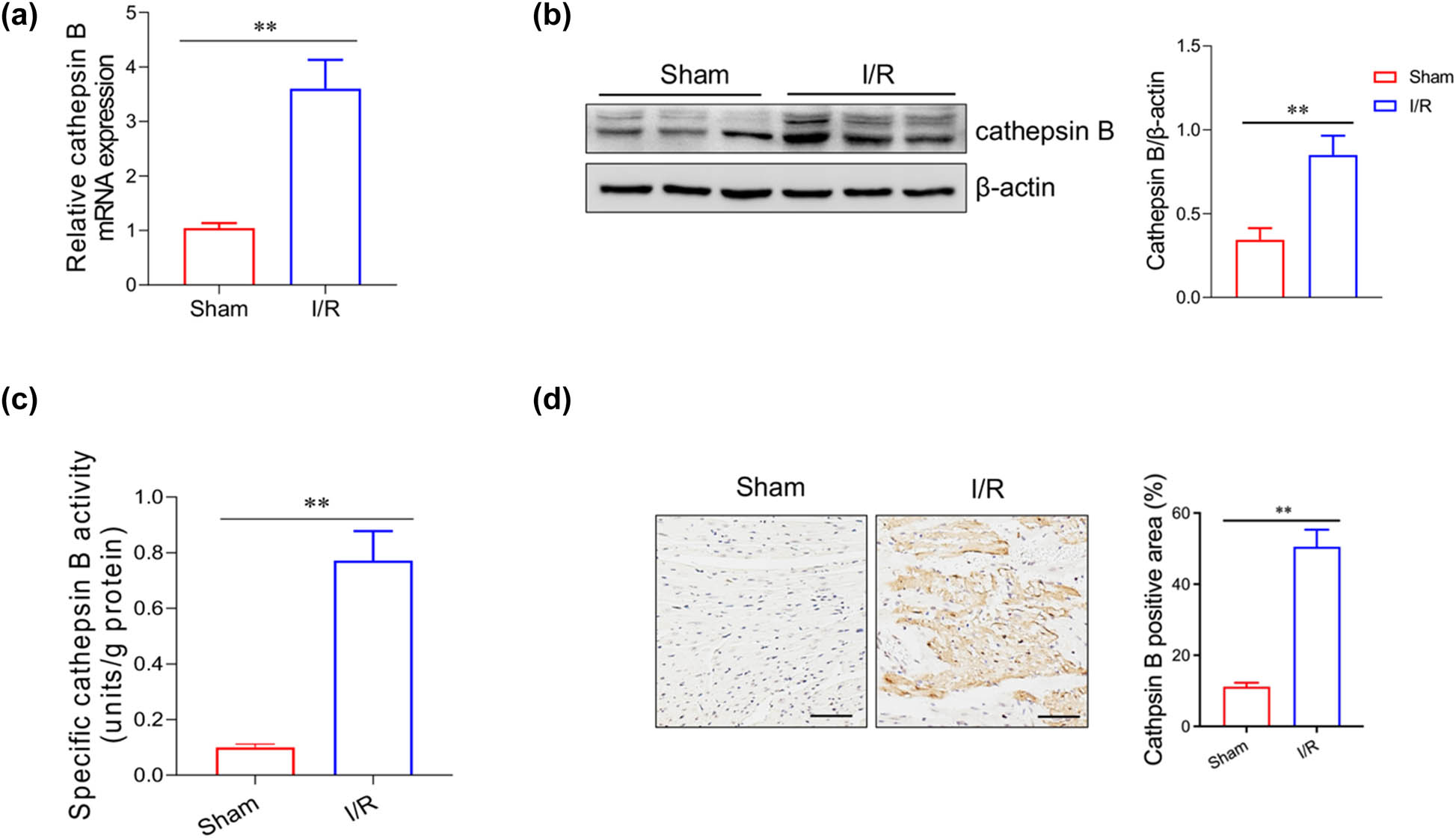
Cathepsin B levels were increased in the heart tissue of myocardial I/R mice. (a) The relative mRNA expression level of cathepsin B in the heart tissue of the myocardial I/R mice was analyzed by qRT-PCR. The protein level (b) and the activity (c) of cathepsin B were evaluated by western blotting analysis and ELISA, respectively. (d) The localization of cathepsin B in the mouse heart tissue was determined by immunohistochemistry. **p < 0.01. Data are representative of three independent experiments with similar results.
3.2 Inhibition of cathepsin B lessens the severity of myocardial injury in mice
To clarify the role of cathepsin B in myocardial injury, we pretreated mice with Ca-074 Me, a specific inhibitor of cathepsin B, before I/R treatment. H&E staining showed that the cardiomyocytes were arranged disorderly and enlarged in I/R treated mice. Meanwhile, the infiltration of inflammatory cells was also observed. After Ca-074 Me treatment, the degree of myocardial injury was significantly alleviated compared with the I/R group (Figure 2a). Ca-074 Me can also reduce the area of heart infarction upon hypoxia (Figure 2b). TUNEL staining showed a higher proportion of apoptotic cells in the infarcted area of the I/R group, which was significantly alleviated by Ca-074 Me (Figure 2c). Moreover, inflammatory cytokine levels (IL-1β, IL-6, TNF-α, and IFN-γ) were significantly lower in the Ca-074 Me-treated I/R group compared to the untreated I/R group (Figure 2d). Additionally, Ca-074 Me can reduce the levels of CK-MB, cTnl, and LDH in serum (Figure 2e–g). The data suggest that cathepsin B may modulate the pathogenesis of myocardial I/R injury.
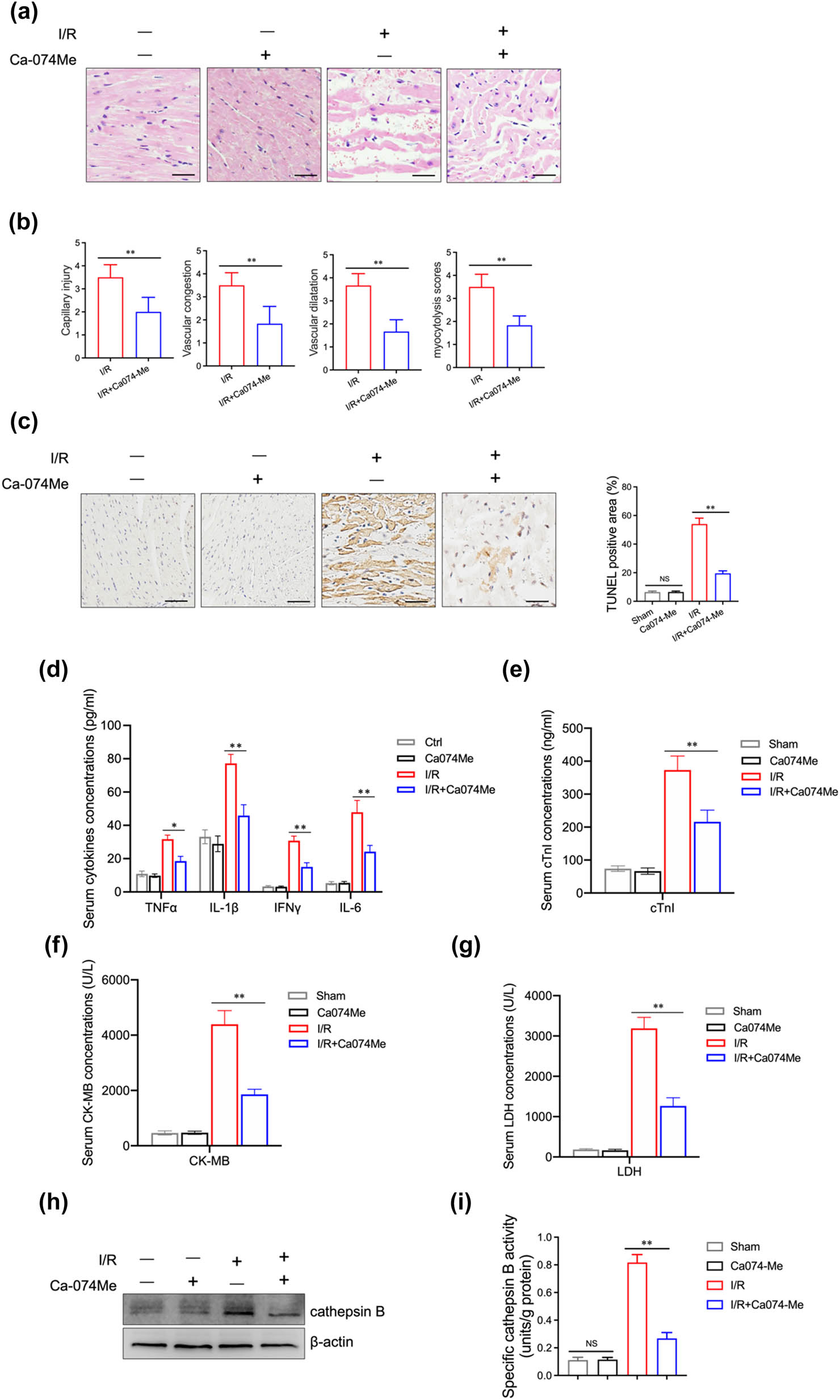
Effects of cathepsin B inhibitor on myocardial injury in myocardial I/R mice. Myocardial ischemia/reperfusion surgery and intraperitoneal injections of Ca-074 Me (50 mg/kg) were simultaneously used to treat mice (n = 6 per group). (a) The histological analysis of the mouse heart on day 1 after surgery was performed by H&E. (b) Capillary injury, vascular congestion, vascular dilatation, and myocytolysis scores of myocardial injury were evaluated. (c) Cell death in the mouse heart tissue was examined by TUNEL staining. Scale bar = 100 μm. (d) The expression level of inflammatory cytokines (TNF-α, IL-1β, IFN-γ, IL-6) in serum was measured by ELISA. The levels of cTnl (e), CK-MB (f), and LDH (g) were detected by ELISA. The protein level (h) and the activity (i) of cathepsin B were evaluated by western blotting analysis and ELISA, respectively. *p < 0.05 and **p < 0.01. Data are representative of three independent experiments with similar results.
3.3 Cathepsin B expression is increased in AC16 cells under hypoxic condition
Myocardial cell damage is the leading cause of cardiac I/R injury [18]. Hypoxia is an essential factor in causing myocardial damage. To investigate whether cathepsin B release is related to hypoxia, we used the human myocardial cell AC16 cells to be cultured in the 1% O2 condition and Cocl2-induced hypoxic environment at different times. The mRNA and protein levels of cathepsin B significantly increased after incubation with 1% O2 (Figure 3a–c) or treatment with varying concentrations of CoCl2 (Figure 3d–f). Furthermore, the activity of cathepsin B was also increased in a time- and dose-dependent manner under hypoxic conditions. Above all, these data indicate that cathepsin B expression was increased in myocardial cells under hypoxia stimulation.
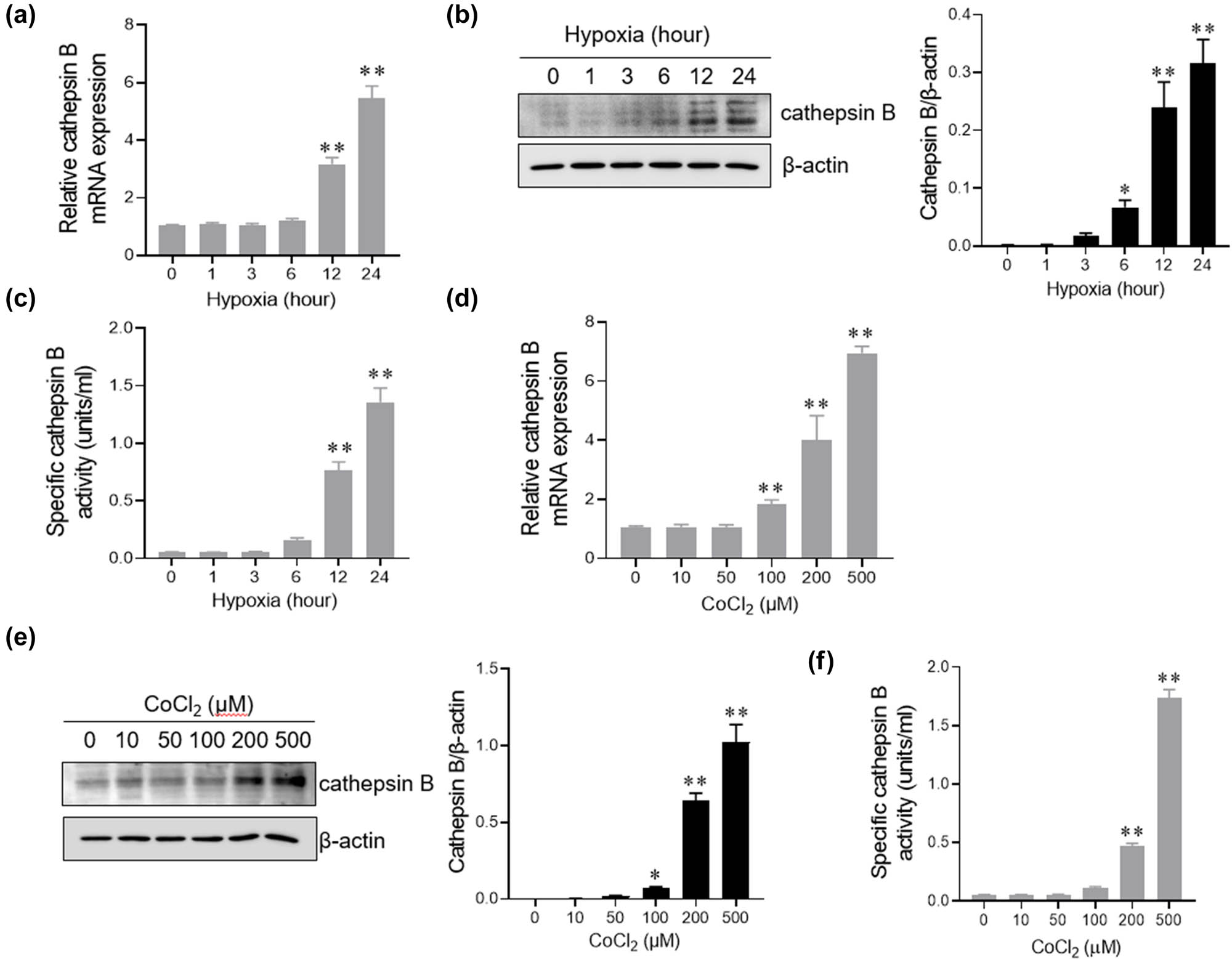
Cathepsin B expression is increased in AC16 cells under hypoxic conditions. (a–c) AC16 cells were incubated with 1% O2 for 0, 1, 3, 6, 12, and 24 h. The mRNA level of cathepsin B was analyzed by q-PCR (a); the protein level of cathepsin B was evaluated by western blotting analysis (b); and the activity of cathepsin B was assessed by ELISA (c). (d–f) AC16 cells were treated with different CoCl2 concentrations (0, 10, 50, 100, 200, and 500 μM) for 24 h. The mRNA level of cathepsin B was analyzed by q-PCR (d). The protein level of cathepsin B was evaluated by western blotting analysis (e). The activity of cathepsin B was assessed by ELISA (f). **p < 0.01. Data are representative of three independent experiments with similar results.
3.4 Inhibition of cathepsin B reduces AC16 cells’ apoptosis under hypoxic condition
To further validate the role of cathepsin B in hypoxia-mediated cardiomyocyte survival, AC16 cells were pretreated with a specific cathepsin B inhibitor, Ca-074 Me, before CoCl2 or 1% O2 treatment. Compared to 1% O2 and CoCl2 treatment, Ca-074 Me pretreatment significantly reversed the proportion of cardiomyocyte apoptosis (Figure 4a and b). Similarly, cathepsin B deficiency decreased hypoxia-induced cardiomyocyte apoptosis (Figure 4c and d). Previous studies have found that cathepsin B is related to endogenous apoptosis in the process of myocardial remodeling [19]. In addition, exogenous cathepsin B can promote cell apoptosis in a dose-dependent manner (Figure S1). These data suggest that cathepsin B inhibition prevents hypoxia-induced cardiomyocyte apoptosis.
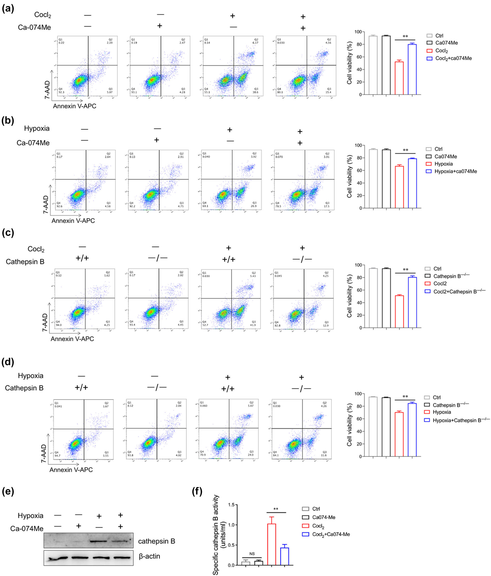
Inhibition of cathepsin B reduces AC16 cell apoptosis under hypoxic conditions. (a) AC16 cells were treated with CoCl2 (200 μM) in combination with or without Ca-074 Me (10 μM) for 24 h. Cell viability of AC16 cells was evaluated by flow cytometry. (b) AC16 cells were cultured under 1% O2 condition in combination with or without Ca-074 Me (10 μM) for 24 h. Cell viability of AC16 cells was determined by flow cytometry. (c) Cathepsin B+/+ and cathepsin B−/− AC16 cells were stimulated with CoCl2 (200 μM) with or without Ca-074 Me (10 μM) for 24 h, and then, the cell viability of AC16 cells was evaluated by flow cytometry. (d) Cathepsin B+/+ and cathepsin B−/− AC16 cells were cultured under 1% O2 condition with or without Ca-074 Me (10 μM) for 24 h, and then the cell viability of AC16 cells was determined by flow cytometry. (e and f) AC16 cells were treated with CoCl2 (200 μM) in combination with or without Ca-074 Me (10 μM) for 24 h. The protein level of cathepsin B was evaluated by western blotting analysis (e); the activity of cathepsin B was assessed by ELISA (f). **p < 0.01. Data are representative of three independent experiments with similar results.
3.5 Cathepsin B promotes AC16 cell apoptosis under hypoxic conditions through the caspase-3 pathway
To verify the conjecture of cathepsin B with cardiomyocyte apoptosis under hypoxia conditions, the caspase-3 pathway-related proteins were analyzed by western blotting analysis. The expression level of caspase-3/8 and FADD was increased after hypoxia induction and significantly reduced upon Ca-074 Me treatment (Figure 5a and b). Similar results were observed in cathepsin B–/– AC16 cells under hypoxia stimulation (Figure 5c and d). To further confirm the role of caspase 3 in cathepsin B-mediated apoptosis, AC16 cells were pretreated with a specific caspase-3 inhibitor, Z-DEVD-FMK. Of note, the cell viability was similar in AC16 cells treated with Ca-074 Me alone or Ca-074 Me plus Z-DEVD-FMK (Figure S2). Overexpression of cathepsin B significantly elevated apoptosis-related protein levels following hypoxia (Figure 5e and f). In conclusion, we determined that cathepsin B promotes the apoptosis of myocardial cells under hypoxic conditions via activating the caspase-3 pathway. In addition, inhibiting the expression of cathepsin B can alleviate the degree of apoptosis in myocardial cells under hypoxic conditions.
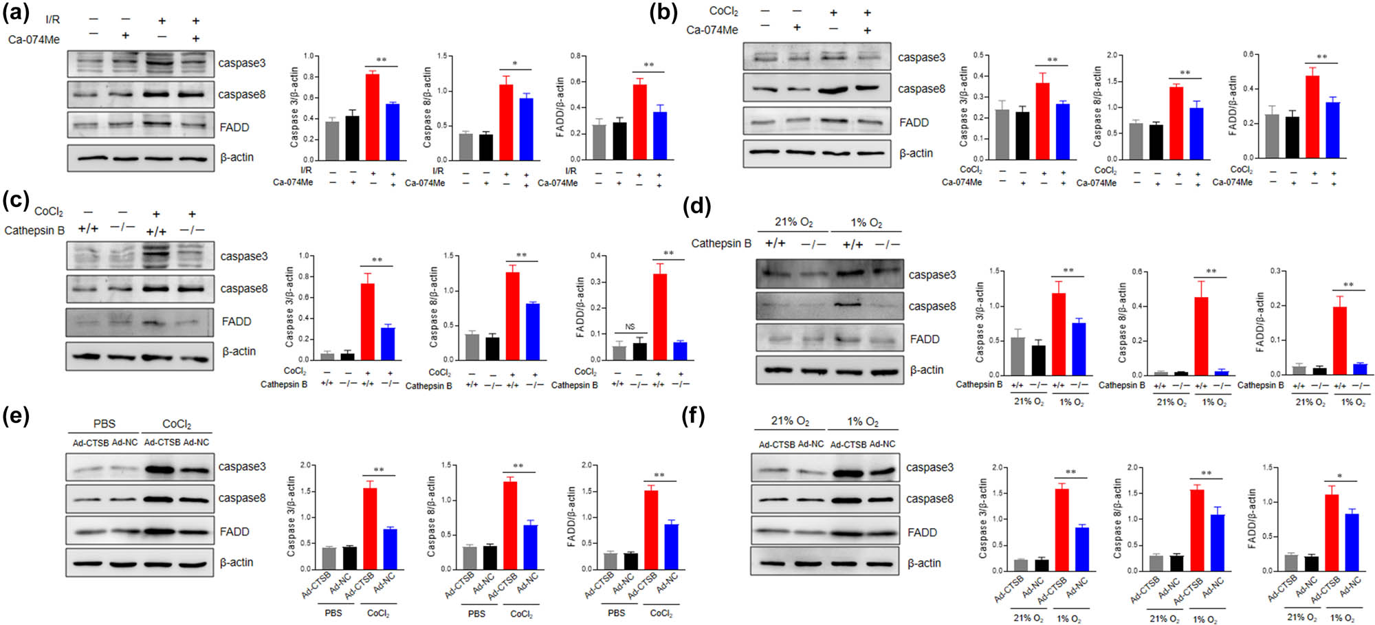
Cathepsin B promotes apoptosis of AC16 cells under hypoxic conditions through the caspase-3 pathway. (a) The protein levels of caspase-3, caspase-8, and FADD in AC16 cells cultured under 1% O2 condition with or without Ca-074 Me (10 μM) were evaluated by western blotting analysis. (b) The protein levels of caspase-3, caspase-8, and FADD in AC16 cells stimulated with CoCl2 (200 μM) for 24 h with or without Ca-074 Me (10 μM) were evaluated by western blotting analysis. (c) Cathepsin B+/+ and cathepsin B−/− AC16 cells were cultured under 1% O2 condition with or without Ca-074 Me (10 μM) for 24 h, then the expression of caspase 3, caspase 8, and FADD was determined by western blotting analysis. (d) Cathepsin B+/+ and cathepsin B−/− AC16 cells were stimulated with CoCl2 (200 μM) for 24 h with or without Ca-074 Me (10 μM), then the expression of caspase 3, caspase 8, and FADD was determined by western blotting analysis. (e and f) AC16 cells were seeded into six-well plates and allowed to grow at 50–70% confluence. The cells were transfected with the expression plasmid of cathepsin B for 48 h. (e) The protein levels of caspase-3, caspase-8, and FADD in AC16 cells stimulated with CoCl2 (200 μM) for 24 h were evaluated by western blotting analysis. (f) The protein levels of caspase-3, caspase-8, and FADD in AC16 cells cultured under 21% O2 or 1% O2 condition were evaluated by western blotting analysis. *p < 0.05, **p < 0.01, ns not significant. Data are representative of three independent experiments with similar results.
4 Discussion
Increased cathepsin gene expression is associated with cardiac stress, remodeling, and dysfunction [20]. In this study, we observed a marked elevation in cathepsin B in murine cardiac tissue in response to I/R-induced myocardial injury. Moreover, our study delineates the beneficial impact of cathepsin B inhibition on myocardial preservation, as evidenced by enhanced cardiomyocyte viability. Concomitantly, cathepsin B inhibition was found to significantly attenuate the induction of pro-inflammatory cytokines in the context of a murine model of cardiac I/R injury. Mechanistic studies further revealed that cathepsin B inhibitor affects cardiomyocyte apoptosis by limiting the activation of the caspase 3 pathway.
Lysosomal proteases are implicated in the pathogenesis of various diseases, including autoimmune, metabolic, cardiovascular, and neurodegenerative conditions [21]. Cathepsins are the major lysosomal proteases localized predominantly in the acidic milieu of endo/lysosomal compartments and are integral to many physiological processes, such as proteolysis, energy metabolism, and immunological responses. Among all the lysosomal proteases, cathepsin B has been identified as a critical agent in lysosomal destabilization and the induction of cellular apoptosis [10,22]. The translocation of cathepsin B into the cytoplasm following lysosomal rupture leads to the initiation of apoptotic signaling cascades. Previous studies demonstrated that cathepsin B contributes to fulminant hepatic failure and acetaminophen hepatotoxicity via hepatocyte apoptosis [23,24]. Furthermore, the involvement of cathepsin B-dependent pathways in TNF-α-mediated hepatocyte apoptosis has been demonstrated by using cathepsin B-deficient mice [25]. This was further validated in a study that pharmacological inhibition of cathepsin B attenuated TNF-α-induced liver injury in mice [26]. Cathepsin B seems to participate in producing pro-apoptotic reactants to protect anti-apoptotic reactants from degradation and removal [14]. Herein, we observed that cathepsin inhibition prevented myocardial I/R injury and cardiomyocyte apoptosis, which is related to reduced caspase 3 activity.
Myocardial cell apoptosis is associated with the production of cytokines that promote inflammation during myocardial injury [4]. We also found that cathepsin B-mediated I/R cardiac damage leads to apoptosis and stimulates the production of pro-inflammatory chemokines. Indeed, cathepsins participate in the production of immune mediators through a limited proteolysis process [9]. Cathepsin B enhances the activation of NLRP3 inflammasome, thereby upregulating IL-1β release from endothelial cells [11]. Also, cathepsin B was required for optimal processing of TNF-α and IL-6 in response to inflammatory stimulation [27,28]. Cathepsin B promotes IκBα degradation and NF-κB nuclear translocation in microglia cells in neonatal mice with hypoxic-ischemic brain injury [29]. Indeed, cathepsin B has been implicated in the pathophysiology of numerous inflammatory diseases and cancer. Therefore, it is interesting to reveal the modulatory function of cathepsin B in the initiation and activation of the signaling pathway related to inflammatory cytokines production, and the role of cathepsin B in the cellular inflammatory response during cardiomyocyte damage remains to be further investigated.
The cardiomyocytes play crucial roles in maintaining myocardial function [30]. Aberrant cardiomyocyte function is linked to the development of myocardial injury. Cardiomyocytes are highly susceptible to apoptosis under hypoxic conditions [31]. Hypoxia induces cellular apoptosis, with the caspase cascade playing a critical role in the induction, transduction, and amplification of intracellular apoptotic signals [32]. Our studies demonstrated that cathepsin B inhibition protects against myocardial injury and hypoxia-induced cardiomyocyte apoptosis. Deng et al. observed that hypoxia-ischemia stimulation significantly increases caspase-3 expression and neuronal apoptosis in the brains of neonatal mice [33]. By attenuating the activation of the caspase-3 enzyme, it is possible to impede the progression of apoptosis, thus alleviating the harmful effects of hypoxia-ischemia on cellular integrity [34]. In the Fas signaling pathway, FADD is an essential adaptor protein critical for transmitting apoptotic signals [35]. FADD recruits caspase-8 through interactions of the death effect domain, activating the apical caspase [36]. In the present study, Ca-074 Me treatment significantly decreased the enhanced expression of caspase-3, caspase-8, and FADD in cardiomyocytes under hypoxia conditions. Furthermore, Ca-074 Me treatment reduced the expression of endogenous apoptosis-related proteins in cardiomyocytes under hypoxia conditions. The data suggested that cathepsin B inhibition-mediated prevention of cardiomyocyte apoptosis was associated with the downregulation of caspase activation.
In summary, our study demonstrated that inhibition of cathepsin B mitigates I/R-induced myocardial injury in murine models by preserving cardiomyocyte viability. Mechanistic studies elucidated that cathepsin B inhibition reduces the expression of proteins associated with the apoptotic pathway, thereby enhancing cardiomyocyte survival. Most importantly, we have provided experimental evidence showing a potential therapeutic efficacy of cathepsin B inhibition towards attenuating myocardial injury in mice.
-
Funding information: This work was supported by the National Key R&D Program of China (grant no: 2023YFC2507500).
-
Author contributions: RS, JZ, and LX designed the research. RS, ZS, and TC performed experiments. HW, ZL, and ZT analyzed data. RS, ZS, JZ, and LX wrote the manuscript.
-
Conflict of interest: The authors have declared that no conflict of interest exists.
-
Data availability statement: Data and materials supporting this research article are available on reasonable request.
References
[1] Roth GA, Mensah GA, Fuster V. The global burden of cardiovascular diseases and risks a compass for global action. J Am Coll Cardiol. 2020;76(25):2980–1.10.1016/j.jacc.2020.11.021Suche in Google Scholar PubMed
[2] Jensen RV, Hjortbak MV, Botker HE. Ischemic heart disease: An update. Semin Nucl Med. 2020;50(3):195–207.10.1053/j.semnuclmed.2020.02.007Suche in Google Scholar PubMed
[3] Heusch G. Myocardial ischaemia-reperfusion injury and cardioprotection in perspective. Nat Rev Cardiol. 2020;17(12):773–89.10.1038/s41569-020-0403-ySuche in Google Scholar PubMed
[4] Markousis-Mavrogenis G, Tromp J, Ouwerkerk W, Devalaraja M, Filippatos G, Van Der Harst P, et al. The clinical significance of interleukin-6 in heart failure: results from the BIOSTAT-CHF study. Eur J Heart Fail. 2019;21:111.10.1002/ejhf.1482Suche in Google Scholar PubMed
[5] Kang SJ, Kishimoto T. Interplay between interleukin-6 signaling and the vascular endothelium in cytokine storms. Exp Mol Med. 2021;53(7):1116–23.10.1038/s12276-021-00649-0Suche in Google Scholar PubMed PubMed Central
[6] Zacho J, Tybjaerg-Hansen A, Jensen JS, Grande P, Sillesen H, Nordestgaard BG. Genetically elevated C-reactive protein and ischemic vascular disease. N Engl J Med. 2008;359(18):1897–908.10.1056/NEJMoa0707402Suche in Google Scholar PubMed
[7] Ji E, Lee S. Antibody-based therapeutics for atherosclerosis and cardiovascular diseases. Int J Mol Sci. 2021;22(11):5770.10.3390/ijms22115770Suche in Google Scholar PubMed PubMed Central
[8] Patel S, Homaei A, El-Seedi HR, Akhtar N. Cathepsins: Proteases that are vital for survival but can also be fatal. Biomed Pharmacother. 2018;105:526–32.10.1016/j.biopha.2018.05.148Suche in Google Scholar PubMed PubMed Central
[9] Yan X, Wu Z, Wang B, Yu T, Hu Y, Wang S, et al. Involvement of cathepsins in innate and adaptive immune responses in periodontitis. Evid Based Complement Altern Med. 2020;2020:4517587.10.1155/2020/4517587Suche in Google Scholar PubMed PubMed Central
[10] Dong LJ, Xie JW, Wang YY, Jiang HL, Chen K, Li DT, et al. Mannose ameliorates experimental colitis by protecting intestinal barrier integrity. Nat Commun. 2022;13(1):4804.10.1038/s41467-022-32505-8Suche in Google Scholar PubMed PubMed Central
[11] Zhang Y, Chen Y, Zhang YZ, Li PL, Li X. Contribution of cathepsin B-dependent Nlrp3 inflammasome activation to nicotine-induced endothelial barrier dysfunction. Eur J Pharmacol. 2019;865:172795.10.1016/j.ejphar.2019.172795Suche in Google Scholar PubMed PubMed Central
[12] Talukdar R, Sareen A, Zhu H, Yuan Z, Dixit A, Cheema H, et al. Release of cathepsin B in cytosol causes cell death in acute pancreatitis. Gastroenterology. 2016;151(4):747–58 e5.10.1053/j.gastro.2016.06.042Suche in Google Scholar PubMed PubMed Central
[13] Guicciardi ME, Deussing J, Miyoshi H, Bronk SF, Svingen PA, Peters C, et al. Cathepsin B contributes to TNF-alpha-mediated hepatocyte apoptosis by promoting mitochondrial release of cytochrome C. J Clin Investigation. 2000;106(9):1127–37.10.1172/JCI9914Suche in Google Scholar PubMed PubMed Central
[14] de Castro MA, Bunt G, Wouters FS. Cathepsin B launches an apoptotic exit effort upon cell death-associated disruption of lysosomes. Cell Death Discov. 2016;2:16012.10.1038/cddiscovery.2016.12Suche in Google Scholar PubMed PubMed Central
[15] Ben-Ari Z, Mor E, Azarov D, Sulkes J, Tor R, Cheporko Y, et al. Cathepsin B inactivation attenuates the apoptotic injury induced by ischemia/reperfusion of mouse liver. Apoptosis. 2005;10(6):1261–9.10.1007/s10495-005-2358-1Suche in Google Scholar PubMed
[16] Feng Y, Ni L, Wang Q. Administration of cathepsin B inhibitor CA-074 Me reduces inflammation and apoptosis in polymyositis. J Dermatol Sci. 2013;72(2):158–67.10.1016/j.jdermsci.2013.06.014Suche in Google Scholar PubMed
[17] Elshikha AS, Teng XY, Kanda N, Li W, Choi SC, Abboud G, et al. TLR7 activation accelerates cardiovascular pathology in a mouse model of lupus. Front Immunol. 2022;13:914468.10.3389/fimmu.2022.914468Suche in Google Scholar PubMed PubMed Central
[18] Susan D, Kent FG. Cellular pathways of death and survival in acute myocardial infarction. J Clin Exp Cardiol. 2013.10.4172/2155-9880.S6-003Suche in Google Scholar
[19] Wu QQ, Xu M, Yuan Y, Li FF, Yang Z, Liu Y, et al. Cathepsin B deficiency attenuates cardiac remodeling in response to pressure overload via TNF-alpha/ASK1/JNK pathway. Am J Physiol Heart Circ Physiol. 2015;308(9):H1143–54.10.1152/ajpheart.00601.2014Suche in Google Scholar PubMed
[20] Blondelle J, Lange S, Greenberg BH, Cowling RT. Cathepsins in heart disease-chewing on the heartache? Am J Physiol Heart Circ Physiol. 2015;308(9):H974–6.10.1152/ajpheart.00125.2015Suche in Google Scholar PubMed PubMed Central
[21] Ge W, Li D, Gao Y, Cao X. The roles of lysosomes in inflammation and autoimmune diseases. Int Rev Immunol. 2015;34(5):415–31.10.3109/08830185.2014.936587Suche in Google Scholar PubMed
[22] Xie Z, Zhao MY, Yan CX, Kong W, Lan F, Zhao SX, et al. Cathepsin B in programmed cell death machinery: Mechanisms of execution and regulatory pathways. Cell Death Dis. 2023;14(4):255.10.1038/s41419-023-05786-0Suche in Google Scholar PubMed PubMed Central
[23] Yan BZ, Wang W, Chen LY, Bi MR, Lu YJ, Li BX, et al. Role of cathepsin B-mediated apoptosis in fulminant hepatic failure in mice. World J Gastroenterol. 2009;15(10):1231–6.10.3748/wjg.15.1231Suche in Google Scholar PubMed PubMed Central
[24] Woolbright BL, Ramachandran A, McGill MR, Yan HM, Bajt ML, Sharpe MR, et al. Lysosomal instability and cathepsin B release during acetaminophen hepatotoxicity. Basic Clin Pharmacol Toxicol. 2012;111(6):417–25.10.1111/j.1742-7843.2012.00931.xSuche in Google Scholar PubMed PubMed Central
[25] Guicciardi ME, Miyoshi H, Bronk SF, Gores GJ. Cathepsin B knockout mice are resistant to tumor necrosis factor-alpha-mediated hepatocyte apoptosis and liver injury: implications for therapeutic applications. Am J Pathol. 2001;159(6):2045–54.10.1016/S0002-9440(10)63056-8Suche in Google Scholar
[26] Baskin-Bey ES, Canbay A, Bronk SF, Werneburg N, Guicciardi ME, Nyberg SL, et al. Cathepsin B inactivation attenuates hepatocyte apoptosis and liver damage in steatotic livers after cold ischemia-warm reperfusion injury. Am J Physiol Gastrointest Liver Physiol. 2005;288(2):G396–402.10.1152/ajpgi.00316.2004Suche in Google Scholar PubMed
[27] Ha SD, Martins A, Khazaie K, Han JH, Chan BMC, Kim SO. Cathepsin B is involved in the trafficking of TNF-alpha-containing vesicles to the plasma membrane in macrophages. J Immunol. 2008;181(1):690–7.10.4049/jimmunol.181.1.690Suche in Google Scholar PubMed
[28] Rasid O, Meriaux V, Khan EM, Borde C, Ciulean IS, Fitting C, et al. Cathepsin B-deficient mice resolve leishmania major inflammation faster in a T cell-dependent manner. PLoS Negl Trop Dis. 2016;10(5):e0004716.10.1371/journal.pntd.0004716Suche in Google Scholar PubMed PubMed Central
[29] Ni JJ, Wu Z, Peterts C, Yamamoto K, Qing H, Nakanishi H. The critical role of proteolytic relay through cathepsins B and E in the phenotypic change of microglia/macrophage. J Neurosci. 2015;35(36):12488–501.10.1523/JNEUROSCI.1599-15.2015Suche in Google Scholar PubMed PubMed Central
[30] Cheng XW, Shi GP, Kuzuya M, Sasaki T, Okumura K, Murohara T. Role for cysteine protease cathepsins in heart disease: Focus on biology and mechanisms with clinical implication. Circulation. 2012;125(12):1551–62.10.1161/CIRCULATIONAHA.111.066712Suche in Google Scholar PubMed
[31] Ren DZ, Li F, Gao A, Cao QW, Liu YR, Zhang JR. Hypoxia-induced apoptosis of cardiomyocytes is restricted by ginkgolide B-downregulated microRNA-29. Cell Cycle. 2020;19(10):1067–76.10.1080/15384101.2020.1731651Suche in Google Scholar PubMed PubMed Central
[32] Sendoel A, Hengartner MO. Apoptotic cell death under hypoxia. Physiology. 2014;29(3):168–76.10.1152/physiol.00016.2013Suche in Google Scholar PubMed
[33] Deng C, Li J, Li L, Sun F, Xie J. Effects of hypoxia ischemia on caspase-3 expression and neuronal apoptosis in the brain of neonatal mice. Exp Ther Med. 2019;17(6):4517–21.10.3892/etm.2019.7487Suche in Google Scholar PubMed PubMed Central
[34] Hu BR, Liu CL, Ouyang Y, Blomgren K, Siesjo BK. Involvement of caspase-3 in cell death after hypoxia-ischemia declines during brain maturation. J Cereb Blood Flow Metab. 2000;20(9):1294–300.10.1097/00004647-200009000-00003Suche in Google Scholar PubMed
[35] Tourneur L, Chiocchia G. FADD: A regulator of life and death. Trends Immunol. 2010;31(7):260–9.10.1016/j.it.2010.05.005Suche in Google Scholar PubMed
[36] Scott FL, Stec B, Pop C, Dobaczewska MK, Lee JJ, Monosov E, et al. The Fas-FADD death domain complex structure unravels signalling by receptor clustering. Nature. 2009;457(7232):1019–22.10.1038/nature07606Suche in Google Scholar PubMed PubMed Central
© 2025 the author(s), published by De Gruyter
This work is licensed under the Creative Commons Attribution 4.0 International License.
Artikel in diesem Heft
- Research Articles
- Network pharmacological analysis and in vitro testing of the rutin effects on triple-negative breast cancer
- Impact of diabetes on long-term survival in elderly liver cancer patients: A retrospective study
- Knockdown of CCNB1 alleviates high glucose-triggered trophoblast dysfunction during gestational diabetes via Wnt/β-catenin signaling pathway
- Risk factors for severe adverse drug reactions in hospitalized patients
- Analysis of the effect of ALA-PDT on macrophages in footpad model of mice infected with Fonsecaea monophora based on single-cell sequencing
- Development and validation of headspace gas chromatography with a flame ionization detector method for the determination of ethanol in the vitreous humor
- CMSP exerts anti-tumor effects on small cell lung cancer cells by inducing mitochondrial dysfunction and ferroptosis
- Predictive value of plasma sB7-H3 and YKL-40 in pediatric refractory Mycoplasma pneumoniae pneumonia
- Antiangiogenic potential of Elaeagnus umbellata extracts and molecular docking study by targeting VEGFR-2 pathway
- Comparison of the effectiveness of nurse-led preoperative counseling and postoperative follow-up care vs standard care for patients with gastric cancer
- Comparing the therapeutic efficacy of endoscopic minimally invasive surgery and traditional surgery for early-stage breast cancer: A meta-analysis
- Adhered macrophages as an additional marker of cardiomyocyte injury in biopsies of patients with dilated cardiomyopathy
- Association between statin administration and outcome in patients with sepsis: A retrospective study
- Exploration of the association between estimated glucose disposal rate and osteoarthritis in middle-aged and older adults: An analysis of NHANES data from 2011 to 2018
- A comparative analysis of the binary and multiclass classified chest X-ray images of pneumonia and COVID-19 with ML and DL models
- Lysophosphatidic acid 2 alleviates deep vein thrombosis via protective endothelial barrier function
- Transcription factor A, mitochondrial promotes lymph node metastasis and lymphangiogenesis in epithelial ovarian carcinoma
- Serum PM20D1 levels are associated with nutritional status and inflammatory factors in gastric cancer patients undergoing early enteral nutrition
- Hydromorphone reduced the incidence of emergence agitation after adenotonsillectomy in children with obstructive sleep apnea: A randomized, double-blind study
- Vitamin D replacement therapy may regulate sleep habits in patients with restless leg syndrome
- The first-line antihypertensive nitrendipine potentiated the therapeutic effect of oxaliplatin by downregulating CACNA1D in colorectal cancer
- Health literacy and health-related quality of life: The mediating role of irrational happiness
- Modulatory effects of Lycium barbarum polysaccharide on bone cell dynamics in osteoporosis
- Mechanism research on inhibition of gastric cancer in vitro by the extract of Pinellia ternata based on network pharmacology and cellular metabolomics
- Examination of the causal role of immune cells in non-alcoholic fatty liver disease by a bidirectional Mendelian randomization study
- Clinical analysis of ten cases of HIV infection combined with acute leukemia
- Investigating the cardioprotective potential of quercetin against tacrolimus-induced cardiotoxicity in Wistar rats: A mechanistic insights
- Clinical observation of probiotics combined with mesalazine and Yiyi Baitouweng Decoction retention enema in treating mild-to-moderate ulcerative colitis
- Diagnostic value of ratio of blood inflammation to coagulation markers in periprosthetic joint infection
- Sex-specific associations of sex hormone binding globulin and risk of bladder cancer
- Core muscle strength and stability-oriented breathing training reduces inter-recti distance in postpartum women
- The ERAS nursing care strategy for patients undergoing transsphenoidal endoscopic pituitary tumor resection: A randomized blinded controlled trial
- The serum IL-17A levels in patients with traumatic bowel rupture post-surgery and its predictive value for patient prognosis
- Impact of Kolb’s experiential learning theory-based nursing on caregiver burden and psychological state of caregivers of dementia patients
- Analysis of serum NLR combined with intraoperative margin condition to predict the prognosis of cervical HSIL patients undergoing LEEP surgery
- Commiphora gileadensis ameliorate infertility and erectile dysfunction in diabetic male mice
- The correlation between epithelial–mesenchymal transition classification and MMP2 expression of circulating tumor cells and prognosis of advanced or metastatic nasopharyngeal carcinoma
- Tetrahydropalmatine improves mitochondrial function in vascular smooth muscle cells of atherosclerosis in vitro by inhibiting Ras homolog gene family A/Rho-associated protein kinase-1 signaling pathway
- A cross-sectional study: Relationship between serum oxidative stress levels and arteriovenous fistula maturation in maintenance dialysis patients
- A comparative analysis of the impact of repeated administration of flavan 3-ol on brown, subcutaneous, and visceral adipose tissue
- Identifying early screening factors for depression in middle-aged and older adults: A cohort study
- Perform tumor-specific survival analysis for Merkel cell carcinoma patients undergoing surgical resection based on the SEER database by constructing a nomogram chart
- Unveiling the role of CXCL10 in pancreatic cancer progression: A novel prognostic indicator
- High-dose preoperative intraperitoneal erythropoietin and intravenous methylprednisolone in acute traumatic spinal cord injuries following decompression surgeries
- RAB39B: A novel biomarker for acute myeloid leukemia identified via multi-omics and functional validation
- Impact of peripheral conditioning on reperfusion injury following primary percutaneous coronary intervention in diabetic and non-diabetic STEMI patients
- Clinical efficacy of azacitidine in the treatment of middle- and high-risk myelodysplastic syndrome in middle-aged and elderly patients: A retrospective study
- The effect of ambulatory blood pressure load on mitral regurgitation in continuous ambulatory peritoneal dialysis patients
- Expression and clinical significance of ITGA3 in breast cancer
- Single-nucleus RNA sequencing reveals ARHGAP28 expression of podocytes as a biomarker in human diabetic nephropathy
- rSIG combined with NLR in the prognostic assessment of patients with multiple injuries
- Toxic metals and metalloids in collagen supplements of fish and jellyfish origin: Risk assessment for daily intake
- Exploring causal relationship between 41 inflammatory cytokines and marginal zone lymphoma: A bidirectional Mendelian randomization study
- Gender beliefs and legitimization of dating violence in adolescents
- Effect of serum IL-6, CRP, and MMP-9 levels on the efficacy of modified preperitoneal Kugel repair in patients with inguinal hernia
- Effect of smoking and smoking cessation on hematological parameters in polycythemic patients
- Pathogen surveillance and risk factors for pulmonary infection in patients with lung cancer: A retrospective single-center study
- Necroptosis of hippocampal neurons in paclitaxel chemotherapy-induced cognitive impairment mediates microglial activation via TLR4/MyD88 signaling pathway
- Celastrol suppresses neovascularization in rat aortic vascular endothelial cells stimulated by inflammatory tenocytes via modulating the NLRP3 pathway
- Cord-lamina angle and foraminal diameter as key predictors of C5 palsy after anterior cervical decompression and fusion surgery
- GATA1: A key biomarker for predicting the prognosis of patients with diffuse large B-cell lymphoma
- Influencing factors of false lumen thrombosis in type B aortic dissection: A single-center retrospective study
- MZB1 regulates the immune microenvironment and inhibits ovarian cancer cell migration
- Integrating experimental and network pharmacology to explore the pharmacological mechanisms of Dioscin against glioblastoma
- Trends in research on preterm birth in twin pregnancy based on bibliometrics
- Four-week IgE/baseline IgE ratio combined with tryptase predicts clinical outcome in omalizumab-treated children with moderate-to-severe asthma
- Single-cell transcriptomic analysis identifies a stress response Schwann cell subtype
- Acute pancreatitis risk in the diagnosis and management of inflammatory bowel disease: A critical focus
- Effect of subclinical esketamine on NLRP3 and cognitive dysfunction in elderly ischemic stroke patients
- Interleukin-37 mediates the anti-oral tumor activity in oral cancer through STAT3
- CA199 and CEA expression levels, and minimally invasive postoperative prognosis analysis in esophageal squamous carcinoma patients
- Efficacy of a novel drainage catheter in the treatment of CSF leak after posterior spine surgery: A retrospective cohort study
- Comprehensive biomedicine assessment of Apteranthes tuberculata extracts: Phytochemical analysis and multifaceted pharmacological evaluation in animal models
- Relation of time in range to severity of coronary artery disease in patients with type 2 diabetes: A cross-sectional study
- Dopamine attenuates ethanol-induced neuronal apoptosis by stimulating electrical activity in the developing rat retina
- Correlation between albumin levels during the third trimester and the risk of postpartum levator ani muscle rupture
- Factors associated with maternal attention and distraction during breastfeeding and childcare: A cross-sectional study in the west of Iran
- Mechanisms of hesperetin in treating metabolic dysfunction-associated steatosis liver disease via network pharmacology and in vitro experiments
- The law on oncological oblivion in the Italian and European context: How to best uphold the cancer patients’ rights to privacy and self-determination?
- The prognostic value of the neutrophil-to-lymphocyte ratio, platelet-to-lymphocyte ratio, and prognostic nutritional index for survival in patients with colorectal cancer
- Factors affecting the measurements of peripheral oxygen saturation values in healthy young adults
- Comparison and correlations between findings of hysteroscopy and vaginal color Doppler ultrasonography for detection of uterine abnormalities in patients with recurrent implantation failure
- The effects of different types of RAGT on balance function in stroke patients with low levels of independent walking in a convalescent rehabilitation hospital
- Causal relationship between asthma and ankylosing spondylitis: A bidirectional two-sample univariable and multivariable Mendelian randomization study
- Correlations of health literacy with individuals’ understanding and use of medications in Southern Taiwan
- Correlation of serum calprotectin with outcome of acute cerebral infarction
- Comparison of computed tomography and guided bronchoscopy in the diagnosis of pulmonary nodules: A systematic review and meta-analysis
- Curdione protects vascular endothelial cells and atherosclerosis via the regulation of DNMT1-mediated ERBB4 promoter methylation
- The identification of novel missense variant in ChAT gene in a patient with gestational diabetes denotes plausible genetic association
- Molecular genotyping of multi-system rare blood types in foreign blood donors based on DNA sequencing and its clinical significance
- Exploring the role of succinyl carnitine in the association between CD39⁺ CD4⁺ T cell and ulcerative colitis: A Mendelian randomization study
- Dexmedetomidine suppresses microglial activation in postoperative cognitive dysfunction via the mmu-miRNA-125/TRAF6 signaling axis
- Analysis of serum metabolomics in patients with different types of chronic heart failure
- Diagnostic value of hematological parameters in the early diagnosis of acute cholecystitis
- Pachymaran alleviates fat accumulation, hepatocyte degeneration, and injury in mice with nonalcoholic fatty liver disease
- Decrease in CD4 and CD8 lymphocytes are predictors of severe clinical picture and unfavorable outcome of the disease in patients with COVID-19
- METTL3 blocked the progression of diabetic retinopathy through m6A-modified SOX2
- The predictive significance of anti-RO-52 antibody in patients with interstitial pneumonia after treatment of malignant tumors
- Exploring cerebrospinal fluid metabolites, cognitive function, and brain atrophy: Insights from Mendelian randomization
- Development and validation of potential molecular subtypes and signatures of ocular sarcoidosis based on autophagy-related gene analysis
- Widespread venous thrombosis: Unveiling a complex case of Behçet’s disease with a literature perspective
- Uterine fibroid embolization: An analysis of clinical outcomes and impact on patients’ quality of life
- Discovery of lipid metabolism-related diagnostic biomarkers and construction of diagnostic model in steroid-induced osteonecrosis of femoral head
- Serum-derived exomiR-188-3p is a promising novel biomarker for early-stage ovarian cancer
- Enhancing chronic back pain management: A comparative study of ultrasound–MRI fusion guidance for paravertebral nerve block
- Peptide CCAT1-70aa promotes hepatocellular carcinoma proliferation and invasion via the MAPK/ERK pathway
- Electroacupuncture-induced reduction of myocardial ischemia–reperfusion injury via FTO-dependent m6A methylation modulation
- Hemorrhoids and cardiovascular disease: A bidirectional Mendelian randomization study
- Cell-free adipose extract inhibits hypertrophic scar formation through collagen remodeling and antiangiogenesis
- HALP score in Demodex blepharitis: A case–control study
- Assessment of SOX2 performance as a marker for circulating cancer stem-like cells (CCSCs) identification in advanced breast cancer patients using CytoTrack system
- Risk and prognosis for brain metastasis in primary metastatic cervical cancer patients: A population-based study
- Comparison of the two intestinal anastomosis methods in pediatric patients
- Factors influencing hematological toxicity and adverse effects of perioperative hyperthermic intraperitoneal vs intraperitoneal chemotherapy in gastrointestinal cancer
- Endotoxin tolerance inhibits NLRP3 inflammasome activation in macrophages of septic mice by restoring autophagic flux through TRIM26
- Lateral transperitoneal laparoscopic adrenalectomy: A single-centre experience of 21 procedures
- Petunidin attenuates lipopolysaccharide-induced retinal microglia inflammatory response in diabetic retinopathy by targeting OGT/NF-κB/LCN2 axis
- Procalcitonin and C-reactive protein as biomarkers for diagnosing and assessing the severity of acute cholecystitis
- Factors determining the number of sessions in successful extracorporeal shock wave lithotripsy patients
- Development of a nomogram for predicting cancer-specific survival in patients with renal pelvic cancer following surgery
- Inhibition of ATG7 promotes orthodontic tooth movement by regulating the RANKL/OPG ratio under compression force
- A machine learning-based prognostic model integrating mRNA stemness index, hypoxia, and glycolysis‑related biomarkers for colorectal cancer
- Glutathione attenuates sepsis-associated encephalopathy via dual modulation of NF-κB and PKA/CREB pathways
- FAHD1 prevents neuronal ferroptosis by modulating R-loop and the cGAS–STING pathway
- Association of placenta weight and morphology with term low birth weight: A case–control study
- Investigation of the pathogenic variants induced Sjogren’s syndrome in Turkish population
- Nucleotide metabolic abnormalities in post-COVID-19 condition and type 2 diabetes mellitus patients and their association with endocrine dysfunction
- TGF-β–Smad2/3 signaling in high-altitude pulmonary hypertension in rats: Role and mechanisms via macrophage M2 polarization
- Ultrasound-guided unilateral versus bilateral erector spinae plane block for postoperative analgesia of patients undergoing laparoscopic cholecystectomy
- Profiling gut microbiome dynamics in subacute thyroiditis: Implications for pathogenesis, diagnosis, and treatment
- Delta neutrophil index, CRP/albumin ratio, procalcitonin, immature granulocytes, and HALP score in acute appendicitis: Best performing biomarker?
- Anticancer activity mechanism of novelly synthesized and characterized benzofuran ring-linked 3-nitrophenyl chalcone derivative on colon cancer cells
- H2valdien3 arrests the cell cycle and induces apoptosis of gastric cancer
- Prognostic relevance of PRSS2 and its immune correlates in papillary thyroid carcinoma
- Association of SGLT2 inhibition with psychiatric disorders: A Mendelian randomization study
- Motivational interviewing for alcohol use reduction in Thai patients
- Luteolin alleviates oxygen-glucose deprivation/reoxygenation-induced neuron injury by regulating NLRP3/IL-1β signaling
- Polyphyllin II inhibits thyroid cancer cell growth by simultaneously inhibiting glycolysis and oxidative phosphorylation
- Relationship between the expression of copper death promoting factor SLC31A1 in papillary thyroid carcinoma and clinicopathological indicators and prognosis
- CSF2 polarized neutrophils and invaded renal cancer cells in vitro influence
- Proton pump inhibitors-induced thrombocytopenia: A systematic literature analysis of case reports
- The current status and influence factors of research ability among community nurses: A sequential qualitative–quantitative study
- OKAIN: A comprehensive oncology knowledge base for the interpretation of clinically actionable alterations
- The relationship between serum CA50, CA242, and SAA levels and clinical pathological characteristics and prognosis in patients with pancreatic cancer
- Identification and external validation of a prognostic signature based on hypoxia–glycolysis-related genes for kidney renal clear cell carcinoma
- Engineered RBC-derived nanovesicles functionalized with tumor-targeting ligands: A comparative study on breast cancer targeting efficiency and biocompatibility
- Relationship of resting echocardiography combined with serum micronutrients to the severity of low-gradient severe aortic stenosis
- Effect of vibration on pain during subcutaneous heparin injection: A randomized, single-blind, placebo-controlled trial
- The diagnostic performance of machine learning-based FFRCT for coronary artery disease: A meta-analysis
- Comparing biofeedback device vs diaphragmatic breathing for bloating relief: A randomized controlled trial
- Serum uric acid to albumin ratio and C-reactive protein as predictive biomarkers for chronic total occlusion and coronary collateral circulation quality
- Multiple organ scoring systems for predicting in-hospital mortality of sepsis patients in the intensive care unit
- Single-cell RNA sequencing data analysis of the inner ear in gentamicin-treated mice via intraperitoneal injection
- Suppression of cathepsin B attenuates myocardial injury via limiting cardiomyocyte apoptosis
- Influence of sevoflurane combined with propofol anesthesia on the anesthesia effect and adverse reactions in children with acute appendicitis
- Identification of hub genes related to acute kidney injury caused by sevoflurane anesthesia and endoplasmic reticulum stress
- Efficacy and safety of PD-1/PD-L1 inhibitors in pancreatic ductal adenocarcinoma: a systematic review and Meta-analysis of randomized controlled trials
- The value of diagnostic experience in O-RADS MRI score for ovarian-adnexal lesions
- Health education pathway for individuals with temporary enterostomies using patient journey mapping
- Serum TLR8 as a potential diagnostic biomarker of coronary heart disease
- Intraoperative temperature management and its effect on surgical outcomes in elderly patients undergoing lichtenstein unilateral inguinal hernia repair
- Immunohistochemical profiling and neuroepithelial heterogeneity in immature ovarian teratomas: a retrospective digital pathology-based study
- Associated risk factors and prevalence of human papillomavirus infection among females visiting tertiary care hospital: a cross-sectional study from Nepal
- Comparative evaluation of various disc elution methods for the detection of colistin-resistant gram-negative bacteria
- Effect of timing of cholecystectomy on weight loss after sleeve gastrectomy in morbidly obese individuals with cholelithiasis: a retrospective cohort study
- Causal association between ceramide levels and central precocious puberty: a mendelian randomization study
- Novel predictive model for colorectal liver metastases recurrence: a radiomics and clinical data approach
- Relationship between resident physicians’ perceived professional value and exposure to violence
- Multiple sclerosis and type 1 diabetes: a Mendelian randomization study of European ancestry
- Rapid pathogen identification in peritoneal dialysis effluent by MALDI-TOF MS following blood culture enrichment
- Comparison of open and percutaneous A1 pulley release in pediatric trigger thumb: a retrospective cohort study
- Impact of combined diaphragm-lung ultrasound assessment on postoperative respiratory function in patients under general anesthesia recovery
- Development and internal validation of a nomogram for predicting short-term prognosis in ICU patients with acute pyelonephritis
- The association between hypoxic burden and blood pressure in patients with obstructive sleep apnea
- Promotion of asthenozoospermia by C9orf72 through suppression of spermatogonia activity via fructose metabolism and mitophagy
- Review Articles
- The effects of enhanced external counter-pulsation on post-acute sequelae of COVID-19: A narrative review
- Diabetes-related cognitive impairment: Mechanisms, symptoms, and treatments
- Microscopic changes and gross morphology of placenta in women affected by gestational diabetes mellitus in dietary treatment: A systematic review
- Review of mechanisms and frontier applications in IL-17A-induced hypertension
- Research progress on the correlation between islet amyloid peptides and type 2 diabetes mellitus
- The safety and efficacy of BCG combined with mitomycin C compared with BCG monotherapy in patients with non-muscle-invasive bladder cancer: A systematic review and meta-analysis
- The application of augmented reality in robotic general surgery: A mini-review
- The effect of Greek mountain tea extract and wheat germ extract on peripheral blood flow and eicosanoid metabolism in mammals
- Neurogasobiology of migraine: Carbon monoxide, hydrogen sulfide, and nitric oxide as emerging pathophysiological trinacrium relevant to nociception regulation
- Plant polyphenols, terpenes, and terpenoids in oral health
- Laboratory medicine between technological innovation, rights safeguarding, and patient safety: A bioethical perspective
- End-of-life in cancer patients: Medicolegal implications and ethical challenges in Europe
- The maternal factors during pregnancy for intrauterine growth retardation: An umbrella review
- Intra-abdominal hypertension/abdominal compartment syndrome of pediatric patients in critical care settings
- PI3K/Akt pathway and neuroinflammation in sepsis-associated encephalopathy
- Screening of Group B Streptococcus in pregnancy: A systematic review for the laboratory detection
- Giant borderline ovarian tumours – review of the literature
- Leveraging artificial intelligence for collaborative care planning: Innovations and impacts in shared decision-making – A systematic review
- Cholera epidemiology analysis through the experience of the 1973 Naples epidemic
- Risk factors of frailty/sarcopenia in community older adults: Meta-analysis
- Supplement strategies for infertility in overweight women: Evidence and legal insights
- Scurvy, a not obsolete disorder: Clinical report in eight young children and literature review
- A meta-analysis of the effects of DBS on cognitive function in patients with advanced PD
- Protective role of selenium in sepsis: Mechanisms and potential therapeutic strategies
- Strategies for hyperkalemia management in dialysis patients: A systematic review
- C-reactive protein-to-albumin ratio in peripheral artery disease
- Research progress on autophagy and its roles in sepsis induced organ injury
- Neuronutrition in autism spectrum disorders
- Pumilio 2 in neural development, function, and specific neurological disorders
- Antibiotic prescribing patterns in general dental practice- a scoping review
- Clinical and medico-legal reflections on non-invasive prenatal testing
- Smartphone use and back pain: a narrative review of postural pathologies
- Targeting endothelial oxidative stress in hypertension
- Exploring links between acne and metabolic syndrome: a narrative review
- Case Reports
- Delayed graft function after renal transplantation
- Semaglutide treatment for type 2 diabetes in a patient with chronic myeloid leukemia: A case report and review of the literature
- Diverse electrophysiological demyelinating features in a late-onset glycogen storage disease type IIIa case
- Giant right atrial hemangioma presenting with ascites: A case report
- Laser excision of a large granular cell tumor of the vocal cord with subglottic extension: A case report
- EsoFLIP-assisted dilation for dysphagia in systemic sclerosis: Highlighting the role of multimodal esophageal evaluation
- Molecular hydrogen-rhodiola as an adjuvant therapy for ischemic stroke in internal carotid artery occlusion: A case report
- Coronary artery anomalies: A case of the “malignant” left coronary artery and its surgical management
- Combined VAT and retroperitoneoscopy for pleural empyema due to nephro-pleuric fistula in xanthogranulomatous pyelonephritis
- A rare case of Opalski syndrome with a suspected multiple sclerosis etiology
- Newly diagnosed B-cell acute lymphoblastic leukemia demonstrating localized bone marrow infiltration exclusively in the lower extremities
- Rapid Communication
- Biological properties of valve materials using RGD and EC
-
A single oral administration of flavanols enhances short
-term memory in mice along with increased brain-derived neurotrophic factor - Repeat influenza incidence across two consecutive influenza seasons
- Letter to the Editor
- Role of enhanced external counterpulsation in long COVID
- Expression of Concern
- Expression of concern “A ceRNA network mediated by LINC00475 in papillary thyroid carcinoma”
- Expression of concern “Notoginsenoside R1 alleviates spinal cord injury through the miR-301a/KLF7 axis to activate Wnt/β-catenin pathway”
- Expression of concern “circ_0020123 promotes cell proliferation and migration in lung adenocarcinoma via PDZD8”
- Corrigendum
- Corrigendum to “Empagliflozin improves aortic injury in obese mice by regulating fatty acid metabolism”
- Corrigendum to “Comparing the therapeutic efficacy of endoscopic minimally invasive surgery and traditional surgery for early-stage breast cancer: A meta-analysis”
- Corrigendum to “The progress of autoimmune hepatitis research and future challenges”
- Retraction
- Retraction of “miR-654-5p promotes gastric cancer progression via the GPRIN1/NF-κB pathway”
- Retraction of: “LncRNA CASC15 inhibition relieves renal fibrosis in diabetic nephropathy through downregulating SP-A by sponging to miR-424”
- Retraction of: “SCARA5 inhibits oral squamous cell carcinoma via inactivating the STAT3 and PI3K/AKT signaling pathways”
- Special Issue Advancements in oncology: bridging clinical and experimental research - Part II
- Unveiling novel biomarkers for platinum chemoresistance in ovarian cancer
- Lathyrol affects the expression of AR and PSA and inhibits the malignant behavior of RCC cells
- The era of increasing cancer survivorship: Trends in fertility preservation, medico-legal implications, and ethical challenges
- Bone scintigraphy and positron emission tomography in the early diagnosis of MRONJ
- Meta-analysis of clinical efficacy and safety of immunotherapy combined with chemotherapy in non-small cell lung cancer
- Special Issue Computational Intelligence Methodologies Meets Recurrent Cancers - Part IV
- Exploration of mRNA-modifying METTL3 oncogene as momentous prognostic biomarker responsible for colorectal cancer development
- Special Issue The evolving saga of RNAs from bench to bedside - Part III
- Interaction and verification of ferroptosis-related RNAs Rela and Stat3 in promoting sepsis-associated acute kidney injury
- The mRNA MOXD1: Link to oxidative stress and prognostic significance in gastric cancer
- Special Issue Exploring the biological mechanism of human diseases based on MultiOmics Technology - Part II
- Dynamic changes in lactate-related genes in microglia and their role in immune cell interactions after ischemic stroke
- A prognostic model correlated with fatty acid metabolism in Ewing’s sarcoma based on bioinformatics analysis
- Red cell distribution width predicts early kidney injury: A NHANES cross-sectional study
- Special Issue Diabetes mellitus: pathophysiology, complications & treatment
- Nutritional risk assessment and nutritional support in children with congenital diabetes during surgery
- Correlation of the differential expressions of RANK, RANKL, and OPG with obesity in the elderly population in Xinjiang
- A discussion on the application of fluorescence micro-optical sectioning tomography in the research of cognitive dysfunction in diabetes
- A review of brain research on T2DM-related cognitive dysfunction
- Metformin and estrogen modulation in LABC with T2DM: A 36-month randomized trial
- Special Issue Innovative Biomarker Discovery and Precision Medicine in Cancer Diagnostics
- CircASH1L-mediated tumor progression in triple-negative breast cancer: PI3K/AKT pathway mechanisms