Surface-engineered quantum dot nanocomposites for neurodegenerative disorder remediation and avenue for neuroimaging
-
Muhanna K. A. Al-Muhanna
 ,
Amani Ahmed Alghamdi
,
Amani Ahmed Alghamdi

Abstract
This review investigates the potential of surface-engineered quantum dot (QD) nanocomposites in the treatment of neurodegenerative disorders and their use in neuroimaging. QDs possess distinctive properties such as adjustable fluorescence and adaptable surface modification, making them useful for both targeted drug administration and precise imaging in the complex nervous system. The report provides a concise overview of progress and obstacles, with a particular focus on concerns regarding biocompatibility, potential toxicity, and the ability to cross the blood–brain barrier. The importance of continuous research in surface engineering, biocompatibility investigations, and standardization of synthesis procedures is emphasized as essential measures to overcome these obstacles. Although there are challenges, the review highlights the significant potential of surface-engineered QD nanocomposites to transform our understanding of neurodegenerative disorders and improve neuroimaging techniques. This could lead to better diagnostic and therapeutic approaches for neurological health in the future.
Graphical abstract
1 Introduction
Neurodegenerative disorders encompass a wide range of debilitating conditions characterized by a progressive loss of the nervous system, namely the neurons in the brain. Disorders including Alzheimer’s disease (AD), Parkinson’s disease (PD), and Huntington’s disease are characterized by a variety of symptoms, including cognitive decline, motor dysfunction, and, occasionally, psychological abnormalities [1,2,3]. The fundamental cause of the condition frequently entails the buildup of abnormal proteins or the deterioration of particular groups of nerve cells. Although age is a notable risk factor, many neurodegenerative illnesses can affect individuals at a younger age as a result of genetic factors. At present, there are no conclusive therapies for these conditions. Thus, it is crucial to recognize and manage them early. Current research endeavours aim to elucidate the complex mechanisms behind neurodegeneration, providing optimism for advancements in treatment and preventative approaches [4,5]. Technological advancements, such as brain imaging and biomarker identification, play a role in the early detection and monitoring of disease progression. Furthermore, promising improvements in the field of therapy, such as targeted pharmaceuticals and gene-based treatments, offer the potential to alter the progression of many conditions. The significance of promptly identifying and addressing neurodegenerative diseases cannot be exaggerated, as these disorders typically progress gradually and covertly. Timely diagnosis enables prompt intervention and the adoption of techniques to effectively manage symptoms, thereby enhancing the quality of life for affected persons [6,7,8]. Early identification is crucial for protecting cognitive function and motor ability in numerous neurodegenerative illnesses, as irreparable damage gradually develops over time [6]. Furthermore, the prompt recognition enables the timely implementation of suitable medical, psychological, and supportive measures, empowering patients and their families to strategize for the future. Moreover, timely identification plays a crucial role in promoting progress in scientific investigations by offering a chance to investigate potential treatment interventions and strategies that can influence the course of the disease [9]. As the quest for targeted medicines and personalized medicine persists, early detection remains a fundamental aspect of the overall strategy to tackle the difficulties presented by neurodegenerative diseases.
Nanomaterials have demonstrated great potential in the field of treating neurodegenerative disorders [10]. Nanostructure materials such as nanotubes, nanowires (1D), and graphene, as well as transition metal dichalcogenides (2D), contain distinct characteristics that make them appealing for therapeutic applications [11,12,13,14,15]. Their extensive surface area, tuneable characteristics, and capacity to engage with biological systems on a nanoscale level present promising prospects for medication administration, imaging, etc. [16,17,18]. Nevertheless, these nanomaterials possess certain limitations that require meticulous consideration for their effective implementation in the treatment of neurodegenerative disorders. One significant issue is the potential toxicity of these nanomaterials. Furthermore, biodistribution and clearance concerns impede their successful usage, as ensuring that nanomaterials reach specific areas in the brain while avoiding accumulating in key organs is difficult. Nanomaterials face a significant challenge in efficiently crossing the blood–brain barrier (BBB) to access neural tissue [19]. Ensuring long-term stability is a significant challenge, as nanomaterials have the potential to aggregate or degrade over time, which can compromise their effectiveness in therapy. To fully exploit the potential of different nanomaterials in developing safe and effective therapeutics for neurodegenerative illnesses, it is essential to address these constraints as research advances.
Quantum dots (QDs) constitute a cutting-edge technology with great potential for applications in diverse sectors, such as medicine and neuroscience [20,21,22,23,24]. The tiny semiconductor particles often made of cadmium selenide or indium phosphide display distinctive optical and electrical characteristics due to quantum confinement effects. QDs in the field of neuroimaging have attracted significant interest due to their capacity to potentially transform the visualization and monitoring of brain structures [25,26]. Due to their small dimensions and tuneable optical characteristics, they are highly suitable for marking and visualizing specific cells or structures within the intricate neural network [27,28]. The functionalization of QDs to target particular biomolecules permits the labelling of neurons and other relevant components with extreme precision and selectivity. This capacity has tremendous potential for enhancing our comprehension of brain anatomy and operation. Moreover, the durability and luminescence of QDs render them highly valuable tools for conducting extended imaging investigations, providing a vital understanding of the ever-changing processes occurring within the brain. As research advances in this area, QDs could become essential instruments for high-resolution and real-time neuroimaging, providing new opportunities to investigate brain disorders and aiding the creation of precise therapeutic approaches [21,29]. Neuroimaging techniques and the study of neurodegenerative diseases stand to benefit greatly from surface-engineered QD nanocomposites, which are highlighted in the review as having enormous promise. This review focuses on the innovative use of surface-engineered QDs for both repair and imaging in neurodegenerative illnesses. The uniqueness of this strategy is found in the versatile abilities of QDs, which provide exceptional optical characteristics, allowing for enhanced imaging precision and accuracy in identifying initial pathogenic alterations in neural tissues. QDs, unlike conventional imaging methods, can be customized with precise surface alterations that improve their compatibility with living organisms and allow them to be delivered specifically to damaged areas of the brain. This enables both diagnostic and therapeutic interventions. Incorporating QDs into therapeutic frameworks offers a revolutionary approach to both monitoring and reducing the progression of diseases, particularly neurodegenerative diseases. This has the potential to significantly change how we treat these types of diseases. This study highlights the notable progress and potential of QDs in addressing current constraints in neuroimaging and treatment. It positions QDs as an innovative and adaptable tool in combating debilitating neurological illnesses.
2 Surface engineering of QDs
Surface engineering of QDs plays an essential role in tailoring their characteristics for specific applications in optoelectronics, bioimaging, and theranostics [30,31,32,33,34]. It is widely acknowledged that surface passivation with functional groups not only creates surface defects that give rise to fluorescence but also offers reactive sites for targeted functional modifications. Utilizing these reactive groups, a wide range of materials, including organic, polymeric, inorganic, and biological substances, can be attached to QD surfaces [35,36,37]. This is achieved through covalent bonds, hydrogen bonds, and electrostatic interactions [38,39]. These attachments serve as a foundation for targeted sensing, drug delivery, and other specialized functions [40]. Experimental evidence indicates that colloidal QDs comprise an inorganic core (that includes inner atoms) that retains the bulk crystal lattice’s geometry, an outer inorganic atom surface (which includes outer atoms) that has a distinct morphology from the inner core, and a layer of ligands bonded to the outer surface [41]. The QD’s photophysical properties are determined by its core, which is influenced by factors such as bulk materials, shape, and confinement effects. On the other hand, the surface of the QDs can also have an impact on these properties. Figure 1 depicts the classification of nanostructures into two limiting cases: clusters and bulk crystals, as well as the potential effects of surface and ligand layers on their electronic structure.
![Figure 1
(a) Graphical representation for classification of clusters, QDs, and bulk and (b) the effect of ligand passivation on QD band gap. Reproduced with permission from Kilina et al. [42], © 2015 American Chemical Society.](/document/doi/10.1515/ntrev-2024-0128/asset/graphic/j_ntrev-2024-0128_fig_001.jpg)
(a) Graphical representation for classification of clusters, QDs, and bulk and (b) the effect of ligand passivation on QD band gap. Reproduced with permission from Kilina et al. [42], © 2015 American Chemical Society.
There is a distinct difference between bulk materials and QDs. QDs are nanostructures that fall within the range where quantum confinement effects come into play. In other words, the size of the QD is such that the exciton Bohr radius is either equal to or greater than the QD radius. The nanosystem’s electronic structure is determined by its geometry. This includes factors such as the size of the system, which can have confinement effects, as well as the stoichiometry and the interface between passivating ligands and the QD surface, which can have surface effects.
Cho et al. successfully devised a simple and efficient technique for controlling precisely the surface characteristics of luminescent graphene quantum dots (GQDs) [43]. The surfaces of the hydrophilic GQDs were modified based on the number of grafted hydrophobic hexylamine during the synthesis process. Unlike the pure GQDs, the modified GQDs can stabilize oil-in-water Pickering emulsions and submicron-sized colloidal particles in mini-emulsion polymerization. The authors demonstrated (Figure 2) full-range control of the surface properties of the GQDs, ranging from very hydrophilic to completely hydrophobic, using a series of four modified GQDs with gradually increasing levels of grafted hexylamine. The efficiency of modified GQDs as particle surfactants in various immiscible blends was investigated using a controlled hydrophilic/hydrophobic balance. Furthermore, all emulsions and particles stabilized by GQD surfactants were highly stable and fluorescent, providing a successful example of synergistic inclusion of the unique GQD characteristics into composites. The synthesis of QDs−antibody bioconjugates (QDs-mAb) involved the incorporation of polyethylene glycol (PEG) cross-linkers and Fc-shielding mAb fragments [44]. This was done to enhance the circulation times and targeting efficiency of the bioconjugates in vivo. When the QDs-mAb directed against cell adhesion molecules was used to incubate endothelial cell cultures and Fc-mediated interaction was reduced, the microscopy results showed increased specificity for the molecular targets.
![Figure 2
Method of preparing surface-engineered GQDs using hexylamines. Reproduced with the permission from Cho et al. [43], © 2015 American Chemical Society.](/document/doi/10.1515/ntrev-2024-0128/asset/graphic/j_ntrev-2024-0128_fig_002.jpg)
Method of preparing surface-engineered GQDs using hexylamines. Reproduced with the permission from Cho et al. [43], © 2015 American Chemical Society.
There are different surface engineering procedures that are utilized to improve stability, biocompatibility, and usage [44]. QD surfaces can be tailored through ligand exchange, encapsulation with polymers, and inorganic shell growth processes. These methods enhance the QD’s ability to dissolve in various solvents and offer chances for interaction with biomolecules or other functional groups, allowing precise manipulation of their interactions with biological systems or other materials [43]. Furthermore, surface engineering can efficiently tackle issues such as photobleaching and toxicity, hence improving the use of QDs in various future technologies.
The incorporation of green biomaterial concepts in the production of QDs signifies a notable progress in terms of environmental sustainability and biomedical safety [45]. Conventional QD synthesis processes commonly employ heavy metals and toxic solvents, resulting in the production of dangerous waste and posing possible health hazards. The principles of green biomaterials promote the utilization of renewable, non-toxic resources and ecologically friendly procedures. This move not only decreases the environmental impact of QD manufacture but also improves their ability to interact with living organisms, making them more secure for use in medical applications, such as seeing biological structures and administering medication [31,35,46]. Green QDs are specifically engineered to retain the desirable traits of traditional QDs, such as strong luminescence and adjustable electrical properties, while mitigating the potential risks to the environment and human health [47]. Green QD production utilizes natural precursors and aqueous-based synthesis processes to minimize the emission of hazardous by-products, hence promoting a more environmentally friendly and secure manufacturing process. Moreover, these green QDs frequently exhibit biodegradability, implying that they can decompose into harmless constituents gradually, so mitigating the ecological consequences in the long run [48]. By using green biomaterial principles in the synthesis of QDs, not only do we support worldwide initiatives to create a circular economy, but we also guarantee the safe and efficient utilization of these advanced materials in various applications, including medical diagnostics and environmental sensing.
3 Nanocomposites in neurodegenerative disorder remediation
Neurological diseases, which are a significant cause of disability and mortality globally, contribute to 12% of all fatalities. Neuroinflammation (INF) is widely recognized as a common feature of various neurological disorders, including PD, AD, and multiple sclerosis (MS), among others. Due to the presence of the BBB, numerous drugs with potential therapeutic effects for neurological diseases are unable to penetrate the brain in sufficient amounts to be effective [49,50,51]. The discovery of medicines for central nervous system (CNS) illnesses has a lengthy history. However, because of low absorption rates, inadequate concentrations, and the absence of personalized therapy, these medications may have limited efficacy for the majority of patients. Hence, it is imperative to develop distinct therapeutic approaches that effectively target and destroy diseased areas while sparing normal tissue. The CNS undergoes immunological activation in response to viral infections, immune-mediated illnesses, and neurodegenerative diseases [52]. This activation entails the operation of microglia and astrocytes, which are the immune cells residing in the CNS [53,54,55]. Furthermore, myeloid cells not only contribute to the inflammatory response but also play a role in regulating neuronal function by facilitating developmental synaptic pruning and plasticity in the healthy brain [56]. The generation of neurotoxic substances such as cytokines and interleukins has been linked to neurodegenerative disorders like AD, MS, PD, and amyotrophic lateral sclerosis, among others.
Although nanomedicines possess exceptional physical and chemical characteristics that offer significant benefits in diagnosing CNS illnesses, there are still unresolved issues regarding potential health risks and the implementation of nanomedicines in clinical surroundings [57]. Nanotechnology offers groundbreaking applications in the diagnosis of brain diseases, leveraging the unique properties of nanoparticles (NPs) to enhance imaging precision and therapeutic targeting. Table 1 shows different types of NPs and their applications in comparison to QDs. Among the most notable are iron-oxide nanoparticles (IONPs), which are extensively used due to their superparamagnetic characteristics, making them highly effective in magnetic resonance imaging (MRI) [58]. IONPs functionalized with molecules such as citric acid and RGD peptides exhibit improved permeability and retention, specifically targeting glioblastomas. These functionalized NPs can cross the BBB, allowing for detailed imaging of brain tumours in both in vitro studies with U87-MG glioblastoma cells and in vivo models such as BALB/c mice. The integration of IONPs into MRI protocols provides high-contrast images, crucial for the early detection and monitoring of glioblastomas and INF, underscoring their significance in neurodiagnostic applications. Gold nanoparticles (AuNPs) present another innovative approach, particularly effective for their optical and electronic properties that are utilized in various imaging techniques. When conjugated with PEG or antibodies such as anti-CD133, AuNPs enhance the targeting specificity for glioblastomas, facilitating advanced imaging techniques like laser scanning microscopy and X-ray computed tomography (CT) [59]. These conjugated AuNPs provide enhanced signal contrast and enable the identification of tumour margins with greater accuracy, making them invaluable in the pre-surgical planning and real-time monitoring of tumour progression. Studies involving AuNPs have demonstrated promising results in vitro with glioblastoma cell lines and in animal models, indicating their potential for clinical translation. Additionally, manganese-based NPs offer distinct advantages due to their paramagnetic properties, which make them suitable for use in MRI [60]. These NPs, when encapsulated in hollow structures or combined with other targeting ligands, improve the imaging of hypoxic regions within brain tumours, aiding in the early detection and treatment planning of glioblastomas [61]. Carbon-based NPs, such as carbon dots, are another promising category, particularly for their biocompatibility and multifunctionality [62,63]. When conjugated with specific peptides, carbon dots can serve as dual-modality probes for both positron emission tomography (PET) and CT imaging. This dual functionality is crucial for comprehensive brain imaging, as it allows for the simultaneous acquisition of metabolic and structural data, which is especially useful in the diagnosis and monitoring of complex conditions like AD and glioblastoma [64,65]. Liposomal formulations have also emerged as significant players in the realm of nanotechnology-based diagnostics for brain diseases. These formulations can encapsulate various imaging agents, including heptamethine cyanine dye IR780, to enhance near-infrared (NIR) fluorescence imaging of glioblastomas. The use of liposomes ensures targeted delivery and prolonged circulation time, which is pivotal for achieving high-resolution images of brain tumours [66]. Moreover, the versatility of liposomes allows for their adaptation in targeting other neurological conditions, making them a flexible tool in neurodiagnostics. Other NP-based strategies include the use of polysaccharide-based NPs for MRI and CT imaging and micellar systems for the delivery of imaging probes across the BBB [67]. These NPs can be tailored for specific diagnostic applications, ensuring high biocompatibility and minimal toxicity. The incorporation of gadolinium-loaded liposomes and micelles further enhances the imaging capabilities, providing detailed insights into brain pathologies [68]. Overall, the diverse array of nanotechnology-based applications in brain disease diagnostics underscores the potential of NPs in transforming the field of neuroimaging. From high-resolution MRI to multifunctional PET/CT imaging, these advancements highlight the importance of NPs in achieving precise, non-invasive diagnosis and targeted treatment of brain diseases [69]. The continued development and integration of these technologies hold promise for improving patient outcomes through early detection and personalized treatment strategies.
An overview of the applications of nanotechnology in diagnosing brain illnesses
| NPs | Targeting strategy | Neuroimaging technique | Targeted brain disease | Ref. |
|---|---|---|---|---|
| Iron oxides | Caffeic acid functionalized | MRI | Gioblastoma | [70] |
| Iron oxides | Cyclic RGD-covalently coupled IONPs functionalized with phosphonate PEG chains | MRI | Gioblastoma | [71] |
| Superparamagnetic IONPs | Microwave | Emergent stroke | [65] | |
| Gold | AuNPs coated with CBP4 peptide and PEG | Laser scanning confocal microscope | Gioblastoma | [67] |
| Manganese oxide | N-(trimethoxysilylpropyl) ethylene diamine triacetic acid silane and folic acid-conjugated | MRI | Gioblastoma | [68] |
| MWCNTs | Conjugated with Pittsburgh Compound B and gadolinium | SPECT and CT | AD | [66] |
| Polysiloxane | AGuIX | MRI | Gioblastoma | [58] |
| Liposomes | Heptamethine cyanine dye IR780 | NIR fluorescence imaging | Gioblastoma | [64] |
| Liposomes | IONPs and DiR in PEG liposomes functionalized with PGN635 F(ab′)2 fragments | MRI, NIR optical imaging | Gioblastoma | [57] |
| Liposomes | Gadolinium-diethylenetriaminepentaacetic acid-loaded PEG-coated liposomes | MRI | Gioblastoma | [61] |
| Liposomes | QD and doxorubicin-loaded liposomes | MRI | Gioblastoma | [62] |
| Liposomes | QD and docetaxel-loaded | MRI | Gioblastoma | [59] |
| Liposomes | PEG-b-poly(l-lysine-DOTA-gadolinium) | MRI | Ischaemia–reperfusion injury | [60] |
| Nanobodies | Anti-Aβ and anti-pTau | 2PFI | AD | [63] |
| QDs | Encapsulated in poly(styrene-co-maleic anhydride) and conjugated with PEG | 2PFI | AD | [69] |
3.1 Nanotheranostics and its usages in neurodegenerative disorders
The advancements in technology across various fields have clearly showcased their beneficial influence on the advancement of medical research. These breakthroughs have enabled the development of cutting-edge and efficient methods, apparatus, and systems that may address a diverse array of intricate diseases and disorders within the human body. Moreover, the increase in research endeavours in this field has led to the development of a wide range of approaches that can efficiently tackle different diseases [72,73,74]. Nanotheranostics, as defined by Zhang et al., involves the administration of a nanotheranostic agent through various drug particles. Once the agent reaches the intended area of the body, the medication’s outer layer tends to break down, leading to the release of the agents. This facilitates the targeting of specific chemicals or neurons that are responsible for or contribute to the illness [75]. Tripathy et al. assert that the treatment is receiving significant interest in the medical sector due to its very aggressive nature and its precise targeting of the damaged location in patients’ bodies [76]. Furthermore, it may be customized based on the specific ailment and requirements of each person, resulting in the improved practical implementation of the strategy and offering personalized therapy and medication choices [77]. The therapy or treatment has become viable as a result of recent breakthroughs in chemistry and technology. The application of different metal NPs enables the efficient conversion of electromagnetic waves into medically relevant processes at the nanoscale [78]. This chemical phenomenon is paired with laser technology to enhance the penetration of substances into deeper levels of human tissue, resulting in a simpler and more effective method. Although these advancements appear to greatly enhance the effectiveness of the nanotheranostic process, it is debateable whether the current results are solely derived from laboratory studies and the practical use in living organisms still poses a hurdle [79]. The therapy has undergone testing in various scenarios and has been refined through the use of animal, cell, and human modelling. A range of experiments have been carried out, and the results are outlined below to elucidate the current advancements in the field [80,81,82].
In nanotheranostics, the diagnostic agents utilized for optical imaging, MRI, nuclear imaging, CT, and radionuclides are QDs or fluorescent dyes, iron oxides, radionuclides, and heavy metals, respectively. Therapeutic agents can take the form of hydrophobic chemical medicines, proteins, peptides, genetic elements, or even diagnostic tools [83]. The main components of a nanotheranostic system are nanocarriers in addition to imaging or diagnostic agents. Drug efficacy is increased with decreased toxicity owing to reduced non-specific biodistribution, and nanocarriers promote pharmacokinetics and improve biodistribution of loaded therapeutic and diagnostic moieties at the target tissues [84,85]. Functionalization of these nanocarriers with biomarkers or other targeting ligands has been identified, enabling target-specific treatments and real-time diagnostic capabilities. Nanocarrier loading increases the stability of therapeutic substances, including tiny hydrophobic compounds, peptide medicines, and oligonucleotides [86]. Radiation therapy, immunotherapy, chemotherapy, and surgery are some of the current options for treating neurological illnesses. Additional contemporary treatment modalities for neurological diseases have recently been investigated, including gene therapy, hyperthermia therapy, and stem cell therapy [87]. However, early diagnosis is essential for achieving effective therapy [88]. The utilization of nanotechnology enables the creation of specialized NPs, which can be employed to boost biomedical capabilities for many chronic diseases. This novel method is now being adopted for therapy and diagnostics. NPs ranging in size from 1 to 100 nm can readily be linked with various ligands such as pharmaceuticals, nucleic acids, proteins, and enzymes, depending on their surface properties, physical and chemical attributes, efficiency in loading medications, drug release capabilities, and, significantly, their low or nonexistent toxicity as carriers [89]. The use of NPs in disease therapy is not new, and ongoing research is being conducted to comprehend the role of these particles in both treatment and diagnostics. The emergence of NPs in the field of nanoscale drug delivery systems has been highly beneficial. These particles not only provide targeted and enhanced drug delivery but also have the ability to overcome the significant obstacle of the BBB. This is crucial for the development of an ensuring cure for neurodegenerative diseases [90].
3.2 Effect of BBB in the path of drug delivery
While there is a wide range of techniques available to suppress INF, some treatments are more advantageous than others. This review specifically examines the therapeutic potential of targeting cytokines such as interleukin 1 (IL-1) and tumour necrosis factor alpha (TNFα), inflammation-mediated receptors, oxidative stress-related enzymes inducible nitric oxide synthase, peripheral immunity, and specific classes of drugs for the treatment of neurodegenerative disorders by suppressing glial activation (Figure 3).

Regulation of the balance between pro-inflammatory and anti-inflammatory responses in the brain, in response to oxidative stress and antioxidants, to maintain homeostasis.
Despite mounting evidence of INF’s critical role, the BBB frequently prevents anti-inflammatory medications from reaching their intended target. The BBB is located anatomically at the surface of the choroid plexus’s epithelial cells as well as the endothelial cells of arterioles, capillaries, veins, and other similar structures [91]. Although it serves to shield the brain from harmful substances, the BBB also makes it more difficult for medications to cross. Carriers that have been utilized in studies to aid in the transfer of neuroinflammatory medications over these barriers are also highlighted in this review. There has been a lot of research into potential medicinal alternatives, but thus far, very few have made it into clinical trials. Therefore, to effectively treat neurodegenerative illnesses, it is of utmost relevance to discover medicines that can reduce INF while minimizing negative effects. Drug distribution to the brain is a highly complex task. The BBB maintains the integrity of the brain by regulating the passage of substances in and out of it. Despite the utilization of multiple methods to traverse the BBB, medication delivery to the brain remains less effective compared to other areas of the human body. To overcome this complex barrier, it is necessary to possess a thorough understanding of the mechanisms and functionalities involved in brain delivery for various components [92]. Additionally, this article discusses the obstacles that exist in drug delivery systems, the tactics that can be used to overcome these obstacles, and various pathways for reaching the brain [93,94,95]. A wide range of biomolecules, including amino acids, peptides, proteins, antibodies, and carbohydrates, can be utilized for bioconjugation. Bioconjugated vesicles, such as liposomes and niosomes, employ the corresponding receptor or transporter to traverse the BBB. Nitric oxide (NO) is involved in the pathogenesis of neurological diseases. Lithium can modulate NO levels in CNS circumstances by influencing the NMDAR/NO pathway [96]. Low lithium concentrations in relapsing–remitting MS may lead to increased generation of NO, which could potentially affect the progression and treatment of the disease.
It is believed that the BBB is the main obstacle to medication delivery to the brain. Because there are very tight endothelial connections between the brain parenchyma and the microvasculature, the BBB is an impenetrable barrier to the passage of many medications. The composition of the BBB includes a monolayer of endothelial cells, pericytes that are encased within the basal lamina of endothelial cells, and astrocytes that touch the abluminal side of the brain arteries with their end feet. A very resistant paracellular seal is formed when endothelial cells of the CNS adhere to one another through tight connections; this seal prevents many chemicals and ions from entering the neural tissues [88]. Besides endothelial cells, the astrocytic end feet that surrounds the BBB also control the permeability of the barrier and determine the positioning of transporters. The passage of chemicals, nutrients, and medications across the BBB is facilitated by different transport processes, including passive diffusion, endocytosis, receptor-mediated transcytosis, active transport, and carrier-mediated transport. The transportation processes involve several transport proteins that facilitate the absorption and removal of numerous metabolites and substances across the BBB. Curiously, the BBB is not a literal barrier; it is really dynamic and permits the passage of important nutrients like amino acids, hexoses, proteins, peptides, and ions to support the brain’s regular operations, while at the same time preventing harmful chemicals from entering. Furthermore, the existence of efflux pumps, such as P-glycoprotein, expeditiously eliminates any exogenous molecule that manages to evade the BBB. Consequently, only small, fat-soluble molecules, like O2 or steroid hormones, can easily pass through the BBB with the help of their concentration gradient. Studies have shown that over 99.9% of macromolecules and almost 98% of small molecules with a size larger than 500 Da are unable to cross the BBB. Hence, the distinctive characteristics of the BBB microvasculature control the movement of molecules over the barrier and have a substantial impact on the transport of drugs to the brain, presenting a difficulty for the efficacy of neural treatments [97]. The utilization of NPs is a medicinal strategy that has attracted considerable interest. The nanomaterials utilized in drug delivery research encompass nanotubes, micelles, nanocapsules, nanoemulsion, liposomes, dendrimers, and a diverse range of organic and inorganic NPs [98,99]. The encapsulation or conjugation of pharmaceuticals to NPs has been found to increase their ability to penetrate the brain, a characteristic that has been widely utilized in numerous research investigations. Nevertheless, NPs are recognized for their ability to trigger INF, neurotoxicity, and cognitive impairment [100]. It is crucial to take into account the potentially harmful consequences that these carriers could have on the body. For instance, zinc oxide NPs can readily enter the brain by many pathways and cause the death of microglial cells by means of ROS activity that is not dependent on NADPH oxidase [101]. The established mechanisms by which chemicals can traverse the BBB include passive diffusion, carrier-mediated transport, and endocytosis facilitated by specific proteins for drug transportation across the BBB (Figure 4).
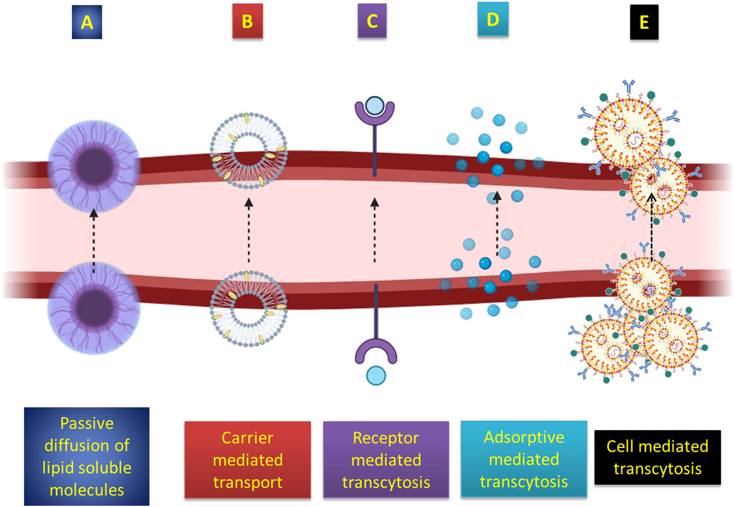
Illustration depicting the ways by which drugs traverse the BBB.
The utilization of intrinsic transport networks is a significant and unexplored approach in delivering drugs to the brain. The vascular BBB and blood-cerebrospinal fluid barrier are both highly equipped with known transporters. However, it is anticipated that the bulk of BBB transporters remain unidentified. Transporters for numerous peptides and regulatory proteins often have a heterogeneous distribution, which can be exploited to selectively deliver drugs to specific regions of the brain. For instance, the BBB transporter responsible for transporting IL-1 is highly concentrated in the posterior division of the septum. Similarly, the leptin transporter is mostly found in the arcuate nucleus of the hypothalamus, while the transporter for APP-directed antisense is predominantly located in the hippocampus. The BBB preserves cerebral equilibrium and restricts the infiltration of harmful substances and microorganisms into the brain. Despite its significance, there is limited understanding of the molecular mechanisms that govern the development and operation of this vital barrier. Researchers conducted a study where they devised techniques to purify and analyse the genetic characteristics of endothelial cells from various tissues [102]. By comparing the gene activity of brain endothelial cells with those from the liver and lung, they created a valuable collection of transcripts that are specifically abundant in the endothelial cells responsible for forming the BBB. By doing this comparison, we have discovered new proteins involved in tight junctions, transporters, metabolic enzymes, signalling components, and unidentified transcripts that are more abundant in endothelial cells of the CNS. The investigation has determined that the RXRalpha signalling cascade is particularly abundant at the BBB, suggesting that this route plays a role in regulating this crucial barrier. This dataset is a valuable resource for comprehending CNS endothelial cells and their interaction with neural and haematogenous cells. Leptin, also known as OB protein, is synthesized by adipocytes and potentially controls body weight through its influence on the CNS. To access the brain, leptin that is circulating in the body must traverse the BBB. The presence of unlabelled leptin hindered the entry of 125I-leptin in a manner that depended on the dosage. Conversely, unlabelled tyrosine and insulin, which possess transport mechanisms that can reach saturation, did not have any impact. Around 75% of the extravascular 125I-leptin successfully traversed the BBB and entered the brain parenchyma. Uptake was seen at the choroid plexus, arcuate nuclei of the hypothalamus, and the median eminence using autoradiography. No saturable transport was observed exiting the brain [103]. Amyloid β protein (Aβ) is perhaps a causative factor in the development of AD. Prior research has demonstrated that the cognitive impairments that arise in ageing SAMP8 mice, a breed that produces excessive amounts of Aβ, can be reversed with intracerebroventricular injections of an Aβ antisense phosphorothiolate oligonucleotide (Olg). In this study, we demonstrated that 32P-labelled oligonucleotide (P-Olg) may pass through the BBB of mice without being altered. This transport is facilitated by a specific system called oligonucleotide transport system-1, which operates in a saturable manner [104]. The results of the regression analysis revealed that the blood-to-brain unidirectional inflow rate for P-Olg was determined to be 1.4 ± 0.39 μL/g min. Additionally, the capillary depletion experiment demonstrated that P-Olg was able to fully traverse the BBB and reach the brain’s parenchymal region. The technique of film autoradiography was employed to illustrate the movement and specific locations of blood-borne radioiodinated IL-1α and other cytokines into the brain following intravenous delivery [105]. The choroid plexus and the capillary network were labelled with [125I]Il-1α, [125I]Il-1β, [125I]interleukin-1 receptor antagonist (Il-1ra), and [125I] TNFα 30 min after being injected into the blood. This indicates that these areas may function as locations for the transit of substances from the blood to the brain. Several medications utilized in the treatment of AD, ranging from clinical applications to preclinical research, have been identified to exploit transport systems. Donepezil, along with other cholinesterase inhibitors, is one of the two classes of medications licenced for treating AD. It is transported through the BBB by an organic cation transporter, most likely the one responsible for transporting choline. A team of researchers studied the choline transport system and investigated how different amine medications affect the choline transporter using a rat brain capillary endothelial cell line (TR-BBB) that can be grown indefinitely in a laboratory setting. The ratio of [3H]choline between the cell and the surrounding medium in TR-BBB cells rose over time. The rate at which [3H]choline is initially absorbed depends on its concentration and can be described by the Michaelis–Menten constant, Km, which has a value of 26.2 ± 2.7 μM. The uptake of [3H]choline into TR-BBB was not reliant on the presence of sodium ions, although it did depend on the membrane potential [106]. The uptake of [3H]choline was vulnerable to suppression by hemicholinium-3 and tetraethylammonium (TEA), both of which are substrates of organic cation transporters. In addition, the absorption of [3H]choline was hindered in a competitive manner, with inhibition constants (Ki values) of 274, 251, and 180 μM when donepezil hydrochloride, tacrine, and α-phenyl-n-tert-butyl nitrone were present, respectively. A targeted oligophosphorothioate antisense molecule that specifically binds to APP has been found to effectively decrease the amounts of APP in the brain. This leads to an increase in the removal of A-beta from the brain, a reduction in oxidative stress, and an improvement in cognitive function in aged SAMP8 mice [107].
4 Application of QD-based nanocomposites for targeted drug delivery
Investigating the manipulation of materials at the nanoscale is a prominent focus of the current study. Owing to the remarkable advancement of technology, there have been a significant surge in life expectancy and a substantial improvement in the overall quality of life. The advent of nanotechnology has propelled research, particularly in the realm of medical, to unprecedented levels. Given the multitude of benefits associated with nano drug delivery carriers, they can be effectively utilized as vehicles for administering medicines to treat a diverse array of medical conditions. Neurodegenerative disorders in an organism arise from a multitude of factors such as brain injuries, malfunctioning proteins, poisons, and others. However, the causes and origins of many disorders have not yet been definitively established. The primary challenge in treating brain-related conditions lies in the limited permeability of medicinal substances across the BBB. The barrier that separates the blood from the cerebral fluid has a high degree of selective permeability [108]. PD is characterized by the loss of dopaminergic neurons, resulting in a reduction of dopamine levels in the brain. Consequently, the patient’s motor function is compromised. Over time, the standard of living declines. Currently, the primary treatment for the initial stages of the condition is the administration of l-DOPA, which serves as the precursor for dopamine [109]. l-DOPA readily crosses the BBB, where it is enzymatically converted to dopamine by l-amino acid decarboxylase or DOPA decarboxylase [110]. In recent studies, NPs have been found to facilitate drug administration over the BBB and into the CNS, surpassing traditional drug delivery methods. A computational biology technique was used in a study to investigate the activity of α-synuclein, a key factor in the development of PD, in the presence of its inhibitor, cerium oxide (CeO2) NPs. A computer analysis was performed to examine the effectiveness and level of inhibition caused by the CeO2 NP on the activity of α-synuclein, using biocompatible metal NPs such as Au NPs and SPIONs. The results indicated that CeO2 NPs exhibited optimal fitting into the active site of α-synuclein, establishing strong contacts and interactions. Furthermore, they demonstrated potential inhibition of PD in the planned biochemical pathway, as seen by their effect on the l-DOPA medication, which was used as a positive control. Therefore, CeO2 NPs have been proposed as a promising inhibitor of α-synuclein and can be utilized as a nanodrug to combat PD [111]. In their 2017 study, Yadav et al. found that chitosan serves as a medium to facilitate the transport of doxycycline hydrochloride via the BBB [112]. The progress of innovative materials for precise and efficient drug delivery is crucial in the field of biomedical research. The research was led to observe the effectiveness of a chitosan/carbon dot (CS/CD)-based nanocomposite as a transporter for the controlled release of dopamine medication. The CDs were produced by carbonizing chitosan and subsequently combined with chitosan (CS) to form a CS/CD matrix. Subsequently, dopamine was enclosed within the matrix to create a dopamine@CS/CD nanocomposite. The study investigated the cytotoxicity of the IC-21 and SH-SY5Y cell lines at different concentrations of the nanocomposite. The findings indicate a cell viability of around 97%. The photoluminescence (PL) property demonstrated the distinctive property of the CDs. Upon excitation at a wavelength of 510 nm, a distinct emission peak was detected at 550 nm, indicating that carbon dots can be effectively utilized as a tracer for bioimaging purposes. The HRTEM images and Raman spectra analysis validate the successful synthesis of carbon dots. Additionally, the particle size is determined to be approximately 3 nm using dynamic light scattering. An in vitro analysis was conducted to study the release of the encapsulated medication from the composite material at various pH levels [113]. CDs have garnered significant interest because of their little cytotoxicity and strong biocompatibility, in contrast to metal-based NPs [114]. Furthermore, the synthetic methods used to produce CDs are straightforward, and their significant surface-area-to-volume ratio enables them to possess a substantial drug-loading capacity. Furthermore, owing to their exceptional PL, the distribution of CDs can be monitored in both laboratory-based and living organism-based research [115,116]. Surgical excision is widely acknowledged as a fundamental component in the treatment of malignant brain tumours. Surgeons encounter significant difficulties in clinical practice when it comes to determining tumour boundaries. This is mostly owing to the infiltrating and diverse characteristics of neoplastic tissues. Contrast-enhanced MRI is widely utilized in clinical settings to accurately identify brain tumours. Regrettably, the commercially accessible (MR) contrast agents exhibit a short-lived circulation lifespan and inadequate penetration across the BBB, significantly impeding their effectiveness in visualizing tumours [117,118,119]. The study focused on examining the potential of red fluorescent carbonized polymer dots (CPDs) to penetrate the BBB (Figure 5). To summarize, CPDs exhibit extended excitation/emission wavelengths, minimal toxicity, exceptional photostability, and outstanding biocompatibility [116]. Cyclopentadienes demonstrate significant uptake within glioma cells, with the extent of uptake depending on both the duration and dosage of exposure. Once internalized, CPDs mostly accumulate within endolysosomal structures. CPDs have been shown to permeate the BBB in both laboratory and living organism experiments. This finding is significant because it allows for the early detection of brain diseases and the noninvasive imaging of brain tumours, all without causing any harm to the BBB. Moreover, due to the significant disparity between tumour and normal tissue in terms of CPDs, the nanoprobe has the potential to assist in brain tumour removal by providing real-time fluorescence imaging during surgery. MRI was employed to assess the advancement of the tumour 13 days after implantation, confirming the successful formation of the orthotopic glioma model (Figure 5a). Following the administration of CPDs through intravenous injection in the tail, both in vivo and ex vivo imaging of the brain tissues of rats with C6 glioma were conducted utilizing an in vivo optical imaging system. Furthermore, the contour of the brain glioma matched the ex vivo imaging shown in Figure 5b and c, providing additional confirmation of the targeting capability of CPDs for brain tumours. Our observation revealed that CPDs were able to quickly cross the BBB and reach the tumour site within 15 min of injection. This suggests that the distribution of CPDs inside the brain may be effectively monitored due to their strong ability to permeate tissues, as indicated by their red fluorescence. The fluorescence intensity of cyclobutane pyrimidine dimers in brain tumours peaked after 60 min and remained strong even at 120 min. This observation aligns well with the in vitro BBB penetration studies of CPDs, indicating their great photostability and efficient targeting of gliomas.
![Figure 5
Imaging of glioma-bearing rats using ex vivo technique following intravenous injection of CPDs through the tail. (a) MRI was employed to ascertain the presence of tumour formation 13 days after implantation; the tumour tissues were indicated by the red arrows. (b) Images of individual brains displaying tumour tissues are depicted, with the tumour tissues highlighted by dotted circles. (c) Fluorescence imaging of brains ex vivo at certain time intervals. Reproduced with permission from Liu et al. [116], © 2018 American Chemical Society.](/document/doi/10.1515/ntrev-2024-0128/asset/graphic/j_ntrev-2024-0128_fig_005.jpg)
Imaging of glioma-bearing rats using ex vivo technique following intravenous injection of CPDs through the tail. (a) MRI was employed to ascertain the presence of tumour formation 13 days after implantation; the tumour tissues were indicated by the red arrows. (b) Images of individual brains displaying tumour tissues are depicted, with the tumour tissues highlighted by dotted circles. (c) Fluorescence imaging of brains ex vivo at certain time intervals. Reproduced with permission from Liu et al. [116], © 2018 American Chemical Society.
To assess the capacity of CDs and CD-based products to traverse the BBB, researchers have established both in vitro and in vivo models. Lu and colleagues employed polyethylenimine (PEI) as the starting material to fabricate nitrogen-doped carbon dots (N-CDs) by a single-step solvothermal process. They assessed the N-CDs’ capability to traverse the BBB by utilizing an in vitro model consisting of rat microvascular endothelial cells and astrocytes [120]. The intense blue PL of N-CDs when exposed to ultraviolet (UV) radiation indicated that N-CDs were transported across the BBB in a manner that depended on their concentration. The utilization of the transwell model in the study of N-CDs serves as a commendable illustration of in vitro investigations. The biomimetic model offers the benefits of changeable parameters and restricted changes. However, its fundamental drawback is its limited ability to accurately replicate the BBB in a live animal. Several types of CDs have been tabulated for neurodegenerative disorders in Table 2.
Potential uses of QDs in the treatment of neurodegenerative disorders
| Type of dots | Disease | Outcomes | Type of study | Ref. |
|---|---|---|---|---|
| GQDs | AD | GQDs combined with tramiprosate exhibit a synergistic effect in inhibiting the buildup of amyloid β. | In vitro | [121] |
| GQDs | PD | Breaks down α-synuclein proteins into smaller components | In vitro | [122] |
| GQDs | PD | Anti-aggregation agents hinder the buildup of α-syn and fully formed fibrils, as well as the creation of Lewy bodies. They also prevent neuronal death, the spread of disease, and deficiencies in mitochondrial function | In vivo | [123] |
| CdTe/CdS/ZnS QDs | PD | Protects against MPP+-driven α-synuclein fibrillation, cellular damage, and decreases cytotoxicity, apoptosis, and oxidative stress | In vitro | [124] |
| Glycine-proline-glutamate on GQDs | AD | Reduced Aβ aggregation improves memory and learning capacity, while reducing TNF-A and pro-inflammatory cytokines (e.g. IL-1, IL-6, IL-33, IL-17a) | In vitro | [125] |
| Branched PEI QDs | AD | Prevents A β plaque development is facilitated by negative targeting and enhanced PL | In vitro | [126] |
| N-doped CQDs | AD | Prevents amyloid beta protein photooxygenation and Cu+2-driven aggregation | In vitro | [127] |
Several peptides, organic compounds, and peptide mimetics have demonstrated promising outcomes in AD by either eliminating aggregation or disrupting the formation of accumulations in preclinical investigations. Nevertheless, these compounds encounter obstacles such as limited ability to pass across the BBB, reduced stability inside living organisms, decreased effectiveness, and intricate production methods, which ultimately hinder their potential for AD therapy [128]. GQDs are now effective for treating AD because of their capacity to inhibit the development of amyloid plaques and mitigate the cytotoxic effects induced by Aβ oligomers. This is possible because GQDs can easily pass the BBB due to their tiny size (2–10 nm) and exhibit low cytotoxicity [129]. The hydrophobic interactions between carbon materials and Aβ1–42 peptides can limit the formation of amyloid plaques by reducing the negative surface potential. This, in turn, enhances the inhibitory efficacy of QDs [130]. An observable inhibitory impact was found when tramiprosate was covalently linked with GQDs, resulting in a synergistic effect against Aβ aggregation in AD [121]. Xiao et al. developed a novel nanomaterial called GQDs that were combined with a neuroprotective peptide called glycine–proline–glutamate (GQDG). They then administered this combined material to mice that were genetically modified to have the APP/PS1 mutation. The in vitro experiments, specifically ThT and CD, demonstrated that GQDs and GQDG possess the ability to impede the aggregation of Aβ1–42 fibrils. The Morris water maze test was conducted to assess the cognitive abilities and memory capacity of APP/PS1 transgenic mice. The GQDG group exhibited a decrease in the surface area of Aβ plaque deposition compared to the Tg Ctrl groups. In addition, the freshly formed neural precursor cells and neurons were examined using immunohistochemistry testing. In addition, neurons were labelled with DiI using a gene gun to visualize dendritic spines. The results demonstrated an improvement in learning and memory ability, as well as an increase in the number of dendritic spines. The inflammation factors and amyloid-β (Aβ) were assessed by suspension array and enzyme-linked immunosorbent assay (ELISA), respectively. The levels of various pro-inflammatory cytokines (IL-1α, IL-1β, IL-6, IL-33, IL-17α, MIP-1β, and TNF-α) were shown to be lower in the GQDG group as compared to the control group. In contrast, the levels of anti-inflammatory cytokines (IL-4, IL-10) were higher in the GQDG group compared to the control group [131]. Clitoria ternatea graphene QDs (ctGQDs) were synthesized using a one-pot microwave-assisted green synthesis procedure by Tak and their team. They specifically focused on extracting the flowers of Clitoria ternatea for the purpose of AD therapy. The results indicated that there was a high level of stability. Additionally, the memory acquisition and cognitive function tests, including the radial arm maze and water Morris maze assay, demonstrated a decrease in the time it took to transfer to the hidden platform. The ctGQDs are capable of crossing the BBB due to their smaller size [132]. This enables them to exhibit greater inhibition of the acetylcholinesterase enzyme (Figure 6), reduced levels of lipid peroxidation and nitric oxide, and an increased level of glutathione protein. The positive alterations in the biochemical indicators suggest improved preservation, making them potentially beneficial in the treatment of AD.
![Figure 6
Clitoria ternatea was responsible for the synthesis of GQDs, which were then used in the treatment of AD. Reproduced with permission from Tak et al. [132], © 2020 American Chemical Society.](/document/doi/10.1515/ntrev-2024-0128/asset/graphic/j_ntrev-2024-0128_fig_006.jpg)
Clitoria ternatea was responsible for the synthesis of GQDs, which were then used in the treatment of AD. Reproduced with permission from Tak et al. [132], © 2020 American Chemical Society.
To administer the fluorescent QD probes, an anti-Aβ antibody is coupled with them. These probes are then injected intra-cerebroventricularly into both healthy mice and transgenic mice that carry human APP695swe and APP717 V–F mutations. The fluorescent of Aβ1–42 was seen in several locations of APP transgenic mice, such as the hippocampus, cerebral cortex, sagittal septum, and striatum, as illustrated in fluorescence microscopy. Furthermore, in vivo imaging demonstrated a decrease in fluorescence intensity in comparison to the APP transgenic animals [133]. Similar to the previous example, CdSe@ZnS QDs were investigated for their potential use as a biosensing carrier in the electrochemical detection of the ApoE-significant biomarker in AD. Through the utilization of an immune-complex assay that was carried out in a chip with flow mode, the evaluation of ApoE was carried out. Indicating the significance of the tosyl-activated magnetic beads platform as a biomarker in AD, the platform consists of a polydimethylsiloxane-polycarbonate microfluidic chip with integrated screen-printed electrodes. Square wave anodic stripping voltammetry demonstrated that CdSe@ZnS QDs exhibited a limited range limit of detection, resulting in a greater level of accuracy among diluted human plasma samples [134]. The CdSe@ZnS QDs were synthesized by the identical research team to detect apolipoprotein E (ApoE). The sandwich immunocomplex microarray was evaluated using the fluorescent dye Alexa 647 to conduct comparative analyses. The experiment was conducted under comparable conditions, employing the ELISA that specifically targeted ApoE, for the purpose of comparison. The excitation wavelength results demonstrated that QDs are extremely effective biosensing reporters in microarray assays, with a lower limit of detection that is five times more sensitive than Alexa microarrays and seven times more sensitive than ELISA [135].
Spongiform encephalopathy (SE), often known as prion disease, is characterized by the gradual development of brain disease that has a sponge-like structure [136]. Prion disease is a neurodegenerative condition characterized by the formation of cysts or vacuoles, resulting in the replacement of healthy brain tissue with a sponge-like structure [137]. The development of prion disease is rooted in the accumulation of misfolded prion proteins mostly in the cerebral cortex and cerebellum. Prions are contagious polypeptides found in the cell membrane of specific neurons. They are composed of around 253 amino acids arranged in three α-helix and two β-pleated sheets. The precise roles of prion proteins remain unknown; however, they do have a role in the absorption of Cu from neuronal cells. Mammalian prions possess the capacity to replicate by using regular proteins, resulting in their transformation into the disease-causing variant known as PrPSc [138]. The misfolded proteins underwent minimal proteolysis, resulting in the creation of tiny, protease-resistant fragments consisting of 142 amino acids. This molecule then polymerizes into amyloid, characterized by two α-helix structures and an increased number of β-strands. The infectious variant prion proteins exhibit a strong attraction to brain activity, leading to their accumulation in neuronal cells and triggering the apoptosis process, ultimately resulting in cell death [139]. Therefore, the brain exhibits a porous structure as a result of the frequent cell death caused by the development of cysts, and the detrimental aggregation of prion proteins on the neurons’ membrane leads to the formation of large plaques [140]. The manifestations of prion disease encompass dementia, insomnia, paralysis of the lower limbs, lack of coordination, abnormal sensations, and aberrant conduct. However, the primary clinical presentation of SE is Creutzfeldt–Jakob disease caused by genetic abnormalities in the PRNP gene.
5 Neuroimaging applications of surface-engineered QD nanocomposites
Neuroimaging is crucial for the timely detection of several neurological illnesses, offering essential information about the brain’s structure and function [141]. The importance of neuroimaging techniques, such as MRI, CT, and PET, cannot be exaggerated in the prompt detection and treatment of neurological diseases [117,142,143]. Neuroimaging has a key benefit in early diagnosis as it may effectively detect structural abnormalities in the brain. Structural imaging modalities such as MRI and CT enable clinicians to observe the intricate details of the brain’s anatomy [119]. It is essential to recognize lesions, tumours, or abnormalities that could suggest illnesses such as brain tumours, vascular malformations, or neurodegenerative diseases [144]. Timely identification of these structural alterations allows healthcare practitioners to immediately react, perhaps averting more harm and enhancing patient outcomes [145]. Furthermore, neuroimaging plays a crucial role in the diagnosis of neurological illnesses at an early stage when symptoms may not be detectable from clinical observation alone. Several neurological disorders, including AD and MS, display inconspicuous or vague symptoms during their initial phases [146,147]. Advanced imaging methods, such as functional MRI (fMRI) and PET scans, allow for the evaluation of brain activity and metabolism [148]. By detecting alterations in blood flow, glucose metabolism, or neurotransmitter activity, these methods can reveal irregularities prior to the appearance of clinical symptoms, enabling prompt intervention and control.
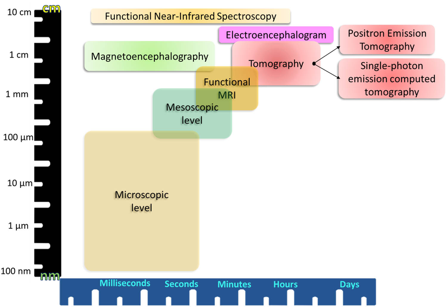
For stroke, prompt diagnosis is crucial for optimal therapy. Neuroimaging techniques such as CT and MRI are essential for differentiating between ischaemic and haemorrhagic strokes, assisting doctors in selecting the most suitable treatment measures [149]. The prompt identification of the kind and site of a stroke can have a substantial impact on the selection and timing of therapies, such as thrombolytic therapy or clot retrieval techniques [150]. This, in turn, can result in enhanced results and decreased disability. Moreover, neuroimaging plays a crucial role in enhancing our comprehension of the advancement and pathological aspects of several neurological illnesses. Imaging techniques used in longitudinal research offer useful data on the progression of diseases over time, providing insights into potential biomarkers and targets for therapeutic interventions [151]. This information is extremely valuable for the advancement of new therapies and the improvement of current ones. Neuroimaging is helpful in understanding the intricate connection between brain structure, function, and mental health in psychiatric diseases. Conditions such as schizophrenia, bipolar disorder, and depression have been linked to distinct brain abnormalities that can be observed by imaging investigations. Timely identification of these alterations in structure and function enables more accurate diagnosis and focused treatment approaches, such as pharmaceutical and psychotherapy therapies [152]. To summarize, the significance of neuroimaging in the early detection of medical conditions cannot be emphasized enough. It significantly transforms the discipline of neurology by offering non-intrusive, comprehensive understanding of the structure and functionality of the brain [153]. Neuroimaging is crucial for promptly diagnosing and treating neurological and psychiatric illnesses, as it allows for the identification of anatomical abnormalities and the detection of minute changes in brain activity [154,155,156]. With the progress of technology, the possibility of diagnosing medical conditions at an earlier stage and with greater precision is increasing, which brings optimism for enhanced patient outcomes and quality of life. Figure 7 illustrates the currently most frequently utilized noninvasive functional imaging techniques and their spatial and temporal capabilities. It is evident that among these very advanced technologies, fMRI achieves the greatest level of spatial resolution. fMRI is capable of evaluating the entire brain and capturing the hemodynamic processes occurring at the layered and columnar levels of the human cortex. This is achieved by utilizing a high-intensity magnetic field, which operates at a submillimeter level [157]. However, its temporal resolution for viewing the dynamics of neuronal populations is comparatively weaker. Both electroencephalogram and magnetoencephalography are capable of detecting electromagnetic fluctuations at the millisecond level. Nevertheless, their spatial resolution/uncertainty exceeds several millimetres [158]. Most imaging methods lack quantitativeness and thus rely on other data to obtain complementary information. The inclusion of this supplementary data enables the standardization of signals, attainment of absolute measurements, and comparison across different subjects. For instance, the fMRI signal quantifies the activity of neurons through hemodynamic changes resulting from intricate physical and physiological processes. Across other disciplines or regions of the brain, equivalent levels of neural activity can elicit distinct related fMRI findings. Therefore, fMRI data may only be regarded as approximately proportionate to the neural activity. In 2008, Ances conducted experiments that discovered a correlation between cerebral blood flow and fluctuations in fMRI signals in specific brain areas, age groups of patients, and their health conditions. The wide applicability of fMRI results in a heightened sensitivity of the signal it produces [159].
Functional neuroimaging techniques and modalities.
Surface-engineered QD nanocomposites are an innovative advancement in the field of neuroimaging, enabling more advanced and precise investigation of the intricate workings of the brain. QDs, which are nanocrystals made of semiconductors, have unique optical and electronic characteristics that make them very suitable for biomedical purposes [160]. To fully unleash their capabilities, surface engineering is crucial [161]. This involves modifying the outer layer of QDs to enhance their interactions with biological systems. This debate explores the various uses of surface-engineered QD nanocomposites and their ability to greatly impact neuroimaging techniques.
The main attraction of QD nanocomposites is their capacity to adjust their fluorescence, stability, and biocompatibility based on their size [145,162,163]. Their features render them very suitable for neuroimaging applications, where precision and sensitivity are of utmost importance. By altering the surfaces of QDs, scientists are able to precisely adjust their characteristics, thereby improving qualities such as the ability to target specific areas, minimizing harmful effects, and enhancing contrast in imaging. The main benefit provided by surface-engineered QD nanocomposites is the enhancement of contrast and sensitivity in neuroimaging techniques. Conventional contrast agents frequently lack the necessary capability to provide the desired level of resolution for precise imaging of brain structures. Through the modification of the surface characteristics of QDs, scientists can surpass these constraints, facilitating the identification of small alterations at the cellular and molecular tiers. This increased sensitivity provides opportunities for a deeper comprehension of neuronal activity and the complex networks within the brain. Moreover, the capacity to modify the surface of QD nanocomposites with certain ligands enables precise imaging of brain structures or disease indicators. The focused methodology is crucial for acquiring knowledge about particular areas of interest inside the brain, enabling researchers to comprehend the intricacies of neuronal networks and disease causes. Furthermore, QDs can fulfil a dual function by serving as carriers for medicinal medicines. This distinctive feature allows for the simultaneous visualization and administration of drugs, representing notable progress in the field of neuroimaging where precise treatments are essential. The surface-engineered QD nanocomposites possess a wide range of applications in multimodal imaging, which involves the use of multiple imaging modalities to gain a more thorough understanding of the structure and function of the brain. QD nanocomposites enable researchers to easily integrate data from several sources, including MRI, PET, CT, and optical imaging. By employing a multimodal method, the accuracy and comprehensiveness of the acquired information is improved, resulting in a comprehensive understanding of neurological processes. Although surface-engineered QD nanocomposites show promise in neuroimaging, there are obstacles that need to be overcome for their widespread use. Thorough analysis and mitigation measures are necessary to address concerns related to long-term biocompatibility, potential toxicity, and regulatory considerations. Ongoing research efforts are focused on optimizing the synthesis and surface modification methods to generate QD nanocomposites that are homogenous and reproducible.
Semiconductor fluorescent QDs are tiny particles that have been modified to exhibit distinct physical characteristics. These attributes make them highly suitable for observing and monitoring molecular events in cells using conventional fluorescence techniques. Existing diagnostic techniques for traumatic brain injury (TBI) which constitutes 15% of all emergency room visits are restricted to neuroimaging approaches. The complexities associated with precise diagnosis and monitoring of TBI have necessitated the development of a straightforward and highly responsive blood test capable of identifying brain-specific biomarkers. A study was conducted to develop a method for detecting S100B, a potential biomarker for TBI. The method involved employing magnetic beads that were attached to antibodies to trap the protein, and QDs that were attached to antibodies for optical detection [164]. The Western blot analysis demonstrates the successful collection and concentration of antigens using magnetic beads. By employing magnetic bead capture and QD detection techniques on serum samples, we demonstrate a broad range of detection and a detection limit that is lower than the clinical cut-off level. Pathak et al. quantified the quantity of operative antibodies (i.e. antibodies that are physically accessible for effective attachment to target proteins) linked to semiconductor QDs. This is crucial for the analysis of biological data that has been labelled using these techniques (Figure 8). Their investigation revealed substantial variation in the quantity of functional antibodies that are accessible using different conjugation techniques, and these quantities are lower than previously thought. These findings may indicate possible approaches for enhancing the use of QDs in labelling biological samples [165]. The structure consists of three components: a 25 kD light chain, which contains half of the specific antigen binding site for a specific IgG molecule, a 50 kD heavy chain, which contains the other half of the binding site, and a 75 kD partially cleaved chain made up of a heavy chain and a light chain that are connected by an unreduced disulphide bond (Figure 8a). Subsequently, each separate fragment was chemically attached to QDs using an SMCC linkage bond that is resistant to being disrupted by DTT treatment. This is a crucial factor to take into account when analysing the experimental findings that come next. Three potential scenarios for the binding of antibody fragments to QDs can arise: covalent attachment of light chains, covalent attachment of heavy chains, and covalent attachment of partial fragments consisting of heavy and light chains. Among these scenarios, only the last one allows for further reduction with DTT to remove the light chain fragment from the heavy chains that are still bound to the QDs or to remove heavy chains from light chains that are bound to the QDs. Biotinylated antibodies are modified with four to eight biotin molecules that are randomly bonded to various positions on the entire antibody. This allows the IgG molecules to be linked to QDs, presumably in all feasible spatial configurations (Figure 8c). In contrast to the direct covalent conjugation procedure, the biotin−streptavidin system allows for the complete conjugation of the antibody molecule to the QD. This means that the antibody molecule is not broken down into light chain and heavy chain fragments before attaching.
![Figure 8
Reducing and conjugating antibodies to QDs. (a) DTT disulphide linkage antibody cleavage sites schematic. DTT reduction can produce light chain, heavy chain, and incompletely cleaved fragments. (b) Direct SMCC covalent antibody-QD conjugation schematic. After the conjugation procedure, DTT reduction creates light chains, which are counted to calculate the average number of functional IgG molecules on QDs. (c) Biotinylated antibodies conjugated to streptavidin-coated QDs schematic. Reproduced with permission from Pathak et al. [165], © 2007 American Chemical Society.](/document/doi/10.1515/ntrev-2024-0128/asset/graphic/j_ntrev-2024-0128_fig_008.jpg)
Reducing and conjugating antibodies to QDs. (a) DTT disulphide linkage antibody cleavage sites schematic. DTT reduction can produce light chain, heavy chain, and incompletely cleaved fragments. (b) Direct SMCC covalent antibody-QD conjugation schematic. After the conjugation procedure, DTT reduction creates light chains, which are counted to calculate the average number of functional IgG molecules on QDs. (c) Biotinylated antibodies conjugated to streptavidin-coated QDs schematic. Reproduced with permission from Pathak et al. [165], © 2007 American Chemical Society.
QDs are extensively employed as fluorescent probes for targeted imaging of cells. Nevertheless, encountering non-specific binding to biological membranes has posed a significant obstacle. A different investigation introduced a novel method to efficiently decrease the occurrence of non-specific binding caused by bovine serum albumin (BSA)-coated QDs in cell targeting [166]. The experimental results demonstrate the successful transfer of hydrophobic QDs from an organic phase to an aqueous phase in the presence of an aqueous solution of BSA under ultrasonication. This method, which uses ultrasonication, is simple, fast, and effective (Figure 9). The stabilization of QDs is primarily accomplished by the presence of several mercapto groups in BSA macromolecules, which act as multidentate ligands. Additionally, the contact between BSA and the fatty ligands attached to the QDs contributes partially to their stabilization through hydrophobic interactions. The presence of amino and carboxyl groups on the surface of QDs enhances their water solubility and also offers sites for the attachment of targeted ligands by conjugation. The QDs covered with BSA, primarily having a hydrodynamic diameter of approximately 18 nm, demonstrate colloidal stability in both acidic and basic environments and display high levels of fluorescence. BSA-coated QDs efficiently decrease nonspecific cellular binding, in contrast to CdTe QDs coated with mercaptopropionic acid (MPA). QDs coated with BSA are additionally modified with cyclic Arg–Gly–Asp (cRGD) peptide. The cell experiments demonstrate the great specificity of the imaging technique for cells expressing integrin αvβ3.
![Figure 9
Fluorescent imaging comparison (a and b) and quantitative assessment (c and d) were conducted to assess the nonspecific cellular binding of MPA-coated CdTe QDs (a1, b1, ■) and BSA-coated QDs (a2, b2, ●) on HeLa (a) and 3T3 (b) cells. Reproduced with permission from Zhang et al. [166], © 2012 American Chemical Society.](/document/doi/10.1515/ntrev-2024-0128/asset/graphic/j_ntrev-2024-0128_fig_009.jpg)
Fluorescent imaging comparison (a and b) and quantitative assessment (c and d) were conducted to assess the nonspecific cellular binding of MPA-coated CdTe QDs (a1, b1, ■) and BSA-coated QDs (a2, b2, ●) on HeLa (a) and 3T3 (b) cells. Reproduced with permission from Zhang et al. [166], © 2012 American Chemical Society.
GQDs are now being utilized for in vivo brain labelling. Qian and her colleagues utilized GO NPs with a diameter of 40 nm, which is in line with the size of GQDs [167]. The researchers administered GO-PEG NPs to mice through intravenous injection and used two-photon imaging to monitor how the NPs flowed, distributed, and were cleared in the blood vessels. In addition, the researchers introduced the NPs into the brains of mice that had fluorescent oligodendrocytes through microinjection. The imaging data obtained using two-photon and three-photon luminescence techniques demonstrated the ability to differentiate GO NPs located at a depth of 300 mm within the brain. A novel variant of carbon dots (CD-Asp) possessing targeted properties for glioma, a kind of brain cancer, was synthesized utilizing a direct pyrolysis method employing d-glucose and l-aspartic acid as initial substances. The CD-Asp produced in this study demonstrates exceptional biocompatibility and the ability to emit a wide range of colours that can be adjusted. Furthermore, it reveals a notable capacity to specifically target C6 glioma cells, without the need for additional targeting agents. Fluorescence images taken inside a living organism demonstrated a clear and distinct distribution pattern of CD-Asp just 15 min after it was injected into the tail vein (Figure 10). The tumour site exhibited a significantly more intense fluorescent signal compared to the normal brain, suggesting its capacity to easily cross the BBB and accurately target glioma tissue. However, the CDs derived from d-glucose (CD-G), l-aspartic acid (CD-A), or a combination of d-glucose and l-glutamic acid (CD-Glu) do not exhibit significant selectivity for glioma. Thus, CD-Asp has the potential to function as a fluorescent imaging and targeting agent for the noninvasive diagnosis of glioma [168].
![Figure 10
Imaging of glioma-bearing mice was conducted both in vivo and ex vivo following the administration of CD-Asp via intravenous injection into the tail. (a) The temporal distribution of CD-Asp throughout the entire body upon injection. (b) The brain’s distribution of CD-Asp 20 min after injection is visualized by three-dimensional reconstruction. (c) Imaging of the brain ex vivo 90 min post-injection of CD-Asp. Reproduced with permission from Zheng et al. [168], © © 2015 American Chemical Society.](/document/doi/10.1515/ntrev-2024-0128/asset/graphic/j_ntrev-2024-0128_fig_010.jpg)
Imaging of glioma-bearing mice was conducted both in vivo and ex vivo following the administration of CD-Asp via intravenous injection into the tail. (a) The temporal distribution of CD-Asp throughout the entire body upon injection. (b) The brain’s distribution of CD-Asp 20 min after injection is visualized by three-dimensional reconstruction. (c) Imaging of the brain ex vivo 90 min post-injection of CD-Asp. Reproduced with permission from Zheng et al. [168], © © 2015 American Chemical Society.
6 Biocompatibility and toxicity
QDs have a wide range of applications in fields such as sensor technology, bio-imaging, drug delivery, and theranostics. One significant concern regarding the utilization of QDs in biological applications revolves around their potential toxicity. Each individual QD possesses its own unique physicochemical property that plays a crucial role in determining its potential toxicity. The toxicity of QDs is influenced by various factors: (i) size, (ii) compounds utilized in the fabrication process, (iii) doses, (iv) environmental condition, (v) material used for capping, and (vi) administration route of QDs [169].
Carbon, silicon, and group I–III−VI2 materials are utilized for biological applications due to their non-toxic nature. The initial discovery of CQDs was made within a single-walled carbon nanotube. These NPs are harmless and simple to create. They are well-suited for biological and biomedical applications because of their aqueous solubility. The Group I–III–VI2 elements, such as Cu, Al, Ag, S, Se, and Te, possess excellent PL characteristics and have a lower level of toxicity [169]. One of the most commonly used QDs in various applications is CdSe. Cadmium exhibits high levels of toxicity and tends to accumulate in the spleen without undergoing degradation. Recent findings indicate that the use of cadmium-based QDs can lead to inflammation in the lungs. Additionally, the oxidation of cadmium QDs produces a reduced form of cadmium, which ultimately leads to cell death. In addition to cadmium-based QDs, the toxicity assessment of various other QDs, including Ag2Se QDs, CuInS2@ZnS, InP QDs, and hybrid QDs of carbon and CdSe@ZnS are also reported. Every type of QDs possesses unique benefits. Cadmium-based QDs exhibit a wide range of UV excitation, emit a narrow spectrum of light, and have high PL. In contrast, cadmium-free QDs have minimal toxicity. Multiple experiments have consistently demonstrated that the toxicity of QDs is directly influenced by the dosage, regardless of their composition. Therefore, surface modification plays a crucial role in decreasing toxicity while enhancing the biocompatibility, stability, and specificity of QDs. Thus, the method of modifying QDs with inorganic shells (such as CdSe@ZnS, CdSe@SiO2), polymers, and bio- or natural macromolecules is advantageous [170]. Nurunnabi et al. conducted a study on the in vivo dispersion and potential toxicity of carboxylated photoluminescent GQDs. The carboxylated photoluminescent GQDs were tested on KB, MDA-MB231, A549 cancer cells, and MDCK normal cell line as in vitro cell culture models to investigate potential negative impacts [171]. The carboxylated GQDs are advantageous due to their enhanced ability to dissolve in water. The carboxylated GQDs were effectively internalized by all cancer cells. There were no signs of immediate harm or physical changes observed in either system at the amounts of exposure that were examined. The utilization of histological analysis to detect the carboxylated GQD NPs enables a comprehensive microscopic evaluation and histological assessment of tissue interactions. The findings visually depict the presence of inflammation or lesions resulting from the intravenous administration of GQD NPs. Upon histological examination of animal tissues, no evident histopathological abnormalities or lesions were detected in the heart, kidney, and spleen (Figure 11). The histological staining images of these organs exhibited identical characteristics to those of the control animals. The histological investigation revealed no significant signs of toxicity; however, considerable pathological alterations were identified in the liver and lung 21 days after administering carboxylated GQD NPs at a higher dosage of 10 mg/kg.
![Figure 11
Histological examinations of mice’s major organs 22 days after GQD intravenous injection. No inflammation or lesions were seen in the photos. Tissues of 5 and 10 mg/kg GQD-treated mice are identical to saline-treated mice. Photos were taken at 160× magnification with haematoxylin and eosin staining. Reproduced with permission from Nurunnabi et al. [171], © 2013 American Chemical Society.](/document/doi/10.1515/ntrev-2024-0128/asset/graphic/j_ntrev-2024-0128_fig_011.jpg)
Histological examinations of mice’s major organs 22 days after GQD intravenous injection. No inflammation or lesions were seen in the photos. Tissues of 5 and 10 mg/kg GQD-treated mice are identical to saline-treated mice. Photos were taken at 160× magnification with haematoxylin and eosin staining. Reproduced with permission from Nurunnabi et al. [171], © 2013 American Chemical Society.
Tabish et al. conducted a study where they administered high doses of GQDs to rats for a period of 4 weeks [172]. They found that there was only a small amount of toxicity observed in the rats during this time. Additionally, they were able to confirm that the GQDs increased the production of singlet oxygen and other reactive oxygen species. Rat tests assessing in vivo toxicity reveal that our GQDs exhibit negligible dark toxicity, a crucial characteristic of a photosensitizer intended for future application in PDT. Boron-doped GQDs are synthesized using a one-pot acid-free microwave method, where graphene oxide is used as the carbon source and borax is used as the boron source [173]. The conventional MTT assay indicates that the B-GQDs exhibit minimal cytotoxicity and favourable biocompatibility. At a concentration of 4.0 mg/mL, the B-GQDs achieve a cell viability of 87%. B-GQDs are practically applied in bio-analysis through the use of bio-imaging to visualize HeLa cells. Semiconductor QDs have the potential to greatly enhance the effectiveness of biomedical NIR imaging. This is because they can offer fluorescent probes that are both brighter and more resistant to fading compared to traditional organic dyes [174]. The influence of QD composition on the RLTLN is consistent, with injections of CuInS2/ZnS up to 100 pmol not causing significant inflammation, while an injection of 10 pmol of CdTeSe/CdZnS leads to a significant increase in RLTLN weight compared to the control (3.43 ± 0.61 mg vs 2.33 ± 0.41 mg; p < 0.05). Figure 12a displays histological sections of RALNs that were dissected 7 days after injection. The findings from this investigation conducted in a living organism prompted a subsequent examination of cytotoxicity conducted in a controlled laboratory setting. To gain a deeper understanding of the mechanism behind CIS toxicity, we conducted measurements to assess the effects of CIS, partially degraded CIS, CISZ, CIS/ZnS, and CdSe on the HepG2 human liver cancer cell line (Figure 12b and c). The cells were cultured in media containing QDs for a duration of 24 h. Following this, the media was removed, the wells were rinsed with phosphate-buffered saline, and the ATP levels were quantified using a conventional viability assay.
![Figure 12
(a) RALN histological sections resected 7 days after injection for animals injected with saline buffer (control), 20 pmol CuInS2/ZnS QDs, and 20 pmol CdTeSe/CdZnS QDs. Light regions with black arrows suggest inflammation. The scale bar is 1 mm. Reproduced with permission from Pons et al. [174], © 2010 American Chemical Society. Toxicity test in vitro of (b) QD-treated HepG2 viability after 24 h, n = 4 wells per dosage. (c) Dose–response curve from viability testing for two degradation levels fit a 4-parameter Hill equation; shaded region shows 95% confidence interval. Reproduced with permission from Kays et al. [175], © 2020 American Chemical Society.](/document/doi/10.1515/ntrev-2024-0128/asset/graphic/j_ntrev-2024-0128_fig_012.jpg)
(a) RALN histological sections resected 7 days after injection for animals injected with saline buffer (control), 20 pmol CuInS2/ZnS QDs, and 20 pmol CdTeSe/CdZnS QDs. Light regions with black arrows suggest inflammation. The scale bar is 1 mm. Reproduced with permission from Pons et al. [174], © 2010 American Chemical Society. Toxicity test in vitro of (b) QD-treated HepG2 viability after 24 h, n = 4 wells per dosage. (c) Dose–response curve from viability testing for two degradation levels fit a 4-parameter Hill equation; shaded region shows 95% confidence interval. Reproduced with permission from Kays et al. [175], © 2020 American Chemical Society.
The stability, in vivo degradation, and prevention of toxicity of QDs can be influenced by their coating. Group III-IV QDs are commonly utilized in optical probe and biosensor applications due to their reduced toxicity. To reduce the toxicity of QDs, biocompatible functionalization can be adopted. In this approach, the use of (i) mercapto group and (ii) encapsulation in phospholipid micelles, which retain optical properties while causing no surface modifications, are employed to mitigate the harmful effects of the QDs. Other methods include QD conjugation with antibodies [176,177,178].
7 Challenges and future directions
QD nanocomposites that have been modified on the surface are an emerging area of research in the domains of treating neurodegenerative disorders and neuroimaging. They have the potential to greatly transform our approach to these complicated challenges. Nevertheless, there are other significant obstacles that need to be overcome to fully exploit the capabilities of these nanocomposites for therapeutic and diagnostic applications. The primary difficulty is the urgent requirement for thorough investigations on the biocompatibility and probable toxicity. An in-depth analysis of the long-term impact of surface-engineered QD nanocomposites on cellular and organismal health is required due to the complex interactions between these materials and biological systems. Ensuring the safe and effective use of these nanocomposites in clinical applications requires addressing concerns regarding biocompatibility.
An important challenge in using surface-engineered QD nanocomposites for treating neurodegenerative disorders is the need to effectively cross the BBB. Although there have been improvements in surface changes intended to improve the accuracy of targeting, successfully penetrating the BBB is still a difficult task. It is essential to overcome this obstacle to ensure that therapeutic QD nanocomposites can accurately and efficiently reach their designated neuronal targets. Current research efforts are focused on developing strategies to increase the permeability of the BBB without causing damage to neural tissues. The aim is to fully utilize the capabilities of QD nanocomposites for delivering drugs specifically to the brain.
Furthermore, the transition of surface-engineered QD nanocomposites from laboratory testing to practical use requires thorough verification of their effectiveness and safety characteristics. Establishing standardized techniques for the synthesis, characterization, and functionalization of these nanocomposites is crucial to guarantee uniformity and repeatability in various research and clinical environments. Developing rigorous procedures would enhance the dependability of findings and streamline the comparison of results across research, ultimately accelerating the practical application of QD nanocomposites for the treatment of neurodegenerative disorders and neuroimaging purposes. Within the field of neuroimaging, there are ongoing difficulties in enhancing the imaging capabilities of QD nanocomposites. To obtain images with high resolution and little background noise, it is necessary to continuously improve the synthesis and surface engineering procedures. Achieving a harmonious equilibrium between enhanced contrast and sensitivity, while also minimizing any possible cytotoxic consequences, continues to be a challenging endeavour. Researchers are currently investigating novel methods to optimize the characteristics of QDs, which can lead to improved imaging techniques. These advanced imaging modalities have the potential to offer unprecedented understanding of the intricate details of neuronal structures and functions.
To summarize, whereas surface-engineered QD nanocomposites show great promise in tackling the difficulties of treating neurodegenerative disorders and improving neuroimaging methods, there are still significant obstacles that need to be overcome. To fully unlock the therapeutic and diagnostic capabilities of these nanocomposites, it is essential to address concerns related to biocompatibility, improve the ability to penetrate the BBB, and establish standardized techniques for the synthesis and functionalization processes. Future endeavours entail the collaboration of several fields of study and ongoing creativity to address these obstacles and facilitate the ground-breaking use of surface-engineered QDs nanocomposites in neuroscience and clinical practice.
8 Comparative analysis with other NPs and practical trials
Within the field of addressing neurodegenerative disorders and studying brain imaging, many methods that involve NPs demonstrate unique properties, along with their own benefits and difficulties. QDs possess adjustable fluorescence and adaptable surface engineering, making them an exceptional choice for combining imaging and drug delivery functionalities. Ongoing research endeavours to fully use the potential of QDs for simultaneous therapeutic and diagnostic purposes, despite concerns over their biocompatibility and ability to penetrate the BBB. Lipid-based NPs, known for their exceptional biocompatibility, are highly effective in delivering drugs [179]. However, they may encounter challenges in terms of their imaging capabilities. Polymeric NPs, which possess customized drug release characteristics, offer a well-balanced method but may face difficulties in producing high-resolution imaging [180]. IONPs are efficient in enhancing the visibility of MRI images, but their potential for therapeutic use is restricted. AuNPs, possessing distinct optical and therapeutic characteristics, exhibit potential in the integration of imaging and therapy. Every form of NP possesses distinct advantages and disadvantages, which must be carefully evaluated according to the specific needs of investigations on neurodegenerative disorders and neuroimaging applications. The continuous progress in surface engineering and biocompatibility plays a crucial role in determining the future of NP-based methods for comprehensive solutions in neuroscience [26]. Table 3 sums up the contemporary NPs and how the QDs are distinguishable among them.
Different types of NPs and their comparison with QDs
| Characteristics | QDs | Lipid-based NPs | Polymeric NPs | IONPs | AuNPs | Ref. |
|---|---|---|---|---|---|---|
| Optical properties | Tuneable fluorescence | Variable | Limited | Contrast agent for MRI | Unique optical properties | [181] |
| Surface engineering | Highly customizable | Limited adaptability | Tailored for drug release | Variable | Surface functionalization | [182] |
| Biocompatibility | Concerns; ongoing research | Usually high | Generally high | Generally high | Generally high | [183] |
| BBB penetration | Challenging | May face challenges | May face challenges | Limited | Challenging | [184] |
| Drug delivery capability | Yes, with surface engineering | Yes | Yes | Limited | Yes | [185] |
| Imaging capabilities | High-resolution imaging | Inadequate | Limited | MRI contrast agent | Imaging and therapy | [186] |
| Targeting specificity | Feasible through modification | Achievable | Achievable | Achievable | Attainable | [187] |
| Clinical translation | Challenges in biocompatibility | Established in some cases | Ongoing research | Widely used | Escalating applications | [188] |
The application of surface-engineered QDs in the treatment of neurodegenerative illnesses is a unique and disruptive method that has potential benefits in both diagnosis and treatment. QDs, such as the Quantum Dot 800 ITK™ and CdSe/ZnS core–shell QDs, have unique optical and electronic characteristics that make them very suitable for neuroimaging [189]. They have unmatched sensitivity and specificity in detecting molecular-level pathological alterations, delivering extraordinary precision. The QDs that have been modified on their surface can be chemically modified with different biomolecules, which improves their ability to interact with living organisms and allows them to be delivered specifically to certain neuronal regions. This focused strategy shows potential in reducing the advancement of conditions such as Alzheimer’s, Parkinson’s, and Huntington’s illnesses [190]. Although QDs have the potential for clinical translation in the treatment of neurodegenerative disorders, their progress in this area is still in the early stages. Multiple preclinical studies have shown evidence of the effectiveness of nanocomposites based on QDs in animal models. These studies have shown notable enhancements in neuronal function and a decrease in illness indicators. In preclinical backgrounds, in vivo therapeutics’ imaging agents based on QDs have played a crucial role [191]. Nevertheless, there is a scarcity of human clinical studies, as the majority of research is primarily concerned with examining safety profiles and biodistribution. Thermo Fisher Scientific’s Qdot® Nanocrystals, an FDA-approved product, has been used in laboratory settings to identify biomolecules [192]. This demonstrates the regulatory process for these sophisticated materials. The current clinical studies and research endeavours are focused on addressing the issues pertaining to the toxicity and long-term stability of QDs, which are crucial for their extensive use in clinical environments [193]. The incorporation of QDs into therapeutic approaches for neurodegenerative illnesses not only signifies the beginning of a new era in their treatment but also opens up possibilities for cutting-edge neuroimaging methods that have the potential to transform early detection and tracking of these incapacitating disorders.
9 Conclusion
To summarize the main discoveries of this extensive review paper, the investigation of surface-engineered QD nanocomposites for the treatment of neurodegenerative disorders and neuroimaging shows an area full of potential and optimism. QDs possess inherent properties, including adjustable fluorescence and the capability for meticulous surface modification, which make them adaptable instruments suitable for specific drug transportation and detailed imaging within the complex structure of the nervous system. The research synthesis examines the progress and difficulties related to surface-engineered QD nanocomposites. The unique optical properties and tailored surface functionalities of these materials offer significant potential for both therapeutic interventions and precise neuroimaging. Nevertheless, it is crucial to carefully tackle notable obstacles such as biocompatibility, potential toxicity, and efficient permeation through the BBB. The comprehensive assessment underscores the substantial potential of surface-engineered QD nanocomposites to enhance our understanding of neurodegenerative diseases and advance neuroimaging techniques. The landscape is undergoing gradual transformation through ongoing efforts in surface modification techniques, biocompatibility research, and the development of standardized protocols, despite the challenges encountered. The integration of nanotechnology, neurobiology, and medical imaging has resulted in the rise of surface-engineered QD nanocomposites as frontrunners in the advancement of innovative remedies for brain well-being. To summarize, this research predicts that surface-engineered QD nanocomposites will have a crucial role in understanding the intricacies of neurodegenerative illnesses and advancing neuroimaging capabilities. The ongoing research on nanocomposites shows great potential for their incorporation into clinical practice. This holds promise for improving the diagnosis and treatment of neurological illnesses, which is a difficult area to address.
Acknowledgments
The authors extend their appreciation to the King Salman Center for Disability Research for funding this work through Research Group No. KSRG-2022-155.
-
Funding information: The authors extend their appreciation to the King Salman Center for Disability Research for funding this work through Research Group No. KSRG-2022-155.
-
Author contributions: Writing – original draft preparation, M.K.A.-M. (Muhanna K. A. Al-Muhanna); writing – review and editing, A.A.A. (Amani Ahmed Alghamdi) and R.H. (Rania Haddadi); visualization, M.A. (Mohammad Afzal) and R.A.-S. (Reema Al-Subaiee); supervision, M.K.A.-M. (Muhanna K. A. Al-Muhanna) and B.A. (Bahauddeen Alrfaei); funding acquisition, M.K.A.-M. (Muhanna K. A. Al-Muhanna). All authors have accepted responsibility for the entire content of this manuscript and approved its submission.
-
Conflict of interest: The authors state no conflict of interest.
-
Data availability statement: All data generated or analysed during this study are included in this published article.
References
[1] Paoli RA, Botturi A, Ciammola A, Silani V, Prunas C, Lucchiari C, et al. Neuropsychiatric burden in huntington’s disease. Brain Sci. 2017;7(6):67.10.3390/brainsci7060067Suche in Google Scholar PubMed PubMed Central
[2] Lauterbach EC. The neuropsychiatry of Parkinson’s disease and related disorders. Psychiatr Clin. 2004;27(4):801–25.10.1016/j.psc.2004.07.001Suche in Google Scholar PubMed
[3] Starkstein SE, Merello M. Psychiatric and cognitive disorders in Parkinson’s disease. England: Cambridge University Press; 2002.10.1017/CBO9780511544279Suche in Google Scholar
[4] Zhang J, Zhang Y, Wang J, Xia Y, Zhang J, Chen L. Recent insights into neurobiology of alzheimer’s disease and advanced treatment strategies. 2024;9(1).10.1038/s41392-024-01911-3Suche in Google Scholar PubMed PubMed Central
[5] Clemente-Suárez VJ, Redondo-Flórez L, Beltrán-Velasco AI, Ramos-Campo DJ, Belinchón-deMiguel P, Martinez-Guardado I, et al. Mitochondria and brain disease: a comprehensive review of pathological mechanisms and therapeutic opportunities. Biomedicines. 2023;11(9):2488.10.3390/biomedicines11092488Suche in Google Scholar PubMed PubMed Central
[6] Karande S, Kulkarni V. Advancing neurodegenerative disorder diagnosis: a machine learning-driven evaluation of assessment modalities. Int J Intell Syst Appl Eng. 2024;12(5s):309–23.Suche in Google Scholar
[7] Iliffe S, Wilcock J, Drennan V, Goodman C, Griffin M, Knapp M, et al. Changing practice in dementia care in the community: developing and testing evidence-based interventions, from timely diagnosis to end of life (EVIDEM). Program Grants Appl Res. 2015;3(3):1–596.10.3310/pgfar03030Suche in Google Scholar PubMed
[8] Wurcel V, Cicchetti A, Garrison L, Kip M, Koffijberg H, Kolbe A, et al. The value of diagnostic information in personalised healthcare: a comprehensive concept to facilitate bringing this technology into healthcare systems. Public Health Genomics. 2019;22(1–2):8–15.10.1159/000501832Suche in Google Scholar PubMed
[9] Levenson RW, Sturm VE, Haase CM. Emotional and behavioral symptoms in neurodegenerative disease: a model for studying the neural bases of psychopathology. Annu Rev Clin Psychol. 2014;10:581–606.10.1146/annurev-clinpsy-032813-153653Suche in Google Scholar PubMed PubMed Central
[10] Modi G, Pillay V, Choonara YE, Ndesendo VM, du Toit LC, Naidoo D. Nanotechnological applications for the treatment of neurodegenerative disorders. Prog Neurobiol. 2009;88(4):272–85.10.1016/j.pneurobio.2009.05.002Suche in Google Scholar PubMed
[11] Chimene D, Alge DL, Gaharwar AK. Two‐dimensional nanomaterials for biomedical applications: emerging trends and future prospects. Adv Mater. 2015;27(45):7261–84.10.1002/adma.201502422Suche in Google Scholar PubMed
[12] Li X, Shan J, Zhang W, Su S, Yuwen L, Wang L. Recent advances in synthesis and biomedical applications of two‐dimensional transition metal dichalcogenide nanosheets. Small. 2017;13(5):1602660.10.1002/smll.201602660Suche in Google Scholar PubMed
[13] Li BL, Wang J, Zou HL, Garaj S, Lim CT, Xie J, et al. Low‐dimensional transition metal dichalcogenide nanostructures based sensors. Adv Funct Mater. 2016;26(39):7034–56.10.1002/adfm.201602136Suche in Google Scholar
[14] Baig N. Two-dimensional nanomaterials: A critical review of recent progress, properties, applications, and future directions. Compos Part A: Appl Sci Manuf. 2023;165:107362.10.1016/j.compositesa.2022.107362Suche in Google Scholar
[15] Das P, Marvi PK, Ganguly S, Tang XS, Wang B, Srinivasan S, et al. MXene-based elastomer mimetic stretchable sensors: design, properties, and applications. Nano-Micro Lett. 2024;16(1):135.10.1007/s40820-024-01349-wSuche in Google Scholar PubMed PubMed Central
[16] Anju S, Mohanan P. Biomedical applications of transition metal dichalcogenides (TMDCs). Synth Met. 2021;271:116610.10.1016/j.synthmet.2020.116610Suche in Google Scholar
[17] Rohaizad N, Mayorga-Martinez CC, Fojtů M, Latiff NM, Pumera M. Two-dimensional materials in biomedical, biosensing and sensing applications. Chem Soc Rev. 2021;50(1):619–57.10.1039/D0CS00150CSuche in Google Scholar
[18] Das P, Ganguly S, Marvi PK, Sherazee M, Ahmed SR, Tang X, et al. Borophene based 3D extrusion printed nanocomposite hydrogel for antibacterial and controlled release application. Adv Funct Mater. 2024;34:2314520.10.1002/adfm.202314520Suche in Google Scholar
[19] Zlokovic BV. The blood-brain barrier in health and chronic neurodegenerative disorders. Neuron. 2008;57(2):178–201.10.1016/j.neuron.2008.01.003Suche in Google Scholar PubMed
[20] Ali MK, Javaid S, Afzal H, Zafar I, Fayyaz K, Ain QU, et al. Exploring the multifunctional roles of quantum dots for unlocking the future of biology and medicine. Environ Res. 2023;232:116290.10.1016/j.envres.2023.116290Suche in Google Scholar PubMed
[21] Chakraborty P, Das SS, Dey A, Chakraborty A, Bhattacharyya C, Kandimalla R, et al. Quantum dots: The cutting-edge nanotheranostics in brain cancer management. J Control Release. 2022;350:698–715.10.1016/j.jconrel.2022.08.047Suche in Google Scholar PubMed
[22] Das P, Ganguly S, Rosenkranz A, Wang B, Yu J, Srinivasan S, et al. MXene/0D nanocomposite architectures: Design, properties and emerging applications. Mater Today Nano. 2023;24:100428.10.1016/j.mtnano.2023.100428Suche in Google Scholar
[23] Das P, Sherazee M, Marvi PK, Ahmed SR, Gedanken A, Srinivasan S, et al. Waste-derived sustainable fluorescent nanocarbon-coated breathable functional fabric for antioxidant and antimicrobial applications. ACS Appl Mater Interfaces. 2023;15(24):29425–39.10.1021/acsami.3c03778Suche in Google Scholar PubMed
[24] Das P, Ganguly S, Saravanan A, Margel S, Gedanken A, Srinivasan S, et al. Naturally derived carbon dots in situ confined self-healing and breathable hydrogel monolith for anomalous diffusion-driven phytomedicine release. ACS Appl Bio Mater. 2022;5(12):5617–33.10.1021/acsabm.2c00664Suche in Google Scholar PubMed
[25] Zhang M, Bishop BP, Thompson NL, Hildahl K, Dang B, Mironchuk O, et al. Quantum dot cellular uptake and toxicity in the developing brain: Implications for use as imaging probes. Nanoscale Adv. 2019;1(9):3424–42.10.1039/C9NA00334GSuche in Google Scholar PubMed PubMed Central
[26] Kumar A, Tan A, Wong J, Spagnoli JC, Lam J, Blevins BD, et al. Nanotechnology for neuroscience: promising approaches for diagnostics, therapeutics and brain activity mapping. Adv Funct Mater. 2017;27(39):1700489.10.1002/adfm.201700489Suche in Google Scholar PubMed PubMed Central
[27] Shaikh SC, Saboo SG, Tandale PS, Memon FS, Tayade SD, Haque MA, et al. Pharmaceutical and biopharmaceutical aspects of quantum dots-an overview. Int J Appl Pharm. 2021;13:44–53.10.22159/ijap.2021v13i5.41623Suche in Google Scholar
[28] Al-Hetty HRAK, Jalil AT, Al-Tamimi JHZ, Shakier HG, Kandeel M, Saleh MM, et al. Engineering and surface modification of carbon quantum dots for cancer bioimaging. Inorg Chem Commun. 2023;149:110433.10.1016/j.inoche.2023.110433Suche in Google Scholar
[29] Batool S, Nabipour H, Ramakrishna S, Mozafari M. Nanotechnology and quantum science enabled advances in neurological medical applications: diagnostics and treatments. Med Biol Eng Comput. 2022;60(12):3341–56.10.1007/s11517-022-02664-3Suche in Google Scholar PubMed
[30] Bai Y, Hao M, Ding S, Chen P, Wang L. Surface chemistry engineering of perovskite quantum dots: strategies, applications, and perspectives. Adv Mater. 2022;34(4):2105958.10.1002/adma.202105958Suche in Google Scholar PubMed
[31] Das P, Ahmed SR, Srinivasan S, Rajabzadeh AR. Optical properties of quantum dots. In Quantum dots and polymer nanocomposites. United States: CRC Press; 2022. p. 69–85.10.1201/9781003266518-4Suche in Google Scholar
[32] Rajabzadeh AR, Srinivasan S, Das P, Ganguly S. Magnetic quantum dots for bioimaging. United States: CRC Press; 2023.Suche in Google Scholar
[33] Das P, Ganguly S, Saha A, Noked M, Margel S, Gedanken A. Carbon-dots-initiated photopolymerization: An in situ synthetic approach for MXene/poly (norepinephrine)/copper hybrid and its application for mitigating water pollution. ACS Appl Mater Interfaces. 2021;13(26):31038–50.10.1021/acsami.1c08111Suche in Google Scholar PubMed
[34] Das P, Ganguly S, Margel S, Gedanken A. Immobilization of heteroatom-doped carbon dots onto nonpolar plastics for antifogging, antioxidant, and food monitoring applications. Langmuir. 2021;37(11):3508–20.10.1021/acs.langmuir.1c00471Suche in Google Scholar PubMed
[35] Das P, Ganguly S, Ahmed SR, Sherazee M, Margel S, Gedanken A, et al. Carbon dot biopolymer-based flexible functional films for antioxidant and food monitoring applications. ACS Appl Polym Mater. 2022;4(12):9323–40.10.1021/acsapm.2c01579Suche in Google Scholar
[36] Das P, Ganguly S, Margel S, Gedanken A. Tailor made magnetic nanolights: Fabrication to cancer theranostics applications. Nanoscale Adv. 2021;3(24):6762–96.10.1039/D1NA00447FSuche in Google Scholar PubMed PubMed Central
[37] Saravanan A, Maruthapandi M, Das P, Ganguly S, Margel S, Luong J, et al. Carbon dots for heavy-metal sensing, pH-sensitive cargo delivery, and antibacterial applications. ACS Appl Nano Mater. 2020;3(12):11777–90.10.1021/acsanm.0c02305Suche in Google Scholar
[38] Liu W, Li C, Ren Y, Sun X, Pan W, Li Y, et al. Carbon dots: surface engineering and applications. J Mater Chem B. 2016;4(35):5772–88.10.1039/C6TB00976JSuche in Google Scholar PubMed
[39] Ganguly S, Das P, Itzhaki E, Hadad E, Gedanken A, Margel S. Microwave-synthesized polysaccharide-derived carbon dots as therapeutic cargoes and toughening agents for elastomeric gels. ACS Appl Mater Interfaces. 2020;12(46):51940–51.10.1021/acsami.0c14527Suche in Google Scholar PubMed
[40] Das P, Ganguly S, Agarwal T, Maity P, Ghosh S, Choudhary S, et al. Heteroatom doped blue luminescent carbon dots as a nano-probe for targeted cell labeling and anticancer drug delivery vehicle. Mater Chem Phys. 2019;237:121860.10.1016/j.matchemphys.2019.121860Suche in Google Scholar
[41] Kilina SV, Tamukong PK, Kilin DS. Surface chemistry of semiconducting quantum dots: Theoretical perspectives. Acc Chem Res. 2016;49(10):2127–35.10.1021/acs.accounts.6b00196Suche in Google Scholar PubMed
[42] Kilina S, Kilin D, Tretiak S. Light-driven and phonon-assisted dynamics in organic and semiconductor nanostructures. Chem Rev. 2015;115(12):5929–78.10.1021/acs.chemrev.5b00012Suche in Google Scholar PubMed
[43] Cho H-H, Yang H, Kang DJ, Kim BJ. Surface engineering of graphene quantum dots and their applications as efficient surfactants. ACS Appl Mater Interfaces. 2015;7(16):8615–21.10.1021/acsami.5b00729Suche in Google Scholar PubMed
[44] Jayagopal A, Russ PK, Haselton FR. Surface engineering of quantum dots for in vivo vascular imaging. Bioconjug Chem. 2007;18(5):1424–33.10.1021/bc070020rSuche in Google Scholar PubMed PubMed Central
[45] Rabiee N, Dokmeci MR, Zarrabi A, Makvandi P, Saeb MR, Karimi-Maleh H, et al. Green Biomaterials: fundamental principles. United Kingdom: Taylor & Francis; 2023. p. 1–4.10.1080/29934168.2023.2268943Suche in Google Scholar
[46] Ganguly S, Das P, Bose M, Mondal S, Das AK, Das NC. Strongly blue-luminescent N-doped carbogenic dots as a tracer metal sensing probe in aqueous medium and its potential activity towards in situ Ag-nanoparticle synthesis. Sens Actuators B: Chem. 2017;252:735–46.10.1016/j.snb.2017.06.068Suche in Google Scholar
[47] Thakur S, Bains A, Sridhar K, Kaushik R, Chawla P, Sharma M. Valorization of food industrial waste: Green synthesis of carbon quantum dots and novel applications. Chemosphere. 2023;347:140656.10.1016/j.chemosphere.2023.140656Suche in Google Scholar PubMed
[48] Yoon S-Y, Han JN, Lee YJ, Channa AI, Jo DY, Kim HM, et al. Highly emissive green ZnSeTe quantum dots: effects of core size on their optical properties and comparison with InP counterparts. ACS Energy Lett. 2023;8(2):1131–40.10.1021/acsenergylett.2c02924Suche in Google Scholar
[49] Moradi F, Dashti N. Targeting neuroinflammation by intranasal delivery of nanoparticles in neurological diseases: A comprehensive review. Naunyn-Schmiedeberg’s Arch Pharmacol. 2022;395(2):133–48.10.1007/s00210-021-02196-xSuche in Google Scholar PubMed
[50] Rauf A, Badoni H, Abu-Izneid T, Olatunde A, Rahman MM, Painuli S, et al. Neuroinflammatory markers: key indicators in the pathology of neurodegenerative diseases. Molecules. 2022;27(10):3194.10.3390/molecules27103194Suche in Google Scholar PubMed PubMed Central
[51] Rahman MM, Ferdous KS, Ahmed M, Islam MT, Khan MR, Perveen A, et al. Hutchinson-Gilford progeria syndrome: an overview of the molecular mechanism, pathophysiology and therapeutic approach. Curr Gene Ther. 2021;21(3):216–29.10.2174/1566523221666210303100805Suche in Google Scholar PubMed
[52] Chen WW, Zhang X, Huang WJ. Role of neuroinflammation in neurodegenerative diseases. Mol Med Rep. 2016;13(4):3391–6.10.3892/mmr.2016.4948Suche in Google Scholar PubMed PubMed Central
[53] Aloisi F. The role of microglia and astrocytes in CNS immune surveillance and immunopathology. The functional roles of glial cells in health and disease: Dialogue between glia and neurons. Boston, MA: Springer; Vol. 468. 1999. p. 123–33.10.1007/978-1-4615-4685-6_10Suche in Google Scholar PubMed
[54] Mucke L, Eddleston M. Astrocytes in infectious and immune‐mediated diseases of the central nervous system. FASEB J. 1993;7(13):1226–32.10.1096/fasebj.7.13.8405808Suche in Google Scholar PubMed
[55] Ransohoff RM, Brown MA. Innate immunity in the central nervous system. J Clin Invest. 2012;122(4):1164–71.10.1172/JCI58644Suche in Google Scholar PubMed PubMed Central
[56] Gomez-Nicola D, Perry VH. Microglial dynamics and role in the healthy and diseased brain: a paradigm of functional plasticity. Neuroscientist. 2015;21(2):169–84.10.1177/1073858414530512Suche in Google Scholar PubMed PubMed Central
[57] Zhang L, Habib AA, Zhao D. Phosphatidylserine-targeted liposome for enhanced glioma-selective imaging. Oncotarget. 2016;7(25):38693–706.10.18632/oncotarget.9584Suche in Google Scholar PubMed PubMed Central
[58] Thomas E, Colombeau L, Gries M, Peterlini T, Mathieu C, Thomas N, et al. Ultrasmall AGuIX theranostic nanoparticles for vascular-targeted interstitial photodynamic therapy of glioblastoma. Int J Nanomed. 2017;12:7075–88.10.2147/IJN.S141559Suche in Google Scholar PubMed PubMed Central
[59] Sonali, Singh RP, Sharma G, Kumari L, Koch B, Singh S, et al. RGD-TPGS decorated theranostic liposomes for brain targeted delivery. Colloids Surf, B. 2016;147:129–41.10.1016/j.colsurfb.2016.07.058Suche in Google Scholar PubMed
[60] Shiraishi K, Wang Z, Kokuryo D, Aoki I, Yokoyama M. A polymeric micelle magnetic resonance imaging (MRI) contrast agent reveals blood–brain barrier (BBB) permeability for macromolecules in cerebral ischemia-reperfusion injury. J Control Release. 2017;253:165–71.10.1016/j.jconrel.2017.03.020Suche in Google Scholar PubMed
[61] Pacheco-Torres J, Mukherjee N, Walko M, López-Larrubia P, Ballesteros P, Cerdan S, et al. Image guided drug release from pH-sensitive Ion channel-functionalized stealth liposomes into an in vivo glioblastoma model. Nanomed: Nanotechnol Biol Med. 2015;11(6):1345–54.10.1016/j.nano.2015.03.014Suche in Google Scholar PubMed
[62] Lin Q, Mao KL, Tian FR, Yang JJ, Chen PP, Xu J, et al. Brain tumor-targeted delivery and therapy by focused ultrasound introduced doxorubicin-loaded cationic liposomes. Cancer Chemother Pharmacol. 2016;77:269–80.10.1007/s00280-015-2926-1Suche in Google Scholar PubMed
[63] Li T, Vandesquille M, Koukouli F, Dudeffant C, Youssef I, Lenormand P, et al. Camelid single-domain antibodies: A versatile tool for in vivo imaging of extracellular and intracellular brain targets. J Control Release. 2016;243:1–10.10.1016/j.jconrel.2016.09.019Suche in Google Scholar PubMed
[64] Li S, Johnson J, Peck A, Xie Q. Near infrared fluorescent imaging of brain tumor with IR780 dye incorporated phospholipid nanoparticles. J Transl Med. 2017;15:1–12.10.1186/s12967-016-1115-2Suche in Google Scholar PubMed PubMed Central
[65] Hudson JS, Chung TK, Prout BS, Nagahama Y, Raghavan ML, Hasan DM. Iron nanoparticle contrast enhanced microwave imaging for emergent stroke: A pilot study. J Clin Neurosci. 2019;59:284–90.10.1016/j.jocn.2018.10.100Suche in Google Scholar PubMed
[66] Costa PM, Wang JT, Morfin JF, Khanum T, To W, Sosabowski J, et al. Functionalised carbon nanotubes enhance brain delivery of amyloid-targeting Pittsburgh compound B (PiB)-derived ligands. Nanotheranostics. 2018;2(2):168–83.10.7150/ntno.23125Suche in Google Scholar PubMed PubMed Central
[67] Cho J-H, Kim AR, Kim SH, Lee SJ, Chung H, Yoon MY. Development of a novel imaging agent using peptide-coated gold nanoparticles toward brain glioma stem cell marker CD133. Acta Biomater. 2017;47:182–92.10.1016/j.actbio.2016.10.009Suche in Google Scholar PubMed
[68] Chen N, Shao C, Qu Y, Li S, Gu W, Zheng T, et al. Folic acid-conjugated MnO nanoparticles as a T 1 contrast agent for magnetic resonance imaging of tiny brain gliomas. ACS Appl Mater interfaces. 2014;6(22):19850–7.10.1021/am505223tSuche in Google Scholar PubMed
[69] Alifu N, Sun Z, Zebibula A, Zhu Z, Zhao X, Wu C, et al. Deep-red polymer dots with bright two-photon fluorescence and high biocompatibility for in vivo mouse brain imaging. Opt Commun. 2017;399:120–6.10.1016/j.optcom.2017.04.001Suche in Google Scholar
[70] Richard S, Saric A, Boucher M, Slomianny C, Geffroy F, Mériaux S, et al. Antioxidative theranostic iron oxide nanoparticles toward brain tumors imaging and ROS production. ACS Chem Biol. 2016;11(10):2812–9.10.1021/acschembio.6b00558Suche in Google Scholar PubMed
[71] Richard S, Boucher M, Lalatonne Y, Mériaux S, Motte L. Iron oxide nanoparticle surface decorated with cRGD peptides for magnetic resonance imaging of brain tumors. Biochim Biophys Acta, Gen Subj. 2017;1861(6):1515–20.10.1016/j.bbagen.2016.12.020Suche in Google Scholar PubMed
[72] Kumar A, Chaudhary RK, Singh R, Singh SP, Wang SY, Hoe ZY, et al. Nanotheranostic applications for detection and targeting neurodegenerative diseases. Front Neurosci. 2020;14:305.10.3389/fnins.2020.00305Suche in Google Scholar PubMed PubMed Central
[73] Sharma M, Dube T, Chibh S, Kour A, Mishra J, Panda JJ. Nanotheranostics, a future remedy of neurological disorders. Expert Opin Drug Deliv. 2019;16(2):113–28.10.1080/17425247.2019.1562443Suche in Google Scholar PubMed
[74] Padmanabhan P, Palanivel M, Kumar A, Máthé D, Radda GK, Lim KL, et al. Nanotheranostic agents for neurodegenerative diseases. Emerg Top Life Sci. 2020;4(6):645–75.10.1042/ETLS20190141Suche in Google Scholar PubMed
[75] Zhang P, Zhang L, Qin Z, Hua S, Guo Z, Chu C, et al. Genetically engineered liposome‐like nanovesicles as active targeted transport platform. Adv Mater. 2018;30(7):1705350.10.1002/adma.201705350Suche in Google Scholar PubMed
[76] Tripathy N, Ahmad R, Khang G. Inorganic Nanotheranostics: Strategy development and applications. In drug delivery nanosystems for biomedical applications. Netherlands: Elsevier; 2018. p. 377–419.10.1016/B978-0-323-50922-0.00017-1Suche in Google Scholar
[77] Assaraf YG, Leamon CP, Reddy JA. The folate receptor as a rational therapeutic target for personalized cancer treatment. Drug Resist Updat. 2014;17(4–6):89–95.10.1016/j.drup.2014.10.002Suche in Google Scholar PubMed
[78] Hans ML, Lowman AM. Biodegradable nanoparticles for drug delivery and targeting. Curr Opin Solid State Mater Sci. 2002;6(4):319–27.10.1016/S1359-0286(02)00117-1Suche in Google Scholar
[79] Melancon BJ, Hopkins CR, Wood MR, Emmitte KA, Niswender CM, Christopoulos A, et al. Allosteric modulation of seven transmembrane spanning receptors: theory, practice, and opportunities for central nervous system drug discovery. J Med Chem. 2012;55(4):1445–64.10.1021/jm201139rSuche in Google Scholar PubMed PubMed Central
[80] McCarroll J, Teo J, Boyer C, Goldstein D, Kavallaris M, Phillips PA. Potential applications of nanotechnology for the diagnosis and treatment of pancreatic cancer. Front Physiol. 2014;5:2.10.3389/fphys.2014.00002Suche in Google Scholar PubMed PubMed Central
[81] Kievit FM, Zhang M. Cancer nanotheranostics: improving imaging and therapy by targeted delivery across biological barriers. Adv Mater. 2011;23(36):H217–47.10.1002/adma.201102313Suche in Google Scholar PubMed PubMed Central
[82] Zavaleta C, Ho D, Chung EJ. Theranostic nanoparticles for tracking and monitoring disease state. SLAS Technol: Transl Life Sci Innov. 2018;23(3):281–93.10.1177/2472630317738699Suche in Google Scholar PubMed PubMed Central
[83] Janib SM, Moses AS, MacKay JA. Imaging and drug delivery using theranostic nanoparticles. Adv Drug Deliv Rev. 2010;62(11):1052–63.10.1016/j.addr.2010.08.004Suche in Google Scholar PubMed PubMed Central
[84] Wicki A, Witzigmann D, Balasubramanian V, Huwyler J. Nanomedicine in cancer therapy: challenges, opportunities, and clinical applications. J Control Release. 2015;200:138–57.10.1016/j.jconrel.2014.12.030Suche in Google Scholar PubMed
[85] Bhojani MS, Van Dort M, Rehemtulla A, Ross BD. Targeted imaging and therapy of brain cancer using theranostic nanoparticles. Mol Pharm. 2010;7(6):1921–9.10.1021/mp100298rSuche in Google Scholar PubMed PubMed Central
[86] Muthu MS, Mei L, Feng S-S. Nanotheranostics: advanced nanomedicine for the integration of diagnosis and therapy. Nanomedicine. 2014;9(9):1277–80.10.2217/nnm.14.83Suche in Google Scholar PubMed
[87] Delhaye C, Mahmoudi M, Waksman R. Hypothermia therapy: neurological and cardiac benefits. J Am Coll Cardiol. 2012;59(3):197–210.10.1016/j.jacc.2011.06.077Suche in Google Scholar PubMed
[88] Yoo J, Kim HS, Hwang DY. Stem cells as promising therapeutic options for neurological disorders. J Cell Biochem. 2013;114(4):743–53.10.1002/jcb.24427Suche in Google Scholar PubMed
[89] Najahi-Missaoui W, Arnold RD, Cummings BS. Safe nanoparticles: are we there yet? Int J Mol Sci. 2020;22(1):385.10.3390/ijms22010385Suche in Google Scholar PubMed PubMed Central
[90] Puri A, Loomis K, Smith B, Lee JH, Yavlovich A, Heldman E, et al. Lipid-based nanoparticles as pharmaceutical drug carriers: from concepts to clinic. Crit Rev™ Ther Drug Carrier Syst. 2009;26(6):523–80.10.1615/CritRevTherDrugCarrierSyst.v26.i6.10Suche in Google Scholar
[91] Moody DM. The blood-brain barrier and blood-cerebral spinal fluid barrier. In Seminars in cardiothoracic and vascular anesthesia. Thousand Oaks, CA: SAGE Publications; 2006.10.1177/1089253206288992Suche in Google Scholar PubMed
[92] Poustforoosh A, Nematollahi MH, Hashemipour H, Pardakhty A. Recent advances in Bio-conjugated nanocarriers for crossing the Blood-Brain Barrier in (pre-)clinical studies with an emphasis on vesicles. J Control Release. 2022;343:777–97.10.1016/j.jconrel.2022.02.015Suche in Google Scholar PubMed
[93] Rajizadeh MA, Motamedy S, Mir Y, Akhgarandouz F, Nematollahi MH, Nezhadi A. A comprehensive and updated review on the applications of vesicular drug delivery systems in treatment of brain disorders: A shelter against storms. J Drug Deliv Sci Technol. 2023;89:105011.10.1016/j.jddst.2023.105011Suche in Google Scholar
[94] Karimi A, Bahrampour K, Momeni Moghaddam MA, Asadikaram G, Ebrahimi G, Torkzadeh-Mahani M, et al. Evaluation of lithium serum level in multiple sclerosis patients: A neuroprotective element. Mult Scler Relat Disord. 2017;17:244–8.Suche in Google Scholar
[95] Juybari KB, Ebrahimi G, Momeni Moghaddam MA, Asadikaram G, Torkzadeh-Mahani M, Akbari M, et al. Evaluation of serum arsenic and its effects on antioxidant alterations in relapsing-remitting multiple sclerosis patients. Mult Scler Relat Disord. 2018;19:79–84.10.1016/j.msard.2017.11.010Suche in Google Scholar PubMed
[96] Karimi A, Bahrampour K, Momeni Moghaddam MA, Asadikaram G, Ebrahimi G, Torkzadeh-Mahani M, et al. Evaluation of lithium serum level in multiple sclerosis patients: A neuroprotective element. Mult Scler Relat Disord. 2017;17:244–8.10.1016/j.msard.2017.08.019Suche in Google Scholar PubMed
[97] Hajal C, Campisi M, Mattu C, Chiono V, Kamm RD. In vitro models of molecular and nano-particle transport across the blood-brain barrier. Biomicrofluidics. 2018;12(4):042213.10.1063/1.5027118Suche in Google Scholar PubMed PubMed Central
[98] Hubbell JA, Chilkoti A. Nanomaterials for drug delivery. Science. 2012;337(6092):303–5.10.1126/science.1219657Suche in Google Scholar PubMed
[99] Werengowska-Ciećwierz K, Wiśniewski M, Terzyk AP, Furmaniak S. The chemistry of bioconjugation in nanoparticles-based drug delivery system. Adv Condens Matter Phys. 2015;2015:1–27.10.1155/2015/198175Suche in Google Scholar
[100] Leite PEC, Pereira MR, Granjeiro JM. Hazard effects of nanoparticles in central nervous system: searching for biocompatible nanomaterials for drug delivery. Toxicol In Vitro. 2015;29(7):1653–60.10.1016/j.tiv.2015.06.023Suche in Google Scholar PubMed
[101] Sharma AK, Singh V, Gera R, Purohit MP, Ghosh D. Zinc oxide nanoparticle induces microglial death by NADPH-oxidase-independent reactive oxygen species as well as energy depletion. Mol Neurobiol. 2017;54:6273–86.10.1007/s12035-016-0133-7Suche in Google Scholar PubMed
[102] Daneman R, Zhou L, Agalliu D, Cahoy JD, Kaushal A, Barres BA. The mouse blood-brain barrier transcriptome: a new resource for understanding the development and function of brain endothelial cells. PLoS One. 2010;5(10):e13741.10.1371/journal.pone.0013741Suche in Google Scholar PubMed PubMed Central
[103] Banks WA, Kastin AJ, Huang W, Jaspan JB, Maness LM. Leptin enters the brain by a saturable system independent of insulin. Peptides. 1996;17(2):305–11.10.1016/0196-9781(96)00025-3Suche in Google Scholar PubMed
[104] Banks WA, Farr SA, Butt W, Kumar VB, Franko MW, Morley JE. Delivery across the blood-brain barrier of antisense directed against amyloid β: reversal of learning and memory deficits in mice overexpressing amyloid precursor protein. J Pharmacol Exp Ther. 2001;297(3):1113–21.10.1016/S0022-3565(24)29640-3Suche in Google Scholar
[105] Maness LM, Banks WA, Zadina JE, Kastin AJ. Selective transport of blood-borne interleukin-1α into the posterior division of the septum of the mouse brain. Brain Res. 1995;700(1–2):83–8.10.1016/0006-8993(95)00913-BSuche in Google Scholar PubMed
[106] Kang Y-S, Lee KE, Lee NY, Terasaki T. Donepezil, tacrine and α-phenyl-n-tert-butyl nitrone (PBN) inhibit choline transport by conditionally immortalized rat brain capillary endothelial cell lines (TR-BBB). Arch Pharmacal Res. 2005;28:443–50.10.1007/BF02977674Suche in Google Scholar PubMed
[107] Kumar VB, Farr SA, Flood JF, Kamlesh V, Franko M, Banks WA, et al. Site-directed antisense oligonucleotide decreases the expression of amyloid precursor protein and reverses deficits in learning and memory in aged SAMP8 mice. Peptides. 2000;21(12):1769–75.10.1016/S0196-9781(00)00339-9Suche in Google Scholar
[108] Chen Y, Liu L. Modern methods for delivery of drugs across the blood–brain barrier. Adv Drug Deliv Rev. 2012;64(7):640–65.10.1016/j.addr.2011.11.010Suche in Google Scholar PubMed
[109] Grumezescu AM. Design of nanostructures for versatile therapeutic applications. New York: William Andrew; 2018.Suche in Google Scholar
[110] Soursou G, Alexiou A, Ashraf GM, Siyal AA, Mushtaq G, Kamal MA. Applications of nanotechnology in diagnostics and therapeutics of Alzheimer’s and Parkinson’s disease. Curr Drug Metab. 2015;16(8):705–12.10.2174/138920021608151107125049Suche in Google Scholar PubMed
[111] Kaushik AC, Bharadwaj S, Kumar S, Wei DQ. Nano-particle mediated inhibition of Parkinson’s disease using computational biology approach. Sci Rep. 2018;8(1):9169.10.1038/s41598-018-27580-1Suche in Google Scholar PubMed PubMed Central
[112] Yadav M, Parle M, Sharma N, Dhingra S, Raina N, Jindal DK. Brain targeted oral delivery of doxycycline hydrochloride encapsulated Tween 80 coated chitosan nanoparticles against ketamine induced psychosis: behavioral, biochemical, neurochemical and histological alterations in mice. Drug Deliv. 2017;24(1):1429–40.10.1080/10717544.2017.1377315Suche in Google Scholar PubMed PubMed Central
[113] Mathew SA, Praveena P, Dhanavel S, Manikandan R, Senthilkumar S, Stephen A. Luminescent chitosan/carbon dots as an effective nano-drug carrier for neurodegenerative diseases. RSC Adv. 2020;10(41):24386–96.10.1039/D0RA04599CSuche in Google Scholar PubMed PubMed Central
[114] Zhou Y, Peng Z, Seven ES, Leblanc RM. Crossing the blood-brain barrier with nanoparticles. J Control Release. 2018;270:290–303.10.1016/j.jconrel.2017.12.015Suche in Google Scholar PubMed
[115] Zhou Y, Liyanage PY, Devadoss D, Rios Guevara LR, Cheng L, Graham RM, et al. Nontoxic amphiphilic carbon dots as promising drug nanocarriers across the blood–brain barrier and inhibitors of β-amyloid. Nanoscale. 2019;11(46):22387–97.10.1039/C9NR08194ASuche in Google Scholar PubMed
[116] Liu Y, Liu J, Zhang J, Li X, Lin F, Zhou N, et al. Noninvasive brain tumor imaging using red emissive carbonized polymer dots across the blood–brain barrier. ACS Omega. 2018;3(7):7888–96.10.1021/acsomega.8b01169Suche in Google Scholar PubMed PubMed Central
[117] Ganguly S, Margel S. 3D printed magnetic polymer composite hydrogels for hyperthermia and magnetic field driven structural manipulation. Prog Polym Sci. 2022;131:101574.10.1016/j.progpolymsci.2022.101574Suche in Google Scholar
[118] Ganguly S, Das P, Srinivasan S, Rajabzadeh AR, Tang XS, Margel S. Superparamagnetic amine-functionalized maghemite nanoparticles as a thixotropy promoter for hydrogels and magnetic field-driven diffusion-controlled drug release. ACS Appl Nano Mater. 2024;7(5):5272–86.10.1021/acsanm.3c05543Suche in Google Scholar
[119] Ganguly S, Neelam, Grinberg I, Margel S. Layer by layer controlled synthesis at room temperature of tri‐modal (MRI, fluorescence and CT) core/shell superparamagnetic IO/human serum albumin nanoparticles for diagnostic applications. Polym Adv Technol. 2021;32(10):3909–21.10.1002/pat.5344Suche in Google Scholar
[120] Lu S, Guo S, Xu P, Li X, Zhao Y, Gu W, et al. Hydrothermal synthesis of nitrogen-doped carbon dots with real-time live-cell imaging and blood–brain barrier penetration capabilities. Int J Nanomed. 2016;11:6325–36.10.2147/IJN.S119252Suche in Google Scholar PubMed PubMed Central
[121] Liu Y, Xu LP, Wang Q, Yang B, Zhang X. Synergistic inhibitory effect of GQDs–tramiprosate covalent binding on amyloid aggregation. ACS Chem Neurosci. 2017;9(4):817–23.10.1021/acschemneuro.7b00439Suche in Google Scholar PubMed
[122] Kim H, Lee JU, Song S, Kim S, Sim SJ. A shape-code nanoplasmonic biosensor for multiplex detection of Alzheimer’s disease biomarkers. Biosens Bioelectron. 2018;101:96–102.10.1016/j.bios.2017.10.018Suche in Google Scholar PubMed
[123] Kim D, Yoo JM, Hwang H, Lee J, Lee SH, Yun SP, et al. Graphene quantum dots prevent α-synucleinopathy in Parkinson’s disease. Nat Nanotechnol. 2018;13(9):812–8.10.1038/s41565-018-0179-ySuche in Google Scholar PubMed PubMed Central
[124] Wang L, Li X, Han Y, Wang T, Zhao Y, Ali A, et al. Quantum dots protect against MPP + -induced neurotoxicity in a cell model of Parkinson’s disease through autophagy induction. Sci China Chem. 2016;59:1486–91.10.1007/s11426-016-0103-7Suche in Google Scholar
[125] Liu C, Huang H, Ma L, Fang X, Wang C, Yang Y. Modulation of β-amyloid aggregation by graphene quantum dots. R Soc Open Sci. 2019;6(6):190271.10.1098/rsos.190271Suche in Google Scholar PubMed PubMed Central
[126] Malishev R, Arad E, Bhunia SK, Shaham-Niv S, Kolusheva S, Gazit E, et al. Chiral modulation of amyloid beta fibrillation and cytotoxicity by enantiomeric carbon dots. Chem Commun. 2018;54(56):7762–5.10.1039/C8CC03235ASuche in Google Scholar
[127] Chung YJ, Lee BI, Park CB. Multifunctional carbon dots as a therapeutic nanoagent for modulating Cu (ii)-mediated β-amyloid aggregation. Nanoscale. 2019;11(13):6297–306.10.1039/C9NR00473DSuche in Google Scholar
[128] Takahashi T, Mihara H. Peptide and protein mimetics inhibiting amyloid β-peptide aggregation. ACC Chem Res. 2008;41(10):1309–18.10.1021/ar8000475Suche in Google Scholar PubMed
[129] Mahmoudi M, Quinlan-Pluck F, Monopoli MP, Sheibani S, Vali H, Dawson KA, et al. Influence of the physiochemical properties of superparamagnetic iron oxide nanoparticles on amyloid β protein fibrillation in solution. ACS Chem Neurosci. 2013;4(3):475–85.10.1021/cn300196nSuche in Google Scholar PubMed PubMed Central
[130] Liu Y, Xu LP, Dai W, Dong H, Wen Y, Zhang X. Graphene quantum dots for the inhibition of β amyloid aggregation. Nanoscale. 2015;7(45):19060–5.10.1039/C5NR06282ASuche in Google Scholar PubMed
[131] Xiao S, Zhou D, Luan P, Gu B, Feng L, Fan S, et al. Graphene quantum dots conjugated neuroprotective peptide improve learning and memory capability. Biomaterials. 2016;106:98–110.10.1016/j.biomaterials.2016.08.021Suche in Google Scholar PubMed
[132] Tak K, Sharma R, Dave V, Jain S, Sharma S. Clitoria ternatea mediated synthesis of graphene quantum dots for the treatment of Alzheimer’s disease. ACS Chem Neurosci. 2020;11(22):3741–8.10.1021/acschemneuro.0c00273Suche in Google Scholar PubMed
[133] Feng L, Long HY, Liu RK, Sun DN, Liu C, Long LL, et al. A quantum dot probe conjugated with Aβ antibody for molecular imaging of Alzheimer’s disease in a mouse model. Cell Mol Neurobiol. 2013;33:759–65.10.1007/s10571-013-9943-6Suche in Google Scholar PubMed
[134] Medina-Sánchez M, Miserere S, Morales-Narváez E, Merkoçi A. On-chip magneto-immunoassay for Alzheimer’s biomarker electrochemical detection by using quantum dots as labels. Biosens Bioelectron. 2014;54:279–84.10.1016/j.bios.2013.10.069Suche in Google Scholar PubMed
[135] Morales-Narváez E, Montón H, Fomicheva A, Merkoçi A. Signal enhancement in antibody microarrays using quantum dots nanocrystals: application to potential Alzheimer’s disease biomarker screening. Anal Chem. 2012;84(15):6821–7.10.1021/ac301369eSuche in Google Scholar PubMed
[136] Hagiwara K, Sato Y, Yamakawa Y, Hara H, Tobiume M, Okemoto-Nakamura Y, et al. Tracking and clarifying differential traits of classical-and atypical L-type bovine spongiform encephalopathy prions after transmission from cattle to cynomolgus monkeys. PLoS One. 2019;14(5):e0216807.10.1371/journal.pone.0216807Suche in Google Scholar PubMed PubMed Central
[137] Forloni G, Roiter I, Tagliavini F. Clinical trials of prion disease therapeutics. Curr Opin Pharmacol. 2019;44:53–60.10.1016/j.coph.2019.04.019Suche in Google Scholar PubMed
[138] Ross ED, Edskes HK, Terry MJ, Wickner RB. Primary sequence independence for prion formation. Proc Natl Acad. 2005;102(36):12825–30.10.1073/pnas.0506136102Suche in Google Scholar PubMed PubMed Central
[139] Scheckel C, Aguzzi A. Prions, prionoids and protein misfolding disorders. Nat Rev Genet. 2018;19(7):405–18.10.1038/s41576-018-0011-4Suche in Google Scholar PubMed
[140] Nirale P, Paul A, Yadav KS. Nanoemulsions for targeting the neurodegenerative diseases: Alzheimer’s, Parkinson’s and Prion’s. Life Sci. 2020;245:117394.10.1016/j.lfs.2020.117394Suche in Google Scholar PubMed
[141] Biswas K, Sett A, De D, Bandyopadhyay J, Mohanta YK. Smart nanomaterials for bioimaging applications: an overview. Bio-nano interface: Applications in food, healthcare sustainability. Singapore: Springer; 2021. p. 287–306.10.1007/978-981-16-2516-9_16Suche in Google Scholar
[142] Ganguly S, Das P, Saha A, Noked M, Gedanken A, Margel S. Mussel-inspired polynorepinephrine/MXene-based magnetic nanohybrid for electromagnetic interference shielding in X-band and strain-sensing performance. Langmuir. 2022;38(12):3936–50.10.1021/acs.langmuir.2c00278Suche in Google Scholar PubMed
[143] Ganguly S, Margel S. Design of magnetic hydrogels for hyperthermia and drug delivery. Polymers. 2021;13(23):4259.10.3390/polym13234259Suche in Google Scholar PubMed PubMed Central
[144] Bartling SH, Budjan J, Aviv H, Haneder S, Kraenzlin B, Michaely H, et al. First multimodal embolization particles visible on x-ray/computed tomography and magnetic resonance imaging. Investig Radiol. 2011;46(3):178–86.10.1097/RLI.0b013e318205af53Suche in Google Scholar PubMed
[145] Ganguly S, Margel S. Bioimaging probes based on magneto-fluorescent nanoparticles. Pharmaceutics. 2023;15(2):686.10.3390/pharmaceutics15020686Suche in Google Scholar PubMed PubMed Central
[146] Krasodomska K, Lubiński W, Potemkowski A, Honczarenko K. Pattern electroretinogram (PERG) and pattern visual evoked potential (PVEP) in the early stages of Alzheimer’s disease. Doc Ophthalmol. 2010;121:111–21.10.1007/s10633-010-9238-xSuche in Google Scholar PubMed PubMed Central
[147] George DR, Whitehouse PJ, Ballenger J. The evolving classification of dementia: placing the DSM-V in a meaningful historical and cultural context and pondering the future of “Alzheimer’s”. Cult Med Psychiatry. 2011;35:417–35.10.1007/s11013-011-9219-xSuche in Google Scholar PubMed
[148] Brossard B. Forgetting items: the social experience of alzheimer’s disease. Bloomington, Indiana: Indiana University Press; 2019.10.2307/j.ctvpb3z16Suche in Google Scholar
[149] Nguyen TN, Abdalkader M, Nagel S, Qureshi MM, Ribo M, Caparros F, et al. Noncontrast computed tomography vs computed tomography perfusion or magnetic resonance imaging selection in late presentation of stroke with large-vessel occlusion. JAMA Neurol. 2022;79(1):22–31.10.1001/jamaneurol.2021.4082Suche in Google Scholar PubMed PubMed Central
[150] Vilela P, Rowley HA. Brain ischemia: CT and MRI techniques in acute ischemic stroke. Eur J Radiol. 2017;96:162–72.10.1016/j.ejrad.2017.08.014Suche in Google Scholar PubMed
[151] Lin MP, Liebeskind DS. Imaging of ischemic stroke. Continuum: Lifelong Learn Neurol. 2016;22(5):1399–423.10.1212/CON.0000000000000376Suche in Google Scholar PubMed PubMed Central
[152] Nash C, Nair R, Naqvi SM. Machine learning in ADHD and depression mental health diagnosis: a survey. IEEE Access. 2023;11:86297–317.10.1109/ACCESS.2023.3304236Suche in Google Scholar
[153] Goyal M, Ospel JM, Menon B, Almekhlafi M, Jayaraman M, Fiehler J, et al. Challenging the ischemic core concept in acute ischemic stroke imaging. Stroke. 2020;51(10):3147–55.10.1161/STROKEAHA.120.030620Suche in Google Scholar PubMed
[154] Tatti E, Phillips AL, Paciorek R, Romanella SM, Dettore D, Di Lorenzo G, et al. Boosting psychological change: combining non-invasive brain stimulation with psychotherapy. Neurosci Biobehav Rev. 2022;142:104867.10.1016/j.neubiorev.2022.104867Suche in Google Scholar PubMed
[155] Murnane KS, Edinoff AN, Cornett EM, Kaye AD. Updated perspectives on the neurobiology of substance use disorders using neuroimaging. Subst Abuse Rehabil. 2023;14:99–111.10.2147/SAR.S362861Suche in Google Scholar PubMed PubMed Central
[156] Wang L, Liu F, Fang Y, Ma J, Wang J, Qu L, et al. Advances in zebrafish as a comprehensive model of mental disorders. Depress Anxiety. 2023;2023:2023–48.10.1155/2023/6663141Suche in Google Scholar
[157] Weiskopf N, Mathiak K, Bock SW, Scharnowski F, Veit R, Grodd W, et al. Principles of a brain-computer interface (BCI) based on real-time functional magnetic resonance imaging (fMRI). IEEE Trans Biomed Eng. 2004;51(6):966–70.10.1109/TBME.2004.827063Suche in Google Scholar PubMed
[158] Michel CM, Murray MM. Towards the utilization of EEG as a brain imaging tool. Neuroimage. 2012;61(2):371–85.10.1016/j.neuroimage.2011.12.039Suche in Google Scholar PubMed
[159] Ances BM, Leontiev O, Perthen JE, Liang C, Lansing AE, Buxton RB. Regional differences in the coupling of cerebral blood flow and oxygen metabolism changes in response to activation: implications for BOLD-fMRI. Neuroimage. 2008;39(4):1510–21.10.1016/j.neuroimage.2007.11.015Suche in Google Scholar PubMed PubMed Central
[160] Zhou J, Yang Y, Zhang CY. Toward biocompatible semiconductor quantum dots: from biosynthesis and bioconjugation to biomedical application. Chem Rev. 2015;115(21):11669–717.10.1021/acs.chemrev.5b00049Suche in Google Scholar PubMed
[161] Song J, Qu J, Swihart MT, Prasad PN. Near-IR responsive nanostructures for nanobiophotonics: Emerging impacts on nanomedicine. Nanomed: Nanotechnol Biol Med. 2016;12(3):771–88.10.1016/j.nano.2015.11.009Suche in Google Scholar PubMed
[162] Ganguly S. Reinforcement mechanisms of quantum dot–polymer composites, in quantum dots and polymer nanocomposites. United States: CRC Press; 2022. p. 151–69.10.1201/9781003266518-8Suche in Google Scholar
[163] Ganguly S, Margel S. Fluorescent quantum dots-based hydrogels: Synthesis, Fabrication and multimodal biosensing. Talanta Open. 2023;8:100243.10.1016/j.talo.2023.100243Suche in Google Scholar
[164] Kim C, Searson PC. Magnetic bead-quantum dot assay for detection of a biomarker for traumatic brain injury. Nanoscale. 2015;7(42):17820–6.10.1039/C5NR05608JSuche in Google Scholar PubMed
[165] Pathak S, Davidson MC, Silva GA. Characterization of the functional binding properties of antibody conjugated quantum dots. Nano Lett. 2007;7(7):1839–45.10.1021/nl062706iSuche in Google Scholar PubMed
[166] Zhang B, Wang X, Liu F, Cheng Y, Shi D. Effective reduction of nonspecific binding by surface engineering of quantum dots with bovine serum albumin for cell-targeted imaging. Langmuir. 2012;28(48):16605–13.10.1021/la302758gSuche in Google Scholar PubMed
[167] Qian J, Wang D, Cai FH, Xi W, Peng L, Zhu ZF, et al. Observation of multiphoton‐induced fluorescence from graphene oxide nanoparticles and applications in in vivo functional bioimaging. Angew Chem. 2012;124(42):10722–7.10.1002/ange.201206107Suche in Google Scholar
[168] Zheng M, Ruan S, Liu S, Sun T, Qu D, Zhao H, et al. Self-targeting fluorescent carbon dots for diagnosis of brain cancer cells. ACS Nano. 2015;9(11):11455–61.10.1021/acsnano.5b05575Suche in Google Scholar PubMed
[169] Gidwani B, Sahu V, Shukla SS, Pandey R, Joshi V, Jain VK, et al. Quantum dots: Prospectives, toxicity, advances and applications. J Drug Deliv Sci Technol. 2021;61:102308.10.1016/j.jddst.2020.102308Suche in Google Scholar
[170] Sun H, Zhang F, Wei H, Yang B. The effects of composition and surface chemistry on the toxicity of quantum dots. J Mater Chem B. 2013;1(47):6485–94.10.1039/c3tb21151gSuche in Google Scholar PubMed
[171] Nurunnabi M, Khatun Z, Huh KM, Park SY, Lee DY, Cho KJ, et al. In vivo biodistribution and toxicology of carboxylated graphene quantum dots. ACS Nano. 2013;7(8):6858–67.10.1021/nn402043cSuche in Google Scholar PubMed
[172] Tabish TA, Scotton CJ, Ferguson D, Lin L, der Veen AV, Lowry S, et al. Biocompatibility and toxicity of graphene quantum dots for potential application in photodynamic therapy. Nanomedicine. 2018;13(15):1923–37.10.2217/nnm-2018-0018Suche in Google Scholar PubMed
[173] Hai X, Mao QX, Wang WJ, Wang XF, Chen XW, Wang JH. An acid-free microwave approach to prepare highly luminescent boron-doped graphene quantum dots for cell imaging. J Mater Chem B. 2015;3(47):9109–14.10.1039/C5TB01954KSuche in Google Scholar PubMed
[174] Pons T, Pic E, Lequeux N, Cassette E, Bezdetnaya L, Guillemin F, et al. Cadmium-free CuInS2/ZnS quantum dots for sentinel lymph node imaging with reduced toxicity. ACS Nano. 2010;4(5):2531–8.10.1021/nn901421vSuche in Google Scholar PubMed
[175] Kays JC, Saeboe AM, Toufanian R, Kurant DE, Dennis AM. Shell-free copper indium sulfide quantum dots induce toxicity in vitro and in vivo. Nano Lett. 2020;20(3):1980–91.10.1021/acs.nanolett.9b05259Suche in Google Scholar PubMed PubMed Central
[176] Zhao M-X, Zhu B-J. The research and applications of quantum dots as nano-carriers for targeted drug delivery and cancer therapy. Nanoscale Res Lett. 2016;11:1–9.10.1186/s11671-016-1394-9Suche in Google Scholar PubMed PubMed Central
[177] Ye L, Yong KT, Liu L, Roy I, Hu R, Zha J, et al. A pilot study in non-human primates shows no adverse response to intravenous injection of quantum dots. Nano-enabled medical application. United Square Singapore: Jenny Stanford Publishing; 2020. p. 431–55.10.1201/9780429399039-15Suche in Google Scholar
[178] Bachtold A, Fuhrer MS, Plyasunov S, Forero M, Anderson EH, Zettl A, et al. Scanned probe microscopy of electronic transport in carbon nanotubes. Phys Rev Lett. 2000;84(26):6082–5.10.1103/PhysRevLett.84.6082Suche in Google Scholar PubMed
[179] Hao Y, Ji Z, Zhou H, Wu D, Gu Z, Wang D, et al. Lipid‐based nanoparticles as drug delivery systems for cancer immunotherapy. MedComm. 2023;4(4):e339.10.1002/mco2.339Suche in Google Scholar PubMed PubMed Central
[180] Wischke C. Concepts for efficient preparation of particulate polymer carrier systems by droplet-based microfluidics. Int J Pharm. 2020;584:119401.10.1016/j.ijpharm.2020.119401Suche in Google Scholar PubMed
[181] Sk MP, Dutta A. New-generation quantum dots as contrast agent in imaging. In Nanomaterials in diagnostic tools and devices. Netherlands: Elsevier; 2020. p. 525–56.10.1016/B978-0-12-817923-9.00018-3Suche in Google Scholar
[182] Huang P, Wang C, Deng H, Zhou Y, Chen X. Surface engineering of nanoparticles toward cancer theranostics. Acc Chem Res. 2023;56(13):1766–79.10.1021/acs.accounts.3c00122Suche in Google Scholar PubMed
[183] Naahidi S, Jafari M, Edalat F, Raymond K, Khademhosseini A, Chen P. Biocompatibility of engineered nanoparticles for drug delivery. J Control Release. 2013;166(2):182–94.10.1016/j.jconrel.2012.12.013Suche in Google Scholar PubMed
[184] Zhang W, Mehta A, Tong Z, Esser L, Voelcker NH. Development of polymeric nanoparticles for blood–brain barrier transfer – strategies and challenges. Adv Sci. 2021;8(10):2003937.10.1002/advs.202003937Suche in Google Scholar PubMed PubMed Central
[185] Meyers SR, Grinstaff MW. Biocompatible and bioactive surface modifications for prolonged in vivo efficacy. Chem Rev. 2012;112(3):1615–32.10.1021/cr2000916Suche in Google Scholar PubMed PubMed Central
[186] Wahsner J, Gale EM, Rodríguez-Rodríguez A, Caravan P. Chemistry of MRI contrast agents: current challenges and new frontiers. Chem Rev. 2018;119(2):957–1057.10.1021/acs.chemrev.8b00363Suche in Google Scholar PubMed PubMed Central
[187] Wang M, Thanou M. Targeting nanoparticles to cancer. Pharmacol Res. 2010;62(2):90–9.10.1016/j.phrs.2010.03.005Suche in Google Scholar PubMed
[188] Anselmo AC, Mitragotri S. A review of clinical translation of inorganic nanoparticles. AAPS J. 2015;17:1041–54.10.1208/s12248-015-9780-2Suche in Google Scholar PubMed PubMed Central
[189] Ghosh S, Sachdeva B, Sachdeva P, Chaudhary V, Rani GM, Sinha JK. Graphene quantum dots as a potential diagnostic and therapeutic tool for the management of Alzheimer’s disease. Carbon Lett. 2022;32(6):1381–94.10.1007/s42823-022-00397-9Suche in Google Scholar
[190] Bondarenko O, Saarma M. Neurotrophic factors in Parkinson’s disease: clinical trials, open challenges and nanoparticle-mediated delivery to the brain. Front Cell Neurosci. 2021;15:682597.10.3389/fncel.2021.682597Suche in Google Scholar PubMed PubMed Central
[191] Chen LL, Zhao L, Wang ZG, Liu SL, Pang DW. Near‐infrared‐II quantum dots for in vivo imaging and cancer therapy. Small. 2022;18(8):2104567.10.1002/smll.202104567Suche in Google Scholar PubMed
[192] Young SH, Rozengurt E. Qdot nanocrystal conjugates conjugated to bombesin or ANG II label the cognate G protein-coupled receptor in living cells. Am J Physiol-Cell Physiol. 2006;290(3):C728–32.10.1152/ajpcell.00310.2005Suche in Google Scholar PubMed
[193] Silva Adaya D, Aguirre-Cruz L, Guevara J, Ortiz-Islas E. Nanobiomaterials’ applications in neurodegenerative diseases. J Biomater Appl. 2017;31(7):953–84.10.1177/0885328216659032Suche in Google Scholar PubMed
© 2024 the author(s), published by De Gruyter
This work is licensed under the Creative Commons Attribution 4.0 International License.
Artikel in diesem Heft
- Research Articles
- Tension buckling and postbuckling of nanocomposite laminated plates with in-plane negative Poisson’s ratio
- Polyvinylpyrrolidone-stabilised gold nanoparticle coatings inhibit blood protein adsorption
- Energy and mass transmission through hybrid nanofluid flow passing over a spinning sphere with magnetic effect and heat source/sink
- Surface treatment with nano-silica and magnesium potassium phosphate cement co-action for enhancing recycled aggregate concrete
- Numerical investigation of thermal radiation with entropy generation effects in hybrid nanofluid flow over a shrinking/stretching sheet
- Enhancing the performance of thermal energy storage by adding nano-particles with paraffin phase change materials
- Using nano-CaCO3 and ceramic tile waste to design low-carbon ultra high performance concrete
- Numerical analysis of thermophoretic particle deposition in a magneto-Marangoni convective dusty tangent hyperbolic nanofluid flow – Thermal and magnetic features
- Dual numerical solutions of Casson SA–hybrid nanofluid toward a stagnation point flow over stretching/shrinking cylinder
- Single flake homo p–n diode of MoTe2 enabled by oxygen plasma doping
- Electrostatic self-assembly effect of Fe3O4 nanoparticles on performance of carbon nanotubes in cement-based materials
- Multi-scale alignment to buried atom-scale devices using Kelvin probe force microscopy
- Antibacterial, mechanical, and dielectric properties of hydroxyapatite cordierite/zirconia porous nanocomposites for use in bone tissue engineering applications
- Time-dependent Darcy–Forchheimer flow of Casson hybrid nanofluid comprising the CNTs through a Riga plate with nonlinear thermal radiation and viscous dissipation
- Durability prediction of geopolymer mortar reinforced with nanoparticles and PVA fiber using particle swarm optimized BP neural network
- Utilization of zein nano-based system for promoting antibiofilm and anti-virulence activities of curcumin against Pseudomonas aeruginosa
- Antibacterial effect of novel dental resin composites containing rod-like zinc oxide
- An extended model to assess Jeffery–Hamel blood flow through arteries with iron-oxide (Fe2O3) nanoparticles and melting effects: Entropy optimization analysis
- Comparative study of copper nanoparticles over radially stretching sheet with water and silicone oil
- Cementitious composites modified by nanocarbon fillers with cooperation effect possessing excellent self-sensing properties
- Confinement size effect on dielectric properties, antimicrobial activity, and recycling of TiO2 quantum dots via photodegradation processes of Congo red dye and real industrial textile wastewater
- Biogenic silver nanoparticles of Moringa oleifera leaf extract: Characterization and photocatalytic application
- Novel integrated structure and function of Mg–Gd neutron shielding materials
- Impact of multiple slips on thermally radiative peristaltic transport of Sisko nanofluid with double diffusion convection, viscous dissipation, and induced magnetic field
- Magnetized water-based hybrid nanofluid flow over an exponentially stretching sheet with thermal convective and mass flux conditions: HAM solution
- A numerical investigation of the two-dimensional magnetohydrodynamic water-based hybrid nanofluid flow composed of Fe3O4 and Au nanoparticles over a heated surface
- Development and modeling of an ultra-robust TPU-MWCNT foam with high flexibility and compressibility
- Effects of nanofillers on the physical, mechanical, and tribological behavior of carbon/kenaf fiber–reinforced phenolic composites
- Polymer nanocomposite for protecting photovoltaic cells from solar ultraviolet in space
- Study on the mechanical properties and microstructure of recycled concrete reinforced with basalt fibers and nano-silica in early low-temperature environments
- Synergistic effect of carbon nanotubes and polyvinyl alcohol on the mechanical performance and microstructure of cement mortar
- CFD analysis of paraffin-based hybrid (Co–Au) and trihybrid (Co–Au–ZrO2) nanofluid flow through a porous medium
- Forced convective tangent hyperbolic nanofluid flow subject to heat source/sink and Lorentz force over a permeable wedge: Numerical exploration
- Physiochemical and electrical activities of nano copper oxides synthesised via hydrothermal method utilising natural reduction agents for solar cell application
- A homotopic analysis of the blood-based bioconvection Carreau–Yasuda hybrid nanofluid flow over a stretching sheet with convective conditions
- In situ synthesis of reduced graphene oxide/SnIn4S8 nanocomposites with enhanced photocatalytic performance for pollutant degradation
- A coarse-grained Poisson–Nernst–Planck model for polyelectrolyte-modified nanofluidic diodes
- A numerical investigation of the magnetized water-based hybrid nanofluid flow over an extending sheet with a convective condition: Active and passive controls of nanoparticles
- The LyP-1 cyclic peptide modified mesoporous polydopamine nanospheres for targeted delivery of triptolide regulate the macrophage repolarization in atherosclerosis
- Synergistic effect of hydroxyapatite-magnetite nanocomposites in magnetic hyperthermia for bone cancer treatment
- The significance of quadratic thermal radiative scrutinization of a nanofluid flow across a microchannel with thermophoretic particle deposition effects
- Ferromagnetic effect on Casson nanofluid flow and transport phenomena across a bi-directional Riga sensor device: Darcy–Forchheimer model
- Performance of carbon nanomaterials incorporated with concrete exposed to high temperature
- Multicriteria-based optimization of roller compacted concrete pavement containing crumb rubber and nano-silica
- Revisiting hydrotalcite synthesis: Efficient combined mechanochemical/coprecipitation synthesis to design advanced tunable basic catalysts
- Exploration of irreversibility process and thermal energy of a tetra hybrid radiative binary nanofluid focusing on solar implementations
- Effect of graphene oxide on the properties of ternary limestone clay cement paste
- Improved mechanical properties of graphene-modified basalt fibre–epoxy composites
- Sodium titanate nanostructured modified by green synthesis of iron oxide for highly efficient photodegradation of dye contaminants
- Green synthesis of Vitis vinifera extract-appended magnesium oxide NPs for biomedical applications
- Differential study on the thermal–physical properties of metal and its oxide nanoparticle-formed nanofluids: Molecular dynamics simulation investigation of argon-based nanofluids
- Heat convection and irreversibility of magneto-micropolar hybrid nanofluids within a porous hexagonal-shaped enclosure having heated obstacle
- Numerical simulation and optimization of biological nanocomposite system for enhanced oil recovery
- Laser ablation and chemical vapor deposition to prepare a nanostructured PPy layer on the Ti surface
- Cilostazol niosomes-loaded transdermal gels: An in vitro and in vivo anti-aggregant and skin permeation activity investigations towards preparing an efficient nanoscale formulation
- Linear and nonlinear optical studies on successfully mixed vanadium oxide and zinc oxide nanoparticles synthesized by sol–gel technique
- Analytical investigation of convective phenomena with nonlinearity characteristics in nanostratified liquid film above an inclined extended sheet
- Optimization method for low-velocity impact identification in nanocomposite using genetic algorithm
- Analyzing the 3D-MHD flow of a sodium alginate-based nanofluid flow containing alumina nanoparticles over a bi-directional extending sheet using variable porous medium and slip conditions
- A comprehensive study of laser irradiated hydrothermally synthesized 2D layered heterostructure V2O5(1−x)MoS2(x) (X = 1–5%) nanocomposites for photocatalytic application
- Computational analysis of water-based silver, copper, and alumina hybrid nanoparticles over a stretchable sheet embedded in a porous medium with thermophoretic particle deposition effects
- A deep dive into AI integration and advanced nanobiosensor technologies for enhanced bacterial infection monitoring
- Effects of normal strain on pyramidal I and II 〈c + a〉 screw dislocation mobility and structure in single-crystal magnesium
- Computational study of cross-flow in entropy-optimized nanofluids
- Significance of nanoparticle aggregation for thermal transport over magnetized sensor surface
- A green and facile synthesis route of nanosize cupric oxide at room temperature
- Effect of annealing time on bending performance and microstructure of C19400 alloy strip
- Chitosan-based Mupirocin and Alkanna tinctoria extract nanoparticles for the management of burn wound: In vitro and in vivo characterization
- Electrospinning of MNZ/PLGA/SF nanofibers for periodontitis
- Photocatalytic degradation of methylene blue by Nd-doped titanium dioxide thin films
- Shell-core-structured electrospinning film with sequential anti-inflammatory and pro-neurogenic effects for peripheral nerve repairment
- Flow and heat transfer insights into a chemically reactive micropolar Williamson ternary hybrid nanofluid with cross-diffusion theory
- One-pot fabrication of open-spherical shapes based on the decoration of copper sulfide/poly-O-amino benzenethiol on copper oxide as a promising photocathode for hydrogen generation from the natural source of Red Sea water
- A penta-hybrid approach for modeling the nanofluid flow in a spatially dependent magnetic field
- Advancing sustainable agriculture: Metal-doped urea–hydroxyapatite hybrid nanofertilizer for agro-industry
- Utilizing Ziziphus spina-christi for eco-friendly synthesis of silver nanoparticles: Antimicrobial activity and promising application in wound healing
- Plant-mediated synthesis, characterization, and evaluation of a copper oxide/silicon dioxide nanocomposite by an antimicrobial study
- Effects of PVA fibers and nano-SiO2 on rheological properties of geopolymer mortar
- Investigating silver and alumina nanoparticles’ impact on fluid behavior over porous stretching surface
- Potential pharmaceutical applications and molecular docking study for green fabricated ZnO nanoparticles mediated Raphanus sativus: In vitro and in vivo study
- Effect of temperature and nanoparticle size on the interfacial layer thickness of TiO2–water nanofluids using molecular dynamics
- Characteristics of induced magnetic field on the time-dependent MHD nanofluid flow through parallel plates
- Flexural and vibration behaviours of novel covered CFRP composite joints with an MWCNT-modified adhesive
- Experimental research on mechanically and thermally activation of nano-kaolin to improve the properties of ultra-high-performance fiber-reinforced concrete
- Analysis of variable fluid properties for three-dimensional flow of ternary hybrid nanofluid on a stretching sheet with MHD effects
- Biodegradability of corn starch films containing nanocellulose fiber and thymol
- Toxicity assessment of copper oxide nanoparticles: In vivo study
- Some measures to enhance the energy output performances of triboelectric nanogenerators
- Reinforcement of graphene nanoplatelets on water uptake and thermomechanical behaviour of epoxy adhesive subjected to water ageing conditions
- Optimization of preparation parameters and testing verification of carbon nanotube suspensions used in concrete
- Max-phase Ti3SiC2 and diverse nanoparticle reinforcements for enhancement of the mechanical, dynamic, and microstructural properties of AA5083 aluminum alloy via FSP
- Advancing drug delivery: Neural network perspectives on nanoparticle-mediated treatments for cancerous tissues
- PEG-PLGA core–shell nanoparticles for the controlled delivery of picoplatin–hydroxypropyl β-cyclodextrin inclusion complex in triple-negative breast cancer: In vitro and in vivo study
- Conduction transportation from graphene to an insulative polymer medium: A novel approach for the conductivity of nanocomposites
- Review Articles
- Developments of terahertz metasurface biosensors: A literature review
- Overview of amorphous carbon memristor device, modeling, and applications for neuromorphic computing
- Advances in the synthesis of gold nanoclusters (AuNCs) of proteins extracted from nature
- A review of ternary polymer nanocomposites containing clay and calcium carbonate and their biomedical applications
- Recent advancements in polyoxometalate-functionalized fiber materials: A review
- Special contribution of atomic force microscopy in cell death research
- A comprehensive review of oral chitosan drug delivery systems: Applications for oral insulin delivery
- Cellular senescence and nanoparticle-based therapies: Current developments and perspectives
- Cyclodextrins-block copolymer drug delivery systems: From design and development to preclinical studies
- Micelle-based nanoparticles with stimuli-responsive properties for drug delivery
- Critical assessment of the thermal stability and degradation of chemically functionalized nanocellulose-based polymer nanocomposites
- Research progress in preparation technology of micro and nano titanium alloy powder
- Nanoformulations for lysozyme-based additives in animal feed: An alternative to fight antibiotic resistance spread
- Incorporation of organic photochromic molecules in mesoporous silica materials: Synthesis and applications
- A review on modeling of graphene and associated nanostructures reinforced concrete
- A review on strengthening mechanisms of carbon quantum dots-reinforced Cu-matrix nanocomposites
- Review on nanocellulose composites and CNFs assembled microfiber toward automotive applications
- Nanomaterial coating for layered lithium rich transition metal oxide cathode for lithium-ion battery
- Application of AgNPs in biomedicine: An overview and current trends
- Nanobiotechnology and microbial influence on cold adaptation in plants
- Hepatotoxicity of nanomaterials: From mechanism to therapeutic strategy
- Applications of micro-nanobubble and its influence on concrete properties: An in-depth review
- A comprehensive systematic literature review of ML in nanotechnology for sustainable development
- Exploiting the nanotechnological approaches for traditional Chinese medicine in childhood rhinitis: A review of future perspectives
- Twisto-photonics in two-dimensional materials: A comprehensive review
- Current advances of anticancer drugs based on solubilization technology
- Recent process of using nanoparticles in the T cell-based immunometabolic therapy
- Future prospects of gold nanoclusters in hydrogen storage systems and sustainable environmental treatment applications
- Preparation, types, and applications of one- and two-dimensional nanochannels and their transport properties for water and ions
- Microstructural, mechanical, and corrosion characteristics of Mg–Gd–x systems: A review of recent advancements
- Functionalized nanostructures and targeted delivery systems with a focus on plant-derived natural agents for COVID-19 therapy: A review and outlook
- Mapping evolution and trends of cell membrane-coated nanoparticles: A bibliometric analysis and scoping review
- Nanoparticles and their application in the diagnosis of hepatocellular carcinoma
- In situ growth of carbon nanotubes on fly ash substrates
- Structural performance of boards through nanoparticle reinforcement: An advance review
- Reinforcing mechanisms review of the graphene oxide on cement composites
- Seed regeneration aided by nanomaterials in a climate change scenario: A comprehensive review
- Surface-engineered quantum dot nanocomposites for neurodegenerative disorder remediation and avenue for neuroimaging
- Graphitic carbon nitride hybrid thin films for energy conversion: A mini-review on defect activation with different materials
- Nanoparticles and the treatment of hepatocellular carcinoma
- Special Issue on Advanced Nanomaterials and Composites for Energy Conversion and Storage - Part II
- Highly safe lithium vanadium oxide anode for fast-charging dendrite-free lithium-ion batteries
- Recent progress in nanomaterials of battery energy storage: A patent landscape analysis, technology updates, and future prospects
- Special Issue on Advanced Nanomaterials for Carbon Capture, Environment and Utilization for Energy Sustainability - Part II
- Calcium-, magnesium-, and yttrium-doped lithium nickel phosphate nanomaterials as high-performance catalysts for electrochemical water oxidation reaction
- Low alkaline vegetation concrete with silica fume and nano-fly ash composites to improve the planting properties and soil ecology
- Mesoporous silica-grafted deep eutectic solvent-based mixed matrix membranes for wastewater treatment: Synthesis and emerging pollutant removal performance
- Electrochemically prepared ultrathin two-dimensional graphitic nanosheets as cathodes for advanced Zn-based energy storage devices
- Enhanced catalytic degradation of amoxicillin by phyto-mediated synthesised ZnO NPs and ZnO-rGO hybrid nanocomposite: Assessment of antioxidant activity, adsorption, and thermodynamic analysis
- Incorporating GO in PI matrix to advance nanocomposite coating: An enhancing strategy to prevent corrosion
- Synthesis, characterization, thermal stability, and application of microporous hyper cross-linked polyphosphazenes with naphthylamine group for CO2 uptake
- Engineering in ceramic albite morphology by the addition of additives: Carbon nanotubes and graphene oxide for energy applications
- Nanoscale synergy: Optimizing energy storage with SnO2 quantum dots on ZnO hexagonal prisms for advanced supercapacitors
- Aging assessment of silicone rubber materials under corona discharge accompanied by humidity and UV radiation
- Tuning structural and electrical properties of Co-precipitated and Cu-incorporated nickel ferrite for energy applications
- Sodium alginate-supported AgSr nanoparticles for catalytic degradation of malachite green and methyl orange in aqueous medium
- An environmentally greener and reusability approach for bioenergy production using Mallotus philippensis (Kamala) seed oil feedstock via phytonanotechnology
- Micro-/nano-alumina trihydrate and -magnesium hydroxide fillers in RTV-SR composites under electrical and environmental stresses
- Mechanism exploration of ion-implanted epoxy on surface trap distribution: An approach to augment the vacuum flashover voltages
- Nanoscale engineering of semiconductor photocatalysts boosting charge separation for solar-driven H2 production: Recent advances and future perspective
- Excellent catalytic performance over reduced graphene-boosted novel nanoparticles for oxidative desulfurization of fuel oil
- Special Issue on Advances in Nanotechnology for Agriculture
- Deciphering the synergistic potential of mycogenic zinc oxide nanoparticles and bio-slurry formulation on phenology and physiology of Vigna radiata
- Nanomaterials: Cross-disciplinary applications in ornamental plants
- Special Issue on Catechol Based Nano and Microstructures
- Polydopamine films: Versatile but interface-dependent coatings
- In vitro anticancer activity of melanin-like nanoparticles for multimodal therapy of glioblastoma
- Poly-3,4-dihydroxybenzylidenhydrazine, a different analogue of polydopamine
- Chirality and self-assembly of structures derived from optically active 1,2-diaminocyclohexane and catecholamines
- Advancing resource sustainability with green photothermal materials: Insights from organic waste-derived and bioderived sources
- Bioinspired neuromelanin-like Pt(iv) polymeric nanoparticles for cancer treatment
- Special Issue on Implementing Nanotechnology for Smart Healthcare System
- Intelligent explainable optical sensing on Internet of nanorobots for disease detection
- Special Issue on Green Mono, Bi and Tri Metallic Nanoparticles for Biological and Environmental Applications
- Tracking success of interaction of green-synthesized Carbopol nanoemulgel (neomycin-decorated Ag/ZnO nanocomposite) with wound-based MDR bacteria
- Green synthesis of copper oxide nanoparticles using genus Inula and evaluation of biological therapeutics and environmental applications
- Biogenic fabrication and multifunctional therapeutic applications of silver nanoparticles synthesized from rose petal extract
- Metal oxides on the frontlines: Antimicrobial activity in plant-derived biometallic nanoparticles
- Controlling pore size during the synthesis of hydroxyapatite nanoparticles using CTAB by the sol–gel hydrothermal method and their biological activities
- Special Issue on State-of-Art Advanced Nanotechnology for Healthcare
- Applications of nanomedicine-integrated phototherapeutic agents in cancer theranostics: A comprehensive review of the current state of research
- Smart bionanomaterials for treatment and diagnosis of inflammatory bowel disease
- Beyond conventional therapy: Synthesis of multifunctional nanoparticles for rheumatoid arthritis therapy