Biosynthesis of copper oxide nanoparticles and its therapeutic efficacy against colon cancer
-
Shams Tabrez
 ,
Azhar U. Khan
,
Ahmed A. Mirza
,
Azhar U. Khan
,
Ahmed A. Mirza

Abstract
In the present study, pumpkin seed extract was used to synthesize copper oxide nanoparticles (CuO NPs) along with evaluating its anticancer activity using different molecular biology tools in the human colorectal cancer cell line (HCT-116). Morphological and structural properties of the biogenically synthesized CuO NPs were characterized by UV-visible spectrophotometry (UV-vis), energy-dispersive X-ray spectroscopy (EDS), scanning electron microscopy (SEM), and transmission electron microscopy (TEM). For estimating the anticancer efficacy, 3-(4,5-dimethylthiazol-2-yl)-2,5-diphenyltetrazolium bromide cytotoxicity, morphological alteration, reactive oxygen species (ROS) formation, and alterations in the mitochondrial membrane potential (MMP) were determined. SEM and TEM data revealed the formation of spherical nanoparticles possessing an average size of 20 nm. The CuO NPs showed 50% inhibitory concentration (IC50) at 25 µg/mL against the HCT-116 cell line. The treatment with IC50 concentration of CuO NPs showed significant shrinking, detachment, membrane blebbing, and shape distortion of cancer cells. Similarly, the IC50 dose of CuO NPs showed significantly early apoptosis in cancer cells compared to late apoptosis. The cancer cell line also showed a dose-dependent increase and decrease in ROS formation and MMP, respectively. The results obtained through various assays indicated significant anticancer efficacy of biogenically synthesized CuO NPs. Thus, further studies are recommended to validate our results using ex vivo and in vivo models.
1 Introduction
Nanotechnology and nanostructured materials have contributed to the development of advanced materials with applications in the field of medicine, called nanomedicine [1]. It involves the catalytic synthesis of pharmaceutical drugs in nanoform that offers high surface area and small particle size [2,3]. Utilizing natural resources as the raw material for the synthesis of NPs leads to reduced waste production and enhances the E-factor and efficiency [4,5]. In the last two decades, the researchers have attempted to reduce waste, following a sustainable approach based on the 12 basic principles of green chemistry. Such an approach discourage the use of hazardous products and improve the current chemical synthesis procedures [6,7]. Hence, green nanoscience has paved a way to identify and develop inexpensive, eco-friendly, reliable, and safe nanomaterials, which offer more efficient processes, along with providing a range of applications for the welfare of human beings [8,9].
Copper is an essential micronutrient and plays an important role in several enzymatic activities in plants and animals [10,11]. Copper nanoparticles offer many applications such as in plant disease management, electronics, textiles coating, and can be utilized as antimicrobials [12,13]. Recently, researchers have successfully synthesized biogenic nanoparticles using extracts of various plant seeds [14,15,16].
Pumpkin is an important vegetable consumed as food in the form of soups, rice cakes, bonbons, etc., and possesses beneficial properties [17]. The bioactive constituents in pumpkin include carotenoids (β-carotene), polysaccharides, para-aminobenzoic acid, fixed oils, sterols, proteins, and peptides [18,19]. Other components in pumpkin include flavonoids, vitamins (vitamin A, B2, C, and E), amino acids, and minerals. Pumpkin is known to be a prominent source of carotenoids and is reported to bear significant anticancer properties [20]. Additionally, the pumpkin seeds are rich in calcium, iron, vitamin A, oils (25–55%, oleic and linoleic acids), proteins (25–35%, high in arginine, aspartate, and glutamic acid), nutrients, peptides, dietary fibers, and micro-nutrients [21,22]. Consuming diets rich in pumpkin seeds have been reported to show a reduced risk of gastric, breast, lung, colorectal, and prostate cancer [23,24,25]. This study presents the biosynthesis of CuO NPs from pumpkin seed extract using a green, environmentally friendly, and non-toxic approach and determines the anticancer efficacy of the synthesized NPs in colorectal cancer cell lines.
2 Materials and methods
All chemicals and reagents used in the present study were acquired from commercial sources such as Merck, Sigma, etc., and used without further purification. UV-Vis spectrum was recorded on Shimadzu UV-Vis spectrophotometer (UV-1800, Japan) with a resolution of 1 nm ranging from 200 to 800 nm. The size and morphology of CuO NPs were measured by transmission electron microscopy (TEM; TECNAI G-20) and scanning electron microscopy (SEM; Nova nano FE-SEM 450 FE). The Fourier transform infrared resonance (FTIR) spectra of the pumpkin seed extract and CuO NPs were recorded in the range of 4,000–400 cm−1 via the KBr pellet technique using Perkin Elmer Spectrum 2000 spectrophotometer. On the other hand, the energy dispersive X-Ray analysis (EDX) spectroscopy confirmed the presence of CuO NPs within the prepared sample.
2.1 Preparation of the extract
Pumpkin seeds are small and contain various nutrients. Pumpkin is generally utilized as a vegetable and comprises of 27 species. The most commonly grown pumpkin species include Cucurbita maxima, Cucurbita pepo, and Cucurbita moschata [26]. Pumpkin seeds were collected from a vegetable shop near the Jaipur National University, India. 2.5 g pumpkin seeds were washed, dried, and grinded to a fine powder. The powdered seeds were refluxed for 45 min in deionized water in a round bottom flask and cooled at room temperature (RT). The resultant solution was filtered through a Whatman filter paper to obtain a purified crude extract. The filtrate was stored in a cool environment for further analysis.
2.2 Biosynthesis of CuO NPs from pumpkin seed
A 250 mL of 3 mM copper acetate solution was prepared in double-distilled water. 20 mL of pumpkin seed extract was then added in a dropwise manner in the prepared copper acetate solution kept under a magnetic stirrer to facilitate vigorous mixing for 2 h at RT. The reaction mixture was heated at 80°C for 3 h. Simultaneously, during the reaction, 0.2 g NaOH pellets dissolved in 5 mL of distilled water was added to the reaction mixture. The reaction mixture was heated again until the color changed to black, which indicated the formation of CuO NPs, and then centrifuged at 15,000 rpm for 10 min and washed multiple times with deionized water. The transparent solution was discarded, and a viscous layer consisting of the CuO NPs was collected and subsequently dried in an oven at 50°C.
2.3 Characterization of CuO NPs
The characterization of CuO NPs was done via various spectral techniques such as UV-Vis absorption spectroscopy, FTIR spectroscopy, X-ray diffraction (XRD) spectroscopy, EDX analysis, SEM, and TEM. The absorbance of CuO NPs was recorded within the range of 200–800 nm. The synthesized CuO NPs showed absorbance at 336 nm. The FTIR analysis of CuO showed a peak at 481/cm, which indicates the formation of CuO NPs. A broad peak at 3,452/cm suggests the presence of stretching vibration of the aliphatic –OH of carboxylic acid. The images obtained through X-ray diffraction, SEM, and TEM analyses confirm the spherical shape of CuO NPs with an approximate size of 20 nm.
3 Biological evaluation of CuO NPs
Dulbecco’s modified eagle’s medium (DMEM), fetal bovine serum (FBS), streptomycin, penicillin, l-glutamine, phosphate-buffered saline (PBS), 3-(4,5 dimethylthiozol-2-yl)-2,5-diphenyltetrazolium bromide (MTT), 2′7-diacetyl dichlorofluorescein (DCFH), trypan blue, trypsin-EDTA, acridine orange (AO), ethidium bromide (EO), rhodamine-123 (Rh-123), triton X-100, ethanol, dimethyl sulfoxide (DMSO), and bovine serum albumin (BSA) were purchased from Sigma Aldrich Chemicals Pvt. Ltd, India. All the other chemicals used were of analytical grade and were locally purchased.
3.1 Cell culture maintenance
The human colon cancer cell line (HCT-116) was procured from National Center for Cell Sciences, Pune, India. DMEM was supplemented with 10% FBS, penicillin (100 U/mL), and streptomycin (100 U/mL) and used for maintaining the cell line in a humidified environment at 37°C with 5% CO2.
3.2 MTT-based cytotoxicity assay
The cytotoxicity was estimated based on the reduction of MTT to purple formazan crystals [27]. Briefly, the viable cells were harvested and counted using a hemocytometer. A density of 1 × 10⁴ cells/mL was seeded/well in 96-well microplates. After 24 h, HCT-116 cells were treated with different concentrations (5–35 µg/mL) of the biogenically synthesized CuO NPs and incubated at 37°C in a 5% CO2 incubator. Following the treatment, the HCT-116 cells were washed with a fresh culture medium, and 10 µL of MTT (stock solution 5 mg/mL of PBS) was added to each well, followed by incubating the HCT-116 cells again for 2–3 h at 37°C. The precipitated purple formazan crystals were dissolved in 100 µL of DMSO, and the absorbance was measured at 540 nm using a multi-well plate reader. The results were expressed as the percentage of treated cells with respect to the control cells.
The IC50 value was determined for the CuO NPs from a dose–responsive curve. Based on the IC50 value, the optimum concentrations of the CuO NPs were selected for further experiments.
3.3 Measuring the apoptotic induction of HCT-116 cells via AO/EB dual staining method
The apoptotic induction in HCT-116 cells were analyzed through the microscopic fluorescence according to the method described by Baskic et al. [28]. The cells were seeded in a 6-well plate (5 × 10⁴ cells/well) and then treated with different concentrations of CuO NPs (20 and 25 µg/mL) for 24 h. After the treatment, the cells were washed with cold PBS and stained for 5 min with 100 µg/mL AO/EB staining solution mixed in a ratio of 1:1 and examined immediately under a fluorescent microscope (40× magnification). The number of cells undergoing apoptosis was counted as a function of the total number of cells.
3.4 Measuring the extent of ROS formation
The intracellular level of the ROS in the HCT-116 cells was measured by the dichloro-dihydro-fluorescein diacetate (DCFH-DA) assay [29,30]. The HCT-116 cells were seeded in 6-well plates (2 × 10⁶ cells/well) and then treated with varying concentrations of CuO NPs (20 and 25 µg/mL) for 24 h at 37°C. After the treatment, the cells were subjected to washing with PBS and then treated with 25 µM of DCFH-DA for 30 min at 37°C. Thereafter, the cells were washed with DMEM, and the fluorescence spectra were recorded every 5 min, up to 30 min (excitation 485 nm and emission 535 nm) using a spectrofluorometer (Shimadzu, Columbia, USA). The increase in ROS formation was calculated by a mean slope/min and normalized with respect to the unexposed control.
3.5 Measuring the mitochondrial membrane potential (MMP)
The MMP of the HCT-116 cells was measured by the method described by Bhosle et al. [31]. The cells were seeded into 6-well plates and then treated with varying concentrations of CuO NPs (20 and 25 µg/mL). Following the treatment, the cells were stained with Rh-123 dye and incubated for 15 min. Thereafter, the cells were washed twice with PBS and fixed. The fluorescence intensities were measured at 535 nm.
4 Results and discussion
4.1 Validating the biosynthesis of CuO NPs and their stability by UV-Vis spectroscopy
The synthesis of CuO NPs was confirmed by a characteristic peak in the 200–800 nm range. A continuous rise in the distinct peak with respect to the increase in the reaction time and concentration of the biological extract with salt ions showed a clear indication of CuO NPs synthesis. The distinct peak reflects a feature of the nano-sized particles with a surface plasmon resonance of 336 nm. Figure 1a and b shows UV-Vis spectra of the extract and biosynthesized nanoparticles (CuO NPs).
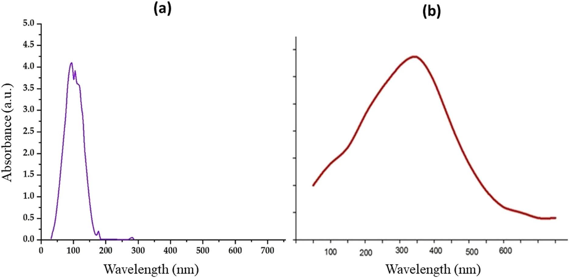
UV-Vis spectrum of (a) pumpkin seed extract and (b) biogenic CuO NPs.
4.2 Spectroscopic analysis of CuO NPs by FTIR
FTIR analysis of the synthesized CuO NPs was recorded in the range of 4,000–400/cm of wavelength. The major Infrared (IR) vibration functional bands in the spectrum of CuO NPs were observed at 2,932, and 2,856/cm depicting the stretching vibration of the aliphatic –CH2 groups, whereas a broad peak at 3,452/cm indicates an aliphatic –OH of carboxylic acid, and a weak peak at 1,733/cm is found to be similar to the keto group stretching vibrational mode. Several functional groups found in the pumpkin seed extract act as reducing and limiting agents for synthesizing stable nanoparticles. The IR, as mentioned above, could be aliphatic –OH carboxylic acid and its derivatives. A peak around 1,634/cm is attributed to the aromatic C═C bond stretching vibrations, whereas a peak at 481/cm indicates the formation of CuO NPs (Figure 2).
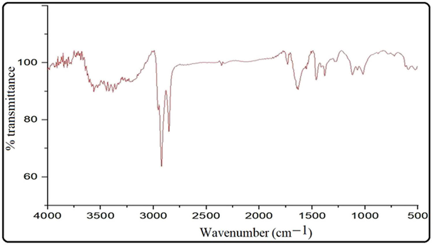
FTIR of biogenic CuO NPs.
4.3 XRD analysis of the CuO NPs
The crystallinity of the biogenically synthesized CuO NPs was determined using the XRD analysis through the generation of various spectra (Figure 3). The observed diffraction sharp peaks at 2θ = 32.22, 35.33, 38.51, 48.44, 61.47, and 65.93 corresponded to miller indices planes of 110, 111, 202, 020, 113, and 311, which are consistent with JCPDS-01-080-0076 and ICSD-087122 of CuO NPs with a monoclinic phase and correspond to the face-centered cubic. Recently, other researchers have also reported the amorphous and less crystalline nature of CuO NPs using the XRD analysis [32,33].
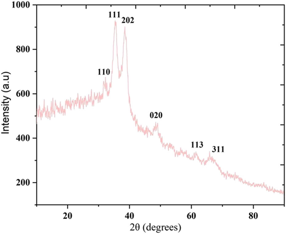
XRD pattern of biogenic CuO NPs.
4.4 SEM and EDX analysis of the CuO NPs
The morphology of the biosynthesized CuO NPs was studied by SEM analysis which showed the dissemination at the surfaces of the CuO NPs (Figure 4a). The SEM micrograph was also analyzed through the EDX spectroscopy to detect the presence of elements in the sample in a qualitative as well as quantitative manner. EDX spectroscopy showed strong peaks corresponding to copper (Cu) and oxygen (O), depicting the formation of nanoparticles. Gold (Au) is also found to be present in the EDX spectrum because the sample was coated with gold for the analysis. The recorded O and Cu values in CuO NPs were 89.42% and 20.11%, respectively (Figure 4b). Hence, the EDX analysis revealed the presence of a lesser amount of Cu and a higher amount of O.
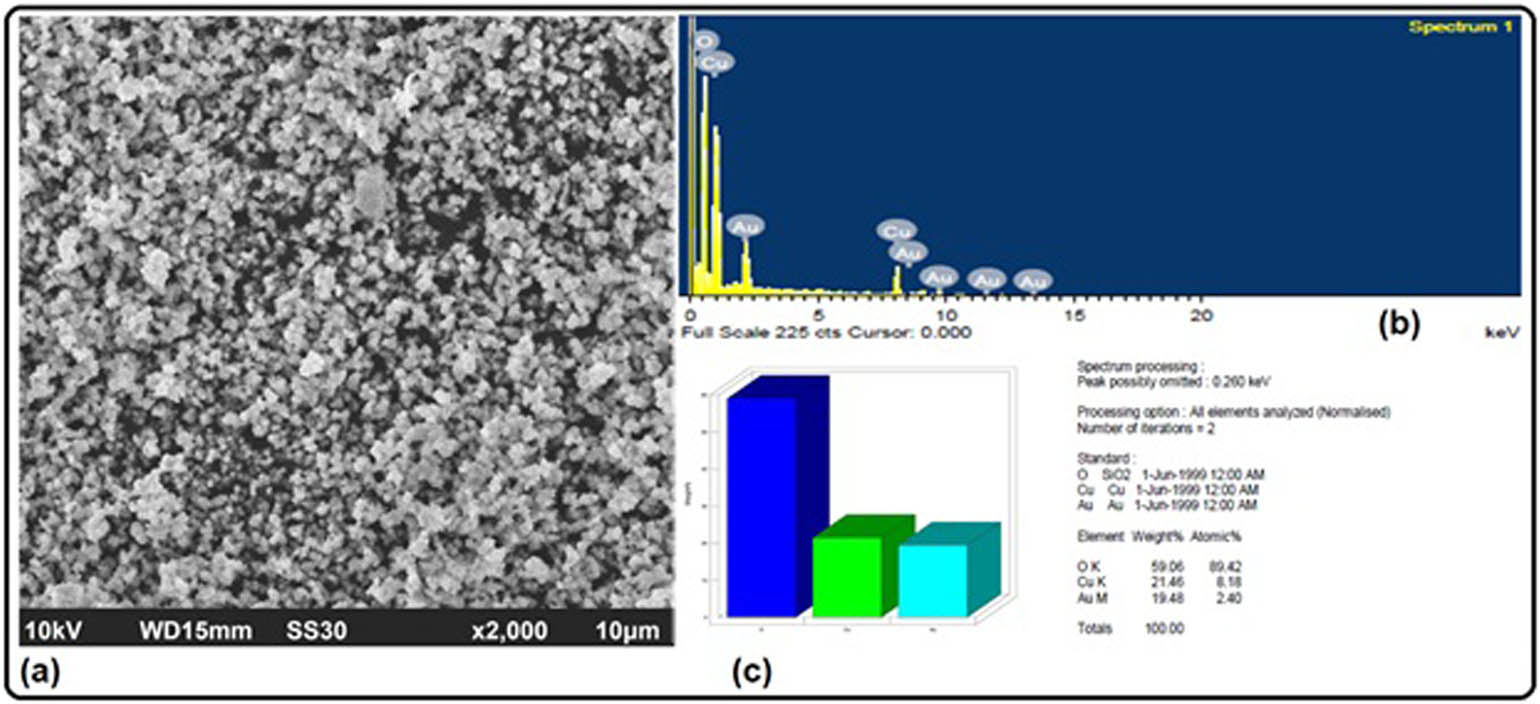
(a) SEM image and (b) EDX profile of the biogenic CuO NPs. (c) a bar chart depicting the weight % of CuO NPs.
4.5 TEM and selected area electron diffraction (SAED) analysis
TEM is a critical technique that provides direct information regarding the particle size distribution, average particle size, and size of the nanoparticles. TEM analysis was carried out to give more insight into the size, shape, and morphology of the biogenically synthesized CuO NPs (Figure 5a). TEM images revealed that the synthesized CuO NPs possess circular shapes with an average size of 20 nm. On the other hand, the SAED images (Figure 5b) determined the nature of CuO NPs revealing circular impression that can be indexed according to the reflection planes such as (110), (111), (202), (020), (113), and (311) planes, as marked by the XRD analysis.
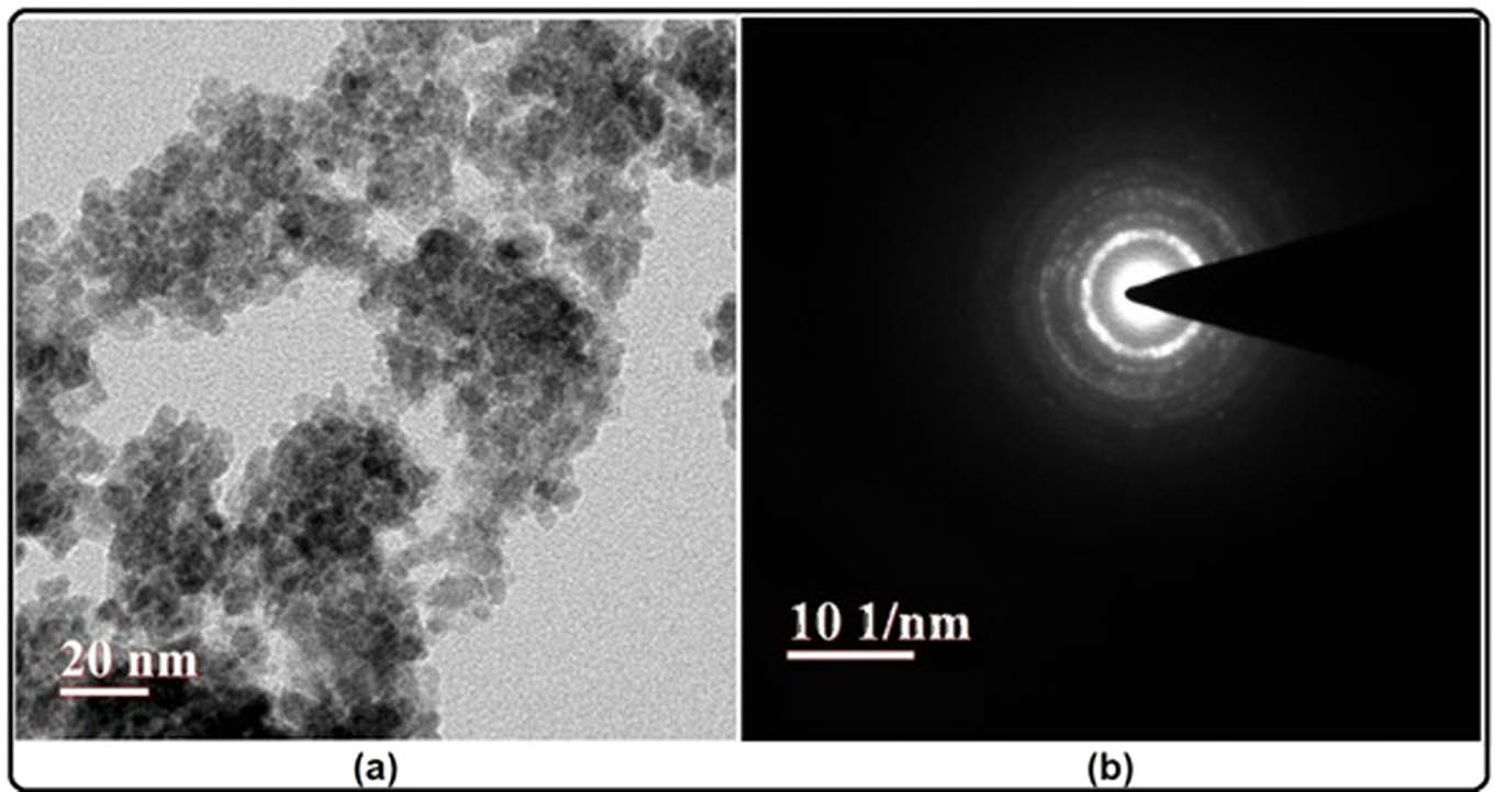
(a) TEM image and (b) SAED pattern of the biogenic CuO NPs.
4.6 Cytotoxicity assay and morphological alterations in the HCT-116 cells
A dose-dependent decrease in the cell viability by the CuO NPs was recorded in the colorectal cancer cells. The synthesized CuO NPs showed an IC50 value at 25 µg/mL (Figure 6). The percentage of cell viability decreased up to 21% at 35 µg/mL. Gnanavel et al. [34] reported the IC50 value of the synthesized CuO NPs to be 40 µg/mL in HCT-116 cell lines. Several other researchers have biosynthesized metal nanoparticles and demonstrated their anticancer potential at much higher concentrations [35,36]. This suggests better anticancer efficacy of our biogenically synthesized CuO NPs. The photomicrograph (10X) represents morphological changes in the HCT-116 cancer cells in the form of shrinking, detachment, membrane blebbing, and distortion in the shape of the cells, which was induced by the treatment of CuO NPs (25 µg and 30 µg/mL). The control cells without any treatment showed normal and intact cellular morphology (Figure 7).
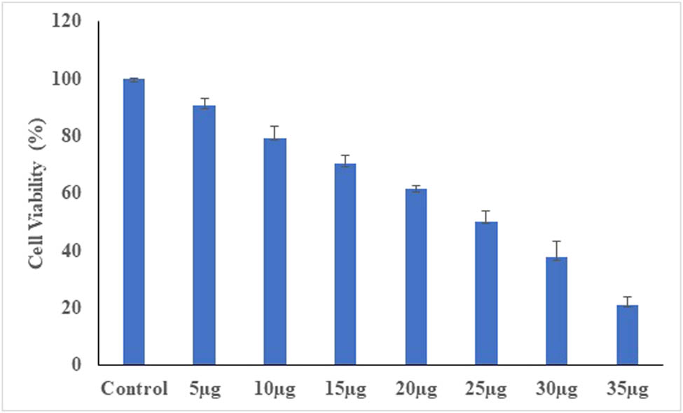
A dose dependent decrease in cell viability of HCT-116 cell lines treated with CuO NPs.
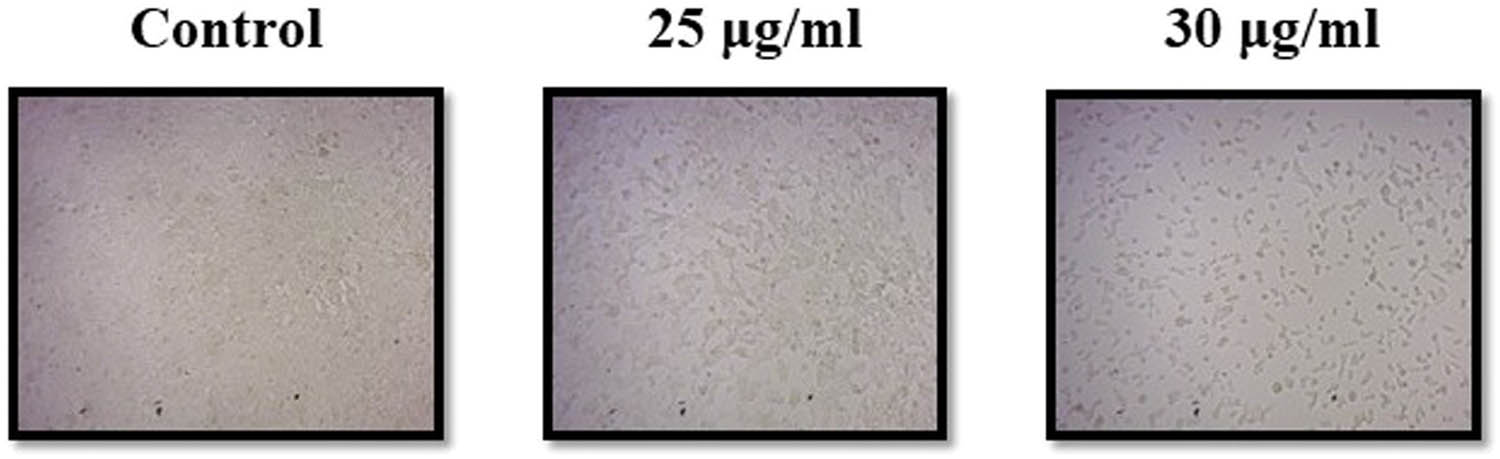
Morphological alterations in HCT-116 cells treated with CuO NPs for 24 h.
4.7 Evaluating the apoptotic induction in the HCT-116 cells by CuO NPs
To study the possible induction of apoptosis, HCT-116 cells were stained with the dual dye, AO/EB, and observed under the fluorescence microscope. Living cells displayed green fluorescence with intact nuclei, while early apoptotic cells showed fragmented nuclei with yellow fluorescence and condensed chromatin. On the other hand, late apoptotic cells displayed orange fluorescence along with the condensation or fragmentation of the chromatin (uniformly red/orange-stained cellular nuclei). Significant apoptotic induction was observed after treating the cells with 25 µg/mL CuO NPs (Figure 8). The results from the current study are in agreement with the earlier reports that observed apoptotic induction in cells treated with green synthesized nanoparticles causing damage to the DNA, and in turn leading to apoptosis/necrosis [37,38]. It is linked to the excessive production of ROS, causing oxidative stress, and eventually leading to the sub-G1 arrest of the cancer cells [37,39]. Similarly, another study also reported apoptotic induction via the upregulation of tumor suppressor genes (p53, Bax, caspase-3, and caspase-9) and the downregulation of oncogenes (Ras and Myc) in response to the green synthesized copper nanoparticles in MCF-7 cells [40].
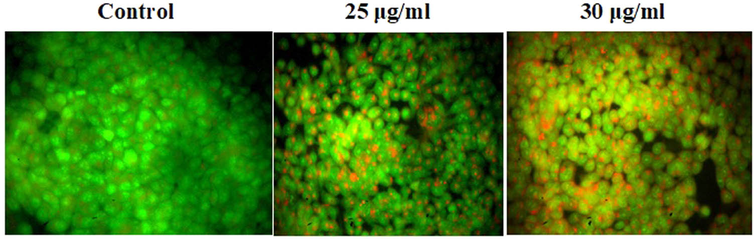
Induction of apoptosis as a result of CuO NPs treatment in HCT-116 cells.
4.8 Inducing the formation of ROS by CuO NPs
We observed a dose-dependent induction in the formation of ROS due to the treatment of HCT-116 cells with CuO NPs (Figure 9). The untreated control cells displayed a dull green fluorescence, while the cells treated with CuO NPs and stained with 2,7-dichlorodihydrofluorescein (DCF) displayed a bright green fluorescence. Our results agree with the previous studies, which reported an increase in the ROS production, following the main toxicity approach of the green synthesized CuO NPs [37,41].
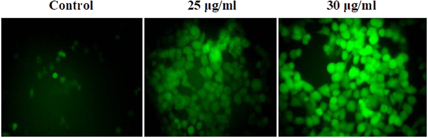
Effect of CuO NPs on the intracellular ROS generation in HCT-116 cells.
4.9 Modulating the MMP
The results show a gradual decrease in the green fluorescence with increasing concentrations of CuO NPs. This indicates a dose-dependent decline in the MMP in HCT-116 cells (Figure 10). The fluorescent images show Rh accumulation in control cells, while it is absent in the treated cells. The green synthesized CuO NPs obtained from the black bean extract have also been reported to alter the mitochondrial structure, accompanied by the loss of membrane potential in HeLa cell lines [42]. Several mechanisms of action associated with green synthesized NPs have been reported in the scientific literature that includes apoptotic induction, increase in the production of ROS/NO, loss of MMP, etc. [43,44,45].
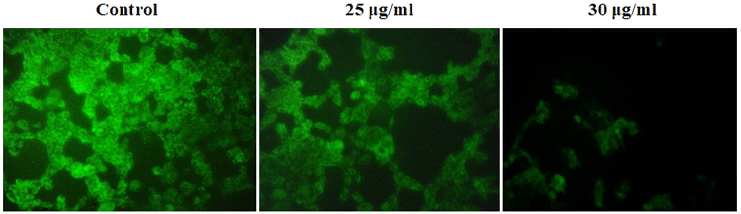
Effects of CuO NPs on MMP in HCT-116 cells.
5 Conclusion
The current study involves the use of an eco-friendly and biogenic method to prepare CuO NPs from pumpkin seed extract. The results of our study show a significant anticancer potential of CuO NPs, which is mediated by the formation of ROS, induction of apoptosis and alteration in MMP in the cancer cells. Considering the benefits associated with plant-based NPs along with the significant anticancer efficacy of our biogenic CuO NPs, it is recommended to validate the results within ex vivo and in vivo models. Successful validation of our results in in vivo modelcould lead to beneficial applications of the biosynthesized CuO NPs in the pharmaceutical, medical, and biotechnological fields. However, the precise mechanism of action of the CuO NPs needs to be studied further to provide a better insight into their application in various fields.
Acknowledgements
The authors gratefully acknowledge the research facility provided by the King Fahd Medical Research Center (KFMRC), King Abdulaziz University, Jeddah, Saudi Arabia.
-
Funding information: This project was funded by the Deanship of Scientific Research (DSR) at King Abdulaziz University, Jeddah, under grant number KEP-1-141-40. The authors, therefore, acknowledge with thanks DSR for technical and financial support.
-
Author contributions: All authors have accepted responsibility for the entire content of this manuscript and approved its submission.
-
Conflict of interest: The authors state no conflict of interest.
References
[1] Sharifi-Rad J, Quispe C, Butnariu M, Rotariu LS, Sytar O, Sestito S, et al. Chitosan nanoparticles as a promising tool in nanomedicine with particular emphasis on oncological treatment. Cancer Cell Int. 2021;21(1):318.10.1186/s12935-021-02025-4Suche in Google Scholar PubMed PubMed Central
[2] Khan I, Saeed K, Khan I. Nanoparticles: properties, applications and toxicities. Arab J Chem. 2019;12(7):908–31.10.1016/j.arabjc.2017.05.011Suche in Google Scholar
[3] Alserihi RF, Mohammed MRS, Kaleem M, Khan MI, Sechi M, Sanna V, et al. Development of (−)-epigallocatechin-3-gallate-loaded folate receptor-targeted nanoparticles for prostate cancer treatment. Nanotechnol Rev. 2022;11(1):298–311.10.1515/ntrev-2022-0013Suche in Google Scholar
[4] Prasad R, Bhattacharyya A, Nguyen QD. Nanotechnology in sustainable agriculture: recent developments, challenges, and perspectives. Front Microbiol. 2017;8:1014.10.3389/fmicb.2017.01014Suche in Google Scholar PubMed PubMed Central
[5] Tabrez S, Jabir NR, Adhami VM, Khan MI, Moulay M, Kamal MA, et al. Nanoencapsulated dietary polyphenols for cancer prevention and treatment: successes and challenges. Nanomed (Lond). 2020;15(11):1147–62.10.2217/nnm-2019-0398Suche in Google Scholar PubMed
[6] Dermatas D, Mpouras T, Panagiotakis I. Application of nanotechnology for waste management: challenges and limitations. Waste Manag Res. 2018;36(3):197–9.10.1177/0734242X18758820Suche in Google Scholar PubMed
[7] Shait Mohammed MR, Ahmad V, Ahmad A, Tabrez S, Choudhry H, Zamzami MA, et al. Prospective of nanoscale metal organic frameworks [NMOFs] for cancer therapy. Semin Cancer Biol. 2021;69:129–39.10.1016/j.semcancer.2019.12.015Suche in Google Scholar PubMed
[8] Hutchison JE. Greener nanoscience: a proactive approach to advancing applications and reducing implications of nanotechnology. ACS Nano. 2008;2(3):395–402.10.1021/nn800131jSuche in Google Scholar PubMed
[9] Oves M, Aslam M, Rauf MA, Qayyum S, Qari HA, Khan MS, et al. Antimicrobial and anticancer activities of silver nanoparticles synthesized from the root hair extract of Phoenix dactylifera. Mater Sci Eng C Mater Biol Appl. 2018;89:429–3.10.1016/j.msec.2018.03.035Suche in Google Scholar PubMed
[10] White PJ, Brown PH. Plant nutrition for sustainable development and global health. Ann Bot. 2010;105(7):1073–80.10.1093/aob/mcq085Suche in Google Scholar PubMed PubMed Central
[11] Izydorczyk G, Sienkiewicz-Cholewa U, Baśladyńska S, Kocek D, Mironiuk M, Chojnacka K. New environmentally friendly bio-based micronutrient fertilizer by biosorption: from laboratory studies to the field. Sci Total Env. 2020;710:136061.10.1016/j.scitotenv.2019.136061Suche in Google Scholar
[12] Bhagat M, Anand R, Sharma P, Rajput P, Sharma N, Singh K. Review – multifunctional copper nanoparticles: synthesis and applications. ECS J Solid State Sci Technol. 2021;10(6):063011.10.1149/2162-8777/ac07f8Suche in Google Scholar
[13] Archana KM, Rajagopal R, Krishnaswamy VG, Aishwarya S. Application of green synthesised copper iodide particles on cotton fabric-protective face mask material against COVID-19 pandemic. J Mater Res Technol. 2021;15:2102–16.10.1016/j.jmrt.2021.09.020Suche in Google Scholar
[14] Khalil AT, Iqbal J, Shah A, Haque MZ, Khan I, Ayaz M, et al. The bio–nano interface as an emerging trend in assembling multi-functional metal nanoparticles: SPR. Nanoscience. 2021;2021:1–24.10.1039/9781839163791-00001Suche in Google Scholar
[15] Khan M, Khan AU, Moon IS, Felimban R, Alserihi R, Alsanie WF, et al. Synthesis of biogenic silver nanoparticles from the seed coat waste of pistachio (Pistacia vera) and their effect on the growth of eggplant. Nanotechnol Rev. 2021;10(1):1789–800.10.1515/ntrev-2021-0107Suche in Google Scholar
[16] Naseer A, Ali A, Ali S, Mahmood A, Kusuma HS, Nazir A, et al. Biogenic and eco-benign synthesis of platinum nanoparticles (Pt NPs) using plants aqueous extracts and biological derivatives: environmental, biological and catalytic applications. J Mater Res Technol. 2020;9(4):9093–107.10.1016/j.jmrt.2020.06.013Suche in Google Scholar
[17] Kim MY, Kim EJ, Kim Y-N, Choi C, Lee B-H. Comparison of the chemical compositions and nutritive values of various pumpkin (Cucurbitaceae) species and parts. Nutr Res Pract. 2012;6(1):21–7.10.4162/nrp.2012.6.1.21Suche in Google Scholar
[18] Dar AH, Sofi SA, Rafiq S. Pumpkin the functional and therapeutic ingredient: a review. Int J Food Sci Nutr. 2017;2(6):165–70.Suche in Google Scholar
[19] Moccia S, Russo M, Durante M, Lenucci MS, Mita G, Russo GL. A carotenoid-enriched extract from pumpkin delays cell proliferation in a human chronic lymphocytic leukemia cell line through the modulation of autophagic flux. Curr Res Biotechnol. 2020;2:74–82.10.1016/j.crbiot.2020.05.001Suche in Google Scholar
[20] Dotto JM, Chacha JS. The potential of pumpkin seeds as a functional food ingredient: A review. Sci Afr. 2020;10:e00575.10.1016/j.sciaf.2020.e00575Suche in Google Scholar
[21] Wang HX, Ng TB. Isolation of cucurmoschin, a novel antifungal peptide abundant in arginine, glutamate and glycine residues from black pumpkin seeds. Peptides. 2003;24(7):969–72.10.1016/S0196-9781(03)00191-8Suche in Google Scholar
[22] Lazos E. Nutritional, fatty acid, and oil characteristics of pumpkin and melon seeds. J Food Sci. 2006;51:1382–3.10.1111/j.1365-2621.1986.tb13133.xSuche in Google Scholar
[23] Huang X-E, Hirose K, Wakai K, Matsuo K, Ito H, Xiang J, et al. Comparison of lifestyle risk factors by family history for gastric, breast, lung and colorectal cancer. Asian Pac J Cancer Prev. 2004;5(4):419–27.Suche in Google Scholar
[24] Jian L, Du C-J, Lee AH, Binns CW. Do dietary lycopene and other carotenoids protect against prostate cancer? Int J Cancer. 2005;113(6):1010–4.10.1002/ijc.20667Suche in Google Scholar PubMed
[25] Nomikos T, Gioti K, Tsoukala M, Tenta R. Pumpkin seed extracts inhibit proliferation and induce autophagy in PC-3 androgen insensitive prostate cancer cells. J Med Food. 2021;24(10):1076–82.10.1089/jmf.2020.0200Suche in Google Scholar PubMed
[26] Ordoñez Lozada MI, Rodrigues Maldonade I, Bobrowski Rodrigues D, Silva Santos D, Ortega Sanchez BA, Narcizo de Souza PE, et al. Physicochemical characterization and nano-emulsification of three species of pumpkin seed oils with focus on their physical stability. Food Chem. 2021;343:128512.10.1016/j.foodchem.2020.128512Suche in Google Scholar PubMed
[27] Khan MS, Alomari A, Tabrez S, Hassan I, Wahab R, Bhat SA, et al. Anticancer potential of biogenic silver nanoparticles: a mechanistic study. Pharmaceutics. 2021;13(5):707.10.3390/pharmaceutics13050707Suche in Google Scholar PubMed PubMed Central
[28] Baskić D, Popović S, Ristić P, Arsenijević NN. Analysis of cycloheximide-induced apoptosis in human leukocytes: fluorescence microscopy using annexin V/propidium iodide versus acridin orange/ethidium bromide. Cell Biol Int. 2006;30(11):924–32.10.1016/j.cellbi.2006.06.016Suche in Google Scholar PubMed
[29] Aranda A, Sequedo L, Tolosa L, Quintas G, Burello E, Castell JV, et al. Dichloro-dihydro-fluorescein diacetate (DCFH-DA) assay: a quantitative method for oxidative stress assessment of nanoparticle-treated cells. Toxicol Vitro. 2013;27(2):954–63.10.1016/j.tiv.2013.01.016Suche in Google Scholar PubMed
[30] Pereira C, Santos MS, Oliveira C. Involvement of oxidative stress on the impairment of energy metabolism induced by A beta peptides on PC12 cells: protection by antioxidants. Neurobiol Dis. 1999;6(3):209–19.10.1006/nbdi.1999.0241Suche in Google Scholar PubMed
[31] Bhosle SM, Huilgol NG, Mishra KP. Enhancement of radiation-induced oxidative stress and cytotoxicity in tumor cells by ellagic acid. Clin Chim Acta. 2005;359(1–2):89–100.10.1016/j.cccn.2005.03.037Suche in Google Scholar PubMed
[32] Al-Dayan N. Biosynthesis of copper oxide nanomaterials using the seeds of date fruits (Phoenix dactylifera L.) and antibacterial activity evaluation. Pak J Biol Sci. 2021;24(10):1034–9.10.3923/pjbs.2021.1034.1039Suche in Google Scholar PubMed
[33] Iliger KS, Sofi TA, Bhat NA, Ahanger FA, Sekhar JC, Elhendi AZ, et al. Copper nanoparticles: green synthesis and managing fruit rot disease of chilli caused by Colletotrichum capsici. Saudi J Biol Sci. 2021;28(2):1477–86.10.1016/j.sjbs.2020.12.003Suche in Google Scholar PubMed PubMed Central
[34] Gnanavel V, Palanichamy V, Roopan SM. Biosynthesis and characterization of copper oxide nanoparticles and its anticancer activity on human colon cancer cell lines (HCT-116). J Photochem Photobiol B. 2017;171:133–8.10.1016/j.jphotobiol.2017.05.001Suche in Google Scholar PubMed
[35] Al Sufyani NM, Hussien NA, Hawsawi YM. Characterization and anticancer potential of silver nanoparticles biosynthesized from olea chrysophylla and lavandula dentata leaf extracts on HCT-116 colon cancer cells. J Nanomater. 2019;2019:e7361695.10.1155/2019/7361695Suche in Google Scholar
[36] Vijay M, Anu Y. Anticancer activity of camellia sinensis mediated copper nanoparticles against HT-29, MCF-7 and MOLT-4 human cancer cell lines. Asian J Pharm Clin Res. 2017;10(2):71–7.10.22159/ajpcr.2017.v10i2.15710Suche in Google Scholar
[37] Al-Sheddi ES, Farshori NN, Al-Oqail MM, Al-Massarani SM, Saquib Q, Wahab R, et al. Anticancer potential of green synthesized silver nanoparticles using extract of nepeta deflersiana against human cervical cancer cells (HeLA). Bioinorg Chem Appl. 2018;2018:9390784.10.1155/2018/9390784Suche in Google Scholar PubMed PubMed Central
[38] Pan Y, Neuss S, Leifert A, Fischler M, Wen F, Simon U, et al. Size-dependent cytotoxicity of gold nanoparticles. Small. 2007;3(11):1941–9.10.1002/smll.200700378Suche in Google Scholar PubMed
[39] Hsin Y-H, Chen C-F, Huang S, Shih T-S, Lai P-S, Chueh PJ. The apoptotic effect of nanosilver is mediated by a ROS- and JNK-dependent mechanism involving the mitochondrial pathway in NIH3T3 cells. Toxicol Lett. 2008;179(3):130–9.10.1016/j.toxlet.2008.04.015Suche in Google Scholar PubMed
[40] Biresaw SS, Taneja P. Copper nanoparticles green synthesis and characterization as anticancer potential in breast cancer cells (MCF7) derived from Prunus nepalensis phytochemicals. Mater Today: Proc. 2022;49(8):3501–9.10.1016/j.matpr.2021.07.149Suche in Google Scholar
[41] Alizadeh SR, Ebrahimzadeh MA. O-Glycoside quercetin derivatives: biological activities, mechanisms of action, and structure-activity relationship for drug design, a review. Phytother Res. 2022;32(12):778–807.10.1002/ptr.7352Suche in Google Scholar PubMed
[42] Nagajyothi PC, Muthuraman P, Sreekanth TVM, Kim DH, Shim J. Green synthesis: in-vitro anticancer activity of copper oxide nanoparticles against human cervical carcinoma cells. Arab J Chem. 2017;10(2):215–25.10.1016/j.arabjc.2016.01.011Suche in Google Scholar
[43] Letchumanan D, Sok SPM, Ibrahim S, Nagoor NH, Arshad NM. Plant-based biosynthesis of copper/copper oxide nanoparticles: an update on their applications in biomedicine, mechanisms, and toxicity. Biomolecules. 2021;11(4):564.10.3390/biom11040564Suche in Google Scholar PubMed PubMed Central
[44] Dey A, Manna S, Chattopadhyay S, Mondal D, Chattopadhyay D, Raj A, et al. Azadirachta indica leaves mediated green synthesized copper oxide nanoparticles induce apoptosis through activation of TNF-α and caspases signaling pathway against cancer cells. J Saudi Chem Soc. 2019;23(2):222–38.10.1016/j.jscs.2018.06.011Suche in Google Scholar
[45] Khursheed A, Quaiser S, Bilal A, Maqsood AS, Javed A, Majed A-S, et al. Bio-functionalized CuO nanoparticles induced apoptotic activities in human breast carcinoma cells and toxicity against Aspergillus flavus: an in vitro approach. Process Biochem. 2020;91:387–97.10.1016/j.procbio.2020.01.008Suche in Google Scholar
© 2022 Shams Tabrez et al., published by De Gruyter
This work is licensed under the Creative Commons Attribution 4.0 International License.
Artikel in diesem Heft
- Research Articles
- Theoretical and experimental investigation of MWCNT dispersion effect on the elastic modulus of flexible PDMS/MWCNT nanocomposites
- Mechanical, morphological, and fracture-deformation behavior of MWCNTs-reinforced (Al–Cu–Mg–T351) alloy cast nanocomposites fabricated by optimized mechanical milling and powder metallurgy techniques
- Flammability and physical stability of sugar palm crystalline nanocellulose reinforced thermoplastic sugar palm starch/poly(lactic acid) blend bionanocomposites
- Glutathione-loaded non-ionic surfactant niosomes: A new approach to improve oral bioavailability and hepatoprotective efficacy of glutathione
- Relationship between mechano-bactericidal activity and nanoblades density on chemically strengthened glass
- In situ regulation of microstructure and microwave-absorbing properties of FeSiAl through HNO3 oxidation
- Research on a mechanical model of magnetorheological fluid different diameter particles
- Nanomechanical and dynamic mechanical properties of rubber–wood–plastic composites
- Investigative properties of CeO2 doped with niobium: A combined characterization and DFT studies
- Miniaturized peptidomimetics and nano-vesiculation in endothelin types through probable nano-disk formation and structure property relationships of endothelins’ fragments
- N/S co-doped CoSe/C nanocubes as anode materials for Li-ion batteries
- Synergistic effects of halloysite nanotubes with metal and phosphorus additives on the optimal design of eco-friendly sandwich panels with maximum flame resistance and minimum weight
- Octreotide-conjugated silver nanoparticles for active targeting of somatostatin receptors and their application in a nebulized rat model
- Controllable morphology of Bi2S3 nanostructures formed via hydrothermal vulcanization of Bi2O3 thin-film layer and their photoelectrocatalytic performances
- Development of (−)-epigallocatechin-3-gallate-loaded folate receptor-targeted nanoparticles for prostate cancer treatment
- Enhancement of the mechanical properties of HDPE mineral nanocomposites by filler particles modulation of the matrix plastic/elastic behavior
- Effect of plasticizers on the properties of sugar palm nanocellulose/cinnamon essential oil reinforced starch bionanocomposite films
- Optimization of nano coating to reduce the thermal deformation of ball screws
- Preparation of efficient piezoelectric PVDF–HFP/Ni composite films by high electric field poling
- MHD dissipative Casson nanofluid liquid film flow due to an unsteady stretching sheet with radiation influence and slip velocity phenomenon
- Effects of nano-SiO2 modification on rubberised mortar and concrete with recycled coarse aggregates
- Mechanical and microscopic properties of fiber-reinforced coal gangue-based geopolymer concrete
- Effect of morphology and size on the thermodynamic stability of cerium oxide nanoparticles: Experiment and molecular dynamics calculation
- Mechanical performance of a CFRP composite reinforced via gelatin-CNTs: A study on fiber interfacial enhancement and matrix enhancement
- A practical review over surface modification, nanopatterns, emerging materials, drug delivery systems, and their biophysiochemical properties for dental implants: Recent progresses and advances
- HTR: An ultra-high speed algorithm for cage recognition of clathrate hydrates
- Effects of microalloying elements added by in situ synthesis on the microstructure of WCu composites
- A highly sensitive nanobiosensor based on aptamer-conjugated graphene-decorated rhodium nanoparticles for detection of HER2-positive circulating tumor cells
- Progressive collapse performance of shear strengthened RC frames by nano CFRP
- Core–shell heterostructured composites of carbon nanotubes and imine-linked hyperbranched polymers as metal-free Li-ion anodes
- A Galerkin strategy for tri-hybridized mixture in ethylene glycol comprising variable diffusion and thermal conductivity using non-Fourier’s theory
- Simple models for tensile modulus of shape memory polymer nanocomposites at ambient temperature
- Preparation and morphological studies of tin sulfide nanoparticles and use as efficient photocatalysts for the degradation of rhodamine B and phenol
- Polyethyleneimine-impregnated activated carbon nanofiber composited graphene-derived rice husk char for efficient post-combustion CO2 capture
- Electrospun nanofibers of Co3O4 nanocrystals encapsulated in cyclized-polyacrylonitrile for lithium storage
- Pitting corrosion induced on high-strength high carbon steel wire in high alkaline deaerated chloride electrolyte
- Formulation of polymeric nanoparticles loaded sorafenib; evaluation of cytotoxicity, molecular evaluation, and gene expression studies in lung and breast cancer cell lines
- Engineered nanocomposites in asphalt binders
- Influence of loading voltage, domain ratio, and additional load on the actuation of dielectric elastomer
- Thermally induced hex-graphene transitions in 2D carbon crystals
- The surface modification effect on the interfacial properties of glass fiber-reinforced epoxy: A molecular dynamics study
- Molecular dynamics study of deformation mechanism of interfacial microzone of Cu/Al2Cu/Al composites under tension
- Nanocolloid simulators of luminescent solar concentrator photovoltaic windows
- Compressive strength and anti-chloride ion penetration assessment of geopolymer mortar merging PVA fiber and nano-SiO2 using RBF–BP composite neural network
- Effect of 3-mercapto-1-propane sulfonate sulfonic acid and polyvinylpyrrolidone on the growth of cobalt pillar by electrodeposition
- Dynamics of convective slippery constraints on hybrid radiative Sutterby nanofluid flow by Galerkin finite element simulation
- Preparation of vanadium by the magnesiothermic self-propagating reduction and process control
- Microstructure-dependent photoelectrocatalytic activity of heterogeneous ZnO–ZnS nanosheets
- Cytotoxic and pro-inflammatory effects of molybdenum and tungsten disulphide on human bronchial cells
- Improving recycled aggregate concrete by compression casting and nano-silica
- Chemically reactive Maxwell nanoliquid flow by a stretching surface in the frames of Newtonian heating, nonlinear convection and radiative flux: Nanopolymer flow processing simulation
- Nonlinear dynamic and crack behaviors of carbon nanotubes-reinforced composites with various geometries
- Biosynthesis of copper oxide nanoparticles and its therapeutic efficacy against colon cancer
- Synthesis and characterization of smart stimuli-responsive herbal drug-encapsulated nanoniosome particles for efficient treatment of breast cancer
- Homotopic simulation for heat transport phenomenon of the Burgers nanofluids flow over a stretching cylinder with thermal convective and zero mass flux conditions
- Incorporation of copper and strontium ions in TiO2 nanotubes via dopamine to enhance hemocompatibility and cytocompatibility
- Mechanical, thermal, and barrier properties of starch films incorporated with chitosan nanoparticles
- Mechanical properties and microstructure of nano-strengthened recycled aggregate concrete
- Glucose-responsive nanogels efficiently maintain the stability and activity of therapeutic enzymes
- Tunning matrix rheology and mechanical performance of ultra-high performance concrete using cellulose nanofibers
- Flexible MXene/copper/cellulose nanofiber heat spreader films with enhanced thermal conductivity
- Promoted charge separation and specific surface area via interlacing of N-doped titanium dioxide nanotubes on carbon nitride nanosheets for photocatalytic degradation of Rhodamine B
- Elucidating the role of silicon dioxide and titanium dioxide nanoparticles in mitigating the disease of the eggplant caused by Phomopsis vexans, Ralstonia solanacearum, and root-knot nematode Meloidogyne incognita
- An implication of magnetic dipole in Carreau Yasuda liquid influenced by engine oil using ternary hybrid nanomaterial
- Robust synthesis of a composite phase of copper vanadium oxide with enhanced performance for durable aqueous Zn-ion batteries
- Tunning self-assembled phases of bovine serum albumin via hydrothermal process to synthesize novel functional hydrogel for skin protection against UVB
- A comparative experimental study on damping properties of epoxy nanocomposite beams reinforced with carbon nanotubes and graphene nanoplatelets
- Lightweight and hydrophobic Ni/GO/PVA composite aerogels for ultrahigh performance electromagnetic interference shielding
- Research on the auxetic behavior and mechanical properties of periodically rotating graphene nanostructures
- Repairing performances of novel cement mortar modified with graphene oxide and polyacrylate polymer
- Closed-loop recycling and fabrication of hydrophilic CNT films with high performance
- Design of thin-film configuration of SnO2–Ag2O composites for NO2 gas-sensing applications
- Study on stress distribution of SiC/Al composites based on microstructure models with microns and nanoparticles
- PVDF green nanofibers as potential carriers for improving self-healing and mechanical properties of carbon fiber/epoxy prepregs
- Osteogenesis capability of three-dimensionally printed poly(lactic acid)-halloysite nanotube scaffolds containing strontium ranelate
- Silver nanoparticles induce mitochondria-dependent apoptosis and late non-canonical autophagy in HT-29 colon cancer cells
- Preparation and bonding mechanisms of polymer/metal hybrid composite by nano molding technology
- Damage self-sensing and strain monitoring of glass-reinforced epoxy composite impregnated with graphene nanoplatelet and multiwalled carbon nanotubes
- Thermal analysis characterisation of solar-powered ship using Oldroyd hybrid nanofluids in parabolic trough solar collector: An optimal thermal application
- Pyrene-functionalized halloysite nanotubes for simultaneously detecting and separating Hg(ii) in aqueous media: A comprehensive comparison on interparticle and intraparticle excimers
- Fabrication of self-assembly CNT flexible film and its piezoresistive sensing behaviors
- Thermal valuation and entropy inspection of second-grade nanoscale fluid flow over a stretching surface by applying Koo–Kleinstreuer–Li relation
- Mechanical properties and microstructure of nano-SiO2 and basalt-fiber-reinforced recycled aggregate concrete
- Characterization and tribology performance of polyaniline-coated nanodiamond lubricant additives
- Combined impact of Marangoni convection and thermophoretic particle deposition on chemically reactive transport of nanofluid flow over a stretching surface
- Spark plasma extrusion of binder free hydroxyapatite powder
- An investigation on thermo-mechanical performance of graphene-oxide-reinforced shape memory polymer
- Effect of nanoadditives on the novel leather fiber/recycled poly(ethylene-vinyl-acetate) polymer composites for multifunctional applications: Fabrication, characterizations, and multiobjective optimization using central composite design
- Design selection for a hemispherical dimple core sandwich panel using hybrid multi-criteria decision-making methods
- Improving tensile strength and impact toughness of plasticized poly(lactic acid) biocomposites by incorporating nanofibrillated cellulose
- Green synthesis of spinel copper ferrite (CuFe2O4) nanoparticles and their toxicity
- The effect of TaC and NbC hybrid and mono-nanoparticles on AA2024 nanocomposites: Microstructure, strengthening, and artificial aging
- Excited-state geometry relaxation of pyrene-modified cellulose nanocrystals under UV-light excitation for detecting Fe3+
- Effect of CNTs and MEA on the creep of face-slab concrete at an early age
- Effect of deformation conditions on compression phase transformation of AZ31
- Application of MXene as a new generation of highly conductive coating materials for electromembrane-surrounded solid-phase microextraction
- A comparative study of the elasto-plastic properties for ceramic nanocomposites filled by graphene or graphene oxide nanoplates
- Encapsulation strategies for improving the biological behavior of CdS@ZIF-8 nanocomposites
- Biosynthesis of ZnO NPs from pumpkin seeds’ extract and elucidation of its anticancer potential against breast cancer
- Preliminary trials of the gold nanoparticles conjugated chrysin: An assessment of anti-oxidant, anti-microbial, and in vitro cytotoxic activities of a nanoformulated flavonoid
- Effect of micron-scale pores increased by nano-SiO2 sol modification on the strength of cement mortar
- Fractional simulations for thermal flow of hybrid nanofluid with aluminum oxide and titanium oxide nanoparticles with water and blood base fluids
- The effect of graphene nano-powder on the viscosity of water: An experimental study and artificial neural network modeling
- Development of a novel heat- and shear-resistant nano-silica gelling agent
- Characterization, biocompatibility and in vivo of nominal MnO2-containing wollastonite glass-ceramic
- Entropy production simulation of second-grade magnetic nanomaterials flowing across an expanding surface with viscidness dissipative flux
- Enhancement in structural, morphological, and optical properties of copper oxide for optoelectronic device applications
- Aptamer-functionalized chitosan-coated gold nanoparticle complex as a suitable targeted drug carrier for improved breast cancer treatment
- Performance and overall evaluation of nano-alumina-modified asphalt mixture
- Analysis of pure nanofluid (GO/engine oil) and hybrid nanofluid (GO–Fe3O4/engine oil): Novel thermal and magnetic features
- Synthesis of Ag@AgCl modified anatase/rutile/brookite mixed phase TiO2 and their photocatalytic property
- Mechanisms and influential variables on the abrasion resistance hydraulic concrete
- Synergistic reinforcement mechanism of basalt fiber/cellulose nanocrystals/polypropylene composites
- Achieving excellent oxidation resistance and mechanical properties of TiB2–B4C/carbon aerogel composites by quick-gelation and mechanical mixing
- Microwave-assisted sol–gel template-free synthesis and characterization of silica nanoparticles obtained from South African coal fly ash
- Pulsed laser-assisted synthesis of nano nickel(ii) oxide-anchored graphitic carbon nitride: Characterizations and their potential antibacterial/anti-biofilm applications
- Effects of nano-ZrSi2 on thermal stability of phenolic resin and thermal reusability of quartz–phenolic composites
- Benzaldehyde derivatives on tin electroplating as corrosion resistance for fabricating copper circuit
- Mechanical and heat transfer properties of 4D-printed shape memory graphene oxide/epoxy acrylate composites
- Coupling the vanadium-induced amorphous/crystalline NiFe2O4 with phosphide heterojunction toward active oxygen evolution reaction catalysts
- Graphene-oxide-reinforced cement composites mechanical and microstructural characteristics at elevated temperatures
- Gray correlation analysis of factors influencing compressive strength and durability of nano-SiO2 and PVA fiber reinforced geopolymer mortar
- Preparation of layered gradient Cu–Cr–Ti alloy with excellent mechanical properties, thermal stability, and electrical conductivity
- Recovery of Cr from chrome-containing leather wastes to develop aluminum-based composite material along with Al2O3 ceramic particles: An ingenious approach
- Mechanisms of the improved stiffness of flexible polymers under impact loading
- Anticancer potential of gold nanoparticles (AuNPs) using a battery of in vitro tests
- Review Articles
- Proposed approaches for coronaviruses elimination from wastewater: Membrane techniques and nanotechnology solutions
- Application of Pickering emulsion in oil drilling and production
- The contribution of microfluidics to the fight against tuberculosis
- Graphene-based biosensors for disease theranostics: Development, applications, and recent advancements
- Synthesis and encapsulation of iron oxide nanorods for application in magnetic hyperthermia and photothermal therapy
- Contemporary nano-architectured drugs and leads for ανβ3 integrin-based chemotherapy: Rationale and retrospect
- State-of-the-art review of fabrication, application, and mechanical properties of functionally graded porous nanocomposite materials
- Insights on magnetic spinel ferrites for targeted drug delivery and hyperthermia applications
- A review on heterogeneous oxidation of acetaminophen based on micro and nanoparticles catalyzed by different activators
- Early diagnosis of lung cancer using magnetic nanoparticles-integrated systems
- Advances in ZnO: Manipulation of defects for enhancing their technological potentials
- Efficacious nanomedicine track toward combating COVID-19
- A review of the design, processes, and properties of Mg-based composites
- Green synthesis of nanoparticles for varied applications: Green renewable resources and energy-efficient synthetic routes
- Two-dimensional nanomaterial-based polymer composites: Fundamentals and applications
- Recent progress and challenges in plasmonic nanomaterials
- Apoptotic cell-derived micro/nanosized extracellular vesicles in tissue regeneration
- Electronic noses based on metal oxide nanowires: A review
- Framework materials for supercapacitors
- An overview on the reproductive toxicity of graphene derivatives: Highlighting the importance
- Antibacterial nanomaterials: Upcoming hope to overcome antibiotic resistance crisis
- Research progress of carbon materials in the field of three-dimensional printing polymer nanocomposites
- A review of atomic layer deposition modelling and simulation methodologies: Density functional theory and molecular dynamics
- Recent advances in the preparation of PVDF-based piezoelectric materials
- Recent developments in tensile properties of friction welding of carbon fiber-reinforced composite: A review
- Comprehensive review of the properties of fly ash-based geopolymer with additive of nano-SiO2
- Perspectives in biopolymer/graphene-based composite application: Advances, challenges, and recommendations
- Graphene-based nanocomposite using new modeling molecular dynamic simulations for proposed neutralizing mechanism and real-time sensing of COVID-19
- Nanotechnology application on bamboo materials: A review
- Recent developments and future perspectives of biorenewable nanocomposites for advanced applications
- Nanostructured lipid carrier system: A compendium of their formulation development approaches, optimization strategies by quality by design, and recent applications in drug delivery
- 3D printing customized design of human bone tissue implant and its application
- Design, preparation, and functionalization of nanobiomaterials for enhanced efficacy in current and future biomedical applications
- A brief review of nanoparticles-doped PEDOT:PSS nanocomposite for OLED and OPV
- Nanotechnology interventions as a putative tool for the treatment of dental afflictions
- Recent advancements in metal–organic frameworks integrating quantum dots (QDs@MOF) and their potential applications
- A focused review of short electrospun nanofiber preparation techniques for composite reinforcement
- Microstructural characteristics and nano-modification of interfacial transition zone in concrete: A review
- Latest developments in the upconversion nanotechnology for the rapid detection of food safety: A review
- Strategic applications of nano-fertilizers for sustainable agriculture: Benefits and bottlenecks
- Molecular dynamics application of cocrystal energetic materials: A review
- Synthesis and application of nanometer hydroxyapatite in biomedicine
- Cutting-edge development in waste-recycled nanomaterials for energy storage and conversion applications
- Biological applications of ternary quantum dots: A review
- Nanotherapeutics for hydrogen sulfide-involved treatment: An emerging approach for cancer therapy
- Application of antibacterial nanoparticles in orthodontic materials
- Effect of natural-based biological hydrogels combined with growth factors on skin wound healing
- Nanozymes – A route to overcome microbial resistance: A viewpoint
- Recent developments and applications of smart nanoparticles in biomedicine
- Contemporary review on carbon nanotube (CNT) composites and their impact on multifarious applications
- Interfacial interactions and reinforcing mechanisms of cellulose and chitin nanomaterials and starch derivatives for cement and concrete strength and durability enhancement: A review
- Diamond-like carbon films for tribological modification of rubber
- Layered double hydroxides (LDHs) modified cement-based materials: A systematic review
- Recent research progress and advanced applications of silica/polymer nanocomposites
- Modeling of supramolecular biopolymers: Leading the in silico revolution of tissue engineering and nanomedicine
- Recent advances in perovskites-based optoelectronics
- Biogenic synthesis of palladium nanoparticles: New production methods and applications
- A comprehensive review of nanofluids with fractional derivatives: Modeling and application
- Electrospinning of marine polysaccharides: Processing and chemical aspects, challenges, and future prospects
- Electrohydrodynamic printing for demanding devices: A review of processing and applications
- Rapid Communications
- Structural material with designed thermal twist for a simple actuation
- Recent advances in photothermal materials for solar-driven crude oil adsorption