Development of (−)-epigallocatechin-3-gallate-loaded folate receptor-targeted nanoparticles for prostate cancer treatment
-


Abstract
In continuation of our previous studies, we developed polymeric epigallocatechin 3-gallate (EGCG)-loaded nanoparticles (NPs) coupled with folic acid (FA), able to dually bind the human folate receptor alpha (FOLR1), and prostate-specific membrane antigen (PSMA+) in prostate cancer (PCa) model. After a preliminary computational molecular recognition of NP′ ligand binding on the FOLR1 active site, we synthesized the biocompatible block-copolymer PLGA–PEG–FA to prepare EGCG-targeted NPs (EGCG-T-NPs). The obtained NPs were characterized by various analytical techniques, and anticancer efficacy was determined by different sets of experiments in a 3D culture of PCa using PC3 and 22Rv1 cell lines. Results showed a significant reduction in spheroid size by EGCG-T-NPs, especially in PSMA+ (22Rv1) cells. The targeted NPs significantly enhanced the antiproliferative activity of EGCG against PCa cell lines, especially toward the PSMA+ cells, known to have higher FOLR1 expression. We did not observe any changes in the reactive oxygen species formation in both studied cell lines. However, significant changes in mitochondrial depolarization (15%) and polarization (18%) were recorded in response to EGCG-T-NP compared to control in 22Rv1. Similarly, EGCG-T-NP treatment also showed an increase in the number of dead apoptotic cells in 22Rv1 spheroids. Collectively, the obtained results support our hypothesis about the role of these targeted nanoprototypes in the increasing cellular uptake of EGCG payload into PCa cells, thus enhancing its antitumor efficacy.
1 Introduction
Prostate cancer (PCa) is the most common neoplastic disorder and the second leading cause of cancer-related deaths among males worldwide [1]. It accounts for 7.3% of all cancer incidences and 3.8% of cancer-related deaths in males in 2020 [2]. The increasing prevalence of PCa has become a global health challenge causing multi-organ spread and poorer prognosis in advanced stages [3]. Extraprostatic or metastatic stage diagnosis and neoplastic heterogeneity often complicate the therapeutic benefits [4]. Therefore, there is a compelling requirement to discover novel, harmless yet highly specific chemotherapeutics against this type of cancer [5,6].
Green tea, the most popular beverage consumed worldwide, demonstrated various degrees of protection against different diseases, including cancer [7,8]. An active catechin component, the (−)-epigallocatechin 3-gallate (EGCG), has shown an effective chemopreventive potential by modulating various cell signaling pathways in several in vitro assays of tumor cell lines and in in vivo preclinical, experimental models of induced carcinogenesis [9,10]. The first pioneering work highlighted that oral administration of green tea catechins extracts could be beneficial in the early stage of PCa, leading to a long-lasting inhibition of cancer progression [11,12]. Further data reported the therapeutic effectiveness of EGCG alone or in combination against PCa and other cancer [13,14]. Despite its efficacy and safety, the role of EGCG in cancer prevention and therapy is still discussed due to poor absorption, rapid metabolism and elimination, and inefficient systemic bioavailability [15].
In this context, nanotechnology-based strategies such as liposomes, nanoparticles (NPs), micelles, and other formulations have shown improvement in the anticancer potential of EGCG by enhancing its potency and reducing toxicity, side effects, and increased the concentration at the cancer site [16,17,18]. Advances in engineered nanovehicles with various materials allowed sustained delivery and controlled release of EGCG with enhanced stability and bioavailability [19,20]. Among these nanosystems, polymeric NPs are largely considered because of good biocompatibility, simple design, preparation, and customization, especially for targeted delivery [21,22]. Previously, Siddiqui et al. introduced the concept of “nanochemoprevention,” where the efficacy of EGCG encapsulated in polylactic acid–polyethylene glycol NPs was determined in a preclinical setting [16,23,24]. A major challenge to improve the pharmacological profile of EGCG can also be pursued by a cell/tissue-specific targeting approach [25]. In our earlier studies, we developed novel NPs targeting prostate-specific membrane antigen (PSMA), a tumor-associated membrane receptor that appeared overexpressed in some specific PCa cells [26,27].
Active targeting exploited by the inclusion of a specific ligand on the NPs’ surface is expected to provide an intriguing strategy for the development of effective antitumor therapeutics. In this study, we focused our attention on folate receptors human folate receptor alpha (FOLR1), well-known transmembrane glycoproteins, highly expressed on the majority of cancer tissues, to exploit the folate demand of rapidly dividing cells under low folate conditions [28,29]. Since PSMA has significantly elevated levels in prostate tissue compared with normal tissues, its upregulation contributed to tumor progression. PSMA exhibits the enzymatic function as a glutamate carboxypeptidase and folate hydrolase, thus implying its role in the metabolism of folates and their subsequent uptake [30].
Functionalization of NPs with ligands, i.e., folic acid (FA), able to bind to the extracellular region of these receptors, can be considered for selective and effective delivery of ECGC to PCa cells. To the best of our knowledge, no previous study has shown the effect of EGCG-loaded PLGA–PEG–FA-based NPs in PCa cells using a 3D culture model. In the present study, we developed a precise targeted polymeric (PLGA–PEG) EGCG-loaded NPs, decorated with FA small molecules on their shell surface to achieve active cellular targeting toward FOLR1 and PSMA, and investigated their antiproliferative properties and compared the efficacy in two PCa cells (i.e., PC3 [PSMA−] and 22Rv1 [PSMA+]). The reason for selecting PC3 and 22Rv1 was the presence (22Rv1) or absence (PC3) of PSMA, albeit both expressed FOLR1. On the other hand, choosing the 3D cell culture system over the traditional 2D model was due to its ability to better represent human tissues outside the body [31].
2 Materials and methods
2.1 Molecular modeling
The three-dimensional structure of the FOLR1 in complex with FA was retrieved from the RCSB Protein Data Bank (pdb: 4LRH, resolution: 2.80 Å) [32] utilizing a well-established docking protocol [33,34]. The docking studies were performed on a personal Macbook installed with the IOS operating system. The ligands FA and PEG–FA were constructed in a neutral form and energy was minimized by standard molecular mechanics protocols. The protein target was prepared by using AutoDock version 4.25 [35]. Briefly, ions and water molecules were removed, while hydrogen atoms were highlighted by the ADT module implemented in AutoDock. The charges were checked using the appropriate Gasteiger module for proteins implemented in AutoDock [36]. The docking was performed using the empirical free energy function and the Lamarckian protocol [37]. Mass-centered grid maps were generated with 40 grid points for every direction. Random starting position on the entire protein surface, orientations, and torsions were used for the ligands. Interactions in all the complexes were analyzed.
2.2 Chemistry of polymer synthesis
2.2.1 Chemistry
The synthesis of the copolymers was followed as described earlier with slight modification [38]. The analyses were done by using different techniques such as nuclear magnetic resonance (1H-NMR), MALDI-TOF mass spectra, UV-vis absorption spectra, and HPLC. The synthesized materials were found to be >95% pure.
2.3 Synthesis of PLGA–PEG–NH2
We prepared a sufficient amount of activated PLGA–N-hydroxysuccinimide (NHS) intermediate. Briefly, NHS (32 mg, 1.1 mmol, ∼4 equiv.) and 1-ethyl-3-(3-dimethylaminopropyl)-carbodiimide (58.2 mg, 1.2 mmol, ∼4.3 equiv.) were added under nitrogen atmosphere to a solution of PLGA–COOH (1.25 g, 0.07 mmol) in anhydrous methylene chloride (5 mL). The resulting solution was magnetically stirred at room temperature for 12 h. The activated polymer of PLGA–NHS was collected by first precipitation with cold diethyl ether (5 mL) as a white solid, which was filtered and repeatedly washed in a cold mixture of diethyl ether and methanol. Then, the product was dried with nitrogen under vacuum to remove the solvent (yield: ∼96%). In the next stage, DIPEA (0.18 mL, 1.034 mmol) was added to a solution of PLGA–NHS (0.10 g, 0.0056 mmol) in anhydrous CHCl3 (2 mL) and NH2–PEG–NH2 (0.098 g, 0.028 mmol) in anhydrous CHCl3 (1 mL). The solution was magnetically stirred at room temperature for 24 h under a nitrogen atmosphere. The PLGA–PEG–NH2 copolymer was obtained by precipitation with cold diethyl ether, dried under vacuum, and used to synthesize the PLGA–PEG–FA copolymer without further treatment (yield, 92%). 1H-NMR (400 MHz, CDCl3): δ 5.30–5.12 (m, 1H, –OC–CH(CH3)O–, PLGA), 4.90–4.62 (m, 2H, –OC–CH2O–, PLGA), 3.65 (brs, 2H, –CH2CH2O–, PEG), 1.58 (brs, 3H, –OC–CH(CH3)O–, PLGA) [38].
2.4 Synthesis of FA–NHS intermediate
The activated FA was synthesized as per the earlier study with few modifications [39]. FA (100 mg; 0.226 mmol), DCC (93.5 mg; 0.453 mmol, 2 equiv.), and NHS (52.08 mg; 0.453 mmol, 2 equiv.) were dissolved and magnetically stirred under light protection and in nitrogen atmosphere in 1.5 mL dimethyl sulfoxide (DMSO) in the presence of a catalytic amount of triethylamine (TEA; 0.045 mL) for 16 h. Then, the reaction mixture was filtered, and the solution was repeatedly treated (decantation and filtering) with cold anhydrous ether to obtain a yellow/orange product. FA–NHS was fully characterized using NMR and mass spectrometry.
2.5 Synthesis of PLGA–PEG–FA
PLGA–PEG–NH2 (500 mg; 0.02736 mmol) and FA–NHS (36.25 mg; 0.08208 mmol) were dissolved in 5 mL DMSO, and the resulting reaction was stirred for 24 h under light-protected conditions in argon atmosphere [39]. The desired tri-block-copolymer was obtained as a solid after precipitation with cold methanol, diethyl ether. The solid was filtered and dried under a vacuum to obtain a crude orange powder. Then, the product was dissolved in 80 mL of dichloromethane (DCM) and filtered, and the solution was concentrated to obtain an amorphous solid, which was used for the preparation of targeted NP (yield, 69%). 1H-NMR (400 MHz, CDCl3): δ 5.28–5.12 (m, –OC–CH(CH3)O–, PLGA), 4.98–4.85 (m, –OC–CH2O–, PLGA), 3.5 (brs, –CH2CH2O–, PEG), 1.48 (brs, –OC–CH(CH3)O–, PLGA). The characteristic pattern of FA (6.6–6.7 ppm, 7.6–7.7 ppm, and 8.6–8.7 ppm) was detected in the spectra.
2.6 Preparation of NPs
NPs were prepared using a nanoprecipitation method as reported earlier with slight modifications [27]. Unloaded NPs were also formulated and used for comparison (vehicle control, Ø-T-NP).
2.7 Characterization of NPs
2.7.1 Morphology and size
Morphological examination of NPs was conducted by utilizing environmental scanning electron microscopy (Zeiss LS10, Germany). Operatively, a drop of an aqueous suspension of NPs was deposited on an aluminum stub and dried until complete solvent (water) evaporation. Then, the samples were submitted to gold sputtering (Sputter Coater Edwards S150A) and analyzed at an acceleration voltage of 10 kV under an argon atmosphere. Mean diameter and polydispersity index (PDI) of NPs were measured using photon correlation spectroscopy (Zetasizer Nano ZS, Malvern Instruments, UK) at 25°C and at a scattering angle of 90° after dilution of samples with Milli-Q water. Each sample was measured in triplicate.
2.7.2 Drug loading content, encapsulation efficiency, and production yields
The amount of the loaded EGCG was determined by dissolving a weighed amount of dried EGCG-loaded NPs in a mixture of acetonitrile/water (50:50, v/v) using a modified HPLC method [40]. Chromatographic analysis was performed on a 1260 Infinity II (Agilent Technologies, US) liquid chromatography system equipped with a diode array detector of the same series, using an Infinity Lab Poroshell 120, EC-C18 column (4 µm, 4.6 mm × 100 mm; Agilent Technologies) with an elution flow rate of 0.8 mL/min, and a linear solvent gradient of A–B [(A, 10 mM KH2PO4 (pH 4.0); B, CH3CN:H2O (65:35 v/v)], as follows: 0 min, 0% B; 5 min, 20% B; 15 min, 30% B.
The injection volume was 20 µL, the wavelength for UV detection was 280 nm, and the retention time of EGCG was 11 min. The calibration curves were found to be linear in the range of 12.5–100 µg/mL (y = 30.453x; R 2 = 0.9999). The production yields were reported and expressed as the weight percentage of the weighed product after drying, considering the initial amount of solid materials used for the preparation of NP.
2.8 Biological evaluation
2.8.1 Cell culture
Tumorigenic cell lines, namely, PC3 and 22Rv1 (prostate carcinoma), were procured from ATCC (Manassas, VA, USA). ATCC used a short tandem repeat DNA profiles database for their identity confirmation. Both cell lines were cultured in a suitable cell culture medium, i.e., DMEM and RPMI-1640 (Gibco, ThermoFisher, USA), respectively, containing 10% fetal bovine serum and 1% penicillin.
2.8.2 3D spheroid assay
We cultured both cell lines on poly-HEMA-covered plates for different time points for the induction of 3D assay. Once formed, the spheroids were treated with either empty targeted nanoparticles (Ø-T-NPs) or with EGCG-targeted nanoparticles (EGCG-T-NPs) continuously for 6 days. The spent media was replaced with fresh media every 2 days. The images of spheroids were captured by using Nikon inverted light microscope, and images were analyzed for size measurement by image J software (https://image j.net/Invasion assay).
2.8.3 Reactive oxygen species measurement in spheroids
CellROX (Life Technologies, Carlsbad, CA, USA) dye was used to quantitate the amount of reactive oxygen species (ROS) formation in spheroids. For ROS quantification, both cell lines were plated on poly-HEMA-coated six-well plates treated with either Ø-T-NP or EGCG-T-NP continuously for 6 days. After reaching the time point, CellROX (500 nM) was added and incubated at 37°C in 5% CO2 for 60 min. After the incubation, the samples were analyzed using a flow cytometer at 488 nm excitation for the CellROX® Green.
2.8.4 Mitochondrial membrane potential measurement in spheroids
For the mitochondrial membrane potential (MMP) analysis, the spheroids were treated with either Ø-T-NP or EGCG-T-NP continuously for 6 days using the procedure described by Mittal et al. [41]. The cells were analyzed for red and green fluorescence in a flow cytometer (Guava easyCyte™ Luminex) coupled with 485 nm excitation filter and 590 nm emission filter.
2.8.5 Live dead assay in spheroids
For this assay, the spheroids were either treated with Ø-T-NP or EGCG-T-NP continuously for 6 days. After completion of the time point, the spheroids were washed with 1× PBS and incubated with 10 µg/mL of propidium iodide (PI) dye in culture medium for 10 min at 37°C. The cells were then analyzed for red fluorescence in a flow cytometer (Guava easyCyte™ Luminex).
3 Statistical analysis
Differences between vehicle control and EGCG targeted NPs treated groups were determined by one-way analysis of variance for multiple groups, using GraphPad Prism 8.0 software (GraphPad Software, La Jolla, CA, USA). Test results with P < 0.05 were considered statistically significant.
4 Results
4.1 Design of FA-targeted NPs
We envisioned obtaining novel FA-targeted polymeric NPs encapsulated with EGCG (EGCG-T-NPs) (Figure 1). A blend of poly(d,l-lactide-co-glycolide) poly(ethylene glycol) (namely, PLGA–PEG) and poly(epsilon-caprolactone) (PCL) was considered as biocompatible/biodegradable polymeric mixture. We used the di-block-copolymer PLGA–PEG–NH2 intermediate to conjugate the FA and obtain the tri-block-copolymer PLGA–PEG–FA, needed for the preparation of targeted NPs.
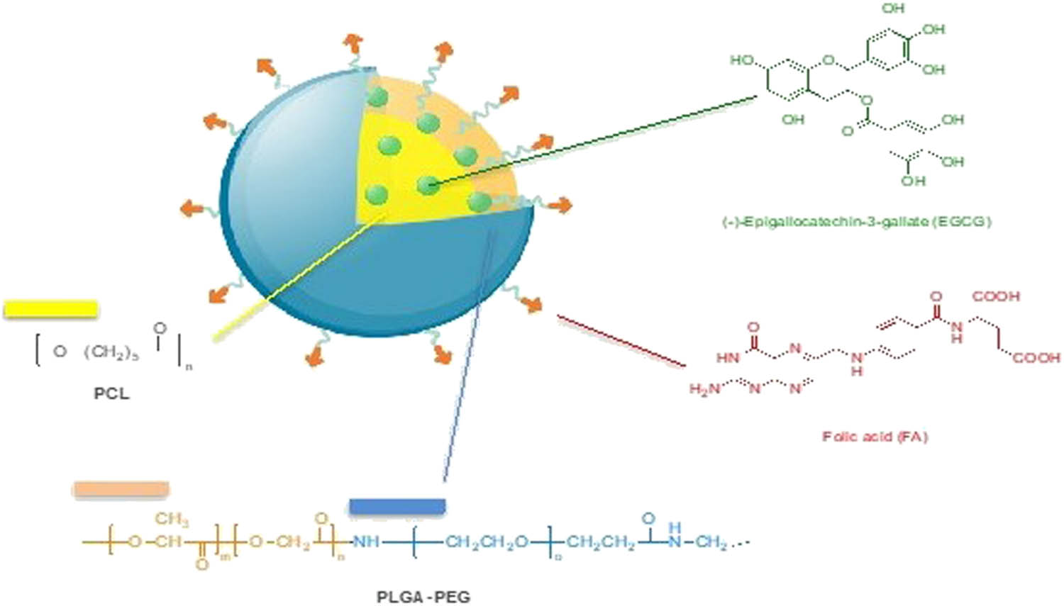
Schematic representation of the designed targeted PLGA–PEG–FA-based (EGCG)-loaded NPs (EGCG-T-NPs). Chemical structure of EGCG, FA targeting ligand, and PLGA–PEG/PCL polymers.
To get information on the impact of FA-PEGylation in primary binding with FOLR1, we conducted a comparative computational docking study between FA (pdb: 4LRH) and a model of PEG–FA to the FOLR1 catalytic site, and peculiar ligand–protein interactions and the docking binding energies were analyzed. Docking analysis revealed that both FA and PEG–FA overlapped and accommodated well within the active site (Figure 2). In contrast, the polymeric aliphatic model (PEG–FA) showed slightly better docking energy to FA binding with FOLR1 (docking energies were −13.232 and −14.126 kcal/mol, for FA and PEG–FA, respectively). More specifically, the folate pteroate moiety was placed inside the receptor, whereas its glutamate moiety extends externally from the cavity to the solvent, sticking out of the pocket entrance, especially the conjugated model. The X-ray cocrystal structure showed stacking of the pterin ring between a pocket formed by Asp81, Tyr85, Trp171, and Ser174 residues, whereas the aminobenzoate portions of both ligands shared hydrophobic interactions with His135, while the glutamate moieties formed hydrogen bonds with Trp102, Gly137, and Trp140. Further interactions with His135, Gly137, and Trp138 contributed to stabilizing the aminobenzoate backbone. This observation supported the use of the PEG–FA constructs for this study.
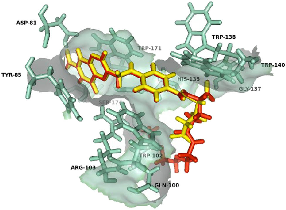
Binding modes of both FA (yellow) and its PEG polymeric conjugated (PEG–FA) model (red) superimposed on the folate receptor active site (pdb: 4LRH).
4.2 Synthesis of tri-block-copolymer PLGA–PEG–FA
The PLGA–PEG–FA tri-block-copolymer was synthesized using a two-step reaction, as described earlier (Scheme 1a–c). First, the carboxyl-terminus PLGA–COOH was activated (with NHS) to generate the intermediate PLGA–NHS, which was then converted on PLGA–PEG–NH2 by conjugating bifunctional PEG, NH2–PEG–NH2, to activated polymer (Scheme 1a). The structure of the copolymer was confirmed by 1H-NMR spectroscopy. More specifically, a signal detected at 1.48 ppm was attributed to the lactide methyl repeat units. Overlapping multiplets observed in the range of 5.30–5.15 and 4.96–4.80 indicated the lactide methine and the glycolide protons, respectively. Moreover, in addition to the signals specific for PLGA, the presence of the peak at about 3.51 ppm correspond to the PEG methylene protons, confirming the successful preparation of the desired copolymer. The desired PLGA–PEG–FA was obtained by conjugating the activated targeting agent, i.e., FA–NHS (Scheme 1b), to the amine PEG terminal group of PLGA–PEG–NH2, which resulted in the structurally and physiologically stable amide bonds (Scheme 1c). The presence of the folate terminal groups was confirmed by 1H-NMR analysis, which displayed the PLGA pattern in the range of 5.1–5.3, 4.8–5, and 1.5 ppm, while the PEG moiety was detected at 3.5 ppm. The characteristic peaks of the folate termini were found to be at 6.6–6.7 ppm, 7.6–7.7 ppm, and 8.6–8.7 ppm, supporting the successful conjugation (Figure 3).
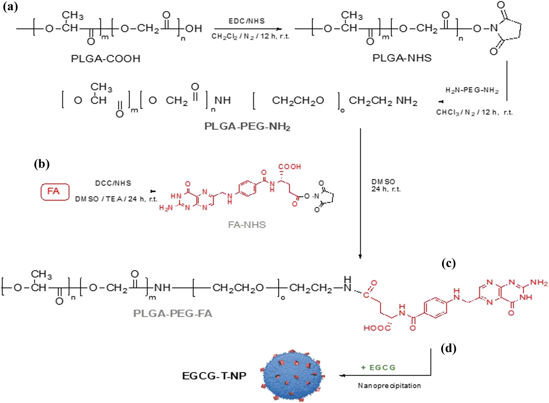
(a) Synthesis of copolymer PLGA–PEG–NH2, (b) FA–NHS intermediate, (c) tri-block-copolymer PLGA–PEG–FA, and (d) nanoformulation for EGCG-T-NP.
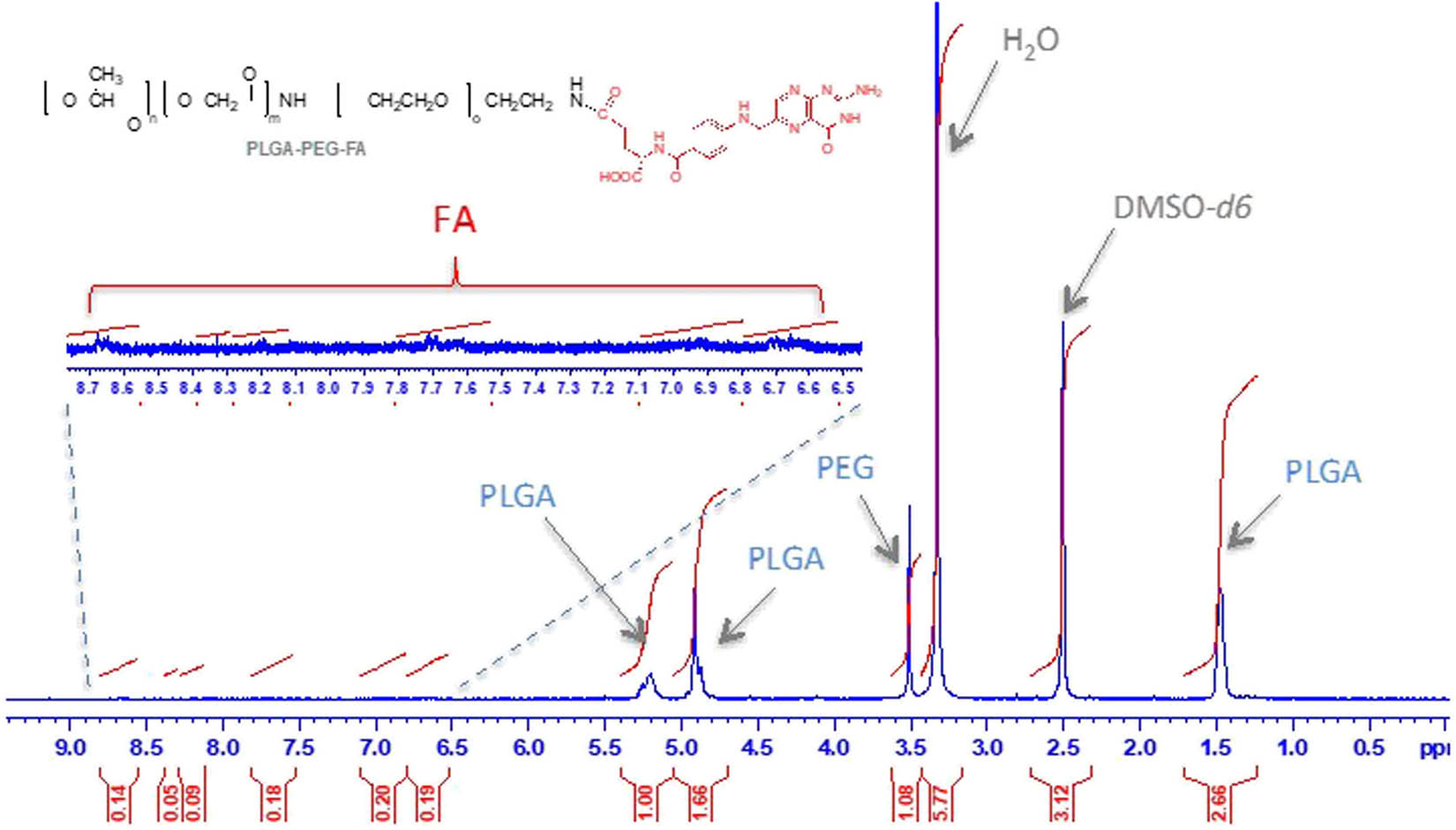
1H-NMR characterization of copolymer PLGA–PEG–FA.
4.3 Preparation and characterization of NPs
NPs were successfully obtained by the nanoprecipitation technique using a blend of hydrophobic polymer PCL and the amphiphilic block copolymers PLGA–PEG–FA (Scheme 1d). We prepared two batches of NPs, which included PLGA–PEG–FA loaded with EGCG NPs (EGCG-T-NPs), and its unloaded ones (Ø-T-NP) for comparison. The blended approach allows the hydrophobic PLGA component and PCL self-assembly to form a core surrounded by hydrophilic PEG chains, thus provide better hydrophilic/hydrophobic balancing. These polymers, particularly PEG, confer a hydrophilic surface to NPs, which provide antibiofouling properties, prevent/reduce nonspecific interaction, and limit macrophage capture. PEG was also able to ensure the optimal distance between FA and NP surface. Conversely, PEGylation allows enhanced distribution and prolonged circulation time of NPs in blood by improved permeability and retention (EPR) effect, resulting in increased accumulation in tumors.
The scanning electron microscope (SEM) observations showed that both NPs batches have well-dispersed particles with a regular spherical shape (Figure 4). The particle size of NPs ranges from 115 nm to about 130 nm, suggesting that the loading of the EGCG into NPs did not result in a significant increase in size compared to the corresponding free counterparts (Table 1). The NP dispersions were characterized by low PDI values (below 0.2), indicating a narrow and unimodal distribution, typical behavior of monodispersed systems (Figure 1). As far as the drug entrapment efficiencies were concerned, the encapsulation efficiency of EGCG-T-NP was found to be 50%, suggesting a good affinity of polymeric blend with the EGCG molecules. Moreover, yields of production obtained were high and were found to be in the range of 75–82%.
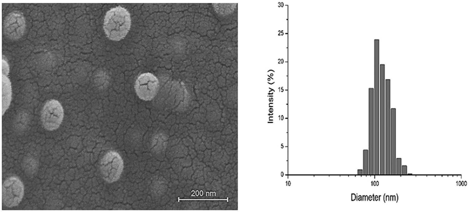
SEM images of EGCG-T-NP (left) and corresponding particle size distribution (right). The scale bar is 200 nm.
Average diameter, PDI, percentage of EE, EGCG loading content (DLC%) ±, and YP% of formulated NPs. Data are represented as mean ± SD, n = 3
| Batch | Mean diameter (nm) | PDI | EE (%) | DCL (%) | YP (%) |
|---|---|---|---|---|---|
| Ø-T-NP | 115.28 ± 6.17 | 0.123 ± 0.04 | — | — | 75.60 ± 3.06 |
| EGCG-T-NP | 128.42 ± 3.57 | 0.114 ± 0.03 | 49.82 ± 3.74 | 2.49 ± 0.19 | 81.73 ± 6.21 |
4.4 EGCG-targeted nanoparticle treatment reduces spheroid size of PCa cell lines
3D spheroids are well-established mimics of in vivo tumors; therefore, we used this highly important and specific model to assess the antitumorigenic capacity of EGCG-targeted NPs. We assessed the impact of both empty targeted NP and EGCG-targeted NP treatment on spheroids size of two well-known PCa cell lines, namely, 22Rv1 and PC3. As shown in Figure 5(a and b upper panel), detachment of both 22Rv1 and PC3 cell lines results in a statistically significant and gradual increase in spheroid size in the presence of empty targeted NP, i.e., from 183 mm3 on the second day to 245 mm3 on the sixth day in 22Rv1 cells and from 236 mm3 on the second day to 438 mm3 on the sixth day in PC3 cells. However, treatment with EGCG-targeted NPs significantly reduces the spheroid size on the sixth day, i.e., from 245 to 96 mm3 in the 22Rv1 cell line (Figure 5a, lower panel). Furthermore, we observed a similar decrease in the spheroid size on the second day and from 438 to 370 mm3 on the sixth day (Figure 5b, lower panel) in the PC3 cell line. Overall, the aforementioned data clearly showed that EGCG-targeted NP significantly attenuates spheroid formation of detached 22Rv1 cells compared to PC3 cells.

EGCG-targeted nanoparticle decreases spheroid size. Both (a) 22Rυ1 and (b) PC3 were fully grown in poly-HEMA-coated plates to form 3D spheroids. Furthermore, spheroids were treated with empty targeted nanoparticle or EGCG-targeted nanoparticle for 2–6 days, and pictures were taken by Nikon inverted light microscope at ×40. Images were analyzed for size measurement using image J software (mean ± S.E.M., n = 100). *P < 0.05, **P < 0.01, and ***P < 0.001. A total number of 100 spheroids were measured for size examination.
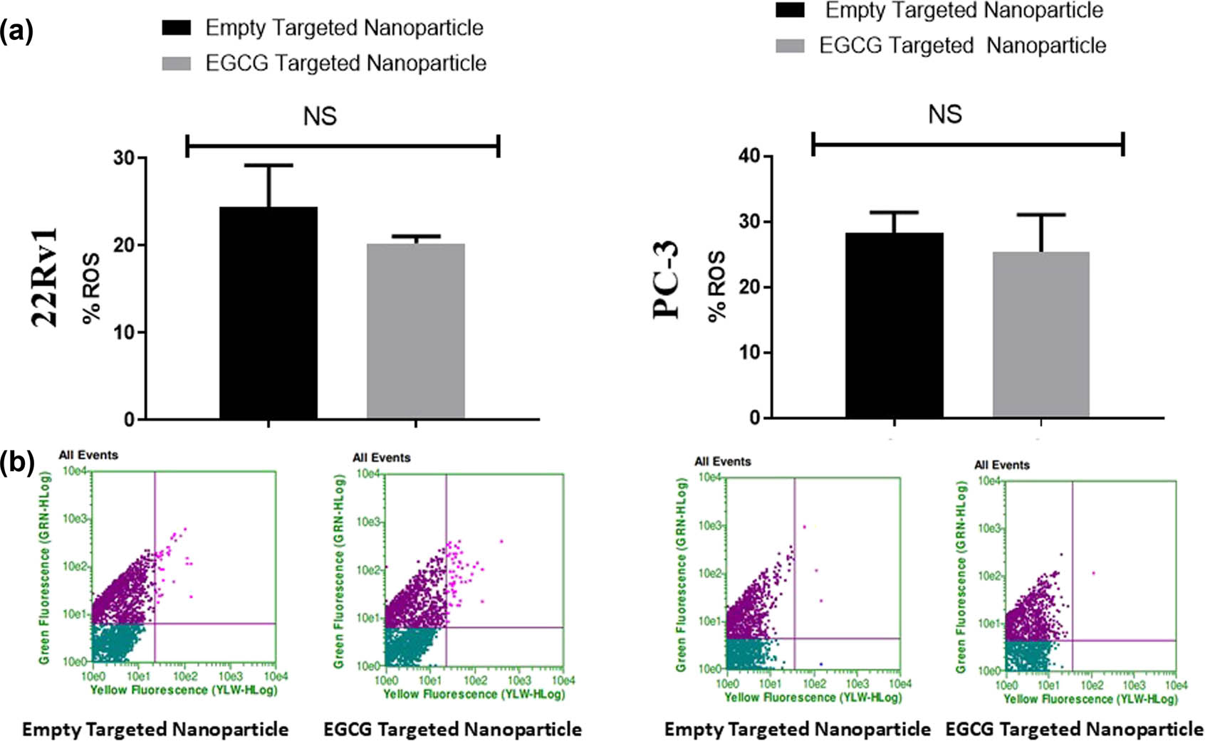
EGCG-targeted nanoparticle fails to induce ROS in spheroids of PCa cell lines. Both (a) 22Rv1 and (b) PC-3 were grown on poly-HEMA-coated plates and treated with either empty targeted nanoparticles or EGCG-targeted nanoparticle (6 µM) for 6 days. At the end of the time point, CellROX dye was added to the spheroids and incubated for 30 min in dark. Fluorescent intensity was measured by using Guava Flow Cytometer at a standardized wavelength provided by the manufacturer. Values are shown as mean ± SEM (n = 6)*P < 0.05, **P < 0.01, and ***P < 0.001.
4.5 EGCG-targeted nanoparticle treatment fails to induce ROS in spheroids of PCa cell lines
We assessed the impact of both empty targeted NPs and EGCG-targeted NPs treatment on ROS production in spheroids of both studied cell lines. As shown in Figure 6(a and b, upper and lower panels), EGCG-targeted NP treatment fails to induce any significant production of ROS in both 22Rv1 and PC3 cell lines compared to empty targeted NPs. These results suggest that the antitumorigenic capabilities of EGCG-targeted NPs were devoid of ROS-mediated mechanism(s).
4.6 EGCG-targeted nanoparticle treatment promotes mitochondrial leakage and cell death
The spheroids of 22Rv1 treated with empty targeted NPs and EGCG-targeted NPs showed a significant (P < 0.001) increase in mitochondrial depolarization. The EGCG-targeted NPs reduce polarization in the mitochondrial membrane in 22Rv1, but no significant difference was observed in PC3 cells as evident by JC-1 dye fluorescence (Figure 7, upper and lower panels). Results showed an approximately 18% reduction in mitochondrial polarization and a 15% increase in mitochondrial depolarization in 22Rv1 spheroids treated with EGCG-targeted NPs compared to empty targeted NPs. Both increases in depolarization and decreases in the polarization of mitochondria membrane potential are hallmarks of mitochondria-mediated apoptosis. As evident in Figure 8 (upper and lower panels), the number of dead apoptotic cells was also found to be increased in 22Rv1 spheroids treated with EGCG-targeted NPs compared to empty targeted NPs. An approximately 12% increase in apoptotic cells was observed in 22Rv1 treated with EGCG-targeted NPs compared to control NPs. Conversely, we did not observe any significant difference in the number of dead apoptotic cells with respect to both control and EGCG-targeted NPs in PC3 cells.
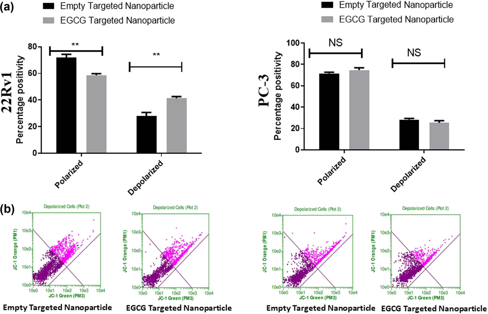
EGCG-targeted nanoparticle induces depolarization in spheroids. Both 22Rv1 (a) and PC3 (b) were grown on poly-HEMA-coated plates and treated with either empty targeted nanoparticles or EGCG-targeted nanoparticles (6 µM) for 6 days. At the end of time point, JC-1 dye was added to the treated spheroids and incubated for 30 min at 37°C. Fluorescent intensity was measured using Guava Flow Cytometer at the standardized wavelength provided by the manufacturer. Values are shown as mean ± SEM (n = 6). *P < 0.05, **P < 0.01, and ***P < 0.001.
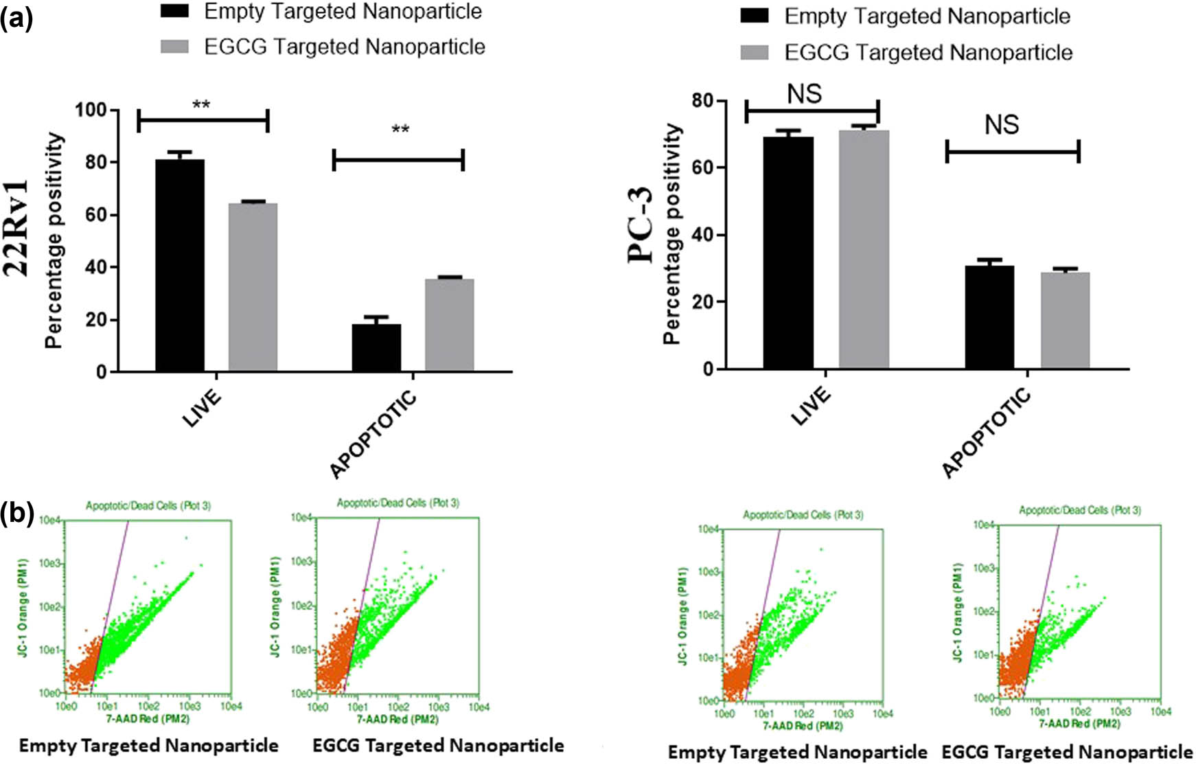
EGCG-targeted nanoparticle induces apoptosis in spheroids. Both 22Rv1 (a) and PC3 (b) were grown on poly-HEMA-coated plates and treated with either empty targeted nanoparticles or EGCG-targeted nanoparticles (6 µM) for 6 days. At the end of time point, PI dye was added to the treated spheroids and incubated for 30 min at 37°C. Fluorescent intensity was measured using Guava Flow Cytometer at standardized wavelength provided by the manufacturer. Values are shown as mean ± SEM (n = 6). *P < 0.05, **P < 0.01, and ***P < 0.001.
5 Discussion
The quest of a potential anticancer agent marks the polyphenolic constituent of green tea, viz, EGCG. Despite its therapeutic potential over a wide range of cancer, its clinical application has been limited because of the low utilization rate with poor stability and systemic bioavailability [42,43,44]. Deprived intestinal stability and low absorption were suggested as the main culprits that influenced its bioavailability [45]. In addition, the lack of specific receptors carrying EGCG into cells and the presence of efflux proteins that pump back EGCG into the intestinal lumen also result in reduced absorption of EGCG in cells [45,46]. Several approaches have been reported in the scientific literature to overcome the reduced bioavailability of EGCG in the therapeutic setup. Earlier works reported significantly increased bioavailability of EGCG by molecular modifications, nanostructure-based drug delivery systems, and the co-administration of EGCG with other bioactive compounds [19,45]. Nanostructure-based drug delivery systems have been suggested as a potential approach that helps to overcome the stability and bioavailability of the enclosed EGCG.
In this study, we used a biocompatible block-copolymer PLGA–PEG–FA for the preparation of targeted EGCG-loaded NPs. A blend of poly(d,l-lactide-co-glycolide) poly(ethylene glycol) (namely, PLGA–PEG) and PCL was considered as biocompatible/biodegradable polymeric mixture for the preparation of NPs [47]. These are the widely considered polyesters for building the core of polymeric micelles and have shown great promises as potential nanocarriers [48]. Compared to other polymeric esters, PCL degradation proceeds very slowly, limiting its utilization in some specific biomedical fields. Modification such as surface decoration or forming composite systems with other natural and synthetic polymers make PCL suitable for a larger application [49,50]. The blended approach allows the hydrophobic PLGA block and PCL self-assembly to form a core surrounded by hydrophilic PEG chains, thus providing a better hydrophilic/hydrophobic balancing. Previously, we reported a precisely controllable EGCG loading content, better encapsulation efficiency, and high production yields by a blend of two polymers, PLGA–PEG–A or PLGA–PEG–DCL, or PLGA–PEG–AG, with PCL [27].
Diverse tumor tissue targeting approaches have been reported in the literature for an accurate and effective anticancer therapy of small molecules [18]. The aggressive upregulation of high-affinity folate receptors in PCa cells with a limited expression in nonmalignant cells makes it an important targeting molecule [51]. The increase in folate receptors reflects the increased nutritional requirement of FA, which is essential for synthesizing nucleotide bases. Several studies suggested folate receptor targeting, a very promising approach in anticancer treatment [49,50,51]. The reduced off-site toxicity, increased selectivity, and enhanced potency by folate-tagged bioactive molecules against tumor cells compared to nontargeted drugs has been reported in the scientific literature [52,53,54]. EGCG combined with FA has also been reported for the chemopreventive effect on gastrointestinal carcinogenesis in rats [55]. Several studies also reported FA-conjugated nano delivery systems as a promising way to improve the stability, solubility, bioavailability, and bioefficacy of EGCG compared to free EGCG [56,57,58]. One study suggested folate conjugation for tumor targeting by chitosan-coated EGCG NPs [56]. The high stability and smaller particle size are achieved by FA–PEG–NPs that provide higher intracellular uptake of EGCG than its free counterpart [57]. A folate peptide decorated PLGA NPs loaded with EGCG reported to strongly bind folate receptor-specific breast cancer cells (MDA-MB-231) to inhibit the progress of breast adenocarcinoma [58]. In an earlier study, Siddiqui et al. [16] reported a 10-fold dose advantage by PLA–PEG–EGCG NPs that showed pro-apoptotic and anti-angiogenesis effects in human PC3 cells.
Lately, the 3D cell culture model has gained attention in preclinical studies because of the possibility of screening a large number of different molecules [59]. This method typically uses an extracellular matrix (ECM) and can mimic cell-to-cell and cell-to-ECM interactions, including paracrine signaling [60]. In the 3D cell culture method, the cells grow in the 3D plane, aggregate, and self-assemble in an environment that prevents attachment to a flat surface, resulting in spheroid formation, which is well-established mimics of in vivo tumors [61]. The 3D tumor spheroids study model often provides comparable results to animal models in many aspects [62]. The predictability of 3D spheroids on in vivo efficacy could resolve the high cost and ethical issues associated with animal usage. It is well known that the spheroid growth impacts cell proliferation, apoptosis, and necrotic/hypoxic core formation, with the outer layer mostly proliferative, the middle layer hypoxic, and the inner core necrotic [63].
The size of tumors is regarded as a crucial factor in determining the drug penetration ability as the larger tumors lead to an increased resistance, which is also observed in 3D tumor spheroid models [64]. We observed a dramatic decrease in the spheroid size as a result of EGCG-targeted NP treatment in 22Rv1 compared to PC3 cell line. Similar results were observed by different research groups using PLGA/lipid NPs in 3D models [63,65,66]. In one study, PLGA-modified NP exhibited increased penetration and distribution in HeLa and SiHa HD spheroids [63]. Another study reported deep penetration by PLGA-encapsulated EtNBS NPs into the hypoxic and acidic cores (usually unresponsive tumor region) of 3D spheroid of gastrointestinal carcinoma [65]. One study reported better growth inhibition by verteporfin-encapsulated lipid nanocarriers compared to a free drug in ovarian spheroid cancer cells [66]. Likewise, biotin-conjugated pullulan acetate NPs treatment also showed similar antitumor activity in 3D culture and xenograft hepatic tumor model [67]. Similar efficacy and deep tumor penetration with reduced tumor growth were reported in both SH-SY5Y spheroids and H22 tumor-bearing mice treated with iRGD-conjugated NPs with doxorubicin [68].
The current study did not observe any induction in ROS production by EGCG-targeted NP treatment in both studied cell lines. These results suggest that the observed antitumorigenic capabilities of EGCG-targeted NPs were devoid of ROS-mediated mechanism(s). Several studies also reported the anticancer activity of bioactive molecules by ROS-independent mechanism [69,70,71]. These evidence suggest that ROS are not obligator effectors in the anticancer mechanisms including apoptosis. This result clearly indicates the better efficacy of EGCG-T-NP toward 22Rv1 compared to PC3. The current result also highlights the strong dual binding of EGCG-T-NP with FOLR1 and PSMA+ in 22Rv1. This was expected because of the presence of PSMA+ and overexpression of FOLR1 in 22Rv1 cells. Mitochondrial metabolism has been reported to play a critical role in tumorigenesis and cancer development [72]. MMP is a key indicator of mitochondrial activity that reflects the process of electron transport and oxidative phosphorylation [73]. We observed increased depolarization and reduced polarization in the mitochondrial membrane potential after the treatment of EGCG-T-NPs in 22Rv1. The mitochondrial membrane depolarization has been reported to induce apoptosis [74]. Our results support earlier findings where increased proton influx leads to ROS production suppression during spontaneous transient depolarization [75]. Moreover, uncoupling of mitochondrial oxidative phosphorylation could also play a role in suppressing ROS production [76]. The current study also reported an increase in the number of dead apoptotic cells in 22Rv1 spheroids treated with EGCG-targeted NPs.
6 Conclusion
Overall, our results clearly indicate the successful synthesis of EGCG-loaded NPs that have the potential to bind FLOR1 and PSMA+ dually. Because of the presence of PSMA+ and overexpression of FLOR1, 22Rv1 showed better efficacy on the targeted delivery compared with PC3 cells (PSMA−). We also observed a significant dose advantage by this EGCG-loaded NPs, indicating their anticancer potential at clinically relevant doses. Further, preclinical in vivo studies are recommended to verify in vitro data.
-
Funding information: The authors extend their appreciation to the Deputyship for Research & Innovation, Ministry of Education in Saudi Arabia for funding this research work through the project number 986.
-
Author contributions: All authors have accepted responsibility for the entire content of this manuscript and approved its submission.
-
Conflict of interest: The authors state no conflict of interest.
References
[1] Siegel RL , Miller KD , Fuchs HE , Jemal A . Cancer statistics, 2021. CA Cancer J Clin. 2021;71(1):7–33.10.3322/caac.21654Search in Google Scholar PubMed
[2] Sung H , Ferlay J , Siegel RL , Laversanne M , Soerjomataram I , Jemal A , et al. Global cancer statistics 2020: GLOBOCAN estimates of incidence and mortality worldwide for 36 cancers in 185 countries. CA Cancer J Clin. 2021;71(3):209–49.10.3322/caac.21660Search in Google Scholar PubMed
[3] Merriel SWD , Funston G , Hamilton W . Prostate cancer in primary care. Adv Ther. 2018;35(9):1285–94.10.1007/s12325-018-0766-1Search in Google Scholar PubMed PubMed Central
[4] Omabe K , Paris C , Lannes F , Taïeb D , Rocchi P . Nanovectorization of prostate cancer treatment strategies: a new approach to improved outcomes. Pharmaceutics. 2021;13(5):591.10.3390/pharmaceutics13050591Search in Google Scholar PubMed PubMed Central
[5] Oves M , Aslam M , Rauf MA , Qayyum S , Qari HA , Khan MS , et al. Antimicrobial and anticancer activities of silver nanoparticles synthesized from the root hair extract of Phoenix dactylifera. Mater Sci Eng C Mater Biol Appl. 2018;89:429–43.10.1016/j.msec.2018.03.035Search in Google Scholar PubMed
[6] Shait Mohammed MR , Ahmad V , Ahmad A , Tabrez S , Choudhry H , Zamzami MA , et al. Prospective of nanoscale metal organic frameworks [NMOFs] for cancer therapy. Semin Cancer Biol. 2021;69:129–39.10.1016/j.semcancer.2019.12.015Search in Google Scholar PubMed
[7] Surh Y-J . Cancer chemoprevention with dietary phytochemicals. Nat Rev Cancer. 2003;3(10):768–80.10.1038/nrc1189Search in Google Scholar PubMed
[8] Mukhtar H . Chemoprevention: making it a success story for controlling human cancer. Cancer Lett. 2012;326(2):123–7.10.1016/j.canlet.2012.05.016Search in Google Scholar PubMed
[9] Du G-J , Zhang Z , Wen X-D , Yu C , Calway T , Yuan C-S , et al. Epigallocatechin gallate (EGCG) is the most effective cancer chemopreventive polyphenol in green tea. Nutrients. 2012;4(11):1679–91.10.3390/nu4111679Search in Google Scholar PubMed PubMed Central
[10] Tabrez S , Jabir NR , Adhami VM , Khan MI , Moulay M , Kamal MA , et al. Nanoencapsulated dietary polyphenols for cancer prevention and treatment: successes and challenges. Nanomedicine (Lond). 2020;15(11):1147–62.10.2217/nnm-2019-0398Search in Google Scholar PubMed
[11] Bettuzzi S , Brausi M , Rizzi F , Castagnetti G , Peracchia G , Corti A . Chemoprevention of human prostate cancer by oral administration of green tea catechins in volunteers with high-grade prostate intraepithelial neoplasia: a preliminary report from a one-year proof-of-principle study. Cancer Res. 2006;66(2):1234–40.10.1158/0008-5472.CAN-05-1145Search in Google Scholar PubMed
[12] Brausi M , Rizzi F , Bettuzzi S . Chemoprevention of human prostate cancer by green tea catechins: two years later. A follow-up update. Eur Urol. 2008;54(2):472–3.10.1016/j.eururo.2008.03.100Search in Google Scholar PubMed
[13] Johnson JJ , Bailey HH , Mukhtar H . Green tea polyphenols for prostate cancer chemoprevention: a translational perspective. Phytomedicine. 2010;17(1):3–13.10.1016/j.phymed.2009.09.011Search in Google Scholar PubMed PubMed Central
[14] Shin CM , Lee DH , Seo AY , Lee HJ , Kim SB , Son W-C , et al. Green tea extracts for the prevention of metachronous colorectal polyps among patients who underwent endoscopic removal of colorectal adenomas: a randomized clinical trial. Clin Nutr. 2018;37(2):452–8.10.1016/j.clnu.2017.01.014Search in Google Scholar PubMed
[15] Gan R-Y , Li H-B , Sui Z-Q , Corke H . Absorption, metabolism, anticancer effect and molecular targets of epigallocatechin gallate (EGCG): an updated review. Crit Rev Food Sci Nutr. 2018;58(6):924–41.10.1080/10408398.2016.1231168Search in Google Scholar PubMed
[16] Siddiqui IA , Adhami VM , Bharali DJ , Hafeez BB , Asim M , Khwaja SI , et al. Introducing nanochemoprevention as a novel approach for cancer control: proof of principle with green tea polyphenol epigallocatechin-3-gallate. Cancer Res. 2009;69(5):1712–6.10.1158/0008-5472.CAN-08-3978Search in Google Scholar PubMed PubMed Central
[17] Kim B , Park J-E , Im E , Cho Y , Lee J , Lee H-J , et al. Recent advances in nanotechnology with nano-phytochemicals: molecular mechanisms and clinical implications in cancer progression. Int J Mol Sci. 2021;22(7):3571.10.3390/ijms22073571Search in Google Scholar PubMed PubMed Central
[18] Chen B-H , Hsieh C-H , Tsai S-Y , Wang C-Y , Wang C-C . Anticancer effects of epigallocatechin-3-gallate nanoemulsion on lung cancer cells through the activation of AMP-activated protein kinase signaling pathway. Sci Rep. 2020;10(1):5163.10.1038/s41598-020-62136-2Search in Google Scholar PubMed PubMed Central
[19] Cai Z-Y , Li X-M , Liang J-P , Xiang L-P , Wang K-R , Shi Y-L , et al. Bioavailability of tea catechins and its improvement. Molecules. 2018;23(9):2346.10.3390/molecules23092346Search in Google Scholar PubMed PubMed Central
[20] Li K , Teng C , Min Q . Advanced nanovehicles-enabled delivery systems of epigallocatechin gallate for cancer therapy. Front Chem. 2020;8:573297.10.3389/fchem.2020.573297Search in Google Scholar PubMed PubMed Central
[21] Mitchell MJ , Billingsley MM , Haley RM , Wechsler ME , Peppas NA , Langer R . Engineering precision nanoparticles for drug delivery. Nat Rev Drug Discov. 2021;20(2):101–24.10.1038/s41573-020-0090-8Search in Google Scholar PubMed PubMed Central
[22] Gagliardi A , Giuliano E , Venkateswararao E , Fresta M , Bulotta S , Awasthi V , et al. Biodegradable polymeric nanoparticles for drug delivery to solid tumors. Front Pharmacol. 2021;12:601626.10.3389/fphar.2021.601626Search in Google Scholar PubMed PubMed Central
[23] Siddiqui IA , Adhami VM , Christopher J , Chamcheu , Mukhtar H . Impact of nanotechnology in cancer: emphasis on nanochemoprevention. Int J Nanomedicine. 2012;7:591–605.10.2147/IJN.S26026Search in Google Scholar
[24] Siddiqui IA , Mukhtar H . Nanochemoprevention by bioactive food components: a perspective. Pharm Res. 2010;27(6):1054–60.10.1007/s11095-010-0087-9Search in Google Scholar PubMed PubMed Central
[25] Leonarduzzi G , Testa G , Sottero B , Gamba P , Poli G . Design and development of nanovehicle-based delivery systems for preventive or therapeutic supplementation with flavonoids. Curr Med Chem. 2010;17(1):74–95.10.2174/092986710789957760Search in Google Scholar PubMed
[26] Sanna V , Pintus G , Roggio AM , Punzoni S , Posadino AM , Arca A , et al. Targeted biocompatible nanoparticles for the delivery of (−)-epigallocatechin 3-gallate to prostate cancer cells. J Med Chem. 2011;54(5):1321–32.10.1021/jm1013715Search in Google Scholar PubMed
[27] Sanna V , Singh CK , Jashari R , Adhami VM , Chamcheu JC , Rady I , et al. Targeted nanoparticles encapsulating (−)-epigallocatechin-3-gallate for prostate cancer prevention and therapy. Sci Rep. 2017;7(1):41573.10.1038/srep41573Search in Google Scholar PubMed PubMed Central
[28] Necela BM , Crozier JA , Andorfer CA , Lewis-Tuffin L , Kachergus JM , Geiger XJ , et al. Folate receptor-α (FOLR1) expression and function in triple negative tumors. PLoS One. 2015;10(3):e0122209.10.1371/journal.pone.0122209Search in Google Scholar PubMed PubMed Central
[29] Shen J , Hu Y , Putt KS , Singhal S , Han H , Visscher DW , et al. Assessment of folate receptor alpha and beta expression in selection of lung and pancreatic cancer patients for receptor targeted therapies. Oncotarget. 2018;9(4):4485–95.10.18632/oncotarget.23321Search in Google Scholar PubMed PubMed Central
[30] Ghosh A , Wang X , Klein E , Heston WDW . Novel role of prostate-specific membrane antigen in suppressing prostate cancer invasiveness. Cancer Res. 2005;65(3):727–31.10.1158/0008-5472.727.65.3Search in Google Scholar
[31] Kapałczyńska M , Kolenda T , Przybyła W , Zajączkowska M , Teresiak A , Filas V , et al. 2D and 3D cell cultures – a comparison of different types of cancer cell cultures. Arch Med Sci. 2018;14(4):910–9.10.5114/aoms.2016.63743Search in Google Scholar PubMed PubMed Central
[32] Chen C , Ke J , Zhou XE , Yi W , Brunzelle JS , Li J , et al. Structural basis for molecular recognition of folic acid by folate receptors. Nature. 2013;500(7463):486–9.10.1038/nature12327Search in Google Scholar
[33] Boraei ATA , Singh PK , Sechi M , Satta S . Discovery of novel functionalized 1,2,4-triazoles as PARP-1 inhibitors in breast cancer: design, synthesis and antitumor activity evaluation. Eur J Med Chem. 2019;182:111621.10.1016/j.ejmech.2019.111621Search in Google Scholar
[34] Pala N , Stevaert A , Dallocchio R , Dessì A , Rogolino D , Carcelli M , et al. Virtual screening and biological validation of novel influenza virus PA endonuclease inhibitors. ACS Med Chem Lett. 2015;6(8):866–71.10.1021/acsmedchemlett.5b00109Search in Google Scholar
[35] Morris GM , Huey R , Lindstrom W , Sanner MF , Belew RK , Goodsell DS , et al. AutoDock4 and AutoDockTools4: Automated docking with selective receptor flexibility. J Comput Chem. 2009;30(16):2785–91.10.1002/jcc.21256Search in Google Scholar
[36] Gasteiger J , Marsili M . Iterative partial equalization of orbital electronegativity – a rapid access to atomic charges. Tetrahedron. 1980;36(22):3219–28.10.1016/0040-4020(80)80168-2Search in Google Scholar
[37] Morris GM , Goodsell DS , Halliday RS , Huey R , Hart WE , Belew RK , et al. Automated docking using a Lamarckian genetic algorithm and an empirical binding free energy function. J Comput Chem. 1998;19(14):1639–62.10.1002/(SICI)1096-987X(19981115)19:14<1639::AID-JCC10>3.0.CO;2-BSearch in Google Scholar
[38] Sanna V , Nurra S , Pala N , Marceddu S , Pathania D , Neamati N , et al. Targeted nanoparticles for the delivery of novel bioactive molecules to pancreatic cancer cells. J Med Chem. 2016;59(11):5209–20.10.1021/acs.jmedchem.5b01571Search in Google Scholar
[39] El-Hammadi MM , Delgado ÁV , Melguizo C , Prados JC , Arias JL . Folic acid-decorated and PEGylated PLGA nanoparticles for improving the antitumour activity of 5-fluorouracil. Int J Pharm. 2017;516(1–2):61–70.10.1016/j.ijpharm.2016.11.012Search in Google Scholar
[40] Sanna V , Lubinu G , Madau P , Pala N , Nurra S , Mariani A , et al. Polymeric nanoparticles encapsulating white tea extract for nutraceutical application. J Agric Food Chem. 2015;63(7):2026–32.10.1021/jf505850qSearch in Google Scholar
[41] Mittal S , Sharma PK , Tiwari R , Rayavarapu RG , Shankar J , Chauhan LKS , et al. Impaired lysosomal activity mediated autophagic flux disruption by graphite carbon nanofibers induce apoptosis in human lung epithelial cells through oxidative stress and energetic impairment. Part Fibre Toxicol. 2017;14(1):15.10.1186/s12989-017-0194-4Search in Google Scholar
[42] Jiang Y , Jiang Z , Ma L , Huang Q . Advances in nanodelivery of green tea catechins to enhance the anticancer activity. Molecules. 2021;26(11):3301.10.3390/molecules26113301Search in Google Scholar PubMed PubMed Central
[43] Zhang G , Zhang J . Enhanced oral bioavailability of EGCG using pH-sensitive polymeric nanoparticles: characterization and in vivo investigation on nephrotic syndrome rats. Drug Des Devel Ther. 2018;12:2509–18.10.2147/DDDT.S172919Search in Google Scholar PubMed PubMed Central
[44] Chavva SR , Deshmukh SK , Kanchanapally R , Tyagi N , Coym JW , Singh AP , et al. Epigallocatechin gallate-gold nanoparticles exhibit superior antitumor activity compared to conventional gold nanoparticles: potential synergistic interactions. Nanomaterials (Basel). 2019;9(3):396.10.3390/nano9030396Search in Google Scholar PubMed PubMed Central
[45] Dai W , Ruan C , Zhang Y , Wang J , Han J , Shao Z , et al. Bioavailability enhancement of EGCG by structural modification and nano-delivery: a review. J Funct Foods. 2020;65:103732.10.1016/j.jff.2019.103732Search in Google Scholar
[46] Chan KY , Zhang L , Zuo Z . Intestinal efflux transport kinetics of green tea catechins in Caco-2 monolayer model. J Pharm Pharmacol. 2007;59(3):395–400.10.1211/jpp.59.3.0009Search in Google Scholar PubMed
[47] Łukasiewicz S , Mikołajczyk A , Błasiak E , Fic E , Dziedzicka-Wasylewska M . Polycaprolactone nanoparticles as promising candidates for nanocarriers in novel nanomedicines. Pharmaceutics. 2021;13(2):191.10.3390/pharmaceutics13020191Search in Google Scholar PubMed PubMed Central
[48] Cabral H , Miyata K , Osada K , Kataoka K . Block copolymer micelles in nanomedicine applications. Chem Rev. 2018;118(14):6844–92.10.1021/acs.chemrev.8b00199Search in Google Scholar PubMed
[49] Elmowafy EM , Tiboni M , Soliman ME . Biocompatibility, biodegradation and biomedical applications of poly(lactic acid)/poly(lactic-co-glycolic acid) micro and nanoparticles. J Pharm Investig. 2019;49(4):347–80.10.1007/s40005-019-00439-xSearch in Google Scholar
[50] Abedalwafa M , Wang F , Wang L , Li C . Biodegradable poly-epsilon-caprolactone (PCL) for tissue engineering applications: a review. Rev Adv Mater Sci. 2013;34:123–40.Search in Google Scholar
[51] Bellotti E , Cascone MG , Barbani N , Rossin D , Rastaldo R , Giachino C , et al. Targeting cancer cells overexpressing folate receptors with new terpolymer-based nanocapsules: toward a novel targeted dna delivery system for cancer therapy. Biomedicines. 2021;9(9):1275.10.3390/biomedicines9091275Search in Google Scholar PubMed PubMed Central
[52] Xia W , Low PS . Folate-targeted therapies for cancer. J Med Chem. 2010;53(19):6811–24.10.1021/jm100509vSearch in Google Scholar PubMed
[53] Din F , Aman W , Ullah I , Qureshi OS , Mustapha O , Shafique S , et al. Effective use of nanocarriers as drug delivery systems for the treatment of selected tumors. Int J Nanomed. 2017;12:7291–309.10.2147/IJN.S146315Search in Google Scholar PubMed PubMed Central
[54] Fernández M , Javaid F , Chudasama V . Advances in targeting the folate receptor in the treatment/imaging of cancers. Chem Sci. 2017;9(4):790–810.10.1039/C7SC04004KSearch in Google Scholar PubMed PubMed Central
[55] Xu Q , Yang CH , Liu Q , Jin XF , Xu XT , Tong JL , et al. Chemopreventive effect of epigallocatechin-3-gallate (EGCG) and folic acid on the N-methyl-N’-nitro-N-nitrosoguanidine (MNNG)-induced gastrointestinal cancer in rat model. J Dig Dis. 2011;12(3):181–7.10.1111/j.1751-2980.2011.00494.xSearch in Google Scholar PubMed
[56] Liang J , Cao L , Zhang L , Wan X-C . Preparation, characterization, and in vitro antitumor activity of folate conjugated chitosan coated EGCG nanoparticles. Food Sci Biotechnol. 2014;23(2):569–75.10.1007/s10068-014-0078-4Search in Google Scholar
[57] Luo Y , Zhang B , Cheng W-H , Wang Q . Preparation, characterization and evaluation of selenite-loaded chitosan/TPP nanoparticles with or without zein coating. Carbohydr Polym. 2010;82(3):942–51.10.1016/j.carbpol.2010.06.029Search in Google Scholar
[58] Kazi J , Sen R , Ganguly S , Jha T , Ganguly S , Chatterjee Debnath M . Folate decorated epigallocatechin-3-gallate (EGCG) loaded PLGA nanoparticles; in-vitro and in-vivo targeting efficacy against MDA-MB-231 tumor xenograft. Int J Pharm. 2020;585:119449.10.1016/j.ijpharm.2020.119449Search in Google Scholar PubMed
[59] Lazzari G , Couvreur P , Mura S . Multicellular tumor spheroids: a relevant 3D model for the in vitro preclinical investigation of polymer nanomedicines. Polym Chem. 2017;8(34):4947–69.10.1039/C7PY00559HSearch in Google Scholar
[60] Lovitt CJ , Shelper TB , Avery VM . Advanced cell culture techniques for cancer drug discovery. Biology (Basel). 2014;3(2):345–67.10.3390/biology3020345Search in Google Scholar PubMed PubMed Central
[61] Białkowska K , Komorowski P , Bryszewska M , Miłowska K . Spheroids as a type of three-dimensional cell cultures-examples of methods of preparation and the most important application. Int J Mol Sci. 2020;21(17):E6225.10.3390/ijms21176225Search in Google Scholar PubMed PubMed Central
[62] Pinto B , Henriques AC , Silva PMA , Bousbaa H . Three-dimensional spheroids as in vitro preclinical models for cancer research. Pharmaceutics. 2020;12(12):1186.10.3390/pharmaceutics12121186Search in Google Scholar PubMed PubMed Central
[63] Sims LB , Huss MK , Frieboes HB , Steinbach-Rankins JM . Distribution of PLGA-modified nanoparticles in 3D cell culture models of hypo-vascularized tumor tissue. J Nanobiotechnology. 2017;15(1):67.10.1186/s12951-017-0298-xSearch in Google Scholar PubMed PubMed Central
[64] Perez JE , Nagle I , Wilhelm C . Magnetic molding of tumor spheroids: emerging model for cancer screening. Biofabrication. 2021;13(1):015018.10.1088/1758-5090/abc670Search in Google Scholar PubMed
[65] Hung H-I , Klein OJ , Peterson SW , Rokosh SR , Osseiran S , Nowell NH , et al. PLGA nanoparticle encapsulation reduces toxicity while retaining the therapeutic efficacy of EtNBS-PDT in vitro. Sci Rep. 2016;6(1):33234.10.1038/srep33234Search in Google Scholar PubMed PubMed Central
[66] Michy T , Massias T , Bernard C , Vanwonterghem L , Henry M , Guidetti M , et al. Verteporfin-loaded lipid nanoparticles improve ovarian cancer photodynamic therapy in vitro and in vivo . Cancers (Basel). 2019;11(11):E1760.10.3390/cancers11111760Search in Google Scholar PubMed PubMed Central
[67] Chen H , Wei X , Chen H , Wei H , Wang Y , Nan W , et al. The study of establishment of an in vivo tumor model by three-dimensional cells culture systems methods and evaluation of antitumor effect of biotin-conjugated pullulan acetate nanoparticles. Artif Cells Nanomed Biotechnol. 2019;47(1):123–31.10.1080/21691401.2018.1544142Search in Google Scholar PubMed
[68] Wang X , Zhen X , Wang J , Zhang J , Wu W , Jiang X . Doxorubicin delivery to 3D multicellular spheroids and tumors based on boronic acid-rich chitosan nanoparticles. Biomaterials. 2013;34(19):4667–79.10.1016/j.biomaterials.2013.03.008Search in Google Scholar PubMed
[69] Ivanova D , Zhelev Z , Aoki I , Bakalova R , Higashi T . Overproduction of reactive oxygen species – obligatory or not for induction of apoptosis by anticancer drugs. Chin J Cancer Res. 2016;28(4):383–96.10.21147/j.issn.1000-9604.2016.04.01Search in Google Scholar PubMed PubMed Central
[70] Lin S , Fujii M , Hou D-X . Rhein induces apoptosis in HL-60 cells via reactive oxygen species-independent mitochondrial death pathway. Arch Biochem Biophys. 2003;418(2):99–107.10.1016/j.abb.2003.08.004Search in Google Scholar PubMed
[71] Hou D-X , Uto T , Tong X , Takeshita T , Tanigawa S , Imamura I , et al. Involvement of reactive oxygen species-independent mitochondrial pathway in gossypol-induced apoptosis. Arch Biochem Biophys. 2004;428(2):179–87.10.1016/j.abb.2004.06.007Search in Google Scholar PubMed
[72] Porporato PE , Filigheddu N , Pedro JMB-S , Kroemer G , Galluzzi L . Mitochondrial metabolism and cancer. Cell Res. 2018;28(3):265–80.10.1038/cr.2017.155Search in Google Scholar PubMed PubMed Central
[73] Ma Y-Y , Chen H-W , Tzeng C-R . Low oxygen tension increases mitochondrial membrane potential and enhances expression of antioxidant genes and implantation protein of mouse blastocyst cultured in vitro. J Ovarian Res. 2017;10(1):47.10.1186/s13048-017-0344-1Search in Google Scholar PubMed PubMed Central
[74] Jamali T , Kavoosi G , Safavi M , Ardestani SK . In-vitro evaluation of apoptotic effect of OEO and thymol in 2D and 3D cell cultures and the study of their interaction mode with DNA. Sci Rep. 2018;8(1):15787.10.1038/s41598-018-34055-wSearch in Google Scholar PubMed PubMed Central
[75] Aklima J , Onojima T , Kimura S , Umiuchi K , Shibata T , Kuraoka Y , et al. Effects of matrix pH on spontaneous transient depolarization and reactive oxygen species production in mitochondria. Front Cell Dev Biol. 2021;9:1582.10.3389/fcell.2021.692776Search in Google Scholar PubMed PubMed Central
[76] Vyssokikh MY , Holtze S , Averina OA , Lyamzaev KG , Panteleeva AA , Marey MV , et al. Mild depolarization of the inner mitochondrial membrane is a crucial component of an anti-aging program. PNAS. 2020;117(12):6491–501.10.1073/pnas.1916414117Search in Google Scholar PubMed PubMed Central
© 2022 Read F. Alserihi et al., published by De Gruyter
This work is licensed under the Creative Commons Attribution 4.0 International License.
Articles in the same Issue
- Research Articles
- Theoretical and experimental investigation of MWCNT dispersion effect on the elastic modulus of flexible PDMS/MWCNT nanocomposites
- Mechanical, morphological, and fracture-deformation behavior of MWCNTs-reinforced (Al–Cu–Mg–T351) alloy cast nanocomposites fabricated by optimized mechanical milling and powder metallurgy techniques
- Flammability and physical stability of sugar palm crystalline nanocellulose reinforced thermoplastic sugar palm starch/poly(lactic acid) blend bionanocomposites
- Glutathione-loaded non-ionic surfactant niosomes: A new approach to improve oral bioavailability and hepatoprotective efficacy of glutathione
- Relationship between mechano-bactericidal activity and nanoblades density on chemically strengthened glass
- In situ regulation of microstructure and microwave-absorbing properties of FeSiAl through HNO3 oxidation
- Research on a mechanical model of magnetorheological fluid different diameter particles
- Nanomechanical and dynamic mechanical properties of rubber–wood–plastic composites
- Investigative properties of CeO2 doped with niobium: A combined characterization and DFT studies
- Miniaturized peptidomimetics and nano-vesiculation in endothelin types through probable nano-disk formation and structure property relationships of endothelins’ fragments
- N/S co-doped CoSe/C nanocubes as anode materials for Li-ion batteries
- Synergistic effects of halloysite nanotubes with metal and phosphorus additives on the optimal design of eco-friendly sandwich panels with maximum flame resistance and minimum weight
- Octreotide-conjugated silver nanoparticles for active targeting of somatostatin receptors and their application in a nebulized rat model
- Controllable morphology of Bi2S3 nanostructures formed via hydrothermal vulcanization of Bi2O3 thin-film layer and their photoelectrocatalytic performances
- Development of (−)-epigallocatechin-3-gallate-loaded folate receptor-targeted nanoparticles for prostate cancer treatment
- Enhancement of the mechanical properties of HDPE mineral nanocomposites by filler particles modulation of the matrix plastic/elastic behavior
- Effect of plasticizers on the properties of sugar palm nanocellulose/cinnamon essential oil reinforced starch bionanocomposite films
- Optimization of nano coating to reduce the thermal deformation of ball screws
- Preparation of efficient piezoelectric PVDF–HFP/Ni composite films by high electric field poling
- MHD dissipative Casson nanofluid liquid film flow due to an unsteady stretching sheet with radiation influence and slip velocity phenomenon
- Effects of nano-SiO2 modification on rubberised mortar and concrete with recycled coarse aggregates
- Mechanical and microscopic properties of fiber-reinforced coal gangue-based geopolymer concrete
- Effect of morphology and size on the thermodynamic stability of cerium oxide nanoparticles: Experiment and molecular dynamics calculation
- Mechanical performance of a CFRP composite reinforced via gelatin-CNTs: A study on fiber interfacial enhancement and matrix enhancement
- A practical review over surface modification, nanopatterns, emerging materials, drug delivery systems, and their biophysiochemical properties for dental implants: Recent progresses and advances
- HTR: An ultra-high speed algorithm for cage recognition of clathrate hydrates
- Effects of microalloying elements added by in situ synthesis on the microstructure of WCu composites
- A highly sensitive nanobiosensor based on aptamer-conjugated graphene-decorated rhodium nanoparticles for detection of HER2-positive circulating tumor cells
- Progressive collapse performance of shear strengthened RC frames by nano CFRP
- Core–shell heterostructured composites of carbon nanotubes and imine-linked hyperbranched polymers as metal-free Li-ion anodes
- A Galerkin strategy for tri-hybridized mixture in ethylene glycol comprising variable diffusion and thermal conductivity using non-Fourier’s theory
- Simple models for tensile modulus of shape memory polymer nanocomposites at ambient temperature
- Preparation and morphological studies of tin sulfide nanoparticles and use as efficient photocatalysts for the degradation of rhodamine B and phenol
- Polyethyleneimine-impregnated activated carbon nanofiber composited graphene-derived rice husk char for efficient post-combustion CO2 capture
- Electrospun nanofibers of Co3O4 nanocrystals encapsulated in cyclized-polyacrylonitrile for lithium storage
- Pitting corrosion induced on high-strength high carbon steel wire in high alkaline deaerated chloride electrolyte
- Formulation of polymeric nanoparticles loaded sorafenib; evaluation of cytotoxicity, molecular evaluation, and gene expression studies in lung and breast cancer cell lines
- Engineered nanocomposites in asphalt binders
- Influence of loading voltage, domain ratio, and additional load on the actuation of dielectric elastomer
- Thermally induced hex-graphene transitions in 2D carbon crystals
- The surface modification effect on the interfacial properties of glass fiber-reinforced epoxy: A molecular dynamics study
- Molecular dynamics study of deformation mechanism of interfacial microzone of Cu/Al2Cu/Al composites under tension
- Nanocolloid simulators of luminescent solar concentrator photovoltaic windows
- Compressive strength and anti-chloride ion penetration assessment of geopolymer mortar merging PVA fiber and nano-SiO2 using RBF–BP composite neural network
- Effect of 3-mercapto-1-propane sulfonate sulfonic acid and polyvinylpyrrolidone on the growth of cobalt pillar by electrodeposition
- Dynamics of convective slippery constraints on hybrid radiative Sutterby nanofluid flow by Galerkin finite element simulation
- Preparation of vanadium by the magnesiothermic self-propagating reduction and process control
- Microstructure-dependent photoelectrocatalytic activity of heterogeneous ZnO–ZnS nanosheets
- Cytotoxic and pro-inflammatory effects of molybdenum and tungsten disulphide on human bronchial cells
- Improving recycled aggregate concrete by compression casting and nano-silica
- Chemically reactive Maxwell nanoliquid flow by a stretching surface in the frames of Newtonian heating, nonlinear convection and radiative flux: Nanopolymer flow processing simulation
- Nonlinear dynamic and crack behaviors of carbon nanotubes-reinforced composites with various geometries
- Biosynthesis of copper oxide nanoparticles and its therapeutic efficacy against colon cancer
- Synthesis and characterization of smart stimuli-responsive herbal drug-encapsulated nanoniosome particles for efficient treatment of breast cancer
- Homotopic simulation for heat transport phenomenon of the Burgers nanofluids flow over a stretching cylinder with thermal convective and zero mass flux conditions
- Incorporation of copper and strontium ions in TiO2 nanotubes via dopamine to enhance hemocompatibility and cytocompatibility
- Mechanical, thermal, and barrier properties of starch films incorporated with chitosan nanoparticles
- Mechanical properties and microstructure of nano-strengthened recycled aggregate concrete
- Glucose-responsive nanogels efficiently maintain the stability and activity of therapeutic enzymes
- Tunning matrix rheology and mechanical performance of ultra-high performance concrete using cellulose nanofibers
- Flexible MXene/copper/cellulose nanofiber heat spreader films with enhanced thermal conductivity
- Promoted charge separation and specific surface area via interlacing of N-doped titanium dioxide nanotubes on carbon nitride nanosheets for photocatalytic degradation of Rhodamine B
- Elucidating the role of silicon dioxide and titanium dioxide nanoparticles in mitigating the disease of the eggplant caused by Phomopsis vexans, Ralstonia solanacearum, and root-knot nematode Meloidogyne incognita
- An implication of magnetic dipole in Carreau Yasuda liquid influenced by engine oil using ternary hybrid nanomaterial
- Robust synthesis of a composite phase of copper vanadium oxide with enhanced performance for durable aqueous Zn-ion batteries
- Tunning self-assembled phases of bovine serum albumin via hydrothermal process to synthesize novel functional hydrogel for skin protection against UVB
- A comparative experimental study on damping properties of epoxy nanocomposite beams reinforced with carbon nanotubes and graphene nanoplatelets
- Lightweight and hydrophobic Ni/GO/PVA composite aerogels for ultrahigh performance electromagnetic interference shielding
- Research on the auxetic behavior and mechanical properties of periodically rotating graphene nanostructures
- Repairing performances of novel cement mortar modified with graphene oxide and polyacrylate polymer
- Closed-loop recycling and fabrication of hydrophilic CNT films with high performance
- Design of thin-film configuration of SnO2–Ag2O composites for NO2 gas-sensing applications
- Study on stress distribution of SiC/Al composites based on microstructure models with microns and nanoparticles
- PVDF green nanofibers as potential carriers for improving self-healing and mechanical properties of carbon fiber/epoxy prepregs
- Osteogenesis capability of three-dimensionally printed poly(lactic acid)-halloysite nanotube scaffolds containing strontium ranelate
- Silver nanoparticles induce mitochondria-dependent apoptosis and late non-canonical autophagy in HT-29 colon cancer cells
- Preparation and bonding mechanisms of polymer/metal hybrid composite by nano molding technology
- Damage self-sensing and strain monitoring of glass-reinforced epoxy composite impregnated with graphene nanoplatelet and multiwalled carbon nanotubes
- Thermal analysis characterisation of solar-powered ship using Oldroyd hybrid nanofluids in parabolic trough solar collector: An optimal thermal application
- Pyrene-functionalized halloysite nanotubes for simultaneously detecting and separating Hg(ii) in aqueous media: A comprehensive comparison on interparticle and intraparticle excimers
- Fabrication of self-assembly CNT flexible film and its piezoresistive sensing behaviors
- Thermal valuation and entropy inspection of second-grade nanoscale fluid flow over a stretching surface by applying Koo–Kleinstreuer–Li relation
- Mechanical properties and microstructure of nano-SiO2 and basalt-fiber-reinforced recycled aggregate concrete
- Characterization and tribology performance of polyaniline-coated nanodiamond lubricant additives
- Combined impact of Marangoni convection and thermophoretic particle deposition on chemically reactive transport of nanofluid flow over a stretching surface
- Spark plasma extrusion of binder free hydroxyapatite powder
- An investigation on thermo-mechanical performance of graphene-oxide-reinforced shape memory polymer
- Effect of nanoadditives on the novel leather fiber/recycled poly(ethylene-vinyl-acetate) polymer composites for multifunctional applications: Fabrication, characterizations, and multiobjective optimization using central composite design
- Design selection for a hemispherical dimple core sandwich panel using hybrid multi-criteria decision-making methods
- Improving tensile strength and impact toughness of plasticized poly(lactic acid) biocomposites by incorporating nanofibrillated cellulose
- Green synthesis of spinel copper ferrite (CuFe2O4) nanoparticles and their toxicity
- The effect of TaC and NbC hybrid and mono-nanoparticles on AA2024 nanocomposites: Microstructure, strengthening, and artificial aging
- Excited-state geometry relaxation of pyrene-modified cellulose nanocrystals under UV-light excitation for detecting Fe3+
- Effect of CNTs and MEA on the creep of face-slab concrete at an early age
- Effect of deformation conditions on compression phase transformation of AZ31
- Application of MXene as a new generation of highly conductive coating materials for electromembrane-surrounded solid-phase microextraction
- A comparative study of the elasto-plastic properties for ceramic nanocomposites filled by graphene or graphene oxide nanoplates
- Encapsulation strategies for improving the biological behavior of CdS@ZIF-8 nanocomposites
- Biosynthesis of ZnO NPs from pumpkin seeds’ extract and elucidation of its anticancer potential against breast cancer
- Preliminary trials of the gold nanoparticles conjugated chrysin: An assessment of anti-oxidant, anti-microbial, and in vitro cytotoxic activities of a nanoformulated flavonoid
- Effect of micron-scale pores increased by nano-SiO2 sol modification on the strength of cement mortar
- Fractional simulations for thermal flow of hybrid nanofluid with aluminum oxide and titanium oxide nanoparticles with water and blood base fluids
- The effect of graphene nano-powder on the viscosity of water: An experimental study and artificial neural network modeling
- Development of a novel heat- and shear-resistant nano-silica gelling agent
- Characterization, biocompatibility and in vivo of nominal MnO2-containing wollastonite glass-ceramic
- Entropy production simulation of second-grade magnetic nanomaterials flowing across an expanding surface with viscidness dissipative flux
- Enhancement in structural, morphological, and optical properties of copper oxide for optoelectronic device applications
- Aptamer-functionalized chitosan-coated gold nanoparticle complex as a suitable targeted drug carrier for improved breast cancer treatment
- Performance and overall evaluation of nano-alumina-modified asphalt mixture
- Analysis of pure nanofluid (GO/engine oil) and hybrid nanofluid (GO–Fe3O4/engine oil): Novel thermal and magnetic features
- Synthesis of Ag@AgCl modified anatase/rutile/brookite mixed phase TiO2 and their photocatalytic property
- Mechanisms and influential variables on the abrasion resistance hydraulic concrete
- Synergistic reinforcement mechanism of basalt fiber/cellulose nanocrystals/polypropylene composites
- Achieving excellent oxidation resistance and mechanical properties of TiB2–B4C/carbon aerogel composites by quick-gelation and mechanical mixing
- Microwave-assisted sol–gel template-free synthesis and characterization of silica nanoparticles obtained from South African coal fly ash
- Pulsed laser-assisted synthesis of nano nickel(ii) oxide-anchored graphitic carbon nitride: Characterizations and their potential antibacterial/anti-biofilm applications
- Effects of nano-ZrSi2 on thermal stability of phenolic resin and thermal reusability of quartz–phenolic composites
- Benzaldehyde derivatives on tin electroplating as corrosion resistance for fabricating copper circuit
- Mechanical and heat transfer properties of 4D-printed shape memory graphene oxide/epoxy acrylate composites
- Coupling the vanadium-induced amorphous/crystalline NiFe2O4 with phosphide heterojunction toward active oxygen evolution reaction catalysts
- Graphene-oxide-reinforced cement composites mechanical and microstructural characteristics at elevated temperatures
- Gray correlation analysis of factors influencing compressive strength and durability of nano-SiO2 and PVA fiber reinforced geopolymer mortar
- Preparation of layered gradient Cu–Cr–Ti alloy with excellent mechanical properties, thermal stability, and electrical conductivity
- Recovery of Cr from chrome-containing leather wastes to develop aluminum-based composite material along with Al2O3 ceramic particles: An ingenious approach
- Mechanisms of the improved stiffness of flexible polymers under impact loading
- Anticancer potential of gold nanoparticles (AuNPs) using a battery of in vitro tests
- Review Articles
- Proposed approaches for coronaviruses elimination from wastewater: Membrane techniques and nanotechnology solutions
- Application of Pickering emulsion in oil drilling and production
- The contribution of microfluidics to the fight against tuberculosis
- Graphene-based biosensors for disease theranostics: Development, applications, and recent advancements
- Synthesis and encapsulation of iron oxide nanorods for application in magnetic hyperthermia and photothermal therapy
- Contemporary nano-architectured drugs and leads for ανβ3 integrin-based chemotherapy: Rationale and retrospect
- State-of-the-art review of fabrication, application, and mechanical properties of functionally graded porous nanocomposite materials
- Insights on magnetic spinel ferrites for targeted drug delivery and hyperthermia applications
- A review on heterogeneous oxidation of acetaminophen based on micro and nanoparticles catalyzed by different activators
- Early diagnosis of lung cancer using magnetic nanoparticles-integrated systems
- Advances in ZnO: Manipulation of defects for enhancing their technological potentials
- Efficacious nanomedicine track toward combating COVID-19
- A review of the design, processes, and properties of Mg-based composites
- Green synthesis of nanoparticles for varied applications: Green renewable resources and energy-efficient synthetic routes
- Two-dimensional nanomaterial-based polymer composites: Fundamentals and applications
- Recent progress and challenges in plasmonic nanomaterials
- Apoptotic cell-derived micro/nanosized extracellular vesicles in tissue regeneration
- Electronic noses based on metal oxide nanowires: A review
- Framework materials for supercapacitors
- An overview on the reproductive toxicity of graphene derivatives: Highlighting the importance
- Antibacterial nanomaterials: Upcoming hope to overcome antibiotic resistance crisis
- Research progress of carbon materials in the field of three-dimensional printing polymer nanocomposites
- A review of atomic layer deposition modelling and simulation methodologies: Density functional theory and molecular dynamics
- Recent advances in the preparation of PVDF-based piezoelectric materials
- Recent developments in tensile properties of friction welding of carbon fiber-reinforced composite: A review
- Comprehensive review of the properties of fly ash-based geopolymer with additive of nano-SiO2
- Perspectives in biopolymer/graphene-based composite application: Advances, challenges, and recommendations
- Graphene-based nanocomposite using new modeling molecular dynamic simulations for proposed neutralizing mechanism and real-time sensing of COVID-19
- Nanotechnology application on bamboo materials: A review
- Recent developments and future perspectives of biorenewable nanocomposites for advanced applications
- Nanostructured lipid carrier system: A compendium of their formulation development approaches, optimization strategies by quality by design, and recent applications in drug delivery
- 3D printing customized design of human bone tissue implant and its application
- Design, preparation, and functionalization of nanobiomaterials for enhanced efficacy in current and future biomedical applications
- A brief review of nanoparticles-doped PEDOT:PSS nanocomposite for OLED and OPV
- Nanotechnology interventions as a putative tool for the treatment of dental afflictions
- Recent advancements in metal–organic frameworks integrating quantum dots (QDs@MOF) and their potential applications
- A focused review of short electrospun nanofiber preparation techniques for composite reinforcement
- Microstructural characteristics and nano-modification of interfacial transition zone in concrete: A review
- Latest developments in the upconversion nanotechnology for the rapid detection of food safety: A review
- Strategic applications of nano-fertilizers for sustainable agriculture: Benefits and bottlenecks
- Molecular dynamics application of cocrystal energetic materials: A review
- Synthesis and application of nanometer hydroxyapatite in biomedicine
- Cutting-edge development in waste-recycled nanomaterials for energy storage and conversion applications
- Biological applications of ternary quantum dots: A review
- Nanotherapeutics for hydrogen sulfide-involved treatment: An emerging approach for cancer therapy
- Application of antibacterial nanoparticles in orthodontic materials
- Effect of natural-based biological hydrogels combined with growth factors on skin wound healing
- Nanozymes – A route to overcome microbial resistance: A viewpoint
- Recent developments and applications of smart nanoparticles in biomedicine
- Contemporary review on carbon nanotube (CNT) composites and their impact on multifarious applications
- Interfacial interactions and reinforcing mechanisms of cellulose and chitin nanomaterials and starch derivatives for cement and concrete strength and durability enhancement: A review
- Diamond-like carbon films for tribological modification of rubber
- Layered double hydroxides (LDHs) modified cement-based materials: A systematic review
- Recent research progress and advanced applications of silica/polymer nanocomposites
- Modeling of supramolecular biopolymers: Leading the in silico revolution of tissue engineering and nanomedicine
- Recent advances in perovskites-based optoelectronics
- Biogenic synthesis of palladium nanoparticles: New production methods and applications
- A comprehensive review of nanofluids with fractional derivatives: Modeling and application
- Electrospinning of marine polysaccharides: Processing and chemical aspects, challenges, and future prospects
- Electrohydrodynamic printing for demanding devices: A review of processing and applications
- Rapid Communications
- Structural material with designed thermal twist for a simple actuation
- Recent advances in photothermal materials for solar-driven crude oil adsorption