Characterization, biocompatibility and in vivo of nominal MnO2-containing wollastonite glass-ceramic
-
Samah S. Eldera


Abstract
The present work pointed out the effect of adding different concentrations of MnO2 (0.25, 0.50, 1.00 and 2.00 wt%) on the structure and crystallization performance of wollastonite glass. Nominal MnO2-containing wollastonite glass was prepared with the addition of 10% Na2O to decrease the melting temperature through melt quenching technique. The thermal history of glasses indicated that the crystallization temperature was between 864 and 895°C. The heat treating of glasses at ∼900 and 1,100°C gave combeite (Na4Ca4Si6O18), rankinite (Ca3Si2O7), pseudowollastonite (Ca3Si3O9), bustamite (CaMnSi2O6) and cristobalite. The later sample densities increased with the incorporation of MnO2 from 1.88 to 2.24 g/cm3 concomitant with decrease of porosities from 32.59 to 20.83%. The microstructure showed nano-size crystals in rounded, angular or irregular micro-size clusters, whereas after soaking in simulated body fluid for 1 month showed submicron crystals of carbonated calcium phosphate phase. Both fourier transform infrared spectroscopy and scanning electron microscopy/energy dispersive X-ray delineated the samples’ biocompatibility. Also, the negative zeta potential results enabled bone cell activity. Moreover, the bone healing with complete mineralization was remarked in case of the in vivo implantation of the G0.50 group. These results can be of a great significance in the application of MnO2-containing combeite, rankinite phases for bone treatment and biomedical applications.
1 Introduction
Wollastonite (Ca–Si-based bioceramic, CaSiO3) is an attractive candidate for bioengineering owing to the release of the Si4+ and Ca2+ ions upon degradation, such ions play a significant role in osteogenesis and angiogenesis. Wollastonite doped with bioactive elements is believed to be a good candidate for bone tissue engineering due to their amendable biodegradation and bioinductivity [1,2]. Doping of wollastonite with manganese (Mn2+) can fortify the capacity of the calcium silicate [3,4]. CaSiO3 has been premeditated as a bioactive material due to its excellent bioactivity and degradability [5]. Silicon and calcium act as a potential network modifier for the improvement of the bone substitutions by therapeutic inorganic ions during the early stages of bone development, mineralization and growth [6]. Their good biological properties are accredited to their capacity for alkalinizing activity and the release of calcium which is ideal for the calcium phosphate (Ca-P) nucleation [7]. Calcium silicate is an osteoinductive non-cytotoxic material [8,9].
Manganese dioxide (MnO2), is a non-stoichiometric inorganic material which added an important consideration in the material science field due to its distinctive physicochemical properties [10]. Among metal oxides, MnO2 has received a considerable attention because of its cost-effective, great activity, high stability, low toxicity, large specific surface area, flexible structure and strong oxidizing properties [11]. The dimensions, crystallographic phases, morphology and particle size are the unique physicochemical properties of the MnO2. Manganese can improve several functional materials as it fulfils the strict biomedical necessities [12]. Researchers, revealed that doping of MnO2 can improve the dielectric properties of ceramics [13,14]. The influence of MnO2 mostly impacts the microstructure and the energy storage properties of the calcium silicate. In 2017, Xiu et al. concluded that the addition of a small amount of MnO2 can improve the homogenization and densification of the ceramic material and subsequently improve its microstructure [15]. In 2016, Danewalia and Singh indicated that the existence of MnO2 increases the leakage of the Na+ ions from the glasses and likewise attracts the Ca2+ cations from the simulated body fluid (SBF) [16]. They similarly mentioned that the incidence of MnO2 may improve the glass surface activity to form an apatite layer. In 2015, Kolmas et al. noted that Mn-doping could increase the compressive strength and density of the scaffolds [17].
In the current research, nominal wollastonite glass-ceramic with various concentrations of MnO2 was prepared through the melt quench method. The produced manganese/wollastonite glasses were characterized in terms of morphology and composition via in vitro and in vivo bioactivity. Characterization of the glasses was obtained using differential thermal analysis (DTA), X-ray diffraction (XRD) and scanning electron microscope (SEM) coupled with energy dispersive X-ray microanalysis (EDX). For in vitro biocompatibility, glass powder was pressed into discs before immersion in SBF for 1 month. For the in vivo investigation, the base nominal wollastonite glass (G0), and those combined with 0.25 M and 0.50 M of MnO2 samples were implanted in the femur bone defects of hamster rats for 45–90 days to identify the new bone formation.
2 Experimentation
Wollastonite (CaSiO3) was prepared through the melt quench route. The influence of MnO2 (BDH) on the characterization and biocompatibility of the glass was investigated. The content of MnO2 in glass samples was 0.0, 0.25, 0.50. 1.00 and 2.00 wt%. To reduce the melting temperatures of the glass batches, 10% Na2O was added over the 100% glass oxide composition (Table 1). The glass batch constituents were pure limestone (LS contains CaO: 55.7, Al2O3: 0.22, Fe2O3: 0.02, MgO: 0.1, Na2O: 0.1, K2O: 0.16 and TiO2: 0.02 wt%) as the source of CaO; whereas, silica sand was used as the source of SiO2. Over 100% glass batch oxides were MnO2 (Sigma Aldrich, USA) and sodium carbonate (Na2CO3, BDH) as the source of Na2O. The melting temperature of the batches was between 1,350 and 1,400°C for 2 h in a platinum crucible. After adequate homogeneity, glass melt was poured into distilled water (at room temperature) before dryness and pulverization (<0.083 mm). The pulverized glass powder was shaped into discs of diameter 1.00 cm, using 7% poly vinyl alcohol as a binder, via Paul Weber Maschinen- und Apparatebau, Remshalden, Germany at 20 kN for 20 s.
Chemical composition of the glass batches
| Sample code | Batch in oxides weight% | Batch in gram | ||||||
|---|---|---|---|---|---|---|---|---|
| CaO | SiO2 | Over 100% addition | Starting material | |||||
| Na2O | MnO2 | LS | Silica sand | Na2CO3 | MnO2 | |||
| G0 | 48.28 | 51.72 | 10.00 | 00 | 86.68 | 51.72 | 17.00 | 0.00 |
| G0.25M | 48.28 | 51.72 | 10.00 | 0.25 | 86.68 | 51.72 | 17.00 | 0.25 |
| G0.50M | 48.28 | 51.72 | 10.00 | 0.50 | 86.68 | 51.72 | 17.00 | 0.50 |
| G1.00M | 48.28 | 51.72 | 10.00 | 1.00 | 86.68 | 51.72 | 17.00 | 1.00 |
| G2.00M | 48.28 | 51.72 | 10.00 | 2.00 | 86.68 | 51.72 | 17.00 | 2.00 |
DTA (Perkin Elmer DTA-7, USA) was used to trace the thermal behavior of the glasses, under argon gas condition and a heating rate of 10°C/min up to 1,000°C. Identification of the crystalline phases of glasses at 900 and 1,100°C was performed using XRD analysis (BRUKER, D8 ADVANCED CuO target, Germany) with CuKα radiations (λ = 1.54 Å). XRD patterns were recorded in the range of 2θ = 5–60°. The morphology and microcrystalline structure of samples were verified by SEM equipped with EDX (SEM/EDX, model FEJ Quanta 250 Fei, Holland) at operating voltage of 15 kV. Prior to the SEM measurements, fresh fractured samples were etched with the solution of 1 wt% HNO3 + 1 wt% HF to clear the outlines of the developed crystalline particles.
Standard in vitro bioactivity tests using SBF at 37°C were carried out upon incubation for 1 month. SBF has ion concentrations, pH and temperature almost identical to that of the human blood plasma [18].
SBF was prepared following Kokubo’s protocol by dissolving appropriate amounts of reagent-grade chemicals NaCl, NaHCO3, KCl, Na2HPO4, MgCl2·6H2O, Na2SO4, (CH2OH)3CNH2 and CaCl2·2H2O in deionized water [19,20]. To mimic the concentration of human blood plasma, 1 M of HCl and tris-hydroxymethyl amino methane [(CH2OH)3CNH2] was used to maintain the pH of SBF solution at 7.4 [21]. Bioactivity test was performed for sintered glass discs sintered at 1,100°C/2 h. Discs were immersed in sealed sterilized polyethylene boxes enclosing SBF for 1 month, at a constant ratio of sample to SBF volume without refreshing the solution throughout the immersion. At the end of the immersion period, discs were removed from the solution, rinsed with distilled water to stop the reaction, and then dried at room temperature. Discs were successively characterized using SEM/EDX and fourier transform infrared spectroscopy (FT-IR) reflection (Jasco, FT/IR-4600, USA) to detect the possible formation of the Ca-P crystals and to assess the microanalysis and the Ca/P ratio on the sample. Also, the Zetasizer ([Malvern Instrument Ltd, UK] fortified with a 633 nm laser) was used to determine the electrical surface charges on the sample powder. A well-dispersed sample in deionized water at temperature 25°C was caught for the measurement of the zeta potential (each measurement being the average of 12 runs).
For in vivo tests, the G0, G0.25M and G0.50M sintered glass-ceramic were implanted in the femur bone defects of hamster rats, besides a positive control (empty bone gap). Normal healing of the soft tissues was identified. All rats were healthy and did not show any signs of edema in the tissues all through the post-operative period.
After 45 and 90 days’ post-surgery, the animals were lacerated with an overdose of anesthesia, and both femurs were extracted, cleaned of soft tissues and soaked in 10% phosphate-buffered formalin for 7 days. The rat bones were prepared for histological evaluation via normal light microscope (Leica, model: DM 2500).
3 Results and discussion
3.1 Characterization of samples
The thermal behavior of the glasses is elucidated by means of DTA in Figure 1. The glass transition temperature is near 675°C and softening temperature near 685°C. Such small heat absorption may designate the occurrence of molecular relocation earlier than the glass crystallization. Exothermic peaks demonstrating the crystallization reaction are also revealed [22]. The crystallization temperature is between 864 and 872°C. The onset and offset temperatures of all the glasses crystallization are between 830 and 900°C, respectively. It is manifested that the incorporation of MnO2 in the wollastonite glass does not show noticeable changes in the temperature of the endothermic and exothermic effect.

DTA thermograms of the glasses heated up to 1,000°C.
XRD analysis of the sintered glasses near the crystallization temperature (at 900°C/2 h) is presented in Figure 2. It exhibits main crystalline phases of complex silicates. The crystallization of combeite (Na4Ca4Si6O18) particularly in the G0 (base) [23]; whereas the incorporation of MnO2 enhances the crystallization of both pseudowollastonite (CaSiO3) [24] and rankinite (Ca3Si2O7) [25]. At higher concentration of MnO2 content (i.e., in G2.00M), bustamite (Mn,Ca)3Si3O9 phase is indexed along with the later phases [26]. At higher temperature (1,100°C), the crystallization of rankinite and combeite is noticed as major phases with little cristobalite (SiO2) in all samples [27] (Figure 3). For the parent (G0) sample, the XRD pattern concurs with those reported in the previous work [28,29].
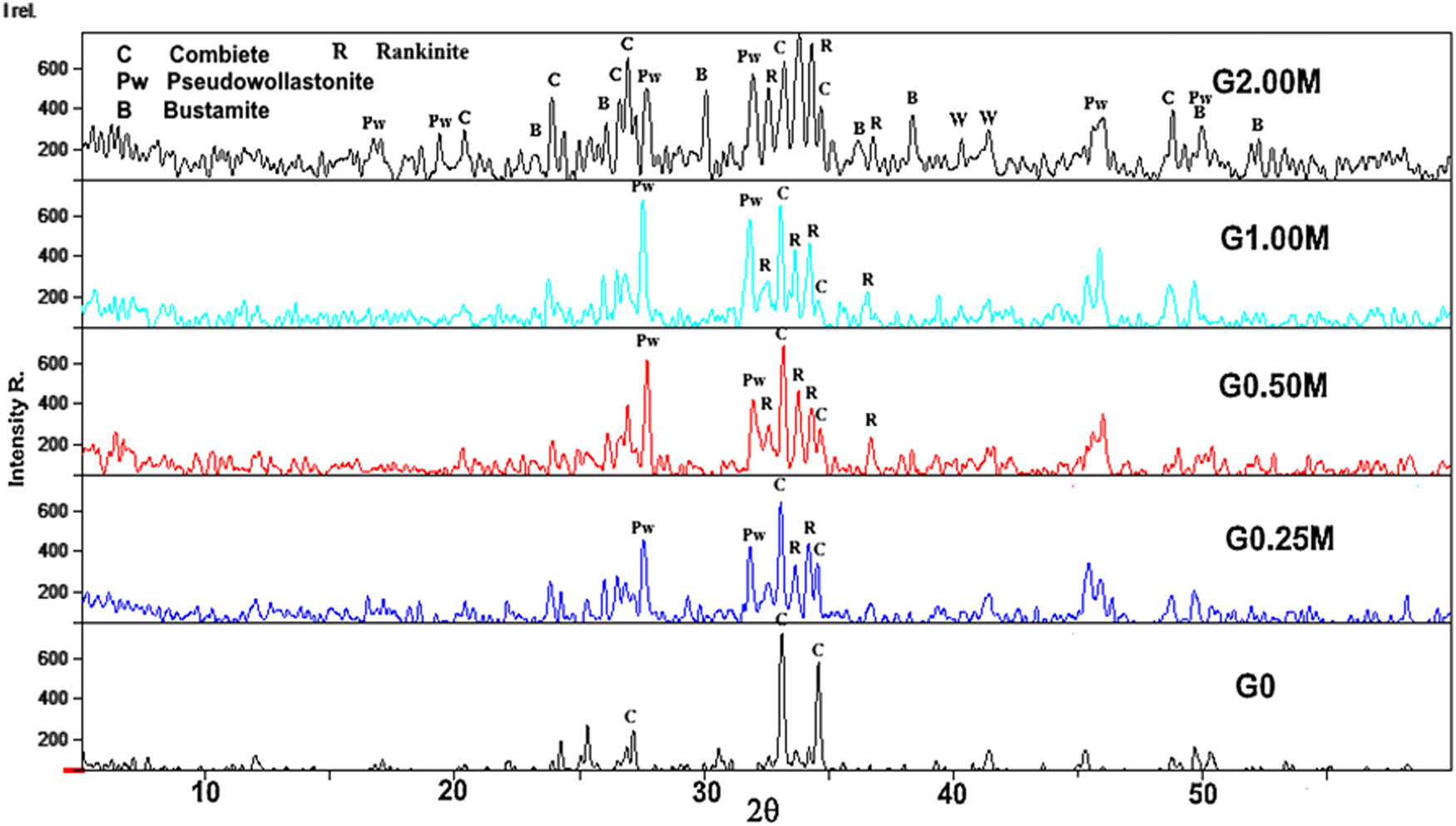
XRD of glasses heated at 900°C/2 h.

XRD of glasses heated at 1,100°C/2 h.
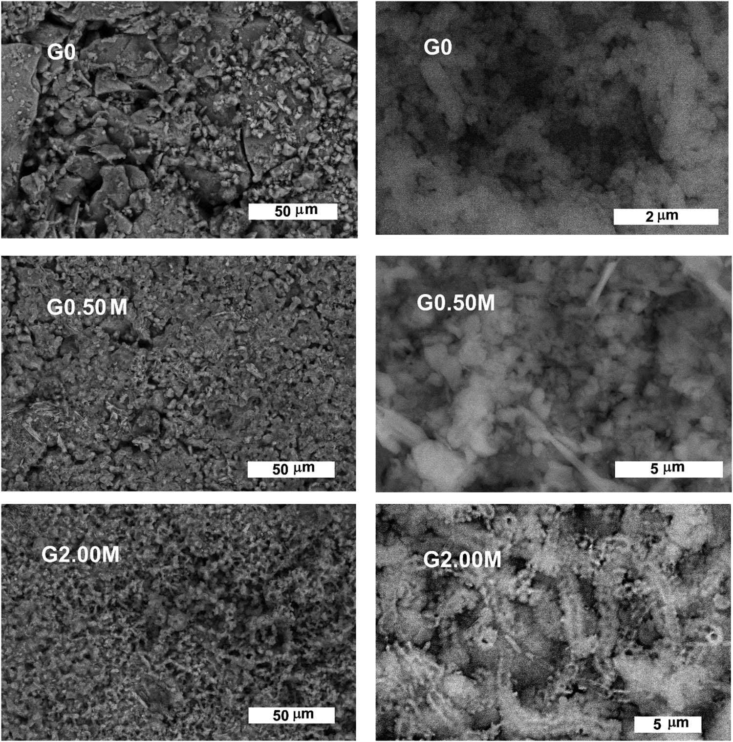
SEM of G0, G0.50M and G2.00M glasses sintered at 1,100°C/2 h are shown in Figure 4. G0 sample shows massive microstructure with some irregular clusters and very little pores. Subsequently at high magnification, connected rounded clusters appear in the range of the nano-size. Once a small amount of MnO2 is added, the microstructure of the sintered samples becomes more uniform and denser having a spheroidal morphology and disordered mesoporous structure [15]. The G0.50M and G2.00M samples present mesoporous glass together with a mix of irregular, angular and fine rod-like crystals with pores in between. At higher magnifications, the microstructure of the later crystals exposes nano-size crystals (30–100 nm) as shown in Figure 4. The EDX microanalysis of G0 indicates the integration of Na, Ca, Si and O. However, the EDX of the G0.50M and G2.00M glass samples confirms the incorporation of the manganese into the glassy matrix. Thus, it exhibits an indication of rankinite crystallization and due to the similarity of ionic radius of calcium and sodium, the possible replacement of Ca (116 pm) by Na (114 pm) may take place (Figure 5 and Table 2).

SEM micrographs of G0, G0.50M and G2.00M glasses sintered at 1,100°C/2 h.
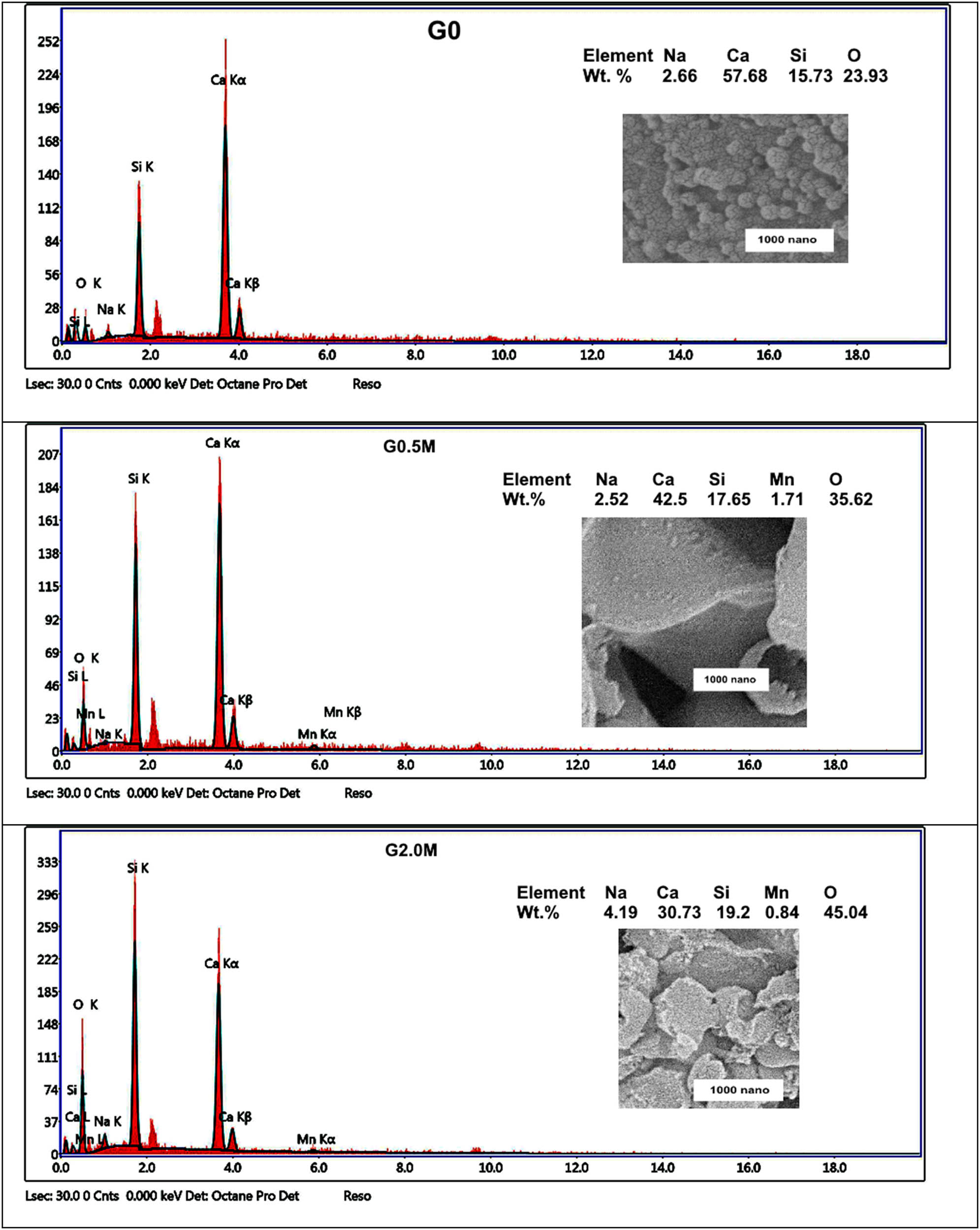
EDX microanalysis of G0, G0.50M and G2.00M glasses sintered at 1,100°C/2 h.
EDX microanalysis of G0, G0.50M and G2.00M samples
| Chemical constituents weight% | |||||
|---|---|---|---|---|---|
| Na | Ca | Si | Mn | O | |
| Nominal rankinite | — | 41.69 | 19.48 | — | 38.83 |
| G0 | 2.66 | 57.68 | 15.73 | — | 23.93 |
| G0.50M | 2.52 | 42.5 | 17.65 | 1.71 | 35.62 |
| G2.00M | 4.19 | 30.73 | 19.2 | 0.83 | 45.04 |
3.2 Densities, porosities and zeta potential
The densities and porosities of the glass samples sintered at 1,100°C are demonstrated in Figure 6. Upon incorporation of MnO2, the densities of the samples relatively increase from 1.88 to 2.24 g/cm3 and the porosities relatively decrease from 32.59 to 20.83%. Although the pre-mentioned densities of rankinite (2.844 g/cm3), combeite (2.690 g/cm3) and cristobalite (2.33 g/cm3) are higher than the present results, it must be mentioned that the increase of the porosities initiate decrease in such values. It is notable that all samples create negative zeta potentials as presented in Table 3. Such negative zeta potentials may be mainly accredited to the negative charge of the samples. Zeta potential of the major combeite, rankinite, containing 0.00, 0.50 and 2.00% of MnO2 has a lower negative value compared to that of the pure parent material (G0). Negative zeta potential may possibly be a useful property for bone-derived appliance when fixed in bone containing viable cells and deliberated for in vivo test (Figure 7). Likewise, it is clear that the conductivity of the electrolyte increases with the increase in the MnO2 content. For that reason, it may possibly be concluded that the deposition of MnO2 leads to a reduction in the zeta potential [30]. The lower values of zeta potential of all investigated samples doped with MnO2 designate the incipient instability of the particles suspension. Scientists stated that the bioactivity of nanocomposite materials can be predicted by the zeta potential of nano-additives [31,32]. From the author’s point of view, the most motivating finding was a negative zeta potential to enable the bone cell activity.
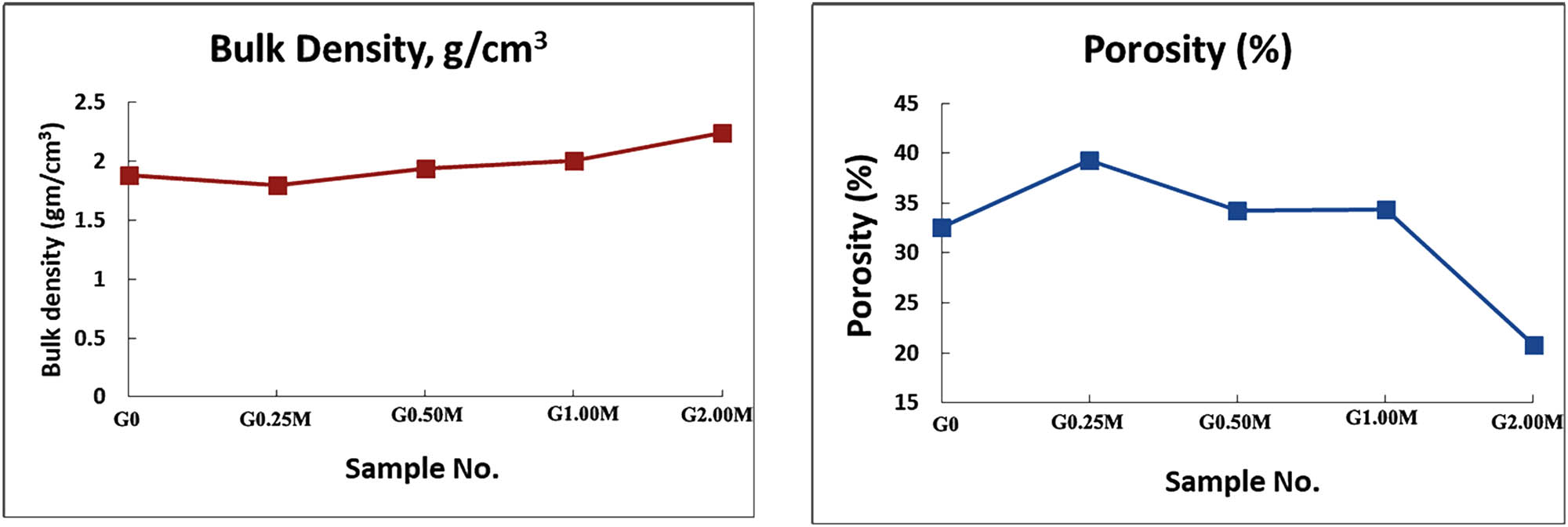
Densities and porosities of the samples sintered at 1,100°C/2 h.
The zeta potential measurements
| Sample/Notation | Zeta potential (mV) | Conductivity (mS/cm) | Std. Dev. |
|---|---|---|---|
| G0 | −40.3 | 0.0402 | 4.64 |
| G0.50M | −25.1 | 0.0574 | 4.61 |
| G2.00M | −27.0 | 0.0888 | 3.78 |
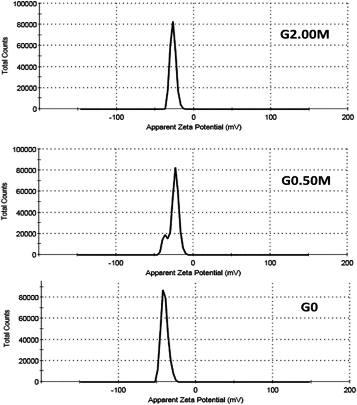
Zeta potential of G0, G0.50M and G2.00M glass samples sintered at 1,100°C/2 h.
3.3 Biocompatibility
3.3.1 In vitro results
To check the biocompatibility of the glasses, in vitro tests by means of the SBF are carried out. SEM micrographs of G0, G0.50M and G2.00M discs sintered at 1,100°C/2 h are soaked in SBF for 4 weeks as shown in Figure 8. G0 sample displays a mix of irregular angular, rod-like crystals with pores in between; whereas, at high magnification rounded, sub-rounded and rod clusters in the nano-size range are scattered all over the sample. On the other hand, G0.50M and G2.00M samples expose fine plate-like crystals mounting on the surface of the immersed samples. As shown in Figure 8, the later plate-like crystals are associated with tiny crystals with little pores in between and at high magnification the later crystals display nano-size crystals between 100 and 200 nm. On high magnification, the microstructure of the G2.00M sample is modified by means of interlocked ribbon containing nano-tiny crystals on its edges. Furthermore, the development of agglomerates is as a consequence of the van der Waals interaction forces between the particles [33]. EDX microanalysis demonstrates a growth in the intensity of the P and Ca peaks together with carbon and decline in the intensity of the Si peaks of all samples, which reflect the deposition of carbonated calcium phosphates (c-CaPs) (Figure 9). Also, the Ca/P ratios were 2.17, 2.30 and 2.97 for G0, G0.50M and G2.00M, respectively. Coating of the material surfaces by Ca-P can be used to increase the biological responses and decrease the toxicity [34]. Ca-P may happen in crystalline and amorphous phases with variable stoichiometry [35]. The in vitro results improve the osteoblast adhesion on the Ca-Ps with elevation in Ca/P ratio (up to 2.9) [36]. In classical condition, as the pH value of SBF was kept constant at 7.40 (i.e., over the isoelectric point of the particles), the surface charge of the particles is of negative charge when immersed in SBF, which managed to adsorb the Ca2+ ions from the SBF solution and ultimately form a crystalline layer of calcium-enriched apatite. In Figure 8, the EDX microanalysis of G2.00M sample shows a minute release of Mn ions, which recommends using such sample as a vehicle for providing therapeutic manganese ions [37].
SEM micrographs of the G0, G0.50M and G2.00M glasses sintered at 1,100°C/2 h and soaked in SBF for a month.

EDX microanalysis of G0, G0.50M and G2.00M glasses sintered at 1,100°C/2 h and soaked in SBF for a month.
Danewalia and Singh [16] mentioned that the incidence of MnO2 proliferates the disordering, thus improving the glass surface activity to form an apatite layer. Such findings approve their fittingness for being applicable in bone tissue engineering. However, in the present work, the increase of the c-CaP ratio monitors the successive incorporation of the MnO2 into the glass network [16].
The in vitro bioactivity studies in SBF designate that samples could prompt the Ca-P layer and propose the opportunity of relating the Mn-modified sample for bone regeneration as comprehended in Figure 9. Earlier, it was informed that the induction of Ca-P layer on surfaces of the bioactive materials was extremely important for the incorporation of materials with normal bone tissue [38]. The steps of the Ca-P layer formation stages on samples surface in SBF may possibly be summarized in the next six steps [36]:
Exchange of Ca2+ from the material surface with H+ or H3O+ in the SBF.
Release of soluble silica in the form of silicic acid [Si(OH)4] into the SBF and the formation of several silanol (Si–OH) groups onto the immersed surface.
Development of hydrated silica rich layer on account of re-polymerization and condensation of the Si–OH groups.
Precipitation of the Ca2+ ions and the
Evolution of the amorphous Ca-P (ACP) rich layer by enhancing more Ca2+ and
The crystallization of ACP rich layer by insertion of OH− and
3.3.2 FT-IR reflection
FT-IR spectra in Figure 10 elucidate the characteristic vibrational bands of the hydroxyapatite (HA) in the G0 and G2.00M glass discs sintered at 1,100°C/2 h and soaked in SBF for a month. Bands at 941 and 561 cm−1 are related to the P–O bending mode of vibration of the HA. At higher Mn content, the 561 band is shifted to 546 cm−1 [40]. Other bands at 1,597 and 845 cm−1 match the carbonate groups of the HA [41]. Likewise, a hump present at the 681 cm−1 is related to the hydroxyl group of the HA. The band at 501 cm−1 is credited to the Si–O–Si symmetric bending mode in the silicate network [42]. The band at 845 cm−1 corresponds to the C–O stretching mode of vibration.

FTIR reflection spectra of G0 and G2.00M glass discs sintered 1,100°C/2 h and soaked in SBF for a month.
Thus it can be concluded that the characteristics of FT-IR reflection spectra in Figure 10 correspond to the development of the carbonated hydroxyapatite (c-HA) layer on the sintered glass-ceramic discs. Consequently, the major combeite, rankinite/MnO2 lead to the development of c-CaP on their surface after immersion in SBF, which is similarly confirmed by the SEM/EDX. Miola et al. concluded that the Mn-leaching test in SBF delineated inconsistent trends probably due to a re-precipitation of the manganese compounds during the process of bioactivity [37]. The study of the in vitro bioactivity designates that all samples are capable to form Ca-P layer above their surfaces as specified by the FT-IR reflection and the SEM/EDX analysis.
3.3.3 In vivo tests
The in vivo results are verified on G0, G0.25M and G0.50M sintered at 1,100°C/2 h, and the positive group (empty bone gap) is the reference for the pre-mentioned sintered samples. Samples are implanted in the femur bone defects of hamster rats, and the results are examined after 45 and 90 days.
Light microscopic examination after 45 days of bone tissues for the control group (Figure 11) reveals normal histology of compact and trabecular bone without any detectible alterations and the positive control group shows an area of bone necrosis with the existence of some newly formed bone tissue that showed an irregular calcification pattern. On the other hand, the bone defect in the (G0) group displays delayed healing with the existence of necrotic tissue filling the bone gap. However, the edges of the defect demonstrate proliferating osteoblasts (PO). The area of bone defect in both G0.25M and G0.50M groups is filled by necrosed tissue debris along with the applied formula as shown in Figure 11.

Microscopic examination of (a) normal bone, (b) positive control and sample containing sintered (c) G0, (d) G0.25M and (e) G0.50M samples. Both G0.25M and G0.50M samples show nearly normal bone after 45 days of implantation. NFB: new formed bone, NT: necrosed tissue and PO: proliferating osteoblasts.
After 90 days of implantation, both the control and positive groups illustrate PO filling the defect area (Figure 12). In the same period, the G0 indicates new formed osteoid tissue filling the defect area with defective mineralization. Moreover, as shown in Figure 12, the G0.25M group elucidates an apparently normal bone tissue at the defect area and perfect bone healing with complete mineralization is noticed in the G0.50 group.

Light microscopic examination of (a) G0, (b) G0.25M and (c) G0.50M demonstrates nearly normal bone after 90 days of implantation. NFO: new formed osteoid.
In general, no inflammation or endothelial swelling or granulation tissue or fibrotic tissue or any rejection in both tested samples is detected. The results show that although the treatment and formation of new bone tissues are slow in the G0 sample, it is fast in case of incorporation of the MnO2 in G0.25M and G0.50M sintered samples. The presence of the major combeite (Na4Ca4Si6O18) and rankinite (Ca3Si2O7) with/without MnO2, as the major phases, not only stimulates the bioactivity but also triggers the activation of the osteoblast cell.
4 Conclusion
Nominal wollastonite with the successive four additions of MnO2 (0.25, 0.50, 1,00 and 2.00 mol%) contents are synthesized through melt quenching route. The incorporation of MnO2 does not show much changes in the temperature of endothermic and exothermic effect. XRD analysis of the sintered glasses near the crystallization temperature (at 900°C/2 h) showed the crystallization of combeite, pseudowollastonite, rankinite, bustamite and cristobalite. At higher temperature (1,100°C) the crystallization of the major rankinite and combeite was noticed as major phases with little cristobalite. In the later samples, with incorporation of MnO2, the densities were increased from 1.88 to 2.24 g/cm3 and the porosities decreased from 32.59 to 20.83%. The negative zeta potential of the present samples could be a useful property enhancing attachment and proliferation of bone cells when fixed in bone containing viable cells. The SEM of samples shows a mix of irregular angular and rod-like crystals with very fine pores in between. At higher magnifications, the microstructure of the later crystals shows nano-size crystals (between 30 and 100 nm). Post-immersion in SBF for 1 month the surface of the samples was studied via SEM/EDX and FT-IR reflection. However, the EDX microanalysis reflected the possible formation of c-HA on the surfaces upon immersion. The in vivo results show that although the formation of new bone tissues was slow in the case of the G0 sample, it was fast in case of G0.25M and G0.50M samples (i.e., the incorporation of MnO2 improved the new bone formation). Such outcomes are important in the application of MnO2-containing wollastonite glass for bone treatment and biomedical applications.
Acknowledgments
The authors extend their appreciation to the Deputyship for Research & Innovation, Ministry of Education in Saudi Arabia for funding this research work through project number (IFPHI-054-247-2020)” and King Abdulaziz University, DSR, Jeddah, Saudi Arabia.
-
Funding information: Deputyship for Research & Innovation, Ministry of Education in Saudi Arabia through project number (IFPHI-054-247-2020)” and King Abdulaziz University, DSR, Jeddah, Saudi Arabia.
-
Author contributions: All authors have accepted responsibility for the entire content of this manuscript and approved its submission.
-
Conflict of interest: The authors state no conflict of interest.
-
Ethical statement: The in vivo research protocol was reviewed and approved by the Animal Care Committee of the National Research Centre (NRC), Egypt, which follows the guidelines of the National Institutes of Health Guide for Care and Use of Laboratory Animals (approval registration No. 16-254).
References
[1] Du Z, Leng H, Guo L, Huang Y, Zheng T, Zhao Z, et al. Calcium silicate scaffolds promoting bone regeneration via the doping of Mg2+ or Mn2+ ion. Compos Part B Eng. 2020;190:107937. 10.1016/j.compositesb.2020.107937.Search in Google Scholar
[2] Beherei HH, Mohamed KR, El-Bassyouni GT. Mechanical and microstructure of reinforced hydroxyapatite/calcium silicate nano-composites materials. Mater Des. 2013;44:461–8. 10.1016/j.matdes.2012.08.020.Search in Google Scholar
[3] Abdel-Fattah WI, Jiang T, El-Bassyouni GT, Laurencin CT. Synthesis, characterization of chitosan and fabrication of sintered chitosan microsphere matrices for bone tissue engineering. Acta Biomater. 2007;3(4):503–14. 10.1016/j.actbio.2006.12.004.Search in Google Scholar PubMed
[4] Gritsch L, Perrin E, Chenal J-M, Fredholm Y, Maçon ALB, Chevalier J, et al. Combining bioresorbable polyesters and bioactive glasses: orthopedic applications of composite implants and bone tissue engineering scaffolds. Appl Mater Today. 2021;22:100923. 10.1016/j.apmt.2020.100923.Search in Google Scholar
[5] Mahdy MA, Kenawy SH, El Zawawi IK, Hamzawy EMA, El-Bassyouni GT. Optical and magnetic properties of wollastonite and its nanocomposite crystalline structure with hematite. Ceram Int. 2020;46(5):6581–93. 10.1016/j.ceramint.2019.11.144.Search in Google Scholar
[6] Hoppe A, Mourino V, Boccaccini AR. Therapeutic inorganic ions in bioactive glasses to enhance bone formation and beyond. Biomater Sci. 2013;1(3):254–6. 10.1039/C2BM00116K.Search in Google Scholar PubMed
[7] Prati C, Gandolfi MG. Calcium silicate bioactive cements: biological perspectives and clinical applications. Dent Mater. 2015;31(4):351–70. 10.1016/j.dental.2015.01.004.Search in Google Scholar PubMed
[8] Almeida MS, Fernandes GVO, Oliveira AM, Granjeiro JM. Calcium silicate as a graft material for bone fractures: a systematic review. J Int Med Res. 2018;46(7):2537–48. 10.1177/0300060518770940.Search in Google Scholar PubMed PubMed Central
[9] Mabrouk M, Taha SK, Abdel Hamid MA, Kenawy SH, Hassan EA, El-Bassyouni GT. Radiological evaluations of low cost wollastonite nano-ceramics graft doped with iron oxide in the treatment of induced defects in canine mandible. J Biomed Mater Res. 2021;109:1029–44. 10.1002/jbm.b.34767.Search in Google Scholar PubMed
[10] Husnain SM, Asim U, Yaqub A, Shahzad F, Abbas N. Recent trends of MnO2-derived adsorbents for water treatment: a review. N J Chem. 2020;44(16):6096–120. 10.1039/C9NJ06392G.Search in Google Scholar
[11] Zhou L, Huang Y, Qiu W, Sun Z, Liu Z, Song Z. Adsorption properties of nano-MnO2-biochar composites for copper in aqueous solution. Molecules. 2017;22(1):173. 10.3390/molecules22010173.Search in Google Scholar PubMed PubMed Central
[12] Qian X, Han X, Yu L, Xu T, Chen Y. Manganese-based functional nanopaltforms: nanosynthetic construction, physiochemical properties, and theranostic applicability. Adv Funct Mater. 2020;30:1907066. 10.1002/adfm.201907066.Search in Google Scholar
[13] Wang CC, Ni W, Zhang D, Sun X, Zhang N. Dielectric properties of pure and Mn-doped CaCu3Ti4O12 ceramics over a wide temperature range. J Electroceram. 2016;36(1–4):46–57. 10.1007/s10832-016-0024-3.Search in Google Scholar
[14] Yao Y, Zhang Y. Fabrication and dielectric properties of LiTaO3 matrix ceramics with added manganese dioxide. J Ceram Sci Technol. 2020;11(1):27–35. m10.4416/JCST2019-00053.Search in Google Scholar
[15] Xiu S, Shen B, Zhai J. The effects of MnO2 addition on the structure and dielectric properties of the strontium barium niobate glass-ceramics. Mater Res Bull. 2017;95:349–53. 10.1016/j.materresbull.2017.08.008.Search in Google Scholar
[16] Danewalia SS, Singh K. Magnetic and bioactive properties of MnO2/Fe2O3 modified Na2O–CaO–P2O5–SiO2 glasses and nanocrystalline glass-ceramics. Ceram Int. 2016;42:11858–68. 10.1016/j.ceramint.2016.04.108.Search in Google Scholar
[17] Kolmas J, Groszyk E, Piotrowska U. Nanocrystalline hydroxyapatite enriched in selenite and manganese ions: physicochemical and antibacterial properties. Nanoscale Res Lett. 2015;10(1):278–87. 10.1186/s11671-015-0989-x.Search in Google Scholar PubMed PubMed Central
[18] Kokubo T, Kushitani H, Sakka S, Kitsugi T, Yamamum T. Solutions able to reproduce in vivo surface-structure changes in bioactive. J Biomed Mater Res. 1990;24(6):721–34. 10.1002/jbm.820240607.Search in Google Scholar PubMed
[19] Kokubo T, Takadama H. How useful is SBF in predicting in vivo bone bioactivity? Biomaterials. 2006;27(15):2907–15. 10.1016/J.BIOMATERIALS.2006.01.017.Search in Google Scholar
[20] Dridi A, Riahi KZ, Somrani S. Mechanism of apatite formation on a poorly crystallized calcium phosphate in a simulated body fluid (SBF) at 37°C. J Phys Chem Solids. 2021;156:110122. 10.1016/j.jpcs.2021.110122.Search in Google Scholar
[21] Jalota S, Bhaduri SB, Tas AC. Effect of carbonate content and buffer type on calcium phosphate formation in SBF solutions. J Mater Sci Mater Med. 2006;17(8):697–707. 10.1007/s10856-006-9680-1.Search in Google Scholar
[22] Khater GA. The use of Saudi slag for the production of glass-ceramic materials. Ceram Int. 2002;28(1):59–67. 10.1016/S0272-8842(01)00058-X.Search in Google Scholar
[23] Fischer RX, Tillmanns E. Die Kristallstrukturen von naturlichem Na2Ca2Si3O9 vom Mt. Shaheru (Zaire) und aus dem Mayener Feld (Eifel) Note: this is the high-temperature form of combeite. Neues Jahrb fur Mineral Monatshefte. 1983;2:49–59.Search in Google Scholar
[24] Yang H, Prewitt CT. On the crystal structure of pseudowollastonite (CaSiO3). Am Miner. 1999; 84(5–6):929–32. 10.2138/am-1999-5-629.Search in Google Scholar
[25] Kusachi I, Henmi C, Kawahara A, Henmi K. The structure of rankinite. Miner J. 1975;8:38–47. 10.2465/MINERJ.8.38.Search in Google Scholar
[26] Ohashi Y, Finger LW. The role of octahedral cations in pyroxenoid crystal chemistry; I, bustamite, wollastonite, and the pectolite-schizolite-serandite series. Am Miner. 1978;63(3–4):274–88.Search in Google Scholar
[27] Dove MT, Craig MS, Keen DA, Marshall WC, Redfern SAT, Trachenko KO, et al. Crystal structure of the high-pressure monoclinic phase-II of cristobalite, SiO2. Locality: synthetic note: P = 3.5 GPa, refinement by unconstrained Rietveld analysis. Miner Magaz. 2000;64(3):569–76.10.1180/002646100549436Search in Google Scholar
[28] Hamzawy EMA, Kenawy SH, Abd El Aty AA, El-Bassyouni GT. Characterization of wollastonite-copper nanoparticles synthesized by a wet method. Interceram. 2018;67(3):20–3.10.1007/s42411-018-0010-7Search in Google Scholar
[29] Mahdy MA, Kenawy SH, Hamzawy EMA, El-Bassyouni GT, Zawawi IKEl. Influence of silicon carbide on structural, optical and magnetic properties of wollastonite/Fe2O3 nanocomposites. Ceram Int. 2021;47(9):12047–55. 10.1016/j.ceramint.2021.01.048.Search in Google Scholar
[30] Zhang H, Xu F, Xue J, Chen S, Wang J, Yang Y. Enhanced removal of heavy metal ions from aqueous solution using manganese dioxide-loaded biochar: behavior and mechanism. Sci Rep. 2020;10(1):6067. 10.1038/s41598-020-63000-z.Search in Google Scholar PubMed PubMed Central
[31] Zych L, Osyczka AM, Łacz A, Rózycka A, Niemiec W, Rapacz-Kmita A, et al. How surface properties of silica nanoparticles influence structural, microstructural and biological properties of polymer nanocomposites. Materials. 2021;14(4):843. 10.3390/ma14040843.Search in Google Scholar PubMed PubMed Central
[32] Mabrouk M, Mousa SM, Abd ElGhany WA, Abo-elfadl MT, El-Bassyouni GT. Bioactivity and cell viability of Ag+- and Zr4+- co-doped biphasic calcium phosphate. Appl Phys A. 2021;127(12):948. 10.1007/s00339-021-05051-1.Search in Google Scholar
[33] Nawaz Q, Ur Rehman MA, Burkovski A, Schmidt J, Beltrán AM, Shahid A, et al. Synthesis and characterization of manganese containing mesoporous bioactive glass nanoparticles for biomedical applications. J Mater Sci Mater Med. 2018;29:64. 10.1007/s10856-018-6070-4.Search in Google Scholar PubMed
[34] Eliaz N, Metoki N. Calcium phosphate bioceramics: a review of their history, structure, properties, coating technologies and biomedical applications. Mater (Basel). 2017;10(4):334.10.3390/ma10040334Search in Google Scholar PubMed PubMed Central
[35] Mancardi G. Computational study of the nucleation of calcium phosphate. Thesis submitted for the degree of doctor of philosophy. University College London, London Department of Chemistry; 2018.Search in Google Scholar
[36] Liu H, Yazici H, Ergun C, Webster TJ, Bermek H. An in vitro evaluation of the Ca/P ratio for the cytocompatibility of nano-to-micron particulate calcium phosphates for bone regeneration. Acta Biomater. 2008;4(5):1472–9. 10.1016/j.actbio.2008.02.025.Search in Google Scholar PubMed
[37] Miola M, Brovarone CV, Maina G, Rossi F, Bergandi L, Ghigo D, et al. In vitro study of manganese-doped bioactive glasses for bone regeneration. Mater Sci Eng C. 2014;38:107–18. 10.1016/j.msec.2014.01.045.Search in Google Scholar PubMed
[38] Baino F. Bioactive glass – when glass science and technology meet regenerative medicine. Ceram Int. 2018;44(13):14953–66. 10.1016/j.ceramint.2018.05.180.Search in Google Scholar
[39] Sayed MK, El-Kady AM, Sallam AM, Talaat MS. In vitro bioactivity evaluation of novel manganese modified calcium silicate ceramics for one regeneration. IJISET – Int J Innovative Sci Eng Technol. 2018;5(10):44–9.Search in Google Scholar
[40] Tripathi H, Hira SK, Kumar AS, Gupta U, Manna PP, Singh SP. Structural characterization and in vitro bioactivity assessment of SiO2–CaO–P2O5–K2O–Al2O3 glass as bioactive ceramic material. Ceram Int. 2015;41:11756–69. 10.1016/J.CERAMINT.2015.05.143.Search in Google Scholar
[41] Beherei HH, El-Bassyouni GT, Mohamed KR. Modulation, characterization and bioactivity of new biocomposites based on apatite. Ceram Int. 2008;34(8):2091–7. 10.1016/j.ceramint.2007.08.003.Search in Google Scholar
[42] Romeis S, Hoppe A, Eisermann C, Schneider N, Boccaccini AR, Schmidt J, et al. Enhancing in vitro bioactivity of melt derived 45S5 bioglass by communication in stirred Media Mill. Am Ceram Soc. 2014;97(1):150–6. 10.1111/jace.12615.Search in Google Scholar
© 2022 Samah S. Eldera et al., published by De Gruyter
This work is licensed under the Creative Commons Attribution 4.0 International License.
Articles in the same Issue
- Research Articles
- Theoretical and experimental investigation of MWCNT dispersion effect on the elastic modulus of flexible PDMS/MWCNT nanocomposites
- Mechanical, morphological, and fracture-deformation behavior of MWCNTs-reinforced (Al–Cu–Mg–T351) alloy cast nanocomposites fabricated by optimized mechanical milling and powder metallurgy techniques
- Flammability and physical stability of sugar palm crystalline nanocellulose reinforced thermoplastic sugar palm starch/poly(lactic acid) blend bionanocomposites
- Glutathione-loaded non-ionic surfactant niosomes: A new approach to improve oral bioavailability and hepatoprotective efficacy of glutathione
- Relationship between mechano-bactericidal activity and nanoblades density on chemically strengthened glass
- In situ regulation of microstructure and microwave-absorbing properties of FeSiAl through HNO3 oxidation
- Research on a mechanical model of magnetorheological fluid different diameter particles
- Nanomechanical and dynamic mechanical properties of rubber–wood–plastic composites
- Investigative properties of CeO2 doped with niobium: A combined characterization and DFT studies
- Miniaturized peptidomimetics and nano-vesiculation in endothelin types through probable nano-disk formation and structure property relationships of endothelins’ fragments
- N/S co-doped CoSe/C nanocubes as anode materials for Li-ion batteries
- Synergistic effects of halloysite nanotubes with metal and phosphorus additives on the optimal design of eco-friendly sandwich panels with maximum flame resistance and minimum weight
- Octreotide-conjugated silver nanoparticles for active targeting of somatostatin receptors and their application in a nebulized rat model
- Controllable morphology of Bi2S3 nanostructures formed via hydrothermal vulcanization of Bi2O3 thin-film layer and their photoelectrocatalytic performances
- Development of (−)-epigallocatechin-3-gallate-loaded folate receptor-targeted nanoparticles for prostate cancer treatment
- Enhancement of the mechanical properties of HDPE mineral nanocomposites by filler particles modulation of the matrix plastic/elastic behavior
- Effect of plasticizers on the properties of sugar palm nanocellulose/cinnamon essential oil reinforced starch bionanocomposite films
- Optimization of nano coating to reduce the thermal deformation of ball screws
- Preparation of efficient piezoelectric PVDF–HFP/Ni composite films by high electric field poling
- MHD dissipative Casson nanofluid liquid film flow due to an unsteady stretching sheet with radiation influence and slip velocity phenomenon
- Effects of nano-SiO2 modification on rubberised mortar and concrete with recycled coarse aggregates
- Mechanical and microscopic properties of fiber-reinforced coal gangue-based geopolymer concrete
- Effect of morphology and size on the thermodynamic stability of cerium oxide nanoparticles: Experiment and molecular dynamics calculation
- Mechanical performance of a CFRP composite reinforced via gelatin-CNTs: A study on fiber interfacial enhancement and matrix enhancement
- A practical review over surface modification, nanopatterns, emerging materials, drug delivery systems, and their biophysiochemical properties for dental implants: Recent progresses and advances
- HTR: An ultra-high speed algorithm for cage recognition of clathrate hydrates
- Effects of microalloying elements added by in situ synthesis on the microstructure of WCu composites
- A highly sensitive nanobiosensor based on aptamer-conjugated graphene-decorated rhodium nanoparticles for detection of HER2-positive circulating tumor cells
- Progressive collapse performance of shear strengthened RC frames by nano CFRP
- Core–shell heterostructured composites of carbon nanotubes and imine-linked hyperbranched polymers as metal-free Li-ion anodes
- A Galerkin strategy for tri-hybridized mixture in ethylene glycol comprising variable diffusion and thermal conductivity using non-Fourier’s theory
- Simple models for tensile modulus of shape memory polymer nanocomposites at ambient temperature
- Preparation and morphological studies of tin sulfide nanoparticles and use as efficient photocatalysts for the degradation of rhodamine B and phenol
- Polyethyleneimine-impregnated activated carbon nanofiber composited graphene-derived rice husk char for efficient post-combustion CO2 capture
- Electrospun nanofibers of Co3O4 nanocrystals encapsulated in cyclized-polyacrylonitrile for lithium storage
- Pitting corrosion induced on high-strength high carbon steel wire in high alkaline deaerated chloride electrolyte
- Formulation of polymeric nanoparticles loaded sorafenib; evaluation of cytotoxicity, molecular evaluation, and gene expression studies in lung and breast cancer cell lines
- Engineered nanocomposites in asphalt binders
- Influence of loading voltage, domain ratio, and additional load on the actuation of dielectric elastomer
- Thermally induced hex-graphene transitions in 2D carbon crystals
- The surface modification effect on the interfacial properties of glass fiber-reinforced epoxy: A molecular dynamics study
- Molecular dynamics study of deformation mechanism of interfacial microzone of Cu/Al2Cu/Al composites under tension
- Nanocolloid simulators of luminescent solar concentrator photovoltaic windows
- Compressive strength and anti-chloride ion penetration assessment of geopolymer mortar merging PVA fiber and nano-SiO2 using RBF–BP composite neural network
- Effect of 3-mercapto-1-propane sulfonate sulfonic acid and polyvinylpyrrolidone on the growth of cobalt pillar by electrodeposition
- Dynamics of convective slippery constraints on hybrid radiative Sutterby nanofluid flow by Galerkin finite element simulation
- Preparation of vanadium by the magnesiothermic self-propagating reduction and process control
- Microstructure-dependent photoelectrocatalytic activity of heterogeneous ZnO–ZnS nanosheets
- Cytotoxic and pro-inflammatory effects of molybdenum and tungsten disulphide on human bronchial cells
- Improving recycled aggregate concrete by compression casting and nano-silica
- Chemically reactive Maxwell nanoliquid flow by a stretching surface in the frames of Newtonian heating, nonlinear convection and radiative flux: Nanopolymer flow processing simulation
- Nonlinear dynamic and crack behaviors of carbon nanotubes-reinforced composites with various geometries
- Biosynthesis of copper oxide nanoparticles and its therapeutic efficacy against colon cancer
- Synthesis and characterization of smart stimuli-responsive herbal drug-encapsulated nanoniosome particles for efficient treatment of breast cancer
- Homotopic simulation for heat transport phenomenon of the Burgers nanofluids flow over a stretching cylinder with thermal convective and zero mass flux conditions
- Incorporation of copper and strontium ions in TiO2 nanotubes via dopamine to enhance hemocompatibility and cytocompatibility
- Mechanical, thermal, and barrier properties of starch films incorporated with chitosan nanoparticles
- Mechanical properties and microstructure of nano-strengthened recycled aggregate concrete
- Glucose-responsive nanogels efficiently maintain the stability and activity of therapeutic enzymes
- Tunning matrix rheology and mechanical performance of ultra-high performance concrete using cellulose nanofibers
- Flexible MXene/copper/cellulose nanofiber heat spreader films with enhanced thermal conductivity
- Promoted charge separation and specific surface area via interlacing of N-doped titanium dioxide nanotubes on carbon nitride nanosheets for photocatalytic degradation of Rhodamine B
- Elucidating the role of silicon dioxide and titanium dioxide nanoparticles in mitigating the disease of the eggplant caused by Phomopsis vexans, Ralstonia solanacearum, and root-knot nematode Meloidogyne incognita
- An implication of magnetic dipole in Carreau Yasuda liquid influenced by engine oil using ternary hybrid nanomaterial
- Robust synthesis of a composite phase of copper vanadium oxide with enhanced performance for durable aqueous Zn-ion batteries
- Tunning self-assembled phases of bovine serum albumin via hydrothermal process to synthesize novel functional hydrogel for skin protection against UVB
- A comparative experimental study on damping properties of epoxy nanocomposite beams reinforced with carbon nanotubes and graphene nanoplatelets
- Lightweight and hydrophobic Ni/GO/PVA composite aerogels for ultrahigh performance electromagnetic interference shielding
- Research on the auxetic behavior and mechanical properties of periodically rotating graphene nanostructures
- Repairing performances of novel cement mortar modified with graphene oxide and polyacrylate polymer
- Closed-loop recycling and fabrication of hydrophilic CNT films with high performance
- Design of thin-film configuration of SnO2–Ag2O composites for NO2 gas-sensing applications
- Study on stress distribution of SiC/Al composites based on microstructure models with microns and nanoparticles
- PVDF green nanofibers as potential carriers for improving self-healing and mechanical properties of carbon fiber/epoxy prepregs
- Osteogenesis capability of three-dimensionally printed poly(lactic acid)-halloysite nanotube scaffolds containing strontium ranelate
- Silver nanoparticles induce mitochondria-dependent apoptosis and late non-canonical autophagy in HT-29 colon cancer cells
- Preparation and bonding mechanisms of polymer/metal hybrid composite by nano molding technology
- Damage self-sensing and strain monitoring of glass-reinforced epoxy composite impregnated with graphene nanoplatelet and multiwalled carbon nanotubes
- Thermal analysis characterisation of solar-powered ship using Oldroyd hybrid nanofluids in parabolic trough solar collector: An optimal thermal application
- Pyrene-functionalized halloysite nanotubes for simultaneously detecting and separating Hg(ii) in aqueous media: A comprehensive comparison on interparticle and intraparticle excimers
- Fabrication of self-assembly CNT flexible film and its piezoresistive sensing behaviors
- Thermal valuation and entropy inspection of second-grade nanoscale fluid flow over a stretching surface by applying Koo–Kleinstreuer–Li relation
- Mechanical properties and microstructure of nano-SiO2 and basalt-fiber-reinforced recycled aggregate concrete
- Characterization and tribology performance of polyaniline-coated nanodiamond lubricant additives
- Combined impact of Marangoni convection and thermophoretic particle deposition on chemically reactive transport of nanofluid flow over a stretching surface
- Spark plasma extrusion of binder free hydroxyapatite powder
- An investigation on thermo-mechanical performance of graphene-oxide-reinforced shape memory polymer
- Effect of nanoadditives on the novel leather fiber/recycled poly(ethylene-vinyl-acetate) polymer composites for multifunctional applications: Fabrication, characterizations, and multiobjective optimization using central composite design
- Design selection for a hemispherical dimple core sandwich panel using hybrid multi-criteria decision-making methods
- Improving tensile strength and impact toughness of plasticized poly(lactic acid) biocomposites by incorporating nanofibrillated cellulose
- Green synthesis of spinel copper ferrite (CuFe2O4) nanoparticles and their toxicity
- The effect of TaC and NbC hybrid and mono-nanoparticles on AA2024 nanocomposites: Microstructure, strengthening, and artificial aging
- Excited-state geometry relaxation of pyrene-modified cellulose nanocrystals under UV-light excitation for detecting Fe3+
- Effect of CNTs and MEA on the creep of face-slab concrete at an early age
- Effect of deformation conditions on compression phase transformation of AZ31
- Application of MXene as a new generation of highly conductive coating materials for electromembrane-surrounded solid-phase microextraction
- A comparative study of the elasto-plastic properties for ceramic nanocomposites filled by graphene or graphene oxide nanoplates
- Encapsulation strategies for improving the biological behavior of CdS@ZIF-8 nanocomposites
- Biosynthesis of ZnO NPs from pumpkin seeds’ extract and elucidation of its anticancer potential against breast cancer
- Preliminary trials of the gold nanoparticles conjugated chrysin: An assessment of anti-oxidant, anti-microbial, and in vitro cytotoxic activities of a nanoformulated flavonoid
- Effect of micron-scale pores increased by nano-SiO2 sol modification on the strength of cement mortar
- Fractional simulations for thermal flow of hybrid nanofluid with aluminum oxide and titanium oxide nanoparticles with water and blood base fluids
- The effect of graphene nano-powder on the viscosity of water: An experimental study and artificial neural network modeling
- Development of a novel heat- and shear-resistant nano-silica gelling agent
- Characterization, biocompatibility and in vivo of nominal MnO2-containing wollastonite glass-ceramic
- Entropy production simulation of second-grade magnetic nanomaterials flowing across an expanding surface with viscidness dissipative flux
- Enhancement in structural, morphological, and optical properties of copper oxide for optoelectronic device applications
- Aptamer-functionalized chitosan-coated gold nanoparticle complex as a suitable targeted drug carrier for improved breast cancer treatment
- Performance and overall evaluation of nano-alumina-modified asphalt mixture
- Analysis of pure nanofluid (GO/engine oil) and hybrid nanofluid (GO–Fe3O4/engine oil): Novel thermal and magnetic features
- Synthesis of Ag@AgCl modified anatase/rutile/brookite mixed phase TiO2 and their photocatalytic property
- Mechanisms and influential variables on the abrasion resistance hydraulic concrete
- Synergistic reinforcement mechanism of basalt fiber/cellulose nanocrystals/polypropylene composites
- Achieving excellent oxidation resistance and mechanical properties of TiB2–B4C/carbon aerogel composites by quick-gelation and mechanical mixing
- Microwave-assisted sol–gel template-free synthesis and characterization of silica nanoparticles obtained from South African coal fly ash
- Pulsed laser-assisted synthesis of nano nickel(ii) oxide-anchored graphitic carbon nitride: Characterizations and their potential antibacterial/anti-biofilm applications
- Effects of nano-ZrSi2 on thermal stability of phenolic resin and thermal reusability of quartz–phenolic composites
- Benzaldehyde derivatives on tin electroplating as corrosion resistance for fabricating copper circuit
- Mechanical and heat transfer properties of 4D-printed shape memory graphene oxide/epoxy acrylate composites
- Coupling the vanadium-induced amorphous/crystalline NiFe2O4 with phosphide heterojunction toward active oxygen evolution reaction catalysts
- Graphene-oxide-reinforced cement composites mechanical and microstructural characteristics at elevated temperatures
- Gray correlation analysis of factors influencing compressive strength and durability of nano-SiO2 and PVA fiber reinforced geopolymer mortar
- Preparation of layered gradient Cu–Cr–Ti alloy with excellent mechanical properties, thermal stability, and electrical conductivity
- Recovery of Cr from chrome-containing leather wastes to develop aluminum-based composite material along with Al2O3 ceramic particles: An ingenious approach
- Mechanisms of the improved stiffness of flexible polymers under impact loading
- Anticancer potential of gold nanoparticles (AuNPs) using a battery of in vitro tests
- Review Articles
- Proposed approaches for coronaviruses elimination from wastewater: Membrane techniques and nanotechnology solutions
- Application of Pickering emulsion in oil drilling and production
- The contribution of microfluidics to the fight against tuberculosis
- Graphene-based biosensors for disease theranostics: Development, applications, and recent advancements
- Synthesis and encapsulation of iron oxide nanorods for application in magnetic hyperthermia and photothermal therapy
- Contemporary nano-architectured drugs and leads for ανβ3 integrin-based chemotherapy: Rationale and retrospect
- State-of-the-art review of fabrication, application, and mechanical properties of functionally graded porous nanocomposite materials
- Insights on magnetic spinel ferrites for targeted drug delivery and hyperthermia applications
- A review on heterogeneous oxidation of acetaminophen based on micro and nanoparticles catalyzed by different activators
- Early diagnosis of lung cancer using magnetic nanoparticles-integrated systems
- Advances in ZnO: Manipulation of defects for enhancing their technological potentials
- Efficacious nanomedicine track toward combating COVID-19
- A review of the design, processes, and properties of Mg-based composites
- Green synthesis of nanoparticles for varied applications: Green renewable resources and energy-efficient synthetic routes
- Two-dimensional nanomaterial-based polymer composites: Fundamentals and applications
- Recent progress and challenges in plasmonic nanomaterials
- Apoptotic cell-derived micro/nanosized extracellular vesicles in tissue regeneration
- Electronic noses based on metal oxide nanowires: A review
- Framework materials for supercapacitors
- An overview on the reproductive toxicity of graphene derivatives: Highlighting the importance
- Antibacterial nanomaterials: Upcoming hope to overcome antibiotic resistance crisis
- Research progress of carbon materials in the field of three-dimensional printing polymer nanocomposites
- A review of atomic layer deposition modelling and simulation methodologies: Density functional theory and molecular dynamics
- Recent advances in the preparation of PVDF-based piezoelectric materials
- Recent developments in tensile properties of friction welding of carbon fiber-reinforced composite: A review
- Comprehensive review of the properties of fly ash-based geopolymer with additive of nano-SiO2
- Perspectives in biopolymer/graphene-based composite application: Advances, challenges, and recommendations
- Graphene-based nanocomposite using new modeling molecular dynamic simulations for proposed neutralizing mechanism and real-time sensing of COVID-19
- Nanotechnology application on bamboo materials: A review
- Recent developments and future perspectives of biorenewable nanocomposites for advanced applications
- Nanostructured lipid carrier system: A compendium of their formulation development approaches, optimization strategies by quality by design, and recent applications in drug delivery
- 3D printing customized design of human bone tissue implant and its application
- Design, preparation, and functionalization of nanobiomaterials for enhanced efficacy in current and future biomedical applications
- A brief review of nanoparticles-doped PEDOT:PSS nanocomposite for OLED and OPV
- Nanotechnology interventions as a putative tool for the treatment of dental afflictions
- Recent advancements in metal–organic frameworks integrating quantum dots (QDs@MOF) and their potential applications
- A focused review of short electrospun nanofiber preparation techniques for composite reinforcement
- Microstructural characteristics and nano-modification of interfacial transition zone in concrete: A review
- Latest developments in the upconversion nanotechnology for the rapid detection of food safety: A review
- Strategic applications of nano-fertilizers for sustainable agriculture: Benefits and bottlenecks
- Molecular dynamics application of cocrystal energetic materials: A review
- Synthesis and application of nanometer hydroxyapatite in biomedicine
- Cutting-edge development in waste-recycled nanomaterials for energy storage and conversion applications
- Biological applications of ternary quantum dots: A review
- Nanotherapeutics for hydrogen sulfide-involved treatment: An emerging approach for cancer therapy
- Application of antibacterial nanoparticles in orthodontic materials
- Effect of natural-based biological hydrogels combined with growth factors on skin wound healing
- Nanozymes – A route to overcome microbial resistance: A viewpoint
- Recent developments and applications of smart nanoparticles in biomedicine
- Contemporary review on carbon nanotube (CNT) composites and their impact on multifarious applications
- Interfacial interactions and reinforcing mechanisms of cellulose and chitin nanomaterials and starch derivatives for cement and concrete strength and durability enhancement: A review
- Diamond-like carbon films for tribological modification of rubber
- Layered double hydroxides (LDHs) modified cement-based materials: A systematic review
- Recent research progress and advanced applications of silica/polymer nanocomposites
- Modeling of supramolecular biopolymers: Leading the in silico revolution of tissue engineering and nanomedicine
- Recent advances in perovskites-based optoelectronics
- Biogenic synthesis of palladium nanoparticles: New production methods and applications
- A comprehensive review of nanofluids with fractional derivatives: Modeling and application
- Electrospinning of marine polysaccharides: Processing and chemical aspects, challenges, and future prospects
- Electrohydrodynamic printing for demanding devices: A review of processing and applications
- Rapid Communications
- Structural material with designed thermal twist for a simple actuation
- Recent advances in photothermal materials for solar-driven crude oil adsorption