Latest developments in the upconversion nanotechnology for the rapid detection of food safety: A review
-
Guangna Ji
 and
Tie Han
and
Tie Han

Abstract
Food safety has become a topic of global concern in the recent decades. The significant food safety incidents occur from time to time around the world, seriously threatening the public health and causing extensive economic losses. In particular, the occurrence of COVID-19 highlights the importance of the food safety for the public health. Therefore, there is an urgent need to establish a fast, simple, sensitive, and efficient method for the detection of food safety. In recent years, the upconversion (UC) nanotechnology has been widely used in the field of food detection. The UC fluorescence analysis technology possesses the advantages of ultra-sensitivity detection, non-invasiveness, light stability, etc., and has broad application prospects in the field of food safety. After cladding and surface modification, it can be combined with other substances through a variety of mechanisms, such as electrostatic interaction, thereby expanding its application in the food safety detection. Thus, overall, there is a vital need to evaluate and utilize the potential of UC nanoparticles in the field of rapid detection of food safety.
1 Introduction
Food safety exhibits that the food is non-toxic, harmless, green, and healthy, along with meeting the required nutritional requirements. In addition, it does not cause any acute, subacute, or chronic harm to the human health. With the rapid economic development, the living standards have also been significantly improved. Thus, the food is also needed to be supplied at an accelerated pace, thereby prospering the food markets [1]. However, owing to the contradiction between the people’s growing needs for a better life and the unbalanced and insufficient development, the food safety incidents occur frequently, such as an excessive content of additives in food [2,3], use of illegal additives [4,5], presence of heavy metals [6,7], excessive veterinary drug residues [8,9], and microbial contamination [10]. These issues have aroused an unprecedented attention of the consumers and industry with respect to the food contamination issues [11,12].
In recent years, the food-borne diseases and food poisoning incidents caused by the pathogenic microorganisms and pesticide residues have been frequently observed. Among these, the food-borne diseases resulting due to the pathogenic microorganisms represent a vital food safety and public health issue [13,14,15]. Recently, the popularity of the functional foods has prompted a large number of food manufacturers to start their production. Though such foods provide consumers with health benefits, the excessive use of some additives is worrying [16]. Diclazuril is a broad-spectrum anti-coccidial drug which is widely used to eliminate insects in chickens, ducks, and other animals. It often causes poisoning and other harmful effects in the human body. Therefore, a rapid and sensitive method for the determination of diclazuril in chickens is urgently needed [17]. Clostridium perfringens is a well-known pathogen leading to food-borne diseases, it can cause diseases such as gas gangrene and food poisoning, thus, causing a serious harm to the human body [18,19,20]. In the coming years, the applications of nanomaterials in the food and food chain are expected to increase, continuously. Although this brings an exciting prospect to the consumers and the food industry, the potential harmful effects on the human body needs to be carefully considered, thus reiterating the need to establish rapid and safe methods for on-site inspection and supervision [21]. Sausages or ham extracted from the deer skin, red deer, or spores are the typical meat products in the high mountainous regions of Germany, Italy, Austria, and Switzerland. However, a considerable number of products do not declare their contents, thus, it is urgent to establish a rapid method for the accurate determination of their content [22]. The pasta salad recall in 2018 and the watermelon recall in 2019 at different places indicate that the fresh produce-borne diseases caused by the pathogens represent a serious food safety issue facing the world [23]. As a global public health problem, food safety has attracted a great deal of attention [24]. Therefore, the efficient analysis of the food safety issues and establishment of the rapid and effective detection methods are needed to protect human health, to ensure the healthy and stable development of the food safety system, as well as realize the food safety strategies [25].
The traditional laboratory detection methods include atomic absorption spectrophotometry, gas phase chromatography, high-performance liquid chromatography, etc. These methods have cumbersome sample preparation, protocols, and long detection time [26,27,28]. Using such methods, it is difficult to meet the needs of high throughput, effective, and low-cost on-site detection and supervision. Therefore, the development of new materials and technologies for the rapid food inspection is becoming increasingly important [4].
The literature studies have reported that the supercritical nanoparticles, especially the rare earth-doped supercritical nanoparticles, possess significant optical properties (including high photobleaching resistance and clear emission band) and demonstrate the eye-catching characteristic spectra [29,30] (fairly long emission lifetime and no autofluorescence background). As a series of functional material, the luminescent material can absorb incident energy and subsequently emit it in the form of photons. The majority of the luminescent materials follow Stokes law, which stipulates that the energy of the emitted light is lower than the energy of the absorbed light, thus, they are also called downconversion materials [31,32]. In contrast, rare earth doped upconversion fluorescent nanoparticles represent a new generation of fluorophores, which can convert low-energy long-wavelength near-infrared light into high-energy short-wavelength light (ultraviolet and visible light). Hence, the upconversion nanoparticles’ (UCNPs) phenomenon follows the anti-Stokes process [33,34,35,36]. The rare-earth-doped UCNPs possess significant advantages. As a result, they are increasingly employed in many emerging applications [37,38,39,40], such as background-free imaging [41,42], biological tags [43,44], magnetic field detection [45], biological detection [46,47], UC micro-lasers [48,49], solar cells [50,51], etc.
In recent years, the upconversion luminescence (UCL) technology has received a significant research attention, and the application of the UCL materials for the detection of the food microorganisms and toxic pollutants [52,53,54,55] has gradually become a research hotspot [56]. In particular, the rare earth-doped UCNPs can excite light in the continuous-wave near-infrared lasers, which increases its penetration depth in the tissues and reduces the damage to the organisms [4,57,58,59,60]. As a powerful and sensitive luminescent nanoprobes, UCNPs have obvious advantages in detecting the toxic ions [61,62], dyes, and drug molecules. For this reason, for overcoming the sample preparation, long detection time, and low detection accuracy [63,64,65]. This study mainly reviews the luminescence mechanism, synthesis methods, surface modification challenges, and applications of the UC luminescent nanomaterials for food safety inspection.
2 Introduction of UC nanomaterials
2.1 Luminous mechanism
The UCL is essentially an anti-Stokes luminescence process, representing the process of obtaining the high-energy photons from the low-energy excitation. As the energy level life of the metastable state is relatively long, photon can stay at a metastable energy level for a longer time, and there is no cross-relaxation before the second photon is absorbed. In other words, the multiphoton absorption process can be used to achieve the transition from the ground state to the high-energy excited state, which in turn produces a high-energy radiation [66]. There are many types of UCL processes associated with the earth ions. The luminescence mechanism has been summarized in detail in Figure 1.
![Figure 1
UCL type [146]; ESA; ETU; PA; CSU; CR.](/document/doi/10.1515/ntrev-2022-0086/asset/graphic/j_ntrev-2022-0086_fig_001.jpg)
UCL type [146]; ESA; ETU; PA; CSU; CR.
2.1.1 Excited state absorption (ESA)
ESA is the continuous absorption of (at least) two photons in a single ion UC process. The electrons that absorb the ions are mainly organized in the ground state electron configuration G. As long as the resonance condition is met, the single-photon absorption takes place, and the ground state electron is simultaneously elevated to the first metastable excited state E1. This transition is called ground state absorption. In case a resonance state is encountered, the long-life characteristic of the metastable excited state allows the absorption of the second further excited electron excite to a higher excited state E2. This transition is termed as the ESA. The radiation relaxation returns the excited electron to the G state, thus, resulting in the emission of a photon with a higher energy (shorter wavelength) than the two absorbed photons. Assuming that the energy difference between G to E1 and E1 to E2 is equal, the excitation can be achieved by a monochromatic excitation source, as the resonance requirements for each electronic transition are the same [67].
2.1.2 Energy transfer upconversion (ETU)
The UC emissions achieved through the ETU approach are similar to the core ESA process. In order to attain the highest metastable excited state E2, two sequential absorptions of the resonant excitation photons are needed. The unique feature of the ESA and ETU approaches is that there is a non-radiative energy transfer channel between the adjacent ions. As shown in Figure 1(b), as a sensitizer, Ion1 absorbs the pumped photons that are excited from the G state to the intermediate energy level E1. Ion2 (denoted as an activator) can correspondingly be excited to the metastable state E2 due to the non-radiative energy transfer from the Ion1 (denoted as a sensitizer), followed by its return to the G state. Once the high-energy electron returns to the G state twice, the Ion2 will be excited to the upper emission state E2. This non-radiative energy transfer process can be replaced by three different ways, including continuous energy transfer, coordinated UC, and cross-relaxation [68].
2.1.3 Cooperative sensitization upconversion (CSU) and cross relaxation (CR)
As shown in Figure 1(c), the CSU process involves three lanthanide ions, among which the Ions1 and 2 are usually of the same type. Both Ions1 and 2 absorb the photons that are excited to an excited state, followed by the interaction with the Ion3 to excite it through the coordinated energy transfer. The excited Ion3 emits the upconverted photons and returns to the G state. Obviously, the efficiency of the CSU process cannot be compared with the efficiency of the ESA or ETU process. CR is a non-radiative relaxation process involving two identical or different ions. In the CR process, as shown in Figure 1(d), the Ion1 transfers a part of the energy of the E2 energy level to the Ion2, such that the Ion2 is transferred to a higher excited state, and Ion1 returns to a lower energy state. Although CR is related to the deleterious concentration quenching effect, it can be utilized to regulate the emission spectrum of UCNPs.
2.1.4 Photon avalanche (PA)
The PA process is a combination of the excited state absorption and cross relaxation (CR). Specifically, ESA is the beginning of the PA process, as shown in Figure 1(f), the Ion2 rises from El to the higher energy state E2. Afterwards, the CR process takes place, resulting in the E1 state of the intermediate Ions1 and 2. In the end, the Ion1 transfers the obtained energy to the G state electron 2 for populating the E1 level, thus, realizing a complete loop process and doubling the population of the Ion2 at the metastable state E1. As the cycle progresses, the avalanche effect takes place, followed by the UC of PA caused by the E2 state of the Ion2. In addition, UCL derived from PA depends on the power of the pump, and only a specific threshold can be generated to produce a significant UCL process [69].
2.2 Synthetic methods
In recent years, the nano-luminescent materials doped with the rare earth or transition metal ions have attracted much attention, and many synthetic methods have been explored. As a result, the rare earth nanomaterials of various shapes and sizes are constantly being developed [70,71],such as spheres, rods, lines flakes, hexagons, etc. [72,73,74,75,76]. Herein the traditional methods to synthesize the rare earth luminescent nanomaterials are summarized, as shown in Table 1.
Traditional synthesis methods of rare earth nanomaterials
| Synthetic method | Reaction system | Reaction conditions | Reaction principle | Ref. |
|---|---|---|---|---|
| Hydrothermal synthesis | Fluids such as water or steam | High temperature and high pressure | Water was used as the reaction medium for the chemical reaction | [146–151] |
| Phase transfer hydrothermal synthesis | Liquid–solid reaction system or solid–solid reaction system | High temperature and high pressure | Hydrophobic outer layer is formed by ion exchange process. The nanoparticles settle down due to gravity | [77] |
| Coprecipitation method | Salt solution system | High temperature | The raw material is dissolved in a certain solvent to form a salt solution, and the precipitant is added to obtain the required powder after treatment | [152,153] |
| Sol–gel method | Transparent sol system | Controllable temperature | In the transparent sol system, the sol forms a three-dimensional network structure gel, and the gel is processed to prepare nano substructure material | [154–156] |
| Thermal decomposition method | Liquid phase reaction system | high temperature | The precursor, surfactant, and organic solvent with high boiling point were mixed and calcined at a certain temperature to obtain rare earth compound nanoparticles | [86,157] |
| Microemulsion method | Emulsion dispersion system | Stable temperature range | Two kinds of insoluble solutions form the emulsion. After a series of processing in microbubbles, nanoparticles are obtained. | [88] |
| Combustion synthesis | Solid state reaction system | Lower furnace temperature | The precursor is placed in an electric furnace, ignited in some way, and then the reaction is maintained by the heat released to synthesize the product | [90] |
2.2.1 Hydrothermal synthesis
The hydrothermal method refers to the nanomaterial synthesis by hydrothermal reaction under high-temperature and high-pressure conditions, using water or steam as the reaction system [77]. A typical hydrothermal reaction is carried out in special reactor. Zhang et al. [78], by hydrothermal reaction, synthesized monodisperse and uniform Y6O5F8:RE3+ (RE = Yb, Er, and Ho) microarchitectures with various morphologies. Hollow hexagonal prisms, microbundle gatherings by rods, and solid hexagonal prisms were designed by employing different additives. Xu et al. [79], using phase change delay, presented that the orthorhombic crystal structure of the UCNPs was stored by hydrothermal method. The optimum doping concentration of Y3+ or Gd3+ was increased from 10% mol to 30% mol merely by using the oleic acid as a solvent instead of H2O, which could efficiently increase UC performance. Because of the breakdown of the symmetry of lanthanide site, this change would happen.
2.2.2 Phase transfer hydrothermal synthesis
The phase transfer synthesis mechanism of alkyl carboxylic acid and its heavy metal brine thermal system is summarized as follows. First, through the ion exchange process, the heavy metal ions containing the rare earth salts enter the solid phase to exchange the sodium ions to form an alkyl chain carboxylic acid complex. Subsequently, it is reduced at the liquid–solid or solid–solid interface, and the periphery of the nanoparticles is wrapped with the alkyl chains to form a hydrophobic outer layer structure. As the nanoparticles develop to a certain extent, they settle down due to the action of gravity, followed by collection at the bottom [80]. By using phase transfer hydrothermal method, the UCNPs with control-label size and shape can be prepared with different nano-dimensional architectures.
2.2.3 Co-precipitation method
The chemical co-precipitation method involves the dissolution of the raw materials in a solvent to form a salt solution containing a variety of soluble cations. A suitable precipitant is subsequently added to form an insoluble co-precipitate, and the required powder is obtained after the post-treatment of the sediment. The synthesis conditions are mild and the equipment is easy to obtain and operate with a uniform particle size distribution and reliable particle size. Marciniak et al. [81], using co-precipitation method, prepared the upconverting nanocrystals of KLa0.95Er0.05YbxP4O12 and La0.95-xEr0.05YbxP5O14. The nanocrystalline powders consist of well crystallized, agglomerated nanograins of average grain size 30 nm. Chen et al. [82], with the method of co-precipitation, successfully obtained the core–shell NaGdF4:Yb,Er@NaGdF4:Yb,Nd UCNPs. The probe of these UCNPs was applied to real sample analysis with satisfactory results.
2.2.4 Sol–gel method
In the sol–gel method, organic or inorganic compounds are prepared from oxides or other solid compounds by sol-gelation and heat treatment to produce molecular or even nanostructured materials [83]. As it is difficult for this method to control the shape and size of the nanoparticles, it cannot achieve monodisperse nanoparticles, and the product is often doped with carbon particles. Hyppänen et al. [84] prepared nanocrystalline upconverting phosphors with zirconium oxide (ZrO2) as the host lattice with sol–gel method. By this method, mixture of both the cubic and monoclinic form and good luminous intensity material can be obtained. Liao et al. [85], by the simple sol–gel method, prepared Gd2TiO5:Yb3+/Er3+ nano-phosphors. To obtain the Yb, Er, and Gd nitrates, the Yb2O3, Er2O3, and Gd2O3 are dissolved in dilute nitric acid. Then, citric acid, as a chelating agent for the metal ions, was added. Subsequently, the tetrabutyl titanate dissolved in ethanol was added to obtain a transparent solution with stirring. At 80°C, the sol is converted into gel, which is dried at 120°C and annealed at 1,100°C.
2.2.5 Thermal decomposition method
The thermal decomposition method involves mixing of the organometallic compounds (trifluoroacetate containing rare earth elements) with the long-chain alkyl surfactants (oleic acid, oleylamine, 1-octadecene, etc.) followed by decomposition at a high temperature. The process involves the formation of the crystal nuclei, along with maintaining a certain temperature for re-growth [86]. Wang et al. [87] fabricated inexpensive and eco-friendly carbon dot fluid by the direct thermal decomposition method. It has great application potential in the field of printable electronics, solid state lighting, and so on. Although pyrolysis has proven to be an effective method for the synthesis of monodisperse, highly crystalline, homogeneous and pure UCNPs, it still has some shortcomings, such as long time, high temperature and the production of toxic substances during the synthesis process.
2.2.6 Microemulsion method
A microemulsion refers to an isotropic, transparent or translucent, and thermodynamically stable dispersion system formed by two mutually insoluble liquids (water and oil phases) by the action of a film composed of the surfactants and co-surfactants [88]. Bensalah et al. [89] reported the synthesis of undoped as well as Yb- or Er-doped CaF2 nanocrystals using a reverse micelle method. The products were single phased and rather monodispersed with an average particle size around 20 nm. By heating the as-obtained nanoparticles at different temperatures, the particles’ size was increased.
2.2.7 Combustion synthesis
The combustion synthesis refers to the combustion reaction of the precursors to obtain the products. The process is as follows: after uniformly mixing the reactants and combustion agent, the precursor is transferred to a crucible and is placed in an electric furnace at a certain temperature. As the precursor attains the temperature sufficient to initiate the exothermic reaction, ignition takes place, and the reaction is subsequently maintained by the released heat. The combustion product represents the desired synthesis product [90]. For the first time, Zhang et al. [91], by using decomposition method, prepared the triangular LaF3 nano powders with plates-like morphology by decomposition method using La(CF3CO2)3 precursor in OA/ODE at 280°C. They discovered the instant decomposition of La(CF3COO)3 followed by simultaneous LaF3 nuclei formation.
2.3 Surface modification
Generally, the UCNPs are hydrophobic and can be dispersed in the non-polar solvents. However, a majority of these cannot be dispersed in water. Their non-dispersibility in water significantly limits their application in the field of food safety testing [92,93]. As an alternative, the surface modification of the UCNPs can overcome this issue. A number of methods have been developed to modify the UCNPs [94,95,96,97,98,99,100,101,102], as classified in Figure 2. After surface modification, the UC fluorescent nanomaterials can be combined with other substances through covalent coupling, such as the coupling of carboxyl and amino ligands, electrostatic adsorption, and van der Waals forces, thereby achieving a rapid detection of food safety.
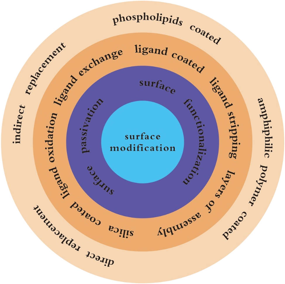
The surface modification methods of UCNPs.
2.3.1 Surface passivation
The surface passivation method involves the coating of a passivation layer (such as a rare earth layer) on the surface of the nanoparticles to protect the exposed doped ions. Such a process effectively prevents the excitation energy from being transferred to the surface of the nanoparticles, thus, improving their luminous efficiency. For instance, the coating of a SiO2 layer on the surface of the UC luminescent nanomaterials, which hydrolyses the precursor of tetraethoxysilane, demonstrates the advantages of high stability, water solubility, and dual compatibility. As a result, this system is widely used for surface modification of the UC fluorescent nanoparticles. Duan et al. [68] obtained meso-porous silica structure by using the CTAB to form micelles during silica shell formation. Then, at certain pH or by refluxing particles, the micelles can be taken away through washing the colloid. The mesoporous silica shell of UCNPs was successfully prepared.
2.3.2 Surface functionalization
The chemical or physical modification of the UCNPs can generate the functional groups with unique chemical or physical properties. For instance, the ligand oxidation method involves the strong oxidants to oxidize the unsaturated bonds on the surface of the nanoparticles into active functional groups (such as carboxyl, aldehyde, etc.), so as to realize the connection and molecular labeling between the nanoparticles and biomolecules. This method has no obvious adverse effects on the morphology, composition, and UCL performance of the UCNPs. However, this method is only suitable for the ligands containing the carbon–carbon unsaturated bonds. As the surface of the nanoparticles is mostly coated with oleic acid or oleylamine ligand molecules, this method can be employed for the surface modification of a majority of the UCNPs. The polymer coating method uses the hydrophobic end of an amphiphilic polymer (such as PEG) and UCNPs so as to coat the surface of the nanoparticles through van der Waals force. The hydrophilic end is compatible with water and forms a hydrophobic/hydrophilic organic core–shell structure [103]. The method can effectively overcome the water solubility issue of the nanoparticles and weakens the fluorescence quenching effect of water on it.
The methods presented in Figure 2 can be divided into two categories: surface passivation and surface functionalization. Surface passivation includes SiO2 coated, while surface functionalization includes ligand oxidation, ligand exchange, amphiphilic ligand coating, ligand stripping, and layer by layer self-assembly. Ligand exchange includes direct and indirect substitution; Ligand coated includes phospholipids coated and amphiphilic polymer package.
3 Application in food safety detection
Food safety is a vital priority related to the human health. With the development of technology, the rapid food safety detection methods are also constantly improving [12]. The unique luminescence mechanism of the UC luminescent nanoparticles makes them the superior biomarker materials. At present, the research on the UC luminescent nanotechnology in the field of food safety is mainly focused on the detection of microorganisms in food [104,105], small molecule organic pollutants [106], heavy metals [107], etc.
3.1 Detection of food-borne pathogens
The food-borne diseases caused by the pathogenic bacteria represent one of the main threats to the food safety. For instance, the investigations reveal that the food-borne diseases are still a public health problem threatening the developing countries like India [108]. In the densely populated provinces of South Africa, the detection rate of the multi-drug resistant Escherichia coli isolated from the fresh vegetables sold by the regular and informal traders is higher than other countries [109]. Therefore, it is of high significance to develop a simple and effective method to identify the food pathogens.
Jin et al. [110] developed a novel detection platform based on the fluorescence resonance energy transfer (FRET) process for the rapid, ultrasensitive, and specific bacteria detection. In this process, the gold nanoparticles (AuNPs, acceptor) were conjugated with aptamers, while the UCNPs (donor) were functionalized with the complementary DNA (cDNA). The spectral overlap between the fluorescence emission of UCNPs and absorption of AuNPs enabled the FRET process to take place, leading to the UC fluorescence quenching. The aptasensor successfully detected E. coli ATCC 8739 (as a model analyte) with the detection range of 5–106 cfu mL−1 and a detection limit of 3 cfu mL−1. It was further used to detect E. coli in the real food and water samples (e.g., tap/pond water, milk, etc.) within 20 min. Comparing to traditional methods, it greatly shortened the detection time and improved the detection efficiency. Overall, the developed sensor demonstrates a high potential in the field of food safety detection and supervision. Due to the specificity of DNA, this method can only detect a single kind of bacteria, thereby limiting its application range.
Wang et al. [111] realized the identification of seven common food-borne pathogens by constructing an UC fluorescent sensor array based on the anti-Stokes luminophores, as shown in Figure 3. The method employed the linear discriminant analysis with an accuracy of 100%. In addition, the developed method accurately identified the bacterial mixture. The bacteria in the real samples (tap water, milk, and beef) could also be effectively identified with an accuracy rate of 92.1%. The sensor overcame the time-consuming bacteria culture and heavy dependence on specific recognition elements. The high efficiency of the whole bacterial cell detection can brighten the application of fluorescence sensor array.
![Figure 3
Based on UCNPs@COPs bacterial pattern recognition of material fluorescent sensor array; (a) the principle of fluorescence emission intensity enhancement of UCNPs materials and (b) schematic diagram of sensor array and the obtained fluorescence response pattern are used to identify and classify bacteria by linear discriminant method [111].](/document/doi/10.1515/ntrev-2022-0086/asset/graphic/j_ntrev-2022-0086_fig_003.jpg)
Based on UCNPs@COPs bacterial pattern recognition of material fluorescent sensor array; (a) the principle of fluorescence emission intensity enhancement of UCNPs materials and (b) schematic diagram of sensor array and the obtained fluorescence response pattern are used to identify and classify bacteria by linear discriminant method [111].
3.2 Biotoxin detection
3.2.1 Bacterial toxin detection
Almost all bacteria synthesize toxins to maintain survival by regulating the different cellular processes as well as cause disease by interacting with the host cell. Generally, the “bacterial toxin” refers to the virulence factors that harm the host [112]. In many countries, the water-borne diarrheal diseases such as the travelers’ diarrhea and cholera still threaten the public health. A rapid diagnosis of the infectious diseases is essential to prevent the disease outbreaks from escalating into epidemics [113].
Wang et al. [114] employed the UC luminescent nanoprobe technology to detect Staphylococcal enterotoxin. Specifically, an ultra-sensitive detection method for Staphylococcal enterotoxin B (SEB) was established based on the nucleic acid aptamer recognition-upconversion fluorescent nanoprobe technology. It combined the resonance energy transfer and enzyme-catalyzed signal amplification technologies. SEB revealed a linear relationship in the concentration range of 0.001–1 ng mL−1, and the detection limit was determined to be up to 0.3 pg mL−1. The detection method demonstrated a positive correlation with the traditional ELISA method and was proven to be a fast and ultra-sensitive method.
3.2.2 Detection of mycotoxins
Mycotoxins are the toxic metabolites of fungi that contaminate the food and feed. These toxins pose acute and chronic health threats to both humans and animals [115]. For instance, Fusarium head blight is a common fungal disease, which not only affects the human health, but also damages the food crops worldwide. It poses a major threat to the food security and world grain trade [116,117]. Therefore, the biosensors are impressive for the detection of mycotoxins through UCNPs [114].
Wang et al. [118] constructed a sensitive aptamer sensor based on the single particle detection (SPD). The analysis of aflatoxin B1 (AFB1) in the peanut samples was carried out using the luminescence resonance energy transfer (LRET) between the aptamer-modified UC (UCNPs-aptamer) and AuNPs, with UCNPs-aptamers acting as the luminescent donors and AuNPs employed as the energy receptors. The linear dynamic detection range of AFB1 was determined to be 3.13–125.00 ng mL−1. The limit of detection was 0.17 ng mL−1, which was much lower than the allowable concentration in the food products. Therefore, the developed method exhibited a broad application potential for the sensitive detection of AFB1 in the food, feed products and promising an important contribution to the food safety and public health.
Kim et al. [119] developed a solid-phase, single-step aptasensor, which revealed a higher performance than the solution-phase aptasensors, along with high reusability (Figure 4d). Specifically, the aptasensor was based on UCNPs (Au/UCNPs) supported by the gold nanocaps partially embedded in a solid substrate polydimethylsiloxane (PDMS). The gold nanocaps allowed the UCNPs to emit the upconverted light only from the restricted area of UCNPs and the area not covered by the nanocaps and PDMS. The functionalization of the restricted region with an aptamer labeled (with a quencher) enabled the effective quenching of the upconverted light from Au/UCNPs via FRET after target (ochratoxin A, OTA) detection. The sensor demonstrated a linear range of 0.1–1,000 ng mL−1, under the optimal condition with 0.022 ng mL−1 detection limit, and a detection time of 30 min. Thus, the developed sensor exhibited a high potential for the development of portable and reusable biosensor platforms, which can be applied to field supervision and detection.
![Figure 4
Schematic diagram of upconversion nanotechnology for detection of agricultural and veterinary drug residues, mycotoxins, and antibiotics: (a) UCNPs@MIP, the preparation and imprinting process of the product are illustrated [123]; (b) schematic diagram of detection principle of upconversion test paper for thiram [124]; (c) schematic diagram of norfloxacin detection principle [129]; (d) based on FRET-induced quenching, the schematic diagram of OTA detection using aptamer (labeled with quenchant) functionalized bare UCNPs on glass substrate [119].](/document/doi/10.1515/ntrev-2022-0086/asset/graphic/j_ntrev-2022-0086_fig_004.jpg)
Schematic diagram of upconversion nanotechnology for detection of agricultural and veterinary drug residues, mycotoxins, and antibiotics: (a) UCNPs@MIP, the preparation and imprinting process of the product are illustrated [123]; (b) schematic diagram of detection principle of upconversion test paper for thiram [124]; (c) schematic diagram of norfloxacin detection principle [129]; (d) based on FRET-induced quenching, the schematic diagram of OTA detection using aptamer (labeled with quenchant) functionalized bare UCNPs on glass substrate [119].
Wu et al. [120] proposed a fumonisin B1 (FB1) sensor based on FRET, process between the NaYF4:Yb,Ho up-conversion fluorescent nanoparticles and AuNPs. AuNPs were connected to the 5’ flanking region of the molecular beacon (MB), whereas UCNPs were connected to the 3,end of MB. Due to the binding of FB1 and FB1 specific aptamers, the separation of the complementary DNA from the magnetic nanoparticles (MNPs) took place. In the presence of the cDNA, MB spontaneously underwent a conformational change, leading to the separation of UCNPs and AuNPs as well as the restoration of the UC fluorescence. Hence, the fluorescence of UCNPs was recovered in a FB1 concentration dependent manner, which formed the basis of the FB1 quantification. The linear range was 0.01–100 ng mL−1, whereas the detection limit in water buffer was determined to be 0.01 ng mL−1. To demonstrate the practical application, the sensor was used to monitor the FB1 level in the natural contaminated corn samples.
Yang et al. [121] developed a smart phone portable device based on the barcode technology of the multi-color UCNPs and fluorescence image processing. The authors used the multicolor upconversion nanoparticle-encoded microspheres (UCNMs) as the coding signals to simultaneously detect the different types of mycotoxins. After using UCNMs for an indirect competitive immune analysis, the images could be captured by the cameras and smartphones. Subsequently, a self-written Android application representing an image recognition program based on HSV (hue, saturation, and value) was installed on the smartphone, which could reliably and accurately analyze the image in less than 1 min. The detection limit was determined to be 1 ng, which was lower than the standard detection system. The developed sensor has the advantages of high sensitivity, low detection limit, wide application, and short detection time.
3.3 Detection of pesticide and veterinary drug residues
The edible agricultural products are the foundation for human’s survival. Thus, activities such as planting, forestry, and aquaculture are closely related to the daily diet. In this respect, the agricultural product safety issues have a significant impact on the human health. With the economic development and gradual improvement in the living standards, stringent requirements with respect to the safety of the agricultural products have also been put forward. The pesticide and veterinary drug residues in the food products pose a serious threat to the human health. Thus, there is an urgent need for a fast, ultra-sensitive, accurate, and cost-effective detection technology [122].
Yu et al. [123] described the fabrication of an imprinted fluorescent nanoprobe, which is based on the SiO2-coated NaYF4:Yb,Er UCNPs encapsulating with a molecularly imprinted polymer (MIP) for the determination of acetamiprid (Figure 4a). The preparation of the fluorescent MIP nanoprobe was achieved by using UCNP as the material for the fluorescent signal readout, acetamiprid as the template molecule, methacrylic acid as the functional monomer, and ethyleneglycol dimethacrylate as the cross-linking agent. Under optimal conditions, this method exhibited a linear relationship between the reduction in the fluorescence intensity (excitation/emission peak at 980/542 nm) and change in acetamiprid concentration in 20–800 ng mL−1 range. The detection limit was determined to be 8.3 ng mL−1. The sensor has the advantages of wide linear range and low detection line. It should be noted, however, that the number of detectable samples is comparatively small.
Hua et al. [106] prepared the water-soluble green β-NaYF4:Yb,Er UCNPs via a facile wet-chemical technique, followed by conjugation with rabbit anti-2,4-dichlorophenoxyacetic acid immunoglobulin G (2,4-D-IgG) and rabbit anti-fenitrothion IgG (fenitrothion-IgG), respectively. The content of 2,4-D-IgG and fenitrothion-IgG in the labeled samples were quantitatively analyzed by the Coomassie Brilliant Blue method. The 2,4-D-IgG (or fenitrothion-IgG) labeled with β-NaYF4:Yb, Er UCNPs conjugates could be used as the fluorescent bio-labels to detect 2,4-D and fenitrothion under 980 nm infrared excitation. Subsequently, the immunochromatographic strip was successfully constructed based on the antibody UCNPs complex. The detection limits for 2,4-D and fenitrothion were determined to be 5 and 11 ng mL−1, respectively. Taken together, the results suggest that the sensor could detect 2,4-D and fenitrothion with both high sensitivity and selectivity.
Mei et al. [124], developed an auxiliary reusable device by using the 3D printing technology. The device assembled a 980 nm micro-laser, a filter, and a micro-cavity in sequence. It was used to digitally image the changes in the luminescence on the test paper to quantitatively analyze thiram (pesticide) through a smart phone (Figure 4b). Specifically, the copper ion modified NaYF4:Yb/Tm UCNPs were immobilized on the filter paper to form a test sheet. After thiram was added through the mechanism of the luminescence resonance energy transfer, the blue luminescence was quenched. The smartphone camera could monitor the changes and calculate by the self-edited Android program installed on the smartphone so as to quantify the amount of thiram, thereby providing reliable and accurate detection limit of 0.1 μM. The study provides a preliminary demonstration for combining the UC nano-sensors with the smart-phone digital imaging for the real-time analysis on a paper-based platform. Compared to the first two sensors, this sensor was portable and more suitable for the onsite rapid detection.
3.4 Antibiotic detection
Antibiotics as the basic therapeutic drugs are widely used in medicine, animal husbandry, aquaculture, etc. However, the residual antibiotics owing to their excessive use also pose a serious threat to the ecological environment, food safety, and human health. The matrix environment of the antibiotic residues is complex. Therefore, the development of cost-effective, rapid, sensitive, efficient, and stable antibiotic detection methods is vitally needed. In addition to regulating the use of antibiotics, it is of great significance to ensure the safety of food and human health through an effective monitoring of the antibiotic residues [125–128].
Ren et al. [129] used the 808 nm near-infrared laser and thermal imaging applications on the smartphone to develop a fast and ultra-sensitive colorimetric/photothermal dual readout detection method (Figure 4c). Norfloxacin was used as a model pollutant to verify the universal rapid detection method. The sample was added to the test strip, and flowed through the strip by the capillary action until it reached the control line, where the immune complexes were formed due to the presence of the secondary antibodies. The added black phosphorus could be captured by the antigens directly exposed to the test line, and the brown band was observable by the naked eye. After 1 min of exposure to the near-infrared light, the thermal imaging analysis was performed by using the phone camera to obtain the real-time temperature for the quantitative analysis. The developed method could detect norfloxacin in the water samples within 20 min, and the detection limit of the colorimetric and photothermal readout reached up to 45 pg mL−1. Compared with the traditional strips, the developed method enhanced the sensitivity by about two orders of magnitude.
Hu et al. [130] developed the NaYF4:Yb/Tm UCNPs bound to the antibodies as the fluorescent signal probes. The authors used the antigen-modified monodisperse magnetic polystyrene microspheres as the immunosensing probes to capture and separate the signal probes (Figure 5a). Overall, a novel fluorescent immunoassay method was established for the detection of sulfaquinoxaline (SQ) in the animal food-derived foods. Based on the competitive immunoassay, the detection limit of the method was 0.1 μg L−1 in buffer and 0.5 μg kg−1 in the food samples. The recovery rate of the added sample was determined to be 69.80–133.00%, with the coefficient of variation in the range of 0.24–25.06%. The developed method demonstrated the advantages of sensitive fluorescence response, simple and rapid extraction process, and environmental friendliness, etc., and could be applied for the determination of sulfamonomethoxine in the animal-derived food.
![Figure 5
Schematic diagram of upconversion nanotechnology applied in the detection of antibiotics and hormones; (a) schematic diagram of detection principle of Sulfaquinoxaline (SQ) [130]; (b) schematic diagram of avidin biotin complex immunosorbent assay for detection of three antibiotics based on controllable assembly of UCNPs [131]; (c) schematic diagram of MIP-coated UCNPs preparation procedure [135].](/document/doi/10.1515/ntrev-2022-0086/asset/graphic/j_ntrev-2022-0086_fig_005.jpg)
Schematic diagram of upconversion nanotechnology applied in the detection of antibiotics and hormones; (a) schematic diagram of detection principle of Sulfaquinoxaline (SQ) [130]; (b) schematic diagram of avidin biotin complex immunosorbent assay for detection of three antibiotics based on controllable assembly of UCNPs [131]; (c) schematic diagram of MIP-coated UCNPs preparation procedure [135].
Wang et al. [131] developed a high-throughput and a high-sensitivity avidin–biotin complex immunoadsorbent assay based on the controlled assembly of the UCNPs (ABC-ULISA). For the detection of antibiotics, the developed system could carry out an accurate quantitative detection within a short period of time (Figure 5b). Streptavidin and biotin labeled UCNPs formed an avidin–biotin-upconversion complex, which was subsequently combined with the biotinylated antibody to achieve a double amplification of the signal and an improved detection sensitivity. ABC-ULISA could detect three antibiotics at the same time. The detection limit of sulfamethazine was 0.15 ng mL−1, whereas the detection limits of sarafloxacin and tetracycline were 0.03 ng mL−1 and 0.05 ng mL−1, respectively. The detection limit of the developed method was much lower than that of the traditional ELISA and ordinary ELISA methods. Further, ABC-ULISA could detect the corresponding target by changing different antibodies, thereby exhibiting its versatility. The obtained findings are observed to be stable and reliable, with the possibility of equipment miniaturization, thus, exhibiting significant commercial application potential. In conclusion, the sensors of antibiotic detection can move towards high sensitivity, short detection time, portable instrument, and high-throughput detection techniques.
3.5 Hormone testing
Illegal addition of low-dose multi-component hormone compounds has become a cunning strategy to evade health supervision; however, it poses hidden dangers to the health and food safety. The temptation of economic benefits leads to the improper use or abuse of the hormonal drugs. Therefore, there is an urgent need for an accurate, effective, and rapid detection technology to overhaul the current protocols [132].
Li et al. [133] developed an ultra-sensitive sensor by employing the FRET between the modified UCNPs and tetramethylrhodamine to detect the trace amounts of bisphenol A (BPA). In order to overcome the challenges of the low efficiency of FRET and low sensitivity of the sensor, the authors quantitatively modified the fluorescence efficiency of the UCNPs. Under optimal detection conditions, the peak intensity at 547 nm had a linear relationship with the logarithm of the BPA concentration, and the detection limit was determined to be 0.05 ng mL−1. Without any pretreatment, the recovery rate of tap, river, and disposable paper cup water are generally between 91.0 and 115.0%. Therefore, the developed sensor is suitable for the effective detection of the trace amounts of BPA in the water samples. Wang et al. [134] successfully developed a fluorescence sensor based on the hybrid chain reaction (HCR) and triple-stranded structure (TS). In the sensor, the nucleic acid aptamer was competitively bound to BPA and cDNA. The unbound cDNA was subsequently obtained by magnetic separation. HCR could be triggered by cDNA to form the long double-stranded DNA products. The UCNPs-modified single-stranded DNA (ssDNA) could form TS with the HCR products. As a result, the distance was shortened, thus, extinguishing the fluorescence and altering the signal. HCR and low background UCNPs could collectively improve the sensitivity of detection. The detection limit of the sensor was as low as 0.057 ng mL−1 and also successfully applied for the actual sample detection. This sensor, relative to the first, improved the sensitivity, specificity, and expanded the range of application.
Wang et al. [135] also developed a novel fluorescent sensor based on the MIP-coated UCNPs to detect diethylstilbestrol (DES). The authors used a simple one-step method to modify the surface of the UCNPs (Figure 5c). Afterwards, the MIP was synthesized on the carbon nanotubes by using the surface grafting molecularly imprinted method. It combined the advantages of UCNPs, including strong fluorescence, stability, reusability, superior adsorption capacity, and high specificity. After removing the template molecules, the nanoparticles could selectively recognize DES, and the fluorescence intensity decreased as the DES concentration was increased. A linear relationship was obtained in the concentration range 50–1,000 ng mL−1, with the correlation coefficient of 0.9989. Further, the detection limit was 12.8 ng mL−1 (S/N = 3). Overall, the developed fluorescence sensor provides a convenient, simple, and highly specific on-site detection of DES, thus, exhibiting a broad application prospect.
3.6 Detection of prohibited additives
The common connection between the food color and quality makes the color one of the most important physical properties of the food. Thus, the color is highly valued by the food industry. Adding colorants to enhance, homogenize, or even change the food color to make it more attractive to the consumers has become a common practice. However, occasionally the prohibited additives are added to the food products in the pursuit of profit. The prohibited additives are harmful to the body as compared to the genuine food additives [136].
Wu et al. [137] developed a melamine nanosensor based on the FRET of UCNPs and AuNPs. UCNPs with a positive charge were used as donors, while AuNPs with a negative charge were used as acceptors. Both UCNPs and AuNPs were bound together through electrostatic interaction, thus, leading to the fluorescence quenching of UCNPs. After adding melamine, owing to the nitrogen–gold interaction between melamine and AuNPs, lead to their aggregation. It resulted in the gradual recovery of the fluorescence of the UCNPs. Under optimal conditions, the concentration of melamine at which the fluorescence enhancement efficiency exhibited a linear response ranged from 32.0 to 500 nM, whereas the detection limit was 18.0 nM. Compared with other fluorescent methods, the developed fluorescence nanosensor demonstrated a sensitivity of up to 0.968, along with simple operation. To demonstrate the actual application, the sensor was used for the determination of melamine in the raw milk. In future, this sensor can be moved toward the high-throughput and commercialization.
3.7 Detection of heavy metal
In recent decades, the heavy metal ions pollution has adversely affected the environment, food safety, and human health. The heavy metals are leached from the industrial wastewater to the water sources and can subsequently enter the food chain of animals. With ever-growing industrialization and urbanization, the presence of heavy metals poses a serious threat to the human health. Owing to this, their detection has attracted a widespread attention [138].
Zhang et al. [139] developed a label free detection scheme based on fluorescence, which employed the use of the photon UCNPs to report the presence of Hg2+. A ssDNA served as the Hg2+ capture element and was covalently connected to the photon upconverting NaYF4:Yb3+,Tm3+ nanoparticles. Under 980 nm laser irradiation, the energy transfer occurred between the NaYF4:Yb3+,Tm3+ nanoparticles as the donor and SYBR Green I as the acceptor. By monitoring the ratio of the acceptor emission to the donor emission, the presence of the mercury ions could be quantitatively detected. The UCNPs biosensor is highly selective for the Hg2+, even in a mixed state with several other metal ions. Correspondingly, the directly observed detection limit was 0.06 nM. The extremely high signal-to-noise ratio of the photon UC particles revealed remarkably high sensitivity and specificity without the need of the fluorescent labeling.
Sun et al. [140] used UCNPs as the energy donor, and single-walled carbon nanotubes or graphene oxide (GO) as the energy acceptor, along with utilizing DNA to connect UCNPs and receptors. In the developed system, the emission could be restored through the G-tetramer-Pb2+ structure, which could be used to determine the Pb2+ content (Figure 6a).
![Figure 6
Schematic diagram of upconversion nanotechnology applied in detection of heavy metals and other pollutants; (a) schematic diagram of Pb2+ detection [140]; (b) schematic diagram of LRET system and HClO detection [144]; (c) dual amplification strategy of upconversion fluorescence aptamer based on HCR and endonuclease for detection of PCB 72/106 [145].](/document/doi/10.1515/ntrev-2022-0086/asset/graphic/j_ntrev-2022-0086_fig_006.jpg)
Schematic diagram of upconversion nanotechnology applied in detection of heavy metals and other pollutants; (a) schematic diagram of Pb2+ detection [140]; (b) schematic diagram of LRET system and HClO detection [144]; (c) dual amplification strategy of upconversion fluorescence aptamer based on HCR and endonuclease for detection of PCB 72/106 [145].
To further refine the UCNPs nanoprobes-based sensor, Yan et al. [141] synthesized a dual colorimetric and fluorometric nanoprobe for the detection of Cu2+ ions. Ligand-free UCNPs and porphyrin hydrate (tetraphenylporphyrin tetrasulfonic acid hydrate, TPPS), based on strong synergism, cause a reduced fluorescence from the UCNPs and green color fluorescence from the TPPS due to the FRET. When the concentration of Cu2+ became higher, the color of UCNPs/TPPS-Cu2+ complex displayed pink color attributed to the inhibition of FRET process. By using a color scanning mobile APP, real-time qualitative and quantitative Cu2+ detection can be achieved. The lowest detection line of the colorimetric and fluorescence methods was 0.13 and 0.32 μM, respectively. The method demonstrated the bright future of onsite rapid detection and supervision.
3.8 Detection of other pollutants
Food is the main substance that all mankind depends on for survival, and its safety is self-evident. Food safety is related to everyone’s health. People are becoming more and more aware of the close connection between food safety and health. Food safety is not only related to the source of human energy but also a feasible way to prevent future human diseases. The ultimate goal of food analysis is to ensure food safety, for this reason, researchers around the world are committed to developing modern and advanced detection and analysis technologies to solve this problem [142,143]. In addition to the earlier-mentioned contaminants, the other miscellaneous contaminants in food include chemicals such as H2S, hypochlorous acid (HClO), and polychlorinated biphenyls (PCBs). The following is a brief introduction of the application of the UC nanotechnology for the detection of these substances.
Zhang et al. [144] prepared a nanoprobe based on the UCNPs for the specific sensing and imaging of HClO(Figure 6b). The Ru@UCNP nanoprobe consisted of two functional components, namely NaYF4:Yb,Tm UCNPs, which could convert the near-infrared light into visible light as an energy donor and HClO-responsive ruthenium(ii) complex [Ru (bpy) 2 (DNCH- bpy)] (PF6) 2 (Ru-DNPH) as an energy acceptor and an UCL quencher. In this system, the on-off emission of UCL was specifically triggered by HClO. One of the advantages of this sensor was high specificity and sensitivity. Thus, the trigger reaction could be used to detect HClO in the aqueous solutions.
Wang et al. [145] developed an UC fluorescent sensor based on hybridization chain reaction and etching endonuclease for the detection of PCBs. It combined the dual advantages of UCNPs and HCR (Figure 6c). First, two harpins (H1 and H2) were designed based on the partial complementary sequence (cDNA) of PCB 72/106. As the aptamer specifically recognized the targets, cDNA was separated from the magnetic microspheres. Second, the gene could trigger a hybrid chain reaction (HCR), then, opening the stems of H1 and H2. After adding endonuclease, UCNPs were pushed away from the quenching enzyme. The fluorescence intensity of the UCNPs could be restored by FRET process, the UC emission intensity rose with the increase in of the PCBs’ concentration. Under optimal detection conditions, PCB 72/106 could be analyzed in the range from 0.004 to 800 ng mL−1, and the detection limit was 0.0035 ng mL−1 (S/N = 3). The developed sensor was also used for the detection of the water and soil samples, with average recovery rates of 93.4–109.7% and 83.2–118.5%, respectively. The findings from this study also provide an opportunity to use the dual amplification strategy to develop the fluorescent aptamer sensor for other targets.
4 Conclusion and future prospects
This study reviews the luminescence mechanism, synthesis methods, and surface modification of the rare earth-doped UCNPs, along with exploring their applications in the field of food safety (Table 2). From the dietary perspectives, the application of the UC nanotechnology has been summarized for the rapid detection of food safety (Figure 7). It can be seen that the UC nanotechnology can effectively provide the safety detection of our daily diet, owing to its wide application spectrum.
Application of UCNPs in food safety detection
| Analyst | Upconversion nanomaterials | detection limit/accuracy | Strategy | Excitation wavelength | Ref. |
|---|---|---|---|---|---|
| Food-borne pathogens | UCNPs@COPs | 92.1% | Electrostatic action | 980 nm | [111] |
| Escherichia coli ATCC 8739 | UCNPs-cDNA | 3 cfu/mL | FRET | 980 nm | [110] |
| Bacterial toxin | |||||
| Staphylococcal enterotoxin | UCNPs probe | 0.3 pg/mL | FRET | 980 nm | [113] |
| Mycotoxin | |||||
| AFB 1 | UCNPs-aptamer | 0.17 ng/mL | FRET | 980 nm | [117] |
| OTA | Au/UCNPs | 0.022 ng/mL | FRET | 980 nm | [118] |
| FB1 | UCNPs (NaYF4:Yb,HO) | 0.01 ng/mL | FRET | 980 nm | [119] |
| Residues of agricultural and veterinary drugs | |||||
| Acetamiprid | UCNPs probe | 8.3 ng/mL | Photo induced electron transfer | 980 nm | [122] |
| 2,4-D | UCNPs-IgG (β-NaYF4:Yb,Er) | 5 ng/mL | Antigen antibody reaction | 980 nm | [123] |
| Fenitrothion | 11 ng/mL | ||||
| Thiram | UCNPs-Cu (NaYF4:Yb,Tm) | 0.1 μM | FRET | 980 nm | [106] |
| Antibiotic | |||||
| SQ | UCNPs probe (NaYF4:Yb,Tm) | 0.5 μg/kg | Competitive immunoassay | 980 nm | [127] |
| Norfloxacin | Magnetic upconversion nanocomposites | 45 pg/mL | Antigen antibody reaction | 808 nm | [158] |
| Sulfamethazine | Avidin–biotin-UCNPs | 0.15 ng/mL | Antigen antibody reaction | 980 nm | [129] |
| Sarafloxacin | 0.03 ng/mL | ||||
| Tetracycline | 0.05 ng/mL | ||||
| Hormone substances | |||||
| Bisphenol A | UCNPs-ssDNA | 0.057 ng/mL | FRET | 980 nm | [133] |
| DES | UCNPs-MIP | 12.8 ng/mL | Molecular imprinting | 980 nm | [134] |
| Prohibited additives | |||||
| Melamine | Positively charged UCNPs | 18.0 nm | FRET | 980 nm | [136] |
| Heavy metal | |||||
| Hg2+ | UCNPs (NaYF4:Yb3+,Tm3+) | 0.06 nm | FRET | 980 nm | [138] |
| Other pollutants | |||||
| PCBs | UCNPs-cDNA | 0.0035 ng/mL | FRET | 980 nm | [144] |

Application of UCNPs in food safety detection based on diet pagoda; the first layer is cereal and potato food: UCNPs can be used to detect mycotoxins. The second layer is fruits and vegetables: UCNPs can be used to detect pesticide residues. The third layer is fish, poultry, meat, eggs, and other animal foods: UCNPs can be used to detect food-borne pathogens, antibiotics, veterinary drug residues, and heavy metals. The fourth layer is milk, soybeans and nuts: UCNPs can be used to detect food-borne pathogens, bacterial toxins, mycotoxins, antibiotics, hormones, and prohibited additives. The fifth layer is cooking oil and salt: UCNPs can be used to detect mycotoxins.
In the recent years, a significant progress has been made in synthesizing UCNPs with controllable shape, size, morphology, and phase by employing various methods. Specifically, the UC luminescent nanoprobe technology has achieved a remarkable success in the fields of medicine, biology, and life sciences, along with food safety detection. For instance, the detection of pesticide residues, antibiotics, biological toxins, and food additives represents a unique application prospect. The potential of UCNPs in the field of rapid detection of food safety is full of infinite possibilities. First, it is expected to achieve rapid detection for field and laboratory detection, thereby increasing efficiency and effectiveness of the routine food safety supervision and inspection, Meanwhile, there is a certain usefulness for improving corporate social responsibility, firm reputation, and firm performance. Second, the technologies promise to the accomplishment of rapid qualitative screening inspection, semi-quantitative, and quantitative assessment. Third, it can avoid the complex process of the sample pretreatment, and achieve advanced and rapid methods of the sample pretreatment. Fourth, hopefully, the lack of rapid detection technology for the disposal of food safety accidents can be made up. After understanding the reaction mechanism, further combinations can be made to expand the application range of the UC technology.
However, some restrictions are yet to be resolved. First, the immunochromatographic test strips, which is based on antibodies, represent the main research direction. However, the related findings are mostly limited to the laboratory stage, with only a few mature commercial products. It is necessary to develop portable on-site screening technologies and equipment that can be applied for rapid food safety testing, to promote a healthy and rapid development of the Food Industry. Second, whether the complex structure of UCNPs can be degraded in the environment is still an environmental safety issue. Therefore, it is necessary to study the long-term degradability of the urban sewage. Third, the biggest challenge in the surface modification of the UC fluorescent nanomaterials is the aggregation and fluorescence quenching of the modified nanomaterials. Therefore, in the future, efforts are required to decrease the fluorescence quenching degree through appropriate chemical modification, thereby expanding the application of the UC materials in food safety detection. Fourth, the UCNPs are related to the bio-safety. One way to overcome this challenge is to use ultra-small UCNPs (<10 nm) as they can be easily excreted or degraded by the body. However, a smaller size will also lead to the enhanced surface quenching effects, thus, designing new methods and new synthetic approaches for the fine dispersity, small and strong fluorescence of UCNPs also need to be studied. Overall, combining the UC technology, enzyme-linked immunoassay, and colloidal gold technology to achieve rapid and sensitive detection is of great significance in food safety and hygiene, medical detection, and other aspects.
-
Funding information: The authors thank the National Key Research and Development Program of China (Grant No. 2018YFC1603500).
-
Author contributions: All authors have accepted responsibility for the entire content of this manuscript and approved its submission.
-
Conflict of interest: The authors state no conflict of interest.
References
[1] Lam HM, Remais J, Fung MC, Xu L, Sun SM. Food supply and food safety issues in China. Lancet. 2013;381(9882):2044–53.10.1016/S0140-6736(13)60776-XSearch in Google Scholar
[2] Bingxue H, Hongbin P, Da-Wen S. Multifunctional cellulose based substrates for SERS smart sensing: Principles, applications and emerging trends for food safety detection. Trends Food Sci Technol. 2021;110:304–20.10.1016/j.tifs.2021.02.005Search in Google Scholar
[3] Oplatowska-Stachowiak M, Elliott CT. Food colors: Existing and emerging food safety concerns. Crit Rev Food Sci Nutr. 2017;57(3):524–48.10.1080/10408398.2014.889652Search in Google Scholar PubMed
[4] Chen Q, Zhang C, Zhao J, Ouyang Q. Recent advances in emerging imaging techniques for non-destructive detection of food quality and safety. Trends Anal Chem. 2013;52:261–74.10.1016/j.trac.2013.09.007Search in Google Scholar
[5] Akhtar S. Food safety challenges – A Pakistan’s perspective. Crit Rev Food Sci Nutr. 2015;55(2):219–26.10.1080/10408398.2011.650801Search in Google Scholar PubMed
[6] Gao F, Gao C, He S, Wang Q, Wu A. Label-free electrochemical lead (II) aptasensor using thionine as the signaling molecule and graphene as signal-enhancing platform. Biosens Bioelectron. 2016;81:15–22.10.1016/j.bios.2016.01.096Search in Google Scholar PubMed
[7] Zhu Y, Zeng G, Zhang Y, Tang L, Chen J, Cheng M, et al. Highly sensitive electrochemical sensor using a MWCNTs/GNPs-modified electrode for lead (II) detection based on Pb2+-induced G-rich DNA conformation. Analyst. 2014;139(19):5014–20.10.1039/C4AN00874JSearch in Google Scholar PubMed
[8] Conzuelo F, Grützke S, Stratmann L, Pingarrón JM, Schuhmann W. Interrogation of immunoassay platforms by SERS and SECM after enzyme-catalyzed deposition of silver nanoparticles. Mikrochim Acta An Int J Phys Chem Methods Anal. 2016;183(1):281–7.10.1007/s00604-015-1654-xSearch in Google Scholar
[9] Song E, Yu M, Wang Y, Hu W, Yang S. Multi-color quantum DoT-based fluorescence immunoassay array for simultaneous visual detection of multiple antibiotic residues in milk. Biosens Bioelectron. 2015;72:320–5.10.1016/j.bios.2015.05.018Search in Google Scholar PubMed
[10] Tzschoppe M, Martin A, Beutin L. A rapid procedure for the detection and isolation of enterohaemorrhagic Escherichia coli (EHEC) serogroup O26, O103, O111, O118, O121, O145 and O157 strains and the aggregative EHEC O104:H4 strain from ready-to-eat vegetables. Int J Food Microbiol. 2012;152(1–2):19–30.10.1016/j.ijfoodmicro.2011.10.009Search in Google Scholar PubMed
[11] Asselt E, Fels-Klerx HVD, Breuer O, Helsloot I. Food safety crisis management – a comparison between Germany and the Netherlands. J Food Sci. 2017;82(1–3):477–83.10.1111/1750-3841.13585Search in Google Scholar PubMed
[12] Zheng H, Ting Y, Donghong L, Yunlei X. Recent advances in gold nanoparticles-based biosensors for food safety detection. Biosens Bioelectron. 2021;179:113076.10.1016/j.bios.2021.113076Search in Google Scholar PubMed
[13] Ji YL, Yoon J, Hovde CJ. A brief overview of Escherichia coli O157:H7 and its plasmid O157. J Microbiol Biotechnol. 2010;20(1):5–14.10.4014/jmb.0908.08007Search in Google Scholar
[14] Xu M, Wang R, Li Y. Electrochemical biosensors for rapid detection of Escherichia coli O157:H7. Talanta. 2017;162:511–22.10.1016/j.talanta.2016.10.050Search in Google Scholar PubMed
[15] Wang PL, Xie LH, Joseph EA, Li JR, Su XO, Zhou HC. Metal-organic frameworks for food safety. Chem Rev. 2019;119(18):10638–90.10.1021/acs.chemrev.9b00257Search in Google Scholar PubMed
[16] Granato D, Barba FJ, Kovacevic DB, Lorenzo JM, Cruz AG, Putnik P. Functional foods: product development, technological trends, efficacy testing, and safety. Annu Rev Food Sci Technol. 2020;11:93–118.10.1146/annurev-food-032519-051708Search in Google Scholar PubMed
[17] Wang Z, Wu X, Liu L, Xu L, Xu C. Rapid and sensitive detection of diclazuril in chicken samples using a gold nanoparticle-based lateral-flow strip. Food Chem. 2019;312:126116.10.1016/j.foodchem.2019.126116Search in Google Scholar PubMed
[18] Elhag M, Abubaker M, Ahmad NM, Haroon EM, Hassan MA. Immunoinformatics prediction of epitope based peptide vaccine against listeria monocytogenes fructose bisphosphate aldolase protein; 2019.10.1101/649111Search in Google Scholar
[19] Sandvang D, Skjoet-Rasmussen L, Cantor M, Mathis GF, Blanch A. Effect of feed supplementation with three different probiotic Bacillus strains and their combination on the performance of broiler chickens challenged with Clostridium perfringens. Poult Sci. 2021;100(4):100982.10.1016/j.psj.2021.01.005Search in Google Scholar PubMed PubMed Central
[20] Ramirez-Hernandez A, Galagarza OA, Álvarez Rodriguez MV, Pachari Vera E, Valdez Ortiz MDC, Deering AJ, et al. Food safety in Peru: a review of fresh produce production and challenges in the public health system. Compr Rev Food Sci Food Saf. 2020;19(6):3323–42.10.1111/1541-4337.12647Search in Google Scholar PubMed
[21] Xavier M, Parente IA, Rodrigues PM, Cerqueira MA, Pastrana L, Goncalves C. Safety and fate of nanomaterials in food: the role of in vitro tests. Trends Food Sci Technol. 2021;109:593–607.10.1016/j.tifs.2021.01.050Search in Google Scholar
[22] Derz W, Pavlovic M, Huber I, Schalch B, Gerdes L. Food fraud in the Alps? – detection of chamois (Rupicapra rupicapra) in firm raw sausages, ham, and meat via qualitative duplex real-time PCR. Food Control. 2021;123:10.10.1016/j.foodcont.2020.107764Search in Google Scholar
[23] Du S, Chen IH, Maclachlan A, Liu Y, Chin BA. 3D phage-based biomolecular filter for effective high throughput capture of salmonella typhimurium in liquid streams. Food Res Int. 2021;142:110181.10.1016/j.foodres.2021.110181Search in Google Scholar PubMed
[24] Huang X, Guo Q, Zhang R, Zhao Z, Leng Y, Lam JWY, et al. AIEgens: an emerging fluorescent sensing tool to aid food safety and quality control. Compr Rev Food Sci Food Saf. 2020;19(4):2297–329.10.1111/1541-4337.12591Search in Google Scholar PubMed
[25] Lan G, Qian L, Nan H, Wang K. Recent developments of photoelectrochemical biosensors for food analysis. J Mater Chem B. 2019;7(46):7283–300.10.1039/C9TB01644ASearch in Google Scholar
[26] Demir R, Ahar U, Deveci R. Determination of terminal glycan and total monosaccharide profiles of reelin glycoprotein in SH-SY5Y neuroblastoma cell line by lectin blotting and capillary liquid chromatography electrospray ionization-ion trap tandem mass spectrometry system – ScienceDi. Biochim et Biophys Acta (BBA) – Proteins Proteom. 2020;1869(2):140559.10.1016/j.bbapap.2020.140559Search in Google Scholar PubMed
[27] Katsoulos PD, Athanasiou LV, Dedousi A, Polizopoulou ZS. Serum calcium determination with atomic absorption spectrophotometry and colorimetric method: association and predictive equations between them in cattle and sheep samples. Comp Clin Pathol. 2020;30(1):1–5.10.1007/s00580-020-03183-zSearch in Google Scholar
[28] Zeki J, Kriman M. Development of gas-chromatographic method for simultaneous determination of cannabinoids and terpenes in hemp. Molecules. 2020;25(24):5872.10.3390/molecules25245872Search in Google Scholar PubMed PubMed Central
[29] Auzel F. Upconversion and anti-Stokes processes with f and d ions in solids. Cheminform. 2004;35(16):139–73.10.1002/chin.200416212Search in Google Scholar
[30] Jayakumar M, Idris NM, Zhang Y. Remote activation of biomolecules in deep tissues using near-infrared-to-UV upconversion nanotransducers. Proc Natl Acad Sci U S A. 2012;109(22):8483–8.10.1073/pnas.1114551109Search in Google Scholar PubMed PubMed Central
[31] He H, Cui Y, Li B, Wang B, Jin C, Yu J, et al. Multiphoton excitation: confinement of perovskite-QDs within a single MOF crystal for significantly enhanced multiphoton excited luminescence (Adv. Mater. 6/2019). Adv Mater. 2019;31(6):e1806897.10.1002/adma.201970036Search in Google Scholar
[32] Zheng X, Kankala RK, Liu C-G, Wang S-B, Chen A-Z, Zhang Y. Lanthanides-doped near-infrared active upconversion nanocrystals: Upconversion mechanisms and synthesis. Coord Chem Rev. 2021;438:213870.10.1016/j.ccr.2021.213870Search in Google Scholar
[33] Anh TK, Thai DV, Ha VTT, Strek W, Minh L. Upconversion luminescence of Er3+ doped Y2O3 and Gd2O3 nanophosphors. Int J Nanotechnol. 2020;17(7–10):689–707.10.1504/IJNT.2020.111334Search in Google Scholar
[34] Hu S, Wu Q, Wu X, Li J, Cao H, Zhan S, et al. Simultaneous luminescence and magnetic control of NaLuF4: Yb3+/Er3+ by introducing NaMnF3 and the application for detecting basic fuchsin. J Alloy Compounds An Interdiscip J Mater Sci Solid-State Chem Phys. 2018;745:490–6.10.1016/j.jallcom.2018.02.181Search in Google Scholar
[35] Kraft M, Würth C, Muhr V, Hirsch T, Resch-Genger U. Particle-size-dependent upconversion luminescence of NaYF4:Yb, Er nanoparticles in organic solvents and water at different excitation power densities. Nano Res. 2018;11(12):6360–74.10.1007/s12274-018-2159-9Search in Google Scholar
[36] Zheng Y, Deng L, Li J, Jia T, Qiu J, Sun Z, et al. Controlling multiphoton excited energy transfer from Tm3+ to Yb3+ ions by a phase-shaped femtosecond laser field. Photonics Res. 2019;7(4):486.10.1364/PRJ.7.000486Search in Google Scholar
[37] Bo Z, Shi B, Jin D, Liu X. Controlling upconversion nanocrystals for emerging applications. Nat Nanotechnol. 2015;10(11):924–36.10.1038/nnano.2015.251Search in Google Scholar PubMed
[38] Qin X, Xu J, Wu Y, Liu X. Energy-transfer editing in lanthanide-activated upconversion nanocrystals: a toolbox for emerging applications. ACS Cent Sci. 2019;5(1):29–42.10.1021/acscentsci.8b00827Search in Google Scholar PubMed PubMed Central
[39] He F, Feng L, Yang P, Liu B, Gai S, Yang G, et al. Enhanced up/downconversion luminescence and heat: simultaneously achieving in one single core–shell structure for multimodal imaging guided therapy. Biomaterials. 2016;105:77–88.10.1016/j.biomaterials.2016.07.031Search in Google Scholar PubMed
[40] Di Z, Liu B, Zhao J, Gu Z, Zhao Y, Li L. An orthogonally regulatable DNA nanodevice for spatiotemporally controlled biorecognition and tumor treatment. Sci Adv. 2020;6(25):eaba9381.10.1126/sciadv.aba9381Search in Google Scholar PubMed PubMed Central
[41] Chhetri BP, Karmakar A, Ghosh A. Recent advancements in ln-ion-based upconverting nanomaterials and their biological applications. Part Part Syst Charact. 2019;36(8):1900153.10.1002/ppsc.201900153Search in Google Scholar
[42] Hong E, Liu L, Bai L, Xia C, Gao L, Zhang L, et al. Control synthesis, subtle surface modification of rare-earth-doped upconversion nanoparticles and their applications in cancer diagnosis and treatment. Mater Sci Eng. 2019;105:110097.1–110097.22.10.1016/j.msec.2019.110097Search in Google Scholar PubMed
[43] Hu J, Zhan S, Wu X, Hu S, Wu S, Liu Y. Core/shell upconversion nanoparticles with intense fluorescence for detecting doxorubicin in vivo. RSC Adv. 2018;8(38):21505–12.10.1039/C8RA02928HSearch in Google Scholar PubMed PubMed Central
[44] Z Q, Li Y, Zhang S, et al. Multicolor core/shell-structured upconversion fluorescent nanoparticles. Adv Mater. 2009;31(36):365705.Search in Google Scholar
[45] Liu Y, Wang D, Shi J, Peng Q, Li Y. Magnetic tuning of upconversion luminescence in lanthanide-doped bifunctional nanocrystals. Angew Chem Int Ed. 2013;125(16):4462–5.10.1002/ange.201209884Search in Google Scholar
[46] He M, Shang N, Zhu Q, Xu J. Paper-based upconversion fluorescence aptasensor for the quantitative detection of immunoglobulin E in human serum. Anal Chim Acta. 2021;1143:93–100.10.1016/j.aca.2020.11.036Search in Google Scholar PubMed
[47] Huang L, Chen F, Zong X, Lu Q, Zhang Y. Near-infrared light excited UCNP-DNAzyme nanosensor for selective detection of Pb2+ and in vivo imaging. Talanta. 2021;227:122156.10.1016/j.talanta.2021.122156Search in Google Scholar PubMed
[48] Igor R, Alexander S. Self-pulsing of a monolithic Tm-doped YAlO3 microlaser. Phys Rev A. 2006;73(5):053815.10.1103/PhysRevA.73.053815Search in Google Scholar
[49] Shi C, Soltani S, Armani AM. Gold nanorod plasmonic upconversion microlaser. Nano Lett. 2013;13(12):5827–31.10.1109/NANO.2014.6967959Search in Google Scholar
[50] Guo Q, Wu J, Yang Y, Liu X, Jia J, Dong J, et al. High performance perovskite solar cells based on β-NaYF4:Yb3+/Er3+/Sc3+ @NaYF4 core–shell upconversion nanoparticles. J Power Sources. 2019;426:178–87.10.1016/j.jpowsour.2019.04.039Search in Google Scholar
[51] Qin R, Jinhua X, Hu J, Zhao L. Effect of luminescent material NaYbF4:Ho on the photovoltaic performance of dye-sensitized solar cells. J Mater Sci Mater Electron. 2021;32(2):1–12.10.1007/s10854-020-04915-1Search in Google Scholar
[52] Guo Y, Zou R, Si F, Liang W, Zhao J. A sensitive immunoassay based on fluorescence resonance energy transfer from up-converting nanoparticles and graphene oxide for one-step detection of imidacloprid. Food Chem. 2020;335:127609.10.1016/j.foodchem.2020.127609Search in Google Scholar
[53] Niazi S, Wang X, Pasha I, Khan IM, Zhao S, Shoaib M, et al. A novel bioassay based on aptamer-functionalized magnetic nanoparticle for the detection of zearalenone using time resolved-fluorescence NaYF4:Ce/Tb nanoparticles as signal probe. Talanta. 2018;186:97–103.10.1016/j.talanta.2018.04.013Search in Google Scholar
[54] Poláchová V, Pastucha M, Mikušová Z, Mickert MJ, Hlaváček A, Gorris HH, et al. Click-conjugated photon-upconversion nanoparticles in an immunoassay for honeybee pathogen Melissococcus plutonius. Nanoscale. 2019;11(17):8343–51.10.1039/C9NR01246JSearch in Google Scholar
[55] Wu S, Liu L, Duan N, Wang W, Yu Q, Wang Z. A test strip for ochratoxin A based on the use of aptamer-modified fluorescence upconversion nanoparticles. Microchim Acta. 2018;185(11):497.10.1007/s00604-018-3022-0Search in Google Scholar
[56] Renero-Lecuna C, Martín-RodríGuez R, Valiente R, GonzáLez J, Rodríguez F, KräMer KW, et al. Origin of the high upconversion green luminescence efficiency in β-NaYF4:2%Er3+,20%Yb3+. Chem Mater. 2011;23(15):3442–8.10.1021/cm2004227Search in Google Scholar
[57] Ansari AA, Thakur VK, Chen G. Functionalized upconversion nanoparticles: New strategy towards FRET-based luminescence bio-sensing. Coord Chem Rev. 2021;436:213821.10.1016/j.ccr.2021.213821Search in Google Scholar
[58] Leiner MJP. Luminescence chemical sensors for biomedical applications: scope and limitations. Analytica Chim Acta. 1991;255(2):209–22.10.1016/0003-2670(91)80049-YSearch in Google Scholar
[59] Liang G, Wang H, Shi H, Wang H, Li G. Recent progress in the development of upconversion nanomaterials in bioimaging and disease treatment. J Nanobiotechnology. 2020;18(1):154.10.1186/s12951-020-00713-3Search in Google Scholar PubMed PubMed Central
[60] Nguyen TN, Ebrahim FM, Stylianou KC. Photoluminescent, upconversion luminescent and nonlinear optical metal-organic frameworks: From fundamental photophysics to potential applications. Coord Chem Rev. 2018;377:259–306.10.1016/j.ccr.2018.08.024Search in Google Scholar
[61] Tian R, Zhao S, Liu G, Chen H, Ma L, You H, et al. Construction of lanthanide-doped upconversion nanoparticle-Uelx Europaeus Agglutinin-I bioconjugates with brightness red emission for ultrasensitive in vivo imaging of colorectal tumor. Biomaterials. 2019;212:64–72.10.1016/j.biomaterials.2019.05.010Search in Google Scholar PubMed
[62] Gu B, Zhou Y, Zhang X, Liu X, Zhang Y, Marks R, et al. Thiazole derivative-modified upconversion nanoparticles for Hg2+ detection in living cells. Nanoscale. 2015;8(1):276–82.10.1039/C5NR05286FSearch in Google Scholar PubMed
[63] Chen D, Wan Z, Liu S. Highly sensitive dual-phase nanoglass-ceramics self-calibrated optical thermometer. Anal Chem. 2016;88(7):4099–106.10.1021/acs.analchem.6b00434Search in Google Scholar PubMed
[64] Luo L, Su JP, Hun-Kuk P. Citric-assisted sol–gel based Er3+/Yb3+-codoped Na0.5Gd0.5MoO4: a novel highly-efficient infrared-to-visible upconversion material for optical temperature sensors and optical heaters. Chem Eng J. 2016;306:840–8.10.1016/j.cej.2016.08.007Search in Google Scholar
[65] Wiesholler LM, Frenzel F, Grauel B, Würth C, Hirsch T. Yb,Nd,Er-doped upconversion nanoparticles: 980nm versus 808 nm excitation. Nanoscale. 2019;11(28):13440–9.10.1039/C9NR03127HSearch in Google Scholar PubMed
[66] Dong H, Sun LD, Yan CH. Basic understanding of the lanthanide related upconversion emissions. Nanoscale. 2013;5(13):5703–14.10.1039/c3nr34069dSearch in Google Scholar PubMed
[67] Chen J, Zhao JX. Upconversion nanomaterials: synthesis, mechanism, and applications in sensing. Sensors. 2012;12(3):2414–35.10.3390/s120302414Search in Google Scholar PubMed PubMed Central
[68] Duan C, Liang L, Li L, Zhang R, Xu ZP. Recent progress in upconversion luminescence nanomaterials for biomedical applications. J Mater Chem B. 2018;6(2):192–209.10.1039/C7TB02527KSearch in Google Scholar PubMed
[69] Xin N, Wei D, Zhu Y, Yang M, Fan H. Upconversion nanomaterials: a platform for biosensing, theranostic and photoregulation. Mater Today Chem. 2020;17:100329.10.1016/j.mtchem.2020.100329Search in Google Scholar
[70] Chaudhuri RG, Paria S. Core/shell nanoparticles: classes, properties, synthesis mechanisms, characterization, and applications. Chem Rev. 2012;112(4):2373–433.10.1021/cr100449nSearch in Google Scholar PubMed
[71] Gnach A, Bednarkiewicz A. Lanthanide-doped up-converting nanoparticles: Merits and challenges. Nano Today. 2012;7(6):532–63.10.1016/j.nantod.2012.10.006Search in Google Scholar
[72] Fan Z, Ying W, Yu T, Zhang F, Shi Y, Xie S, et al. Uniform nanostructured arrays of sodium rare-earth fluorides for highly efficient multicolor upconversion luminescence. Vol. 46(42). John Wiley & Sons, Ltd; 2007. p. 7976–9.10.1002/anie.200702519Search in Google Scholar PubMed
[73] Lou XWD, Archer LA, Yang Z. Hollow micro-nanostructures: synthesis and applications. Adv Mater. 2008;20(21):3987–4019.10.1002/adma.200800854Search in Google Scholar
[74] Na H, Woo K, Lim K, Jang HS. Rational morphology control of β-NaYF4:Yb,Er/Tm upconversion nanophosphors using a ligand, an additive, and lanthanide doping. Nanoscale. 2013;5(10):4242–51.10.1039/c3nr00080jSearch in Google Scholar PubMed
[75] Wu YA, Li CX, Yang DM, Lin J. Rare earth beta-NaGdF4 fluorides with multiform morphologies: hydrothermal synthesis and luminescent properties. J Colloid Interface Sci. 2010;354(2):429–36.10.1016/j.jcis.2010.11.040Search in Google Scholar PubMed
[76] Xu Z, Ma P, Li C, Hou Z, Zhai X, Huang S, et al. Monodisperse core–shell structured up-conversion Yb(OH)CO@YbPO:Er+ hollow spheres as drug carriers. Biomaterials. 2011;32(17):4161–73.10.1016/j.biomaterials.2011.02.026Search in Google Scholar PubMed
[77] Mei X, Yan Q, Lu P, Wang J, Cui Y, Nie Y, et al. Hydrothermal crystallization of a Ln2(OH)4SO4·nH2O layered compound for a wide range of Ln (Ln = La–Dy), thermolysis, and facile transformation into oxysulfate and oxysulfide phosphors. Rsc Adv. 2017;7(22):13331–9.10.1039/C7RA00645DSearch in Google Scholar
[78] Zhang J, Wang Y, Guo L, Dong P. Up-conversion luminescence and near-infrared quantum cutting in Y6O5F8:RE3+ (RE = Yb, Er, and Ho) with controllable morphologies by hydrothermal synthesis. Dalton Trans. 2013;42(10):3542–51.10.1039/c2dt32463fSearch in Google Scholar PubMed
[79] Xu D, Xie F, Yao L, Li Y, Lin H, Li A, et al. Enhancing upconversion luminescence of highly doped lanthanide nanoparticles through phase transition delay. J Alloy Compd. 2020;815:152622.10.1016/j.jallcom.2019.152622Search in Google Scholar
[80] Wang X, Zhuang J, Peng Q, Li Y. A general strategy for nanocrystal synthesis. Nature. 2005;437(7055):121–4.10.1038/nature03968Search in Google Scholar PubMed
[81] Marciniak L, Stefanski M, Tomala R, Hreniak D, Strek W. Synthesis and up-conversion luminescence of Er3+ and Yb3+ codoped nanocrystalline tetra-(KLaP4O12) and pentaphosphates (LaP5O14). J Chem Phys. 2015;143(9):094701.10.1063/1.4929582Search in Google Scholar PubMed
[82] Chen H, Wang W, Ji C, Wang L. Dye-sensitized core–shell NaGdF(4):Yb,Er@NaGdF(4):Yb,Nd upconversion nanoprobe for determination of H(2)S. Spectrochim Acta A Mol Biomol Spectrosc. 2021;248:119281.10.1016/j.saa.2020.119281Search in Google Scholar PubMed
[83] Wei Y, Lu F, Zhang X, Chen D. Synthesis and characterization of efficient near-infrared upconversion Yb and Tm codoped NaYF4 nanocrystal reporter. J Alloy Compd. 2007;427:333–40.10.1016/j.jallcom.2006.03.014Search in Google Scholar
[84] Hyppnen I, Hls J, Kankare J, Lastusaari M, Pihlgren L. Preparation and characterization of nanocrystalline ZrO2:Yb3+,Er3+ up-conversion phosphors. Ann N Y Acad. 2010;1130(1):267–71.10.1196/annals.1430.045Search in Google Scholar PubMed
[85] Liao J, Wang Q, Kong L, Ming Z, Wang Y, Li Y, et al. Effect of Yb3+ concentration on tunable upconversion luminescence and optically temperature sensing behavior in Gd2TiO5:Yb3+/Er3+ phosphors. Optical Mater. 2018;75:841–9.10.1016/j.optmat.2017.12.009Search in Google Scholar
[86] Mai HX, Zhang YW, Si R, Yan ZG, Sun LD, You LP, et al. High-quality sodium rare-earth fluoride nanocrystals: controlled synthesis and optical properties. ChemInform. 2006;128(19):6426–36.10.1021/ja060212hSearch in Google Scholar PubMed
[87] Wang F, Xie Z, Zhang B, Liu Y, Yang W, Liu CY. Down- and up-conversion luminescent carbon dot fluid: inkjet printing and gel glass fabrication. Nanoscale. 2014;6(7):3818–23.10.1039/c3nr05869gSearch in Google Scholar PubMed
[88] Yan X, Alcouffe P, Sudre G, David L, Bernard J, Ganachaud F. Modular construction of single-component polymer nanocapsules through a one-step surfactant-free microemulsion templated synthesis. Chem Commun. 2017;53(8):1401–4.10.1039/C6CC09701DSearch in Google Scholar PubMed
[89] Bensalah A, Mortier M, Patriarche G, Gredin P, Vivien D. Synthesis and optical characterizations of undoped and rare-earth-doped CaF2 nanoparticles. J Solid State Chem. 2006;179(8):2636–44.10.1016/j.jssc.2006.05.011Search in Google Scholar
[90] Kaur G, Dwivedi Y, Rai A, Rai SB. Green and NIR sensitized luminescent nanophosphor: preparation, characterization and application. Spectrochim Acta A Mol Biomol Spectrosc. 2012;95:511–6.10.1016/j.saa.2012.04.040Search in Google Scholar PubMed
[91] Zhang YW, Sun X, Si R, You LP, Yan CH. Single-crystalline and monodisperse LaF3 triangular nanoplates from a single-source precursor. J Am Chem Soc. 2005;127(10):3260–1.10.1021/ja042801ySearch in Google Scholar PubMed
[92] Han J, Zhang C, Liu F, Liu B, Han M, Zou W, et al. Upconversion nanoparticles for ratiometric fluorescence detection of nitrite. Analyst. 2014;139(12):3032–8.10.1039/C4AN00402GSearch in Google Scholar PubMed
[93] Wang C, Cheng L, Liu Y, Wang X, Ma X, Deng Z, et al. Imaging-guided pH-sensitive photodynamic therapy using charge reversible upconversion nanoparticles under near-infrared light. Adv Funct Mater. 2013;23(24):3077–86.10.1002/adfm.201202992Search in Google Scholar
[94] Dong A, Ye X, Chen J, Kang Y, Gordon T, Kikkawa JM, et al. A generalized ligand-exchange strategy enabling sequential surface functionalization of colloidal nanocrystals. J Am Chem Soc. 2011;133(4):998–1006.10.1021/ja108948zSearch in Google Scholar PubMed
[95] Vetrone F, Naccache R, Mahalingam V, Morgan CG, Capobianco JA. The active-core/active-shell approach: a strategy to enhance the upconversion luminescence in lanthanide-doped nanoparticles. Adv Funct Mater. 2009;19(18):2924–9.10.1002/adfm.200900234Search in Google Scholar
[96] Liu Q, Li C, Yang T, Yi T, Li F. “Drawing” upconversion nanophosphors into water through host–guest interaction. Chem Commun. 2010;46(30):5551–3.10.1039/c0cc01352hSearch in Google Scholar PubMed
[97] Mai HX, Zhang YW, Sun LD, Yan CH. Highly efficient multicolor up-conversion emissions and their mechanisms of monodisperse NaYF4:Yb,Er core and core/shell-structured nanocrystals. J Phys Chem C. 2007;111(37):13721–9.10.1021/jp073920dSearch in Google Scholar
[98] Mai HX, Zhang YW, Sun LD, Yan CH. Size- and phase-controlled synthesis of monodisperse NaYF4:Yb,Er nanocrystals from a unique delayed nucleation pathway monitored with upconversion spectroscopy. J Phys Chem C. 2007;111(37):13730–9.10.1021/jp073919eSearch in Google Scholar
[99] Rafik N, Fiorenzo V, Venkataramanan M, et al. Controlled synthesis and water dispersibility of hexagonal phase NaGdF4:Ho3+/Yb3+ nanoparticles. Chem Mater. 2009;21(4):717–23.10.1021/cm803151ySearch in Google Scholar
[100] Schfer H, Ptacek P, Zerzouf O, Haase M. Synthesis and optical properties of KYF4/Yb, Er nanocrystals, and their surface modification with undoped KYF4. Adv Funct Mater. 2010;18(19):2913–8.10.1002/adfm.200800368Search in Google Scholar
[101] Zhang T, Ge J, Hu Y, Yin Y. A general approach for transferring hydrophobic nanocrystals into water. Nano Lett. 2007;7(10):3203–7.10.1021/nl071928tSearch in Google Scholar PubMed
[102] Zhou J, Shirahata N, Sun HT, Ghosh B, Ogawara M, Teng Y, et al. Efficient dual-modal NIR-to-NIR emission of rare earth ions co-doped nanocrystals for biological fluorescence imaging. J Phys Chem Lett. 2013;4(3):402–8.10.1021/jz302122aSearch in Google Scholar PubMed
[103] Chen Z, Chen H, He H, Yu M, Li F. Versatile synthesis strategy for carboxylic acid-functionalized upconverting nanophosphors as biological labels. J Am Chem Soc. 2008;130(10):3023–9.10.1021/ja076151kSearch in Google Scholar PubMed
[104] Cheng K, Zhang J, Zhang L, Wang L, Chen H. Aptamer biosensor for Salmonella typhimurium detection based on luminescence energy transfer from Mn2+-doped NaYF4:Yb, Tm upconverting nanoparticles to gold nanorods. Spectrochim Acta Part A Mol Biomol Spectrosc. 2017;171:168–73.10.1016/j.saa.2016.08.012Search in Google Scholar PubMed
[105] Abdul Hakeem D, Su S, Mo Z, Wen H. Upconversion luminescent nanomaterials: A promising new platform for food safety analysis. Crit Rev Food Sci Nutr. 2021;1–42.10.1080/10408398.2021.1937039Search in Google Scholar PubMed
[106] Yu J, Guo T, Zhang W, Li B, Liu L, Hua R. Green upconversion nanoparticles for 2,4-dichlorophenoxyacetic acid and fenitrothion detection. J Alloy Compd. 2019;771:187–94.10.1016/j.jallcom.2018.08.202Search in Google Scholar
[107] Liu Y, Ouyang Q, Li H, Chen M, Zhang Z, Chen Q. Turn-on fluoresence sensor for Hg2+ in food based on FRET between aptamers-functionalized upconversion nanoparticles and gold nanoparticles. J Agric Food Chem. 2018;66(24):6188–95.10.1021/acs.jafc.8b00546Search in Google Scholar PubMed
[108] Chaturvedi K, Bisht A, Kamble MP, Choudhary P, Taneja NK. A surveillance of food borne disease outbreaks in India: 2009–2018. Food Control. 2020;121:107630.10.1016/j.foodcont.2020.107630Search in Google Scholar
[109] Loandi R, Du PE, Stacey D, Lise K. High prevalence of multidrug resistant Escherichia coli isolated from fresh vegetables sold by selected formal and informal traders in the most densely populated Province of South Africa. J Food Sci. 2020;86(1):161–8.10.1111/1750-3841.15534Search in Google Scholar
[110] Jin B, Wang S, Lin M, Jin Y, Zhang S, Cui X, et al. Upconversion nanoparticles based FRET aptasensor for rapid and ultrasenstive bacteria detection. Biosens Bioelectron. 2017;90:525–33.10.1016/j.bios.2016.10.029Search in Google Scholar PubMed
[111] Yin M, Jing C, Li H, Deng Q, Wang S. Surface chemistry modified upconversion nanoparticles as fluorescent sensor array for discrimination of foodborne pathogenic bacteria. J Nanobiotechnol. 2020;18(1):41.10.1186/s12951-020-00596-4Search in Google Scholar PubMed PubMed Central
[112] Srivastava A, Pati S, Kaushik H, Singh S, Garg LC. Toxin-antitoxin systems and their medical applications: current status and future perspective. Appl Microbiol Biotechnol. 2021;105(5):1803–21.10.1007/s00253-021-11134-zSearch in Google Scholar PubMed
[113] Eduardo AE, S MC, Ziqiu T, H VN, Markus M. A regenerable biosensing platform for bacterial toxins. Biomacromolecules. 2020;22(2):441–53.10.1021/acs.biomac.0c01318Search in Google Scholar
[114] Wu S, Duan N, Ma X, Xia Y, Wang H, Wang Z, et al. Multiplexed fluorescence resonance energy transfer aptasensor between upconversion nanoparticles and graphene oxide for the simultaneous determination of mycotoxins. Anal Chem. 2012;84(14):6263–70.10.1021/ac301534wSearch in Google Scholar PubMed
[115] Godfrey W, Sofie L, Juliet A, Kris A, Richard E, Geert H. Mycotoxin profile of staple grains in northern Uganda: understanding the level of human exposure and potential risks. Food Control. 2021;122:107813.10.1016/j.foodcont.2020.107813Search in Google Scholar
[116] Maroua H, Abdelmoneim M, Noureddine R. Biosensor based on antifouling PEG/Gold nanoparticles composite for sensitive detection of aflatoxin M1 in milk. Microchem J. 2021;165:107630.10.1016/j.microc.2021.106102Search in Google Scholar
[117] Palacios SA, Del Canto A, Erazo J, Torres AM. Fusarium cerealis causing Fusarium head blight of durum wheat and its associated mycotoxins. Int J Food Microbiol. 2021;346:109161.10.1016/j.ijfoodmicro.2021.109161Search in Google Scholar PubMed
[118] Wang F, Han Y, Wang S, Ye Z, Wei L, Xiao L. Single-particle LRET aptasensor for the sensitive detection of Aflatoxin B1 with upconversion nanoparticles. Anal Chem. 2019;91(18):11856–63.10.1021/acs.analchem.9b02599Search in Google Scholar PubMed
[119] Kim K, Jo EJ, Lee KJ, Park J, Kim MG. Gold nanocap-supported upconversion nanoparticles for fabrication of a solid-phase aptasensor to detect ochratoxin A. Biosens Bioelectron. 2019;150:111885.10.1016/j.bios.2019.111885Search in Google Scholar PubMed
[120] Wu S, Duan N, Li X, Tan G, Wang H. Homogenous detection of fumonisin B with a molecular beacon based on fluorescence resonance energy transfer between NaYF:Yb,Ho upconversion nanoparticles and gold nanoparticles. Talanta. 2013;116:611–8.10.1016/j.talanta.2013.07.016Search in Google Scholar PubMed
[121] Yang M, Zhang Y, Cui M, Tian Y, Zhang S, Peng K, et al. A smartphone-based quantitative detection platform of mycotoxins based on multiple-color upconversion nanoparticles. Nanoscale. 2018;10(33):15865–74.10.1039/C8NR04138ESearch in Google Scholar
[122] Mingtao L, Xiang Z. Nanostructure-based surface-enhanced Raman spectroscopy techniques for pesticide and veterinary drug residues screening. Bull Environ Contamin Toxicol. 2020;107(2):194–205.10.1007/s00128-020-02989-5Search in Google Scholar
[123] Yu Q, He C, Li Q, Zhou Y, Duan N, Wu S. Fluorometric determination of acetamiprid using molecularly imprinted upconversion nanoparticles. Mikrochim Acta. 2020;187(4):222.10.1007/s00604-020-4204-0Search in Google Scholar PubMed
[124] Mei Q, Jing H, Li Y, Yisibashaer W, Chen J, Li BN, et al. Smartphone based visual and quantitative assays on upconversional paper sensor. Biosens Bioelectron. 2016;75:427–32.10.1016/j.bios.2015.08.054Search in Google Scholar PubMed
[125] Jing M, Zhang H, Li M, Mao Z, Shi X. Silver nanoparticle-decorated TiO2 nanotube array for solid-phase microextraction and SERS detection of antibiotic residue in milk. Spectrochim Acta Part A Mol Biomolecular Spectrosc. 2021;255:119652.10.1016/j.saa.2021.119652Search in Google Scholar PubMed
[126] Yang Y, Ren G, Yang W, Qin X, Gu D, Liang Z, et al. A new MOF-based fluorescent sensor for the detection of nitrofuran antibiotics. Polyhedron. 2021;194:114923.10.1016/j.poly.2020.114923Search in Google Scholar
[127] Yue F, Li F, Kong Q, Guo Y, Sun X. Recent advances in aptamer-based sensors for aminoglycoside antibiotics detection and their applications. Sci Total Environ. 2021;762:143129.10.1016/j.scitotenv.2020.143129Search in Google Scholar PubMed
[128] Lan L, Yao Y, Ping J, Ying Y. Recent advances in nanomaterial-based biosensors for antibiotics detection. Biosens Bioelectron. 2017;91:504–14.10.1016/j.bios.2017.01.007Search in Google Scholar PubMed
[129] Ren S, Li Q, Wang J, Fan B, Bai J, Peng Y, Li S, et al. Development of a fast and ultrasensitive black phosphorus-based colorimetric/photothermal dual-readout immunochromatography for determination of norfloxacin in tap water and river water. J Hazard Mater. 2021 Jan 15;402:123781.10.1016/j.jhazmat.2020.123781Search in Google Scholar PubMed
[130] Hu G, Sheng W, Zhang Y, Wang J, Wu X, Wang S. Upconversion nanoparticles and monodispersed magnetic polystyrene microsphere based fluorescence immunoassay for the detection of sulfaquinoxaline in animal-derived foods. J Agric Food Chem. 2016;64(19):3908–15.10.1021/acs.jafc.6b01497Search in Google Scholar PubMed
[131] Wang Y, Zhao X, Zhang M, Sun X, Gao Z. Immunosorbent assay based on upconversion nanoparticles controllable assembly for simultaneous detection of three antibiotics. J Hazard Mater. 2021;406(11):124703.10.1016/j.jhazmat.2020.124703Search in Google Scholar PubMed
[132] Hz A, Zc A, By A, Dan FA, Yuan L, Zwac D. Integrated recombinant gene yeast bioassay and HPLC-MS analysis for detection of low-dose multi-component residue of hormone-like compounds in environment. Sci Total Environ. 2021;773:145569.10.1016/j.scitotenv.2021.145569Search in Google Scholar PubMed
[133] Li Q, Bai J, Ren S, Jiang W, Gao Z. An ultrasensitive sensor based on quantitatively modified upconversion particles for trace bisphenol A detection. Anal Bioanal Chem. 2018;411(1–2):139–46.10.1007/s00216-018-1425-8Search in Google Scholar PubMed
[134] Wang Y, Zhao X, Huo B, Ren S, Gao Z. Sensitive fluorescence aptasensor based on hybridization chain reaction with upconversion nanoparticles by triplex DNA formation for bisphenol A detection. ACS Appl Bio Mater. 2020;4(1):763–9.10.1021/acsabm.0c01347Search in Google Scholar
[135] Wang Y, Ren S, Jiang H, Peng Y, Bai J, Li Q, et al. A label-free detection of diethylstilbestrol based on molecularly imprinted polymer-coated upconversion nanoparticles obtained by surface grafting. RSC Adv. 2017;7(36):22215–21.10.1039/C6RA26999KSearch in Google Scholar
[136] Xie Y, Yan L, Li N, Wang H, He Q, Yao W. A novel surface-enhanced Raman scattering sensor to detect prohibited colorants in food by graphene/silver nanocomposite. Talanta. 2012;100:32–7.10.1016/j.talanta.2012.07.080Search in Google Scholar PubMed
[137] Wu Q, Long Q, Li H, Zhang Y, Yao S. An upconversion fluorescence resonance energy transfer nanosensor for one step detection of melamine in raw milk. Talanta. 2015;136:47–53.10.1016/j.talanta.2015.01.005Search in Google Scholar PubMed
[138] Mukherjee S, Bhattacharyya S, Ghosh K, Pal S, Bhattacharyya N. Sensory development for heavy metal detection: a review on translation from conventional analysis to field-portable sensor. Trends Food Sci Technol. 2021;109:674–89.10.1016/j.tifs.2021.01.062Search in Google Scholar
[139] Kumar M, Zhang P. Highly sensitive and selective label-free optical detection of mercuric ions using photon upconverting nanoparticles. Biosens Bioelectron. 2010 Jul 15;25(11):2431–5.10.1016/j.bios.2010.03.038Search in Google Scholar PubMed PubMed Central
[140] Xu Y, Meng X, Liu J, Song D, Sun LN. Luminescent nanoprobes based on upconversion nanoparticles and single-walled carbon nanohorns or graphene oxide for detection of pb2+ ion. CrystEngComm. 2016;18(22):4032–7.10.1039/C5CE02537KSearch in Google Scholar
[141] Yan Q, Chen Z-H, Xue S-F, Han X-Y, Lin Z-Y, Zhang S, et al. Lanthanide-doped nanoparticles encountering porphyrin hydrate: boosting a dual-mode optical nanokit for Cu2+ sensing. Sens Actuators B Chem. 2018;268:108–14.10.1016/j.snb.2018.04.080Search in Google Scholar
[142] Lv M, Liu Y, Geng J, Kou X, Xin Z, Yang D. Engineering nanomaterials-based biosensors for food safety detection. Biosens Bioelectron. 2018;106:122–8.10.1016/j.bios.2018.01.049Search in Google Scholar PubMed
[143] Xiyi Z, Hongbin P, DaWen S. DNA functionalized metal and metal oxide nanoparticles: principles and recent advances in food safety detection. Crit Rev Food Sci Nutr. 2020;61(14):2277–96.10.1080/10408398.2020.1809343Search in Google Scholar
[144] Zhang R, Liang L, Meng Q, Zhao J, Ta HT, Li L, et al. Responsive upconversion nanoprobe for background-free hypochlorous acid detection and bioimaging. Small. 2019;15(2):e1803712.10.1002/smll.201803712Search in Google Scholar PubMed
[145] Wang Y, Bai J, Huo B, Yuan S, Zhang M, Sun X, et al. Upconversion fluorescent aptasensor for polychlorinated biphenyls detection based on nicking endonuclease and hybridization chain reaction dual-amplification strategy. Anal Chem. 2018;90(16):9936–42.10.1021/acs.analchem.8b02159Search in Google Scholar PubMed
[146] Yao J, Huang C, Liu C, Yang M. Upconversion luminescence nanomaterials: a versatile platform for imaging, sensing, and therapy. Talanta. 2020;208:120157.10.1016/j.talanta.2019.120157Search in Google Scholar PubMed
[147] Feng S, Xu R. New materials in hydrothermal synthesis. Acc Chem Res. 2001;34(3):239–47.10.1021/ar0000105Search in Google Scholar PubMed
[148] Jing L, Hao Z, Xia Z, Luo Y, Zhang J. Hydrothermal synthesis and upconversion luminescence properties of β-NaGdF4:Yb3+/Tm3+ and β-NaGdF4:Yb3+/Ho3+ submicron crystals with regular morphologies. J Colloid Interface Sci. 2012;392:206–12.10.1016/j.jcis.2012.09.076Search in Google Scholar
[149] Li CX, Quan ZW, Yang J, Yang PP, Lin J. Highly uniform and monodisperse beta-NaYF(4):Ln(3+) (Ln = Eu, Tb, Yb/Er, and Yb/Tm) hexagonal microprism crystals: hydrothermal synthesis and luminescent properties. Inorg Chem. 2007;46(16):6329–37.10.1021/ic070335iSearch in Google Scholar PubMed
[150] Zhuang J, Liang L, Sung HH, Yang X, Wu M, Williams ID, et al. Controlled hydrothermal growth and up-conversion emission of NaLnF(4) (Ln = Y, Dy-Yb). Inorg Chem. 2007;46(13):5404–10.10.1021/ic070220eSearch in Google Scholar PubMed
[151] Yue T, Hua R, Chen B, Yu N, Na L. Lanthanide dopant-induced phase transition and luminescent enhancement of EuF3 nanocrystals. Crystengcomm. 2012;14(23):8110–6.10.1039/c2ce26101dSearch in Google Scholar
[152] Han ZY. Uniform lanthanide-doped Y2O3 hollow microspheres: Controlled synthesis and luminescence properties. Mater Sci Eng B. 2011;176:1251–6.10.1016/j.mseb.2011.07.004Search in Google Scholar
[153] Raju GSR, Pavitra E, Yu JS. Facile template free synthesis of Gd2O(CO3)2·H2O chrysanthemum-like nanoflowers and luminescence properties of corresponding Gd2O3:RE3+ spheres. Dalton Trans. 2013;42(32):11400–10.10.1039/c3dt51154eSearch in Google Scholar PubMed
[154] Dixit CK, Bhakta S, Kumar A, Suib SL, Rusling JF. Fast nucleation for silica nanoparticle synthesis using a sol–gel method. Nanoscale. 2016;8(47):19662–7.10.1039/C6NR07568ASearch in Google Scholar PubMed PubMed Central
[155] Mialon G, Türkcan S, Dantelle G, Collins DP, Hadjipanayi M, Taylor RA, et al. High up-conversion efficiency of YVO4:Yb,Er nanoparticles in water down to the single-particle level. J Phys Chem C. 2010;114(51):22449–54.10.1021/jp107900zSearch in Google Scholar
[156] Martin N, Boutinaud P, Mahiou R, Cousseins JC, Bouderbala M. Preparation of fluorides at 80°C in the NaF - (Y, Yb, Pr)F∼3 system. J Mater Chem. 1999.10.1039/a804472dSearch in Google Scholar
[157] Cho JS, Jung KY, Kang YC. Yolk–shell structured Gd2O3:Eu3+ phosphor prepared by spray pyrolysis: the effect of preparation conditions on microstructure and luminescence properties. Phys Chem Chem Phys. 2014;17(2):1325–31.10.1039/C4CP03477ESearch in Google Scholar PubMed
[158] Gao M, Wu R, Mei Q, Zhang C, Ling X, Deng S, et al. Upconversional nanoprobes with highly efficient energy transfer for ultrasensitive detection of alkaline phosphatase. ACS Sens. 2019;4(11):2864–8.10.1021/acssensors.9b00858Search in Google Scholar PubMed
© 2022 Guangna Ji et al., published by De Gruyter
This work is licensed under the Creative Commons Attribution 4.0 International License.
Articles in the same Issue
- Research Articles
- Theoretical and experimental investigation of MWCNT dispersion effect on the elastic modulus of flexible PDMS/MWCNT nanocomposites
- Mechanical, morphological, and fracture-deformation behavior of MWCNTs-reinforced (Al–Cu–Mg–T351) alloy cast nanocomposites fabricated by optimized mechanical milling and powder metallurgy techniques
- Flammability and physical stability of sugar palm crystalline nanocellulose reinforced thermoplastic sugar palm starch/poly(lactic acid) blend bionanocomposites
- Glutathione-loaded non-ionic surfactant niosomes: A new approach to improve oral bioavailability and hepatoprotective efficacy of glutathione
- Relationship between mechano-bactericidal activity and nanoblades density on chemically strengthened glass
- In situ regulation of microstructure and microwave-absorbing properties of FeSiAl through HNO3 oxidation
- Research on a mechanical model of magnetorheological fluid different diameter particles
- Nanomechanical and dynamic mechanical properties of rubber–wood–plastic composites
- Investigative properties of CeO2 doped with niobium: A combined characterization and DFT studies
- Miniaturized peptidomimetics and nano-vesiculation in endothelin types through probable nano-disk formation and structure property relationships of endothelins’ fragments
- N/S co-doped CoSe/C nanocubes as anode materials for Li-ion batteries
- Synergistic effects of halloysite nanotubes with metal and phosphorus additives on the optimal design of eco-friendly sandwich panels with maximum flame resistance and minimum weight
- Octreotide-conjugated silver nanoparticles for active targeting of somatostatin receptors and their application in a nebulized rat model
- Controllable morphology of Bi2S3 nanostructures formed via hydrothermal vulcanization of Bi2O3 thin-film layer and their photoelectrocatalytic performances
- Development of (−)-epigallocatechin-3-gallate-loaded folate receptor-targeted nanoparticles for prostate cancer treatment
- Enhancement of the mechanical properties of HDPE mineral nanocomposites by filler particles modulation of the matrix plastic/elastic behavior
- Effect of plasticizers on the properties of sugar palm nanocellulose/cinnamon essential oil reinforced starch bionanocomposite films
- Optimization of nano coating to reduce the thermal deformation of ball screws
- Preparation of efficient piezoelectric PVDF–HFP/Ni composite films by high electric field poling
- MHD dissipative Casson nanofluid liquid film flow due to an unsteady stretching sheet with radiation influence and slip velocity phenomenon
- Effects of nano-SiO2 modification on rubberised mortar and concrete with recycled coarse aggregates
- Mechanical and microscopic properties of fiber-reinforced coal gangue-based geopolymer concrete
- Effect of morphology and size on the thermodynamic stability of cerium oxide nanoparticles: Experiment and molecular dynamics calculation
- Mechanical performance of a CFRP composite reinforced via gelatin-CNTs: A study on fiber interfacial enhancement and matrix enhancement
- A practical review over surface modification, nanopatterns, emerging materials, drug delivery systems, and their biophysiochemical properties for dental implants: Recent progresses and advances
- HTR: An ultra-high speed algorithm for cage recognition of clathrate hydrates
- Effects of microalloying elements added by in situ synthesis on the microstructure of WCu composites
- A highly sensitive nanobiosensor based on aptamer-conjugated graphene-decorated rhodium nanoparticles for detection of HER2-positive circulating tumor cells
- Progressive collapse performance of shear strengthened RC frames by nano CFRP
- Core–shell heterostructured composites of carbon nanotubes and imine-linked hyperbranched polymers as metal-free Li-ion anodes
- A Galerkin strategy for tri-hybridized mixture in ethylene glycol comprising variable diffusion and thermal conductivity using non-Fourier’s theory
- Simple models for tensile modulus of shape memory polymer nanocomposites at ambient temperature
- Preparation and morphological studies of tin sulfide nanoparticles and use as efficient photocatalysts for the degradation of rhodamine B and phenol
- Polyethyleneimine-impregnated activated carbon nanofiber composited graphene-derived rice husk char for efficient post-combustion CO2 capture
- Electrospun nanofibers of Co3O4 nanocrystals encapsulated in cyclized-polyacrylonitrile for lithium storage
- Pitting corrosion induced on high-strength high carbon steel wire in high alkaline deaerated chloride electrolyte
- Formulation of polymeric nanoparticles loaded sorafenib; evaluation of cytotoxicity, molecular evaluation, and gene expression studies in lung and breast cancer cell lines
- Engineered nanocomposites in asphalt binders
- Influence of loading voltage, domain ratio, and additional load on the actuation of dielectric elastomer
- Thermally induced hex-graphene transitions in 2D carbon crystals
- The surface modification effect on the interfacial properties of glass fiber-reinforced epoxy: A molecular dynamics study
- Molecular dynamics study of deformation mechanism of interfacial microzone of Cu/Al2Cu/Al composites under tension
- Nanocolloid simulators of luminescent solar concentrator photovoltaic windows
- Compressive strength and anti-chloride ion penetration assessment of geopolymer mortar merging PVA fiber and nano-SiO2 using RBF–BP composite neural network
- Effect of 3-mercapto-1-propane sulfonate sulfonic acid and polyvinylpyrrolidone on the growth of cobalt pillar by electrodeposition
- Dynamics of convective slippery constraints on hybrid radiative Sutterby nanofluid flow by Galerkin finite element simulation
- Preparation of vanadium by the magnesiothermic self-propagating reduction and process control
- Microstructure-dependent photoelectrocatalytic activity of heterogeneous ZnO–ZnS nanosheets
- Cytotoxic and pro-inflammatory effects of molybdenum and tungsten disulphide on human bronchial cells
- Improving recycled aggregate concrete by compression casting and nano-silica
- Chemically reactive Maxwell nanoliquid flow by a stretching surface in the frames of Newtonian heating, nonlinear convection and radiative flux: Nanopolymer flow processing simulation
- Nonlinear dynamic and crack behaviors of carbon nanotubes-reinforced composites with various geometries
- Biosynthesis of copper oxide nanoparticles and its therapeutic efficacy against colon cancer
- Synthesis and characterization of smart stimuli-responsive herbal drug-encapsulated nanoniosome particles for efficient treatment of breast cancer
- Homotopic simulation for heat transport phenomenon of the Burgers nanofluids flow over a stretching cylinder with thermal convective and zero mass flux conditions
- Incorporation of copper and strontium ions in TiO2 nanotubes via dopamine to enhance hemocompatibility and cytocompatibility
- Mechanical, thermal, and barrier properties of starch films incorporated with chitosan nanoparticles
- Mechanical properties and microstructure of nano-strengthened recycled aggregate concrete
- Glucose-responsive nanogels efficiently maintain the stability and activity of therapeutic enzymes
- Tunning matrix rheology and mechanical performance of ultra-high performance concrete using cellulose nanofibers
- Flexible MXene/copper/cellulose nanofiber heat spreader films with enhanced thermal conductivity
- Promoted charge separation and specific surface area via interlacing of N-doped titanium dioxide nanotubes on carbon nitride nanosheets for photocatalytic degradation of Rhodamine B
- Elucidating the role of silicon dioxide and titanium dioxide nanoparticles in mitigating the disease of the eggplant caused by Phomopsis vexans, Ralstonia solanacearum, and root-knot nematode Meloidogyne incognita
- An implication of magnetic dipole in Carreau Yasuda liquid influenced by engine oil using ternary hybrid nanomaterial
- Robust synthesis of a composite phase of copper vanadium oxide with enhanced performance for durable aqueous Zn-ion batteries
- Tunning self-assembled phases of bovine serum albumin via hydrothermal process to synthesize novel functional hydrogel for skin protection against UVB
- A comparative experimental study on damping properties of epoxy nanocomposite beams reinforced with carbon nanotubes and graphene nanoplatelets
- Lightweight and hydrophobic Ni/GO/PVA composite aerogels for ultrahigh performance electromagnetic interference shielding
- Research on the auxetic behavior and mechanical properties of periodically rotating graphene nanostructures
- Repairing performances of novel cement mortar modified with graphene oxide and polyacrylate polymer
- Closed-loop recycling and fabrication of hydrophilic CNT films with high performance
- Design of thin-film configuration of SnO2–Ag2O composites for NO2 gas-sensing applications
- Study on stress distribution of SiC/Al composites based on microstructure models with microns and nanoparticles
- PVDF green nanofibers as potential carriers for improving self-healing and mechanical properties of carbon fiber/epoxy prepregs
- Osteogenesis capability of three-dimensionally printed poly(lactic acid)-halloysite nanotube scaffolds containing strontium ranelate
- Silver nanoparticles induce mitochondria-dependent apoptosis and late non-canonical autophagy in HT-29 colon cancer cells
- Preparation and bonding mechanisms of polymer/metal hybrid composite by nano molding technology
- Damage self-sensing and strain monitoring of glass-reinforced epoxy composite impregnated with graphene nanoplatelet and multiwalled carbon nanotubes
- Thermal analysis characterisation of solar-powered ship using Oldroyd hybrid nanofluids in parabolic trough solar collector: An optimal thermal application
- Pyrene-functionalized halloysite nanotubes for simultaneously detecting and separating Hg(ii) in aqueous media: A comprehensive comparison on interparticle and intraparticle excimers
- Fabrication of self-assembly CNT flexible film and its piezoresistive sensing behaviors
- Thermal valuation and entropy inspection of second-grade nanoscale fluid flow over a stretching surface by applying Koo–Kleinstreuer–Li relation
- Mechanical properties and microstructure of nano-SiO2 and basalt-fiber-reinforced recycled aggregate concrete
- Characterization and tribology performance of polyaniline-coated nanodiamond lubricant additives
- Combined impact of Marangoni convection and thermophoretic particle deposition on chemically reactive transport of nanofluid flow over a stretching surface
- Spark plasma extrusion of binder free hydroxyapatite powder
- An investigation on thermo-mechanical performance of graphene-oxide-reinforced shape memory polymer
- Effect of nanoadditives on the novel leather fiber/recycled poly(ethylene-vinyl-acetate) polymer composites for multifunctional applications: Fabrication, characterizations, and multiobjective optimization using central composite design
- Design selection for a hemispherical dimple core sandwich panel using hybrid multi-criteria decision-making methods
- Improving tensile strength and impact toughness of plasticized poly(lactic acid) biocomposites by incorporating nanofibrillated cellulose
- Green synthesis of spinel copper ferrite (CuFe2O4) nanoparticles and their toxicity
- The effect of TaC and NbC hybrid and mono-nanoparticles on AA2024 nanocomposites: Microstructure, strengthening, and artificial aging
- Excited-state geometry relaxation of pyrene-modified cellulose nanocrystals under UV-light excitation for detecting Fe3+
- Effect of CNTs and MEA on the creep of face-slab concrete at an early age
- Effect of deformation conditions on compression phase transformation of AZ31
- Application of MXene as a new generation of highly conductive coating materials for electromembrane-surrounded solid-phase microextraction
- A comparative study of the elasto-plastic properties for ceramic nanocomposites filled by graphene or graphene oxide nanoplates
- Encapsulation strategies for improving the biological behavior of CdS@ZIF-8 nanocomposites
- Biosynthesis of ZnO NPs from pumpkin seeds’ extract and elucidation of its anticancer potential against breast cancer
- Preliminary trials of the gold nanoparticles conjugated chrysin: An assessment of anti-oxidant, anti-microbial, and in vitro cytotoxic activities of a nanoformulated flavonoid
- Effect of micron-scale pores increased by nano-SiO2 sol modification on the strength of cement mortar
- Fractional simulations for thermal flow of hybrid nanofluid with aluminum oxide and titanium oxide nanoparticles with water and blood base fluids
- The effect of graphene nano-powder on the viscosity of water: An experimental study and artificial neural network modeling
- Development of a novel heat- and shear-resistant nano-silica gelling agent
- Characterization, biocompatibility and in vivo of nominal MnO2-containing wollastonite glass-ceramic
- Entropy production simulation of second-grade magnetic nanomaterials flowing across an expanding surface with viscidness dissipative flux
- Enhancement in structural, morphological, and optical properties of copper oxide for optoelectronic device applications
- Aptamer-functionalized chitosan-coated gold nanoparticle complex as a suitable targeted drug carrier for improved breast cancer treatment
- Performance and overall evaluation of nano-alumina-modified asphalt mixture
- Analysis of pure nanofluid (GO/engine oil) and hybrid nanofluid (GO–Fe3O4/engine oil): Novel thermal and magnetic features
- Synthesis of Ag@AgCl modified anatase/rutile/brookite mixed phase TiO2 and their photocatalytic property
- Mechanisms and influential variables on the abrasion resistance hydraulic concrete
- Synergistic reinforcement mechanism of basalt fiber/cellulose nanocrystals/polypropylene composites
- Achieving excellent oxidation resistance and mechanical properties of TiB2–B4C/carbon aerogel composites by quick-gelation and mechanical mixing
- Microwave-assisted sol–gel template-free synthesis and characterization of silica nanoparticles obtained from South African coal fly ash
- Pulsed laser-assisted synthesis of nano nickel(ii) oxide-anchored graphitic carbon nitride: Characterizations and their potential antibacterial/anti-biofilm applications
- Effects of nano-ZrSi2 on thermal stability of phenolic resin and thermal reusability of quartz–phenolic composites
- Benzaldehyde derivatives on tin electroplating as corrosion resistance for fabricating copper circuit
- Mechanical and heat transfer properties of 4D-printed shape memory graphene oxide/epoxy acrylate composites
- Coupling the vanadium-induced amorphous/crystalline NiFe2O4 with phosphide heterojunction toward active oxygen evolution reaction catalysts
- Graphene-oxide-reinforced cement composites mechanical and microstructural characteristics at elevated temperatures
- Gray correlation analysis of factors influencing compressive strength and durability of nano-SiO2 and PVA fiber reinforced geopolymer mortar
- Preparation of layered gradient Cu–Cr–Ti alloy with excellent mechanical properties, thermal stability, and electrical conductivity
- Recovery of Cr from chrome-containing leather wastes to develop aluminum-based composite material along with Al2O3 ceramic particles: An ingenious approach
- Mechanisms of the improved stiffness of flexible polymers under impact loading
- Anticancer potential of gold nanoparticles (AuNPs) using a battery of in vitro tests
- Review Articles
- Proposed approaches for coronaviruses elimination from wastewater: Membrane techniques and nanotechnology solutions
- Application of Pickering emulsion in oil drilling and production
- The contribution of microfluidics to the fight against tuberculosis
- Graphene-based biosensors for disease theranostics: Development, applications, and recent advancements
- Synthesis and encapsulation of iron oxide nanorods for application in magnetic hyperthermia and photothermal therapy
- Contemporary nano-architectured drugs and leads for ανβ3 integrin-based chemotherapy: Rationale and retrospect
- State-of-the-art review of fabrication, application, and mechanical properties of functionally graded porous nanocomposite materials
- Insights on magnetic spinel ferrites for targeted drug delivery and hyperthermia applications
- A review on heterogeneous oxidation of acetaminophen based on micro and nanoparticles catalyzed by different activators
- Early diagnosis of lung cancer using magnetic nanoparticles-integrated systems
- Advances in ZnO: Manipulation of defects for enhancing their technological potentials
- Efficacious nanomedicine track toward combating COVID-19
- A review of the design, processes, and properties of Mg-based composites
- Green synthesis of nanoparticles for varied applications: Green renewable resources and energy-efficient synthetic routes
- Two-dimensional nanomaterial-based polymer composites: Fundamentals and applications
- Recent progress and challenges in plasmonic nanomaterials
- Apoptotic cell-derived micro/nanosized extracellular vesicles in tissue regeneration
- Electronic noses based on metal oxide nanowires: A review
- Framework materials for supercapacitors
- An overview on the reproductive toxicity of graphene derivatives: Highlighting the importance
- Antibacterial nanomaterials: Upcoming hope to overcome antibiotic resistance crisis
- Research progress of carbon materials in the field of three-dimensional printing polymer nanocomposites
- A review of atomic layer deposition modelling and simulation methodologies: Density functional theory and molecular dynamics
- Recent advances in the preparation of PVDF-based piezoelectric materials
- Recent developments in tensile properties of friction welding of carbon fiber-reinforced composite: A review
- Comprehensive review of the properties of fly ash-based geopolymer with additive of nano-SiO2
- Perspectives in biopolymer/graphene-based composite application: Advances, challenges, and recommendations
- Graphene-based nanocomposite using new modeling molecular dynamic simulations for proposed neutralizing mechanism and real-time sensing of COVID-19
- Nanotechnology application on bamboo materials: A review
- Recent developments and future perspectives of biorenewable nanocomposites for advanced applications
- Nanostructured lipid carrier system: A compendium of their formulation development approaches, optimization strategies by quality by design, and recent applications in drug delivery
- 3D printing customized design of human bone tissue implant and its application
- Design, preparation, and functionalization of nanobiomaterials for enhanced efficacy in current and future biomedical applications
- A brief review of nanoparticles-doped PEDOT:PSS nanocomposite for OLED and OPV
- Nanotechnology interventions as a putative tool for the treatment of dental afflictions
- Recent advancements in metal–organic frameworks integrating quantum dots (QDs@MOF) and their potential applications
- A focused review of short electrospun nanofiber preparation techniques for composite reinforcement
- Microstructural characteristics and nano-modification of interfacial transition zone in concrete: A review
- Latest developments in the upconversion nanotechnology for the rapid detection of food safety: A review
- Strategic applications of nano-fertilizers for sustainable agriculture: Benefits and bottlenecks
- Molecular dynamics application of cocrystal energetic materials: A review
- Synthesis and application of nanometer hydroxyapatite in biomedicine
- Cutting-edge development in waste-recycled nanomaterials for energy storage and conversion applications
- Biological applications of ternary quantum dots: A review
- Nanotherapeutics for hydrogen sulfide-involved treatment: An emerging approach for cancer therapy
- Application of antibacterial nanoparticles in orthodontic materials
- Effect of natural-based biological hydrogels combined with growth factors on skin wound healing
- Nanozymes – A route to overcome microbial resistance: A viewpoint
- Recent developments and applications of smart nanoparticles in biomedicine
- Contemporary review on carbon nanotube (CNT) composites and their impact on multifarious applications
- Interfacial interactions and reinforcing mechanisms of cellulose and chitin nanomaterials and starch derivatives for cement and concrete strength and durability enhancement: A review
- Diamond-like carbon films for tribological modification of rubber
- Layered double hydroxides (LDHs) modified cement-based materials: A systematic review
- Recent research progress and advanced applications of silica/polymer nanocomposites
- Modeling of supramolecular biopolymers: Leading the in silico revolution of tissue engineering and nanomedicine
- Recent advances in perovskites-based optoelectronics
- Biogenic synthesis of palladium nanoparticles: New production methods and applications
- A comprehensive review of nanofluids with fractional derivatives: Modeling and application
- Electrospinning of marine polysaccharides: Processing and chemical aspects, challenges, and future prospects
- Electrohydrodynamic printing for demanding devices: A review of processing and applications
- Rapid Communications
- Structural material with designed thermal twist for a simple actuation
- Recent advances in photothermal materials for solar-driven crude oil adsorption