Optimization of gallic acid-enriched ultrasonic-assisted extraction from mango peels
-
Tuba Riaz
 ,
Kashif Akram
,
Kashif Akram

Abstract
Gallic acid is recognized as a notable bioactive compound among secondary polyphenolic metabolites. In the current study, gallic acid-enriched extracts were obtained from mango peels using different solvents (ethanol or water) via ultrasound-assisted extraction, and optimized yields were compared with the conventional extraction technique (decoction). Independent variables for the optimization through response surface methodology were ethanol concentration (0–60%), solvent ratio (10–50 mL/g), temperature (30–60℃), and time (10–30 min) for ethanolic extraction. However, extraction carried out by using water had extraction conditions of pH (2–8), solvent ratio (20–0 mL/g), extraction temperature (40–70℃), and time (30–60 min). The optimized yield of gallic acid obtained through ethanol was 5.75 ± 0.21 mg/g, whereas 3.14 ± 0.24 mg/g of gallic acid was quantified in extraction through water. The results were compared with the aforementioned conventional method of decoction, and it was concluded that the ethanolic extracts of mango peels showed the highest gallic acid yield and total flavonoid contents. The obtained extracts could be a potential source of polyphenolics, especially gallic acid, for use in nutraceuticals as well as in other food applications.
1 Introduction
Phenolic compounds have gained a prominent place in the food, cosmetic, and pharmaceutical industries. These compounds possess bioactive properties that can positively impact the immune systems of both humans and animals [1]. Fruits, vegetables, and their agro-waste are the resort of bioactive compounds, and their extraction becomes, even more, cost-effective if extracted for the valorization of agricultural waste since it is economically beneficial as well as reduces pollution in the environment that is caused by this waste [2]. Regarding agro-waste, mango waste has also attracted researchers toward its bioactive compound profile and multiple polyphenols [3]. Mango processing generates huge quantities (40–60%) of mango waste, including seed kernels and peels, which are a great source of bioactive compounds but go to waste [3,4]. Mango waste consists of numerous effective bioactive compounds, which confirms that it is a coin dumped under rocks, not mere waste. This waste contains high levels of phenolics and can be utilized in food and feed applications if these active compounds are correctly extracted [5].
The composition of mango peels comprises 45–80% dietary fiber content, 16–30% soluble and 30–50% insoluble fractions, and it varies based on the cultivar. Apart from this, various proteins, celluloses, hemicelluloses, and polyphenols are also present abundantly [6]. Polyphenolics such as gallotannins, alkyl resorcinol, xanthones, flavonols, and derivatives of benzophenones [7,8], as well as mangiferin, anthocyanins, gallic acid, kaempferol, catechin, quercetin, tannins, and ellagic acid are also present in mango peels [9]. Among all, gallic acid is the most abundant phenolic compound of mango waste [10], followed by p-OH-benzoic acid, m-coumaric acid, p-coumaric acid, and ferulic acid [1,5]. It has major therapeutic benefits such as antioxidant, antibacterial and antiviral, anti-inflammatory, antiallergic, anticarcinogenic and antimutagenic properties [11,12].
The gallic acid content of mango peel differs according to variety, origin of the fruit, and method of extraction. Its concentration was reported to be 0.08–0.59 mg/g when extracted with 80% ethanol [13], 4.54 mg/g of extract by using 70% methanol and acetone [14], and 12.5–25.6 mg/100 g of dry matter when extracted using 80% methanol [15]. Two other studies reported it as 18 µg/100 mL and 20.21 µg/g of extract as a result of extraction with 65% and 50% ethanol, respectively, with a combination of different enzymes [16,17]. However, several studies report the yield of gallic acid from mango peels with different extraction solvents but none has studied the optimization of gallic acid. Furthermore, the extraction processes reported in the studies have higher concentrations of organic solvents such as methanol, acetone, and ethanol. The utilization of organic solvents (ethanol, methanol, diethyl ether, acetone etc.) with or without combination with water has been widely reported for the extraction of bioactive compounds with multiple extraction techniques [2,9,16,18,19,20]. Even so, there are some health and environmental concerns related to the safety of these extraction solvents, which is why it is necessary to adopt green extraction solvents and methods [2]. However, among the organic solvents, ethanol is labeled as generally recognized as safe, by US food and drug administration, to be used in food products [21].
Green extraction principles have been developed to promote the use of viable plant resources and varieties, water as an alternative solvent, reduced energy consumption, safe and speedy processes, and the production of biodegradable and uncontaminated plant extracts [22]. These are also known as “cold extraction techniques,” where the stability of the extracted compounds is not compromised as well as the amount of time or energy required for extraction is kept to a minimum [23]. However, the use of solvents such as methanol, ethanol, acetone, and their aqueous combinations in extraction has been a prominent characteristic of recovering bioactive compounds from plants [24]. Despite the higher extraction efficiency of organic solvents, water has remained the commonly used solvent in industries because it is safe for the environment, cheap, and non-toxic. Water can be the most environment-friendly solvent when used in conjunction with modern extraction techniques to produce sustainable nutraceuticals [25,26].
The extraction process can be significantly improved by utilizing modern techniques and applying mechanical and physical actions to the material. Those that use less solvent and for a shorter period of time have a positive impact on the environment. High hydrostatic pressure, supercritical fluid extraction, microwave hydrodiffusion and gravity, pulsed electric field, ohmic heat, radio frequency, microwave-assisted extraction, and ultrasound-assisted extraction (UAE) are the most commonly used unconventional methods for effective and sustainable extraction of polyphenols from plant matrices [19,27]. Among these, UAE is a successful extraction approach due to the feature of acoustic cavitation, which is caused by the passage of an ultrasound wave produced in the solvent, and may improve extraction capability in plants [28]. UAE is regarded as a good extraction method due to its ease of adoption and application, as well as the lower cost of developing a workable setup when compared to other current procedures [21]. In this line, a study was conducted by our research team to optimize extraction of gallic acid from mango seed kernel via UAE with 19.4% ethanol and the resulting yields were comparable to that of high-solvent extraction systems [29].
The current study was planned to utilize mango peels for extraction of gallic acid by following the same methodology. The study aimed at evaluating the extraction efficiency of two different extractions (with or without ethanol) described in methodology. The obtained extracts were subjected to high-performance liquid chromatography (HPLC) analysis to quantify gallic acid. Both extracts obtained after optimization of extraction conditions were compared for the yield of gallic acid and total flavonoid content (TFC) with conventional extraction of decoction. This meticulous comparison aimed to provide a comprehensive understanding of the characteristics of the extracts obtained from mango peels with different solvents to enable its utilization in food and nutraceutical applications.
2 Materials and methods
2.1 Chemicals
For this experiment, gallic acid standard was acquired from Sigma Aldrich, Germany. Acetonitrile, ethanol, formic acid, methanol, and water, all of HPLC grade, were purchased from Merck, Germany. Additionally, we procured 2,2-diphenyl-1-picrylhydrazyl (DPPH) (Alfa Aesar, USA) and Folin–Ciocalteu (Scharlau, Spain); sodium hydroxide, aluminum chloride, and sodium nitrite were procured from Daejung, South Korea.
2.2 Collection and extraction of mango peels
Mango peels (Chaunsa variety) were procured in June 2021 from Iftekhar Ahmed & Co., a fruit processing unit located in District Sargodha, Punjab, Pakistan. The peels were then air dried until they reached a moisture level of <10%, ground into a fine powder, and screened through a 40-mesh sieve to obtain a particle size of 0.4 mm on the basis of a preliminary analysis. Samples were then stored in labeled plastic bags and stored at 4℃, maintaining their quality until they are needed.
Extraction was executed in an ultrasonic bath (E30H, Elma, Germany) having a frequency of 37 kHz, with an adjustable time and temperature controller. One gram of mango peel powder was weighed precisely and mixed in the extraction solvent in a beaker. After proper mixing, the sample was transferred to an ultrasonic bath and subjected to predetermined irradiation conditions as described in Section 2.3.1. Next, the samples were filtered using Whatmann filter paper (No. 1, England), followed by concentration in a water bath (Memmert, Germany) at 50℃ till it reached one-third of the actual volume. The extracts were then stored at 4℃ until next analysis.
2.3 Experiment design
2.3.1 Single-factor experiments of ethanolic extraction
In order to determine the working ranges for independent variables, initial experiments were conducted. These independent variables included solvent concentration (ethanol), solvent ratio, extraction time, and extraction temperature. The extracts were prepared and tested at different ethanol concentrations (0, 20, 40, and 60%), solvent ratios of 10, 20, 30, 40, and 50 mL/g, temperature (30, 40, 50, and 60℃), and extraction time (10, 15, 20, 25, and 30 min). When not being evaluated, all extraction variables were fixed at 40℃, 10 mL/g, and 10 min. HPLC analysis was performed on the prepared extracts to determine the gallic acid content. Based on the results of these single-factor experiments, the working ranges (maximum and minimum values) were selected.
2.3.2 Single-factor experiments of aqueous extraction
In the second extraction procedure, only water was used for the extraction by changing its pH. Solutions of NaOH and HCl were used to set the pH. The effect of pH along with solvent ratio, temperature, and time of extraction was studied. Selected pH levels were 4, 6, and 8, the solvent ratios at 20, 30, 40, 50, and 60 mL/g, extraction temperatures of 40, 50, 60, and 70℃, and the extraction time range was 30, 40, 50, and 60 min. Whenever not being evaluated, the extraction variables were fixed at 50℃, 30 mL/g, and 40 min. The prepared extracts were then analyzed on HPLC to determine the yield of gallic acid. Selection of the extraction variable ranges (minimum and maximum values) for optimization was done on results of single-factor experiments.
2.3.3 Multiple-factor experiments
After the selection of extraction ranges (highest, middle, and lowest) of all four variables for response surface methodology, three central composite design levels for both extraction procedures were used for extraction optimization [29]. These variables included ethanol concentration and pH (X 1) for both procedures, respectively. The other extraction variables for both procedures were solvent ratio (X 2), temperature (X 3), and time (X 4), with a focus on the yield of gallic acid as response variable. Table 1 shows the actual and coded levels of each variable for extraction procedures based on our preliminary study of single-factor extraction. Twenty-seven experimental runs were carried out with three replications at center point in order to establish the method repeatability index. A second-order polynomial regression model was used to accurately express the contents of gallic acid.
Working ranges of experimental variables
| Factors | −1 | 0 | 1 |
|---|---|---|---|
| (a) Ethanolic extraction | |||
| Ethanol concentration (%) | 10 | 20 | 30 |
| Solvent ratio (mL/g) | 20 | 30 | 40 |
| Temperature (℃) | 30 | 40 | 50 |
| Time (min) | 15 | 20 | 25 |
| (b) Aqueous extraction | |||
| pH | 3 | 4 | 5 |
| Solvent ratio (mL/g) | 20 | 30 | 40 |
| Temperature (℃) | 30 | 40 | 50 |
| Time (min) | 30 | 40 | 50 |
2.4 HPLC analysis
HPLC analysis of extracts was carried out by partitioning of the extracts with n-hexane as previously described by Hayat et al. [29]. Briefly, the extracts underwent a filtration process using a 0.45 μm syringe filter for analysis. The Agilent HPLC (1260 Infinity II, Agilent Technologies Inc., USA) with a VWD-UV visible detector was used to obtain the phenolic profiles of extracts. The analysis was conducted on a Zorbax Eclipse plus C18 analytical column measuring 4.6 mm × 150 mm with a particle size of 5 µm, manufactured by Agilent, USA. The mobile phase consisted of solvent A (distilled water: formic acid, 99:1, v/v) and solvent B (acetonitrile: formic acid, 99:1, v/v), with a flow rate of 0.6 mL/min and following a linear gradient scheme of t in min; %B: (0; 0%), (5; 20%), (10; 50%), (15; 100%), and (20; 0%). Chromatograms were recorded at a wavelength of 280 nm using 20 µL of injection volume at 25°C temperature. Quantification of gallic acid was done based on standard curve comparisons and peak area. Results were presented as a mean value ± SD of the assay for each experiment which was conducted in triplicates.
2.5 Comparison of optimized ethanolic and aqueous extracts of mango peels
The gallic acid-rich extracts obtained after optimization were compared with each other as well as with a conventional extraction method of decoction in order to evaluate the efficacy of the methods. Decoction extracts were prepared by mixing mango peel powder with water and incubating it in boiling water for 30 min, as previously described by Chanda et al. [30]. The extracts were filtered, concentrated, and stored as mentioned in Section 2.2.
The extracts were freeze-dried (−50℃, 24 h) and were labeled as MP-Eth. (ethanolic extracts of mango peels), MP-Aq. (aqueous extracts of mango peels), and MP-Dec. (decoction extracts of mango peels), respectively. To calculate TFC, 1 mL of extracts (at a concentration of 0.01/1 mL) was combined with 5 mL of distilled water as previously described [29]. A volume of 0.3 mL of 5% sodium nitrite was added, followed by the addition of 0.6 mL of 10% aluminum chloride, 2 mL of 1 M sodium hydroxide, and 2.4 mL of distilled water. The reagents were mixed thoroughly and allowed a 5 min interval before adding the next reagent. Analysis was performed in triplicate, and absorbance was recorded at 510 nm via spectrophotometer. TFC was calculated and represented as milligrams of catechin equivalent per gram of dried extract using the catechin calibration curve.
2.6 Statistical analysis
All the experiments were conducted in triplicates and results were presented as mean ± SD. Analysis of variance (ANOVA) was applied to detect the statistical differences (p < 0.05) through SPSS (version 25) on the data generated by the comparison study of extraction techniques. Data analysis for model construction, predicting values, and visualizing the influence of independent variables were all done through Design Expert Software (Version 12, Stat-Ease, Inc., Minneapolis, MN, USA). The three-dimensional graphs were used to plot the yields of gallic acid as a response variable. The non-significant lack of fit was calculated to best fit the model’s regression equation. The coefficient of determination of R 2, lack of fit, and Fisher value test (F-value) were assessed to determine the sufficiency and quality of the model.
3 Results and discussion
3.1 Single-factor experiments of ethanolic and aqueous extraction
3.1.1 Effect of ethanol concentration
The study analyzed the effect of varying ethanol concentrations (0, 20, 40, and 60%) on gallic acid yields from mango peels. The solvent ratio was fixed at 10 mL/g, temperature at 40℃, and time at 10 min. The results showed a notable increase in gallic acid yield (4.21 mg/g) as ethanol concentration went up from 0 to 20% (Figure 1a). However, a gradual decline in yield was observed as the concentration increased to 40 or 60%, unlike the studies that reported the highest phenolic content obtained at 40–60% of aqueous ethanol through UAE [19,27,28,31]. The results suggest that increase in ethanol concentration has a negative effect on the extraction of gallic acid. Polarity of solvents plays an important role in extraction of bioactive compounds; since water is a polar solvent, increase in water percentage with ethanol allowed to extract hydrophilic phenolic or non-phenolic compounds, efficiently [32]. Hence, 20% ethanol concentration was used in successive experiments.
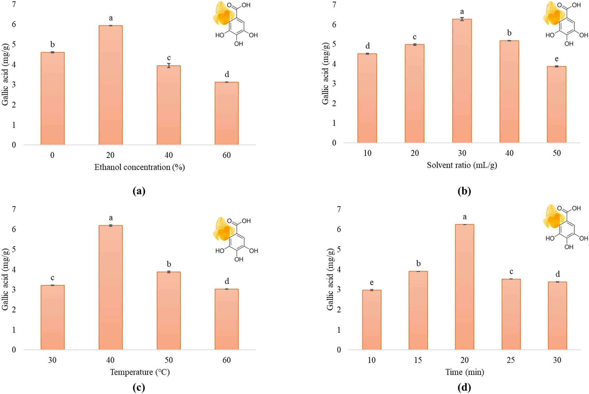
Single-factor experiments of ultrasound-assisted ethanolic extraction of mango peels (a) ethanol concentration (%); (b) solvent ratio (mL/g); (c) temperature (℃); (d) time (min).
3.1.2 Effect of pH
Aqueous extraction of mango peel powder was carried out by adjusting the pH levels of distilled water to 2, 4, 6, and 8. Other extraction parameters were kept constant at pre-determined levels. Results showed that the yield of gallic acid was higher at pH 4 (4.14 mg/g). The yield gradually declined with the increase in pH of the extraction solvent as shown in Figure 2(a). The results are in agreement with the study reporting effect of pH of total phenolic contents extracted from mango peels ranging from pH 2 to 8. The phenolic content increased as the pH increased up to 4 but declined when a pH higher than 4 was used [33]. Acidic pH facilitates the gallic acid yield which is basic in nature as the non-charged form of gallic acid transfers to the ionic liquid-rich phase due to low pH, leading toward the increased solubility of the gallic acid in extraction solvent [34]. As the solubility increases, the concentration of the extractable also increases until the total pool of extractable in the sample is exhausted or the solubility of dissociated form of the extractable exceeded [25]. Based on the results, pH 4 was selected to be used in subsequent experiments.

Single-factor experiments of ultrasound-assisted aqueous extraction of mango peels (a) pH; (b) solvent ratio (mL/g); (c) temperature (℃); (d) time (min).
3.1.3 Effect of solvent ratio
Various solvent ratios were tested, including 10, 20, 30, 40, and 50 mL/g, while adjusting other variables such as 20% solvent concentration, 40℃ temperature, and 10 min. Figure 1(b) illustrates that gallic acid content increased as the ratio increased to 30 mL/g. However, a gradual decline in yield was seen with further increases to 40 or 50 mL/g. The maximum yield was observed at a solvent ratio of 30 mL/g, which was 6.27 mg/g for the ethanolic extraction procedure. A similar effect of solvent ratios ranging from 20 to 60 mL/g for aqueous extraction was observed as the gallic acid yield was highest at 30 mL/g (4.13 mg/g) as depicted in Figure 2(b). There was a gradual decline in gallic acid with the increase in ratio of more than 30 mL/g as similar results were reported for extraction of total phenolic content from mango peels when extracted with 50% ethanol at 30 mL/g solvent ratio [19]. Another study reported the same liquid-to-solid ratio suitable for the maximum extraction yield of mangiferin from mango peels [28]. There are some other studies that reported high solvent ratios ranging from 40 to 50 mL/g to give maximum phenolic recovery [32,33]. As the ratio increases, the compounds present in plants are more efficiently dissolved, which results in increasing the extraction yield [35]. The principle of mass transfer is consistent with raise in yield of gallic acid. The main driving force behind this process is the gradient in sample and solvent concentration, which becomes stronger with a higher solvent ratio [33]. The highest gallic acid yield was observed at a solvent ratio of 30 mL/g, which was used further.
3.1.4 Effect of extraction temperature
Different temperature levels (30, 40, 50, and 60℃) were tested, with other extraction parameters set at 20% ethanol concentration, 30 mL/g of solvent ratio, and 10 min of time. The overall gallic acid yield was increased as the extraction temperature rose to 40℃, then reduced at 50 and 60℃ (Figure 1c). The highest yield, 6.19 mg/g, was achieved at 40℃. Likewise, aqueous extraction was performed on temperatures from 40 to 70℃ by keeping other factors constant or fixed at determined points. Upon analysis of extracts, it was revealed that the extraction temperature at 40℃ was the most efficient one to yield the highest gallic acid (4.22 mg/g) among all (Figure 2c). Similar findings were reported in a study where phenolic contents extracted from mango peels were highest between 35 and 45℃ [19]. The yield was reduced at higher temperatures as higher extraction temperatures increase solvent diffusion into cells, improving desorption and solubility of compounds, and facilitates splitting up of compounds. However, bioactive compounds are often denatured at high temperatures, as observed in the decrease in yield of gallic acid at temperatures between 40 and 60℃ [36]. The results suggest that gallic acid has the highest solubility and desorption equilibrium at 40℃. Additionally, it was found to be thermally stable between 30 and 50℃. These findings have significant implications for the potential applications of gallic acid in various industries [37]. However, higher temperatures have been found to reduce the viscosity of solvent and enhance the plant cell wall permeability, resulting in a positive impact on overall yield. It is important to note that excessively high temperatures can have adverse effects on the quantity and quality of extracted phenolic compounds [28,33]. Thus, for optimization purposes, an extraction temperature of 40℃ was selected for both methods.
3.1.5 Effect of extraction time
The study involved processing samples using UAE for varying time periods (10, 15, 20, 25, and 30 min) at 20% of solvent concentration, 30 mL/g of solvent ratio extracted at 40℃. Based on the results (Figure 1d), the yield gradually increased from 10 to 20 min and decreased thereafter. The significantly higher yield of 6.24 mg/g was obtained at 20 min. Similar time period was reported to gain maximum polyphenols and flavonoids in a study where peels were treated with UAE for 20 min [27,28]. However, gallic acid yield was higher at 40 min in aqueous extraction when different time intervals (30, 40, 50, and 60 min) were studied to determine a center point for the single-factor extraction optimization of gallic acid from mango peels (Figure 2d). It was observed that gallic acid yield was reduced with the increase in extraction time. The study suggests that ultrasonic waves enhance the equilibrium coefficient between the cell walls of plants and solvent, thereby dissolving target compounds more quickly. This makes UAE a more efficient extraction method than conventional approaches [39]. However, the prolonged exposure to ultrasonic waves may lead to the degradation of bioactive compounds [28]. While the extraction time of 10–20 min increased the yield of gallic acid, further increases beyond 20 min yielded diminishing returns. Hence, 20 and 40 min were chosen as extraction times for optimization of MP-Eth. and MP-Aq., respectively.
3.2 Multiple-factor ethanolic extraction optimization of gallic acid-enriched extracts
The gallic acid yield was obtained after running 27 experiments in triplicates (Table 2). The extracts were prepared and analyzed according to the experimental variable combinations generated by the design. The gallic acid content was observed between 4.16 and 5.95 mg/g. The highest gallic acid yield was obtained at the extraction variables of X 1 = 20%, X 2 = 30 mL/g, X 3 = 40℃, and X 4 = 20 min.
Gallic acid yield of ethanolic extraction of mango peels for the experimental design
| Run | X 1 Ethanol concentration | X 2 Solvent ratio | X 3 Temperature | X 4 Time | Gallic acid (mg/g)* |
|---|---|---|---|---|---|
| 1 | 10(−1) | 40(1) | 30(−1) | 15(−1) | 5.25 ± 0.03 |
| 2 | 20(0) | 30(0) | 40(0) | 20(0) | 5.80 ± 0.03 |
| 3 | 20(0) | 30(0) | 40(0) | 20(0) | 5.72 ± 0.02 |
| 4 | 10(−1) | 20(−1) | 50(1) | 25(1) | 4.71 ± 0.02 |
| 5 | 30(1) | 40(1) | 50(1) | 25(1) | 5.03 ± 0.02 |
| 6 | 10(−1) | 40(1) | 50(1) | 25(1) | 4.38 ± 0.07 |
| 7 | 40(2) | 30(0) | 40(0) | 20(0) | 5.76 ± 0.03 |
| 8 | 20(0) | 50(2) | 40(0) | 20(0) | 5.95 ± 0.03 |
| 9 | 30(1) | 20(−1) | 50(1) | 15−1) | 4.77 ± 0.03 |
| 10 | 20(0) | 30(0) | 40(0) | 20(0) | 5.80 ± 0.02 |
| 11 | 30(1) | 20(−1) | 30(−1) | 25(1) | 4.85 ± 0.04 |
| 12 | 10(−1) | 20(−1) | 30(−1) | 25(1) | 4.85 ± 0.03 |
| 13 | 20(0) | 30(0) | 40 (0) | 30(2) | 4.65 ± 0.06 |
| 14 | 20(0) | 30(0) | 60(2) | 20(0) | 4.24 ± 0.03 |
| 15 | 30(1) | 40(1) | 30(−1) | 25(1) | 5.73 ± 0.04 |
| 16 | 10(−1) | 40(1) | 50(1) | 15(−1) | 5.33 ± 0.05 |
| 17 | 30(1) | 40(1) | 50(1) | 15(−1) | 5.38 ± 0.01 |
| 18 | 30(1) | 40(1) | 30(−1) | 15(−1) | 5.24 ± 0.03 |
| 19 | 10(−1) | 20(−1) | 30 (−1) | 15(−1) | 4.45 ± 0.04 |
| 20 | 30(1) | 20(−1) | 50(1) | 25(1) | 4.74 ± 0.03 |
| 21 | 20(0) | 30(0) | 40(0) | 10(−2) | 4.16 ± 0.03 |
| 22 | 10(−1) | 40(1) | 30(−1) | 25(1) | 5.54 ± 0.03 |
| 23 | 0(−2) | 30(0) | 40(0) | 20(0) | 5.64 ± 0.01 |
| 24 | 30(1) | 20(−1) | 30(−1) | 15(−1) | 4.56 ± 0.03 |
| 25 | 10(−1) | 20(−1) | 50(1) | 15(−1) | 5.03 ± 0.04 |
| 26 | 20(0) | 30(0) | 20 (−2) | 20(0) | 4.78 ± 0.05 |
| 27 | 20(0) | 10(−2) | 40(0) | 20(0) | 5.40 ± 0.02 |
*Results are presented as mean ± SD.
Test and response variables were checked via multiple regression analysis of experimental data by following equation (1).
where Y is the gallic acid yield which is the response variable while X 1, X 2, X 3, and X 4 are the factors for extraction optimization.
ANOVA for the regression equation is given in Table 3. The analysis confirmed that lack of fit helped us ensure the model’s adequacy. We found a lack of fit that was not statistically significant (p > 0.05), indicating that the model was fitting the experiment appropriately. Moreover, we accurately measured the signal-to-noise ratio, which was 12.962, a preferred value as it is higher than 4. This indicated a definite design space, implying that this model could be of great help. Additionally, we noticed that the plotted points were clustered finely around the diagonal line, demonstrating the connection between experimental and predicted values. This is a sign of the model’s fitness, as the predicted R-squared value of 0.6883 was in reasonable agreement with the R-squared adjusted value of 0.8820.
ANOVA for the fitted quadratic model
| Source | Sum of square | Degree of freedom | Mean square | F-value | p-value |
|---|---|---|---|---|---|
| Model | 6.95 | 14 | 0.4967 | 14.88 | <0.0001 |
| X 1 | 0.0417 | 1 | 0.0417 | 1.25 | 0.2857 |
| X 2 | 1.05 | 1 | 1.05 | 31.46 | 0.0001 |
| X 3 | 0.1980 | 1 | 0.1980 | 5.93 | 0.0314 |
| X 4 | 0.0267 | 1 | 0.0267 | 0.7990 | 0.3890 |
| X 1 × X 2 | 0.0625 | 1 | 0.0625 | 1.87 | 0.1962 |
| X 1 × X 3 | 0.0020 | 1 | 0.0020 | 0.0607 | 0.8096 |
| X 1 × X 4 | 0.0600 | 1 | 0.0600 | 1.80 | 0.2047 |
| X 2 × X 3 | 0.2970 | 1 | 0.2970 | 8.90 | 0.0114 |
| X 2 × X 4 | 0.0462 | 1 | 0.0462 | 1.39 | 0.2621 |
| X 3 × X 4 | 0.6084 | 1 | 0.6084 | 18.23 | 0.0011 |
| X 1 2 | 0.0220 | 1 | 0.0220 | 0.6580 | 0.4331 |
| X 2 2 | 0.0313 | 1 | 0.0313 | 0.9393 | 0.3516 |
| X 3 2 | 2.32 | 1 | 2.32 | 69.43 | <0.0001 |
| X 4 2 | 2.70 | 1 | 2.70 | 80.93 | <0.0001 |
| Residual | 0.4005 | 12 | 0.0334 | ||
| Lack of fit | 0.3962 | 10 | 0.0396 | 18.57 | 0.0521 |
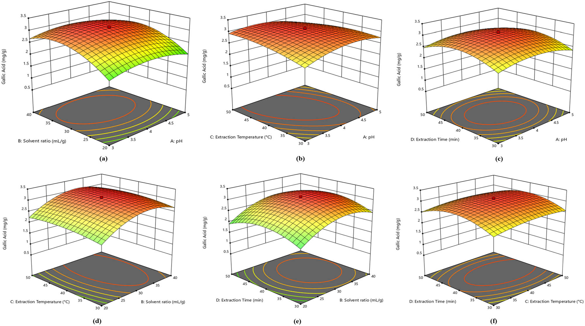
The three-dimensional surface plots of the model exhibit the optimized values to predict and display the relationship of all the processed variables. An elliptical shape of the plot shows a significant interaction of the variables, whereas the circular one depicts the insignificance of interaction between variables [38]. The results obtained from this study of gallic acid extraction optimization from peels of mango showed that ultrasonic treatment during 15–20 min and temperature ranging between 30 and 35°C with the least ethanol concentration proves to be an effective technique to extract bioactive compounds. Furthermore, the results indicate a significant interaction of time of UAE treatment and temperature that can enhance the gallic acid yield (Figure 3). Optimal values for the extraction variables were achieved by solving the regression equation. After running the experiment on the software, the optimal conditions for gallic acid extraction were as follows: 21.2% of ethanol concentration, 30.2 mL/g of solvent ratio, with 33.4°C extraction temperature, and 17.6 min of extraction time, with the corresponding Y = 5.77 ± 0.18 mg/g. Extraction was carried out in triplicates under the optimized conditions to recheck the results further. Gallic acid obtained from the extracts was 5.75 ± 0.21 mg/g, indicating that the model fitted the experimental data very well. Hence, optimization to obtain gallic acid-enriched extracts from mango peels through ethanol was completed (Table 6).

Response surface plots as effected by extraction variables of ethanolic extraction of gallic acid-enriched extracts from mango peels (a) ethanol concentration and solvent ratio; (b) ethanol concentration and temperature; (c) ethanol concentration and time; (d) solvent ratio and temperature; (e) solvent ratio and time; (f) temperature and time.
3.3 Multiple-factor aqueous extraction optimization of gallic acid-enriched extracts
The total number of runs generated was 27, so extracts were prepared according to the variables (Table 4) and gallic acid content was analyzed. The yields of gallic acid ranged from 0.65 to 3.15 mg/g. The highest gallic acid yield was obtained with the extraction variables of X 1 = 4, X 2 = 30 mL/g, X 3 = 40℃, and X 4 = 40 min.
Gallic acid yield of aqueous extraction of mango peels for the experimental design
| Run | X 1 pH | X 2 Solvent ratio | X 3 Temperature | X 4 Time | Gallic acid (mg/g)* |
|---|---|---|---|---|---|
| 1 | 3(−1) | 40(+1) | 50(+1) | 30(−1) | 2.15 ± 0.02 |
| 2 | 4(0) | 30(0) | 20(−2) | 40(0) | 2.41 ± 0.02 |
| 3 | 3(−1) | 20(−1) | 50(+1) | 50(+1) | 1.53 ± 0.04 |
| 4 | 3(−1) | 40(+1) | 30(−1) | 30(−1) | 2.08 ± 0.03 |
| 5 | 2(−2) | 30(0) | 40(0) | 40(0) | 2.10 ± 0.02 |
| 6 | 5(+1) | 40(+1) | 50(+1) | 30(−1) | 2.05 ± 0.03 |
| 7 | 5(+1) | 20(−1) | 50(+1) | 30(−1) | 1.47 ± 0.02 |
| 8 | 3(−1) | 40(+1) | 50(+1) | 50(+1) | 2.57 ± 0.04 |
| 9 | 5(+1) | 40(+1) | 30(−1) | 50(+1) | 2.33 ± 0.02 |
| 10 | 3(−1) | 20(−1) | 30(−1) | 30(−1) | 1.17 ± 0.03 |
| 11 | 3(−1) | 20(−1) | 50(+1) | 30(−1) | 1.70 ± 0.04 |
| 12 | 4(0) | 10(−2) | 40(0) | 40(0) | 0.65 ± 0.05 |
| 13 | 4(0) | 30(0) | 40(0) | 40(0) | 3.14 ± 0.02 |
| 14 | 4(0) | 30(0) | 40(0) | 40(0) | 3.15 ± 0.03 |
| 15 | 5(+1) | 20(−1) | 50(+1) | 50(+1) | 1.30 ± 0.03 |
| 16 | 4(0) | 30(0) | 40(0) | 60(+2) | 1.79 ± 0.02 |
| 17 | 4(0) | 30(0) | 60(+2) | 40(0) | 2.76 ± 0.04 |
| 18 | 4(0) | 50(+2) | 40(0) | 40(0) | 1.66 ± 0.03 |
| 19 | 5(+1) | 40(+1) | 50(+1) | 50(+1) | 1.75 ± 0.03 |
| 20 | 5(+1) | 40(+1) | 30(−1) | 30(−1) | 2.00 ± 0.04 |
| 21 | 3(−1) | 40(+1) | 30(−1) | 50(+1) | 1.64 ± 0.02 |
| 22 | 4(0) | 30(0) | 40(0) | 20(−2) | 1.22 ± 0.09 |
| 23 | 3(−1) | 20(−1) | 30(−1) | 50(+1) | 1.26 ± 0.04 |
| 24 | 5(+1) | 20(−1) | 30(−1) | 30(−1) | 1.5 ± 0.02 |
| 25 | 4(0) | 30(0) | 40(0) | 40(0) | 2.97 ± 0.02 |
| 26 | 6(+2) | 30(0) | 40(0) | 40(0) | 2.03 ± 0.04 |
| 27 | 5(+1) | 20(−1) | 30(−1) | 50(+1) | 1.50 ± 0.02 |
-
*Results are presented as mean ± SD.
Experimental data of extraction variable and gallic acid yield were evaluated through multiple regression analysis using equation (2).
where Y is the gallic acid yield which is the response variable while X 1, X 2, X 3, and X 4 are the factors for extraction optimization.
Table 5 shows the ANOVA for the regression equation. The analysis presented that lack of fit helped in ensuring the model’s adequacy. A lack of fit was found that was not statistically significant (p > 0.05), indicating that the model was fitting the experiment appropriately. Moreover, we accurately measured a higher than 4 signal-to-noise ratio, which was 16.387, a preferred value. This indicated a definite design space, implying that this model could be of great help. Additionally, we noticed that the plotted points were clustered finely around the diagonal line, demonstrating the connection between experimental and predicted values. This is a sign of the model’s fitness, as the predicted R-squared value of 0.7052 was in reasonable agreement with the R-squared adjusted value of 0.8865.
ANOVA for the fitted quadratic model
| Source | Sum of square | Degree of freedom | Mean square | F-value | p-value |
|---|---|---|---|---|---|
| Model | 9.68 | 14 | 0.6917 | 15.50 | <0.0001 |
| X1 | 0.0048 | 1 | 0.0048 | 0.1079 | 0.7482 |
| X2 | 2.14 | 1 | 2.14 | 47.87 | <0.0001 |
| X 3 | 0.1261 | 1 | 0.1261 | 2.83 | 0.1185 |
| X 4 | 0.0337 | 1 | 0.0337 | 0.7564 | 0.4015 |
| X1 × X2 | 0.0110 | 1 | 0.0110 | 0.2471 | 0.6281 |
| X1 × X3 | 0.4096 | 1 | 0.4096 | 9.18 | 0.0105 |
| X1 × X4 | 0.0001 | 1 | 0.0001 | 0.0022 | 0.9630 |
| X2 × X3 | 0.0006 | 1 | 0.0006 | 0.0140 | 0.9077 |
| X2 × X4 | 0.0042 | 1 | 0.0042 | 0.0947 | 0.7636 |
| X3 × X4 | 0.0025 | 1 | 0.0025 | 0.0560 | 0.8169 |
| X12 | 1.54 | 1 | 1.54 | 34.43 | <0.0001 |
| X22 | 5.24 | 1 | 5.24 | 117.54 | <0.0001 |
| X32 | 0.4082 | 1 | 0.4082 | 9.15 | 0.0106 |
| X42 | 3.56 | 1 | 3.56 | 79.72 | <0.0001 |
| Residual | 0.5354 | 12 | 0.0446 | ||
| Lack of fit | 0.5150 | 10 | 0.0515 | 5.03 | 0.1771 |
| Pure error | 0.0205 | 2 | 0.0102 |
The three-dimensional surface plots obtained by the model exhibited the optimized values that showed the interaction between the variables explored in optimization process. The appearance of the plot helps in identifying these interactions between variables as the circular one exhibits the insignificant interaction while an elliptical shape depicts that there is a significant interaction between variables [38]. The three-dimensional surface plots in Figure 4 represent the interaction of extraction factors effecting the yield of gallic acid for aqueous extraction of mango peels. Quadratic polynomial equation was established to quantify the relationship between the extraction parameters and response. The optimized values for extraction were obtained by solving the regression equation.
Response surface plots as effected by extraction variables of aqueous extraction of gallic acid-enriched extracts from mango peels (a) pH and solvent ratio; (b) pH and temperature; (c) pH and time; (d) solvent ratio and temperature; (e) solvent ratio and time; (f) temperature and time.
After running the experiment, the common regions for optimized conditions of gallic acid extraction from mango peels were obtained with the corresponding Y = 3.14 ± 0.21 mg/g. Optimized conditions were used to perform triplicate extraction tests in order to validate the optimization. The gallic acid yield was found to be 3.14 ± 0.24 mg/g indicating that the extraction procedure for gallic acid was fully optimized (Table 6).
Predicted and experimental values of gallic acid yield for both extraction methods
| Extraction method | Optimum conditions | Extraction yield (mg/g) | ||||
|---|---|---|---|---|---|---|
| X 1 | X 2 | X 3 | X 4 | Predicted | Experimental* | |
| UAE-ethanolic | 21.2 | 30.2 | 33.4 | 17.6 | 5.77 ± 0.18 | 5.75 ± 0.21 |
| UAE-aqueous | 3.8 | 33.02 | 43.35 | 40.47 | 3.14 ± 0.21 | 3.14 ± 0.24 |
*Results are presented as mean ± SD.
Hence, the extraction of gallic acid from mango peels through UAE was an efficient green extraction method with lesser power consumption. It yielded comparable amounts of gallic acid to those obtained using organic solvents in previous studies [13–18]. In the current study, the extraction done with 21% ethanol showed significantly higher yield of gallic acid (5.77 ± 0.18 mg/g) as compared to water (3.14 ± 0.21 mg/g) within a lesser time of 17.6 min. Gallic acid yields were quite higher in the current study than the reported studies. A combination of different enzymes with 65% alcohol and 50% ethanol resulted in a concentration of 18 µg/100 mL and 20.21 µg/g of extracts, respectively [15,39]. However, the gallic acid yield obtained with ethanolic extraction was comparable with another study where 80% acetone was used to extract bioactive compounds and reported 4.54–6.29 mg/g of extract gallic acid from mango peels [14]. The solvent concentration to obtain this yield is too high as compared to the solvent percentages used in the current study. Palafox-Carlos et al. [39] studied the major phenolic compounds of mango fruit that contribute to antioxidant activity and reported that mango peels yield in a range from 40 to 60 µg/mL of gallic acid at different maturity stages and showed highest antioxidant potential. In another study, the reported gallic acid values were 12.5, 13, and 25.6 mg/100 g of extract and concluded that the antioxidant potential of mango peel extracts is greatly contributed by gallic acid and makes it useful as a functional ingredient and nutraceutical [6]. It is important to note that mango peels, typically thrown away during fruit juice processing, are actually a rich source of phenolic compounds. The peels contain even more phenolic compounds than the mango pulp itself and gallic acid is one of the most prominent phenolic compounds found in mango peels having reported antioxidant potential [32,37].
3.4 Comparison of optimized ethanolic and aqueous extracts of mango peels
The extracts were compared for their gallic acid content and TFC in order to determine the quality of extraction methods. MP-Eth. showed significantly higher (p < 0.05) gallic acid yield as well as TFC in contrast to MP-Aq. and MP-Dec (Table 7). Gallic acid content extracted from mango peels with 50% ethanol was reported to be 144.18 µg/g through UAE [40] and reported as the abundantly found phenolic acid in mango peels. Similarly, another study reported gallic acid content of three different varieties of mango peels ranging from 12.51 to 25.62 mg/100 g dry matter when the peels were irradiated for 15 min with 80% methanol through UAE [15]. However, comparing the extraction time and temperature to the UAE-optimized conditions in current study obtained higher amount of gallic acid (5.75 ± 0.21 mg/g) and TFC (9.46 ± 0.03 mg/g) in a much shorter time of 17.6 min with a temperature of 33.4℃. Total flavonoids extracted from mango peels through UAE were reported as 556.6 ± 0.3 µg/g with 45% ethanol in a 60 min period of time [21]. Another study reported 18 mg/100 g fresh weight TFC of mango peels when extracted with methanol using UAE [41]. However, Borrás-Enríquez et al. [27] reported a higher TFC (12.28 mg/g) when mango peels were subjected to UAE with 50 mL of 99% HPLC-grade ethanol, which is a higher concentration as compared to the observed optimized variables of current study.
Comparison of extraction methods
| Extraction method | Gallic acid (mg/g) | TFC as CE (mg/g) |
|---|---|---|
| MP-Eth. | 5.75 ± 0.21a | 9.46 ± 0.03a |
| MP-Aq. | 3.14 ± 0.02b | 7.08 ± 0.01b |
| MP-Dec. | 2.57 ± 0.02c | 6.71 ± 0.02c |
Extraction with ethanol (MP-Eth.), water (MP-Aq.), and decoction (MP-Dec.) Results are provided as mean ± SD. Significance (p < 0.05) is depicted by superscripts.
4 Conclusion
The results of the current study report the optimization of extraction conditions to obtain gallic acid-enriched extract from mango peels by using ethanol and water, where extraction using ethanol as a solvent gave preferred results for gallic acid yields and TFCs. The optimized conditions of ethanolic extraction were 21.2% ethanol concentration, 30.3 mL/g of solvent ratio at 33.4℃ for 17.6 min, whereas extraction done with water was optimized at pH 3.8, solvent ratio of 33.02 mL/g at 43.35℃ for 40.47 min with a gallic acid yield of 5.75 ± 0.21 mg/g and 3.14 ± 0.02 mg/g, respectively. Both extraction methods showed higher gallic acid and TFC when compared with conventional extraction of decoction. The reported UAE method may help in designing more efficient extraction methods to extract valuable compounds from mango peels and enable its utilization.
-
Funding information: This work was carried out with financial support from the UK Government’s Department of Health and Social Care, Global AMR Innovation Fund (GAMRIF), and the International Development Research Centre (IDRC), Ottawa, Canada (Grant No. 109051–003). The views expressed herein do not necessarily represent those of IDRC or its Board of Governors.
-
Author contributions: Conceptualization, T.R., Z.H., and K.A.; Formal analysis, T.R., K.S., M.A., and Z.T.; Investigation, Z.H. and S.U.R.; Methodology, Z.H., T.R., K.A., K.S., and A.S.; Project administration, Z.H., S.U.R., A.M., and U.F.; Software, T.R., H.U.R., and A.S.; Supervision, Z.H. and S.U.R.; Validation, T.R., H.U.R., and K.A.; Writing – original draft, T.R.; Writing – review and editing, T.R., Z.H., and K.A.; Funding acquisition, Z.H. and S.U.R.
-
Conflict of interest: The authors declare no conflict of interest.
-
Ethical approval: The conducted research is not related to either human or animal use.
-
Data availability statement: All data generated or analyzed during this study are included in this published article.
References
[1] Masibo M, He Q. Major mango polyphenols and their potential significance to human health. Compr Rev Food Sci Food Saf. 2008;7(4):309–19.10.1111/j.1541-4337.2008.00047.xSearch in Google Scholar PubMed
[2] Tirado-Kulieva V, Atoche-Dioses S, Hernández-Martínez E. Phenolic compounds of mango (Mangifera indica) by-products: Antioxidant and antimicrobial potential, use in disease prevention and food industry, methods of extraction and microencapsulation. Sci Agropecu. 2021;12(2):283–93.10.17268/sci.agropecu.2021.031Search in Google Scholar
[3] Asif A, Farooq U, Akram K, Hayat Z, Shafi A, Sarfraz F, et al. Therapeutic potentials of bioactive compounds from mango fruit wastes. Trends Food Sci Technol. 2016;53:102–12.10.1016/j.tifs.2016.05.004Search in Google Scholar
[4] Castro-Vargas HI, Ballesteros Vivas D, Ortega Barbosa J, Morantes Medina SJ, Aristizabal Gutiérrez F, Parada-Alfonso F. Bioactive phenolic compounds from the agroindustrial waste of Colombian mango cultivars ‘Sugar Mango’and ‘Tommy Atkins’—An alternative for their use and valorization. Antioxidants. 2019;8(2):41.10.3390/antiox8020041Search in Google Scholar PubMed PubMed Central
[5] Masibo M, He Q. Mango bioactive compounds and related nutraceutical properties—a review. Food Rev Int. 2009;25(4):346–70.10.1080/87559120903153524Search in Google Scholar
[6] Ajila C, Naidu K, Bhat S, Rao UP. Bioactive compounds and antioxidant potential of mango peel extract. Food Chem. 2007;105(3):982–8.10.1016/j.foodchem.2007.04.052Search in Google Scholar
[7] Barreto JC, Trevisan MT, Hull WE, Erben G, De Brito ES, Pfundstein B, et al. Characterization and quantitation of polyphenolic compounds in bark, kernel, leaves, and peel of mango (Mangifera indica L.). J Agric Food Chem. 2008;56(14):5599–610.10.1021/jf800738rSearch in Google Scholar PubMed
[8] Berardini N, Carle R, Schieber A. Characterization of gallotannins and benzophenone derivatives from mango (Mangifera indica L. cv.‘Tommy Atkins’) peels, pulp and kernels by high‐performance liquid chromatography/electrospray ionization mass spectrometry. Rapid Commun Mass Spectrom. 2004;18(19):2208–16.10.1002/rcm.1611Search in Google Scholar PubMed
[9] Ribeiro SMR, Queiroz JH, de Queiroz MELR, Campos FM, Sant’Ana HMP. Antioxidant in mango (Mangifera indica L.) pulp. Plant Foods Hum Nutr. 2007;62(1):13–7.10.1007/s11130-006-0035-3Search in Google Scholar PubMed
[10] Kim Y, Brecht JK, Talcott ST. Antioxidant phytochemical and fruit quality changes in mango (Mangifera indica L.) following hot water immersion and controlled atmosphere storage. Food Chem. 2007;105(4):1327–34.10.1016/j.foodchem.2007.03.050Search in Google Scholar
[11] Jung S, Choe JH, Kim B, Yun H, Kruk ZA, Jo C. Effect of dietary mixture of gallic acid and linoleic acid on antioxidative potential and quality of breast meat from broilers. Meat Sci. 2010;86(2):520–6.10.1016/j.meatsci.2010.06.007Search in Google Scholar PubMed
[12] Özçelik B, Kartal M, Orhan I. Cytotoxicity, antiviral and antimicrobial activities of alkaloids, flavonoids, and phenolic acids. Pharm Biol. 2011;49(4):396–402.10.3109/13880209.2010.519390Search in Google Scholar PubMed
[13] Hu K, Dars AG, Liu Q, Xie B, Sun Z. Phytochemical profiling of the ripening of Chinese mango (Mangifera indica L.) cultivars by real-time monitoring using UPLC-ESI-QTOF-MS and its potential benefits as prebiotic ingredients. Food Chem. 2018;256:171–80.10.1016/j.foodchem.2018.02.014Search in Google Scholar PubMed
[14] Ajila C, Rao UP. Mango peel dietary fibre: Composition and associated bound phenolics. J Funct Foods. 2013;5(1):444–50.10.1016/j.jff.2012.11.017Search in Google Scholar
[15] López-Cobo A, Verardo V, Diaz-de-Cerio E, Segura-Carretero A, Fernández-Gutiérrez A, Gómez-Caravaca AM. Use of HPLC-and GC-QTOF to determine hydrophilic and lipophilic phenols in mango fruit (Mangifera indica L.) and its by-products. Food Res Int. 2017;100:423–34.10.1016/j.foodres.2017.02.008Search in Google Scholar PubMed
[16] Coelho EM, de Souza MEAO, Corrêa LC, Viana AC, de Azevêdo LC, dos Santos Lima M. Bioactive compounds and antioxidant activity of mango peel liqueurs (Mangifera indica L.) produced by different methods of maceration. Antioxidants. 2019;8(4):102.10.3390/antiox8040102Search in Google Scholar PubMed PubMed Central
[17] Sharif T, Bhatti HN, Bull ID, Bilal M. Recovery of high-value bioactive phytochemicals from agro-waste of mango (Mangifera indica L.) using enzyme-assisted ultrasound pretreated extraction. Biomass Convers Biorefin. 2021;13(8):6591–9.10.1007/s13399-021-01589-5Search in Google Scholar
[18] Jirasuteeruk C, Theerakulkait C. Ultrasound-assisted extraction of phenolic compounds from mango (Mangifera indica cv. Chok Anan) peel and its inhibitory effect on enzymatic browning of potato puree. Food Technol Biotechnol. 2019;57(3):350–7.10.17113/ftb.57.03.19.5728Search in Google Scholar PubMed PubMed Central
[19] Kaur B, Panesar PS, Anal AK. Standardization of ultrasound assisted extraction for the recovery of phenolic compounds from mango peels. J Food Sci Technol. 2022;59(7):2813–20.10.1007/s13197-021-05304-0Search in Google Scholar PubMed PubMed Central
[20] Ordoñez‐Torres A, Torres‐León C, Hernández‐Almanza A, Flores‐Guía T, Luque‐Contreras D, Aguilar CN, et al. Ultrasound‐microwave‐assisted extraction of polyphenolic compounds from Mexican “Ataulfo” mango peels: Antioxidant potential and identification by HPLC/ESI/MS. Phytochem Anal. 2021;32(4):495–502.10.1002/pca.2997Search in Google Scholar PubMed
[21] Aznar-Ramos MJ, Razola-Díaz MDC, Verardo V, Gómez-Caravaca AM. Comparison between ultrasonic bath and sonotrode extraction of phenolic compounds from mango peel by-products. Horticulturae. 2022;8(11):1014.10.3390/horticulturae8111014Search in Google Scholar
[22] Chemat F, Vian MA, Cravotto G. Green extraction of natural products: concept and principles. Int J Mol Sci. 2012;13(7):8615–27.10.3390/ijms13078615Search in Google Scholar PubMed PubMed Central
[23] Tiwari BK. Ultrasound: A clean, green extraction technology. TrAC, Trends Anal Chem. 2015;71:100–9.10.1016/j.trac.2015.04.013Search in Google Scholar
[24] Boeing JS, Barizão ÉO, e Silva BC, Montanher PF, de Cinque Almeida V, Visentainer JV. Evaluation of solvent effect on the extraction of phenolic compounds and antioxidant capacities from the berries: application of principal component analysis. Chem Cent J. 2014;8:1–9.10.1186/s13065-014-0048-1Search in Google Scholar PubMed PubMed Central
[25] Castro-Puyana M, Marina ML, Plaza M. Water as green extraction solvent: Principles and reasons for its use. Curr Opin Green Sustain Chem. 2017;5:31–6.10.1016/j.cogsc.2017.03.009Search in Google Scholar
[26] Hartonen K, Riekkola ML. Water as the first choice green solvent. The Application Of Green Solvents in Separation Processes. Netherlands: Elsevier; 2017. p. 19–55.10.1016/B978-0-12-805297-6.00002-4Search in Google Scholar
[27] Borrás-Enríquez AJ, Reyes-Ventura E, Villanueva-Rodríguez SJ, Moreno-Vilet L. Effect of ultrasound-assisted extraction parameters on total polyphenols and its antioxidant activity from mango residues (Mangifera indica L. var. Manililla). Separations. 2021;8(7):94.10.3390/separations8070094Search in Google Scholar
[28] Zou T-B, Xia E-Q, He T-P, Huang M-Y, Jia Q, Li H-W. Ultrasound-assisted extraction of mangiferin from mango (Mangifera indica L.) leaves using response surface methodology. Molecules. 2014;19(2):1411–21.10.3390/molecules19021411Search in Google Scholar PubMed PubMed Central
[29] Hayat Z, Riaz T, Saleem K, Akram K, Ur Rehman H, Azam M. Optimization of gallic acid-rich extract from mango (Mangifera indica) Seed kernels through ultrasound-assisted extraction. Separations. 2023;10(7):376.10.3390/separations10070376Search in Google Scholar
[30] Chanda S, Amrutiya N, Rakholiya K. Evaluation of antioxidant properties of some Indian vegetable and fruit peels by decoction extraction method. Am J Food Technol. 2013;8(3):173–82.10.3923/ajft.2013.173.182Search in Google Scholar
[31] Ojeda GA, Sgroppo SC, Sánchez-Moreno C, de Ancos B. Mango ‘criollo’by-products as a source of polyphenols with antioxidant capacity. Ultrasound assisted extraction evaluated by response surface methodology and HPLC-ESI-QTOF-MS/MS characterization. Food Chem. 2022;396:133738.10.1016/j.foodchem.2022.133738Search in Google Scholar PubMed
[32] Castañeda-Valbuena D, Ayora-Talavera T, Luján-Hidalgo C, Álvarez-Gutiérrez P, Martínez-Galero N, Meza-Gordillo R. Ultrasound extraction conditions effect on antioxidant capacity of mango by-product extracts. Food Bioprod Process. 2021;127:212–4.10.1016/j.fbp.2021.03.002Search in Google Scholar
[33] Tunchaiyaphum S, Eshtiaghi M, Yoswathana N. Extraction of bioactive compounds from mango peels using green technology. Int J Chem Eng. 2013;4(4):194.10.7763/IJCEA.2013.V4.293Search in Google Scholar
[34] Eslami AC, Pasanphan W, Wagner BA, Buettner GR. Free radicals produced by the oxidation of gallic acid: An electron paramagnetic resonance study. Chem Cent J. 2010;4(1):1–4.10.1186/1752-153X-4-15Search in Google Scholar PubMed PubMed Central
[35] Liyana-Pathirana C, Shahidi F. Optimization of extraction of phenolic compounds from wheat using response surface methodology. Food Chem. 2005;93(1):47–56.10.1016/j.foodchem.2004.08.050Search in Google Scholar
[36] Setyaningsih W, Saputro IE, Palma M, Barroso CG, editors. Stability of 40 phenolic compounds during ultrasound-assisted extractions (UAE). Advances of Science and Technology for Society Proceedings of the 1st International Conference on Science and Technology; 2016 Nov 11–13. Yogyakarta, Indonesia: AIP Publishing LLC; 2017.10.1063/1.4958517Search in Google Scholar
[37] Polewski K, Kniat S, Slawinska D. Gallic acid, a natural antioxidant, in aqueous and micellar environment: spectroscopic studies. Curr Topics Biophys. 2002;26(2):217–27.Search in Google Scholar
[38] Aware CB, Patil RR, Vyavahare GD, Gurme ST, Jadhav JP. Ultrasound-assisted aqueous extraction of phenolic, flavonoid compounds and antioxidant activity of Mucuna macrocarpa beans: Response surface methodology optimization. J Am Coll Nutr. 2019;38(4):364–72.10.1080/07315724.2018.1524315Search in Google Scholar PubMed
[39] Palafox-Carlos H, Yahia E, González-Aguilar G. Identification and quantification of major phenolic compounds from mango (Mangifera indica, cv. Ataulfo) fruit by HPLC–DAD–MS/MS-ESI and their individual contribution to the antioxidant activity during ripening. Food Chem. 2012;135(1):105–11.10.1016/j.foodchem.2012.04.103Search in Google Scholar
[40] Safdar MN, Kausar T, Nadeem M. Comparison of ultrasound and maceration techniques for the extraction of polyphenols from the mango peel. J Food Process Preserv. 2017;41(4):e13028.10.1111/jfpp.13028Search in Google Scholar
[41] Ramirez JE, Zambrano R, Sepúlveda B, Simirgiotis MJ. Antioxidant properties and hyphenated HPLC-PDA-MS profiling of Chilean Pica mango fruits (Mangifera indica L. Cv. piqueño). Molecules. 2014;19(1):438–58.10.3390/molecules19010438Search in Google Scholar PubMed PubMed Central
© 2023 the author(s), published by De Gruyter
This work is licensed under the Creative Commons Attribution 4.0 International License.
Articles in the same Issue
- Characteristics, source, and health risk assessment of aerosol polyaromatic hydrocarbons in the rural and urban regions of western Saudi Arabia
- Regular Articles
- A network-based correlation research between element electronegativity and node importance
- Pomegranate attenuates kidney injury in cyclosporine-induced nephrotoxicity in rats by suppressing oxidative stress
- Ab initio study of fundamental properties of XInO3 (X = K, Rb, Cs) perovskites
- Responses of feldspathic sandstone and sand-reconstituted soil C and N to freeze–thaw cycles
- Robust fractional control based on high gain observers design (RNFC) for a Spirulina maxima culture interfaced with an advanced oxidation process
- Study on arsenic speciation and redistribution mechanism in Lonicera japonica plants via synchrotron techniques
- Optimization of machining Nilo 36 superalloy parameters in turning operation
- Vacuum impregnation pre-treatment: A novel method for incorporating mono- and divalent cations into potato strips to reduce the acrylamide formation in French fries
- Characterization of effective constituents in Acanthopanax senticosus fruit for blood deficiency syndrome based on the chinmedomics strategy
- Comparative analysis of the metabolites in Pinellia ternata from two producing regions using ultra-high-performance liquid chromatography–electrospray ionization–tandem mass spectrometry
- The assessment of environmental parameter along the desalination plants in the Kingdom of Saudi Arabia
- Effects of harpin and carbendazim on antioxidant accumulation in young jujube leaves
- The effects of in ovo injected with sodium borate on hatching performance and small intestine morphology in broiler chicks
- Optimization of cutting forces and surface roughness via ANOVA and grey relational analysis in machining of In718
- Essential oils of Origanum compactum Benth: Chemical characterization, in vitro, in silico, antioxidant, and antibacterial activities
- Translocation of tungsten(vi) oxide/gadolinium(iii) fluoride in tellurite glasses towards improvement of gamma-ray attenuation features in high-density glass shields
- Mechanical properties, elastic moduli, and gamma ray attenuation competencies of some TeO2–WO3–GdF3 glasses: Tailoring WO3–GdF3 substitution toward optimum behavioral state range
- Comparison between the CIDR or sponge with hormone injection to induce estrus synchronization for twining and sex preselection in Naimi sheep
- Exergetic performance analyses of three different cogeneration plants
- Psoralea corylifolia (babchi) seeds enhance proliferation of normal human cultured melanocytes: GC–MS profiling and biological investigation
- A novel electrochemical micro-titration method for quantitative evaluation of the DPPH free radical scavenging capacity of caffeic acid
- Comparative study between supported bimetallic catalysts for nitrate remediation in water
- Persicaline, an alkaloid from Salvadora persica, inhibits proliferation and induces apoptosis and cell-cycle arrest in MCF-7 cells
- Determination of nicotine content in locally produced smokeless tobacco (Shammah) samples from Jazan region of Saudi Arabia using a convenient HPLC-MS/MS method
- Changes in oxidative stress markers in pediatric burn injury over a 1-week period
- Integrated geophysical techniques applied for petroleum basins structural characterization in the central part of the Western Desert, Egypt
- The impact of chemical modifications on gamma-ray attenuation properties of some WO3-reinforced tellurite glasses
- Microwave and Cs+-assisted chemo selective reaction protocol for synthesizing 2-styryl quinoline biorelevant molecules
- Structural, physical, and radiation absorption properties of a significant nuclear power plant component: A comparison between REX-734 and 316L SS austenitic stainless steels
- Effect of Moringa oleifera on serum YKL-40 level: In vivo rat periodontitis model
- Investigating the impact of CO2 emissions on the COVID-19 pandemic by generalized linear mixed model approach with inverse Gaussian and gamma distributions
- Influence of WO3 content on gamma rays attenuation characteristics of phosphate glasses at low energy range
- Study on CO2 absorption performance of ternary DES formed based on DEA as promoting factor
- Performance analyses of detonation engine cogeneration cycles
- Sterols from Centaurea pumilio L. with cell proliferative activity: In vitro and in silico studies
- Untargeted metabolomics revealing changes in aroma substances in flue-cured tobacco
- Effect of pumpkin enriched with calcium lactate on iron status in an animal model of postmenopausal osteoporosis
- Energy consumption, mechanical and metallographic properties of cryogenically treated tool steels
- Optimization of ultra-high pressure-assisted extraction of total phenols from Eucommia ulmoides leaves by response surface methodology
- Harpin enhances antioxidant nutrient accumulation and decreases enzymatic browning in stored soybean sprouts
- Physicochemical and biological properties of carvacrol
- Radix puerariae in the treatment of diabetic nephropathy: A network pharmacology analysis and experimental validation
- Anti-Alzheimer, antioxidants, glucose-6-phosphate dehydrogenase effects of Taverniera glabra mediated ZnO and Fe2O3 nanoparticles in alloxan-induced diabetic rats
- Experimental study on photocatalytic CO2 reduction performance of ZnS/CdS-TiO2 nanotube array thin films
- Epoxy-reinforced heavy metal oxides for gamma ray shielding purposes
- Black mulberry (Morus nigra L.) fruits: As a medicinal plant rich in human health-promoting compounds
- Promising antioxidant and antimicrobial effects of essential oils extracted from fruits of Juniperus thurifera: In vitro and in silico investigations
- Chloramine-T-induced oxidation of Rizatriptan Benzoate: An integral chemical and spectroscopic study of products, mechanisms and kinetics
- Study on antioxidant and antimicrobial potential of chemically profiled essential oils extracted from Juniperus phoenicea (L.) by use of in vitro and in silico approaches
- Screening and characterization of fungal taxol-producing endophytic fungi for evaluation of antimicrobial and anticancer activities
- Mineral composition, principal polyphenolic components, and evaluation of the anti-inflammatory, analgesic, and antioxidant properties of Cytisus villosus Pourr leaf extracts
- In vitro antiproliferative efficacy of Annona muricata seed and fruit extracts on several cancer cell lines
- An experimental study for chemical characterization of artificial anterior cruciate ligament with coated chitosan as biomaterial
- Prevalence of residual risks of the transfusion-transmitted infections in Riyadh hospitals: A two-year retrospective study
- Computational and experimental investigation of antibacterial and antifungal properties of Nicotiana tabacum extracts
- Reinforcement of cementitious mortars with hemp fibers and shives
- X-ray shielding properties of bismuth-borate glass doped with rare earth ions
- Green supported silver nanoparticles over modified reduced graphene oxide: Investigation of its antioxidant and anti-ovarian cancer effects
- Orthogonal synthesis of a versatile building block for dual functionalization of targeting vectors
- Thymbra spicata leaf extract driven biogenic synthesis of Au/Fe3O4 nanocomposite and its bio-application in the treatment of different types of leukemia
- The role of Ag2O incorporation in nuclear radiation shielding behaviors of the Li2O–Pb3O4–SiO2 glass system: A multi-step characterization study
- A stimuli-responsive in situ spray hydrogel co-loaded with naringenin and gentamicin for chronic wounds
- Assessment of the impact of γ-irradiation on the piperine content and microbial quality of black pepper
- Antioxidant, sensory, and functional properties of low-alcoholic IPA beer with Pinus sylvestris L. shoots addition fermented using unconventional yeast
- Screening and optimization of extracellular pectinase produced by Bacillus thuringiensis SH7
- Determination of polyphenols in Chinese jujube using ultra-performance liquid chromatography–mass spectrometry
- Synergistic effects of harpin and NaCl in determining soybean sprout quality under non-sterile conditions
- Field evaluation of different eco-friendly alternative control methods against Panonychus citri [Acari: Tetranychidae] spider mite and its predators in citrus orchards
- Exploring the antimicrobial potential of biologically synthesized zero valent iron nanoparticles
- NaCl regulates goldfish growth and survival at three food supply levels under hypoxia
- An exploration of the physical, optical, mechanical, and radiation shielding properties of PbO–MgO–ZnO–B2O3 glasses
- A novel statistical modeling of air pollution and the COVID-19 pandemic mortality data by Poisson, geometric, and negative binomial regression models with fixed and random effects
- Treatment activity of the injectable hydrogels loaded with dexamethasone In(iii) complex on glioma by inhibiting the VEGF signaling pathway
- An alternative approach for the excess lifetime cancer risk and prediction of radiological parameters
- Panax ginseng leaf aqueous extract mediated green synthesis of AgNPs under ultrasound condition and investigation of its anti-lung adenocarcinoma effects
- Study of hydrolysis and production of instant ginger (Zingiber officinale) tea
- Novel green synthesis of zinc oxide nanoparticles using Salvia rosmarinus extract for treatment of human lung cancer
- Evaluation of second trimester plasma lipoxin A4, VEGFR-1, IL-6, and TNF-α levels in pregnant women with gestational diabetes mellitus
- Antidiabetic, antioxidant and cytotoxicity activities of ortho- and para-substituted Schiff bases derived from metformin hydrochloride: Validation by molecular docking and in silico ADME studies
- Antioxidant, antidiabetic, antiglaucoma, and anticholinergic effects of Tayfi grape (Vitis vinifera): A phytochemical screening by LC-MS/MS analysis
- Identification of genetic polymorphisms in the stearoyl CoA desaturase gene and its association with milk quality traits in Najdi sheep
- Cold-acclimation effect on cadmium absorption and biosynthesis of polyphenolics, and free proline and photosynthetic pigments in Spirogyra aequinoctialis
- Analysis of secondary metabolites in Xinjiang Morus nigra leaves using different extraction methods with UPLC-Q/TOF-MS/MS technology
- Nanoarchitectonics and performance evaluation of a Fe3O4-stabilized Pickering emulsion-type differential pressure plugging agent
- Investigating pyrolysis characteristics of Shengdong coal through Py-GC/MS
- Extraction, phytochemical characterization, and antifungal activity of Salvia rosmarinus extract
- Introducing a novel and natural antibiotic for the treatment of oral pathogens: Abelmoschus esculentus green-formulated silver nanoparticles
- Optimization of gallic acid-enriched ultrasonic-assisted extraction from mango peels
- Effect of gamma rays irradiation in the structure, optical, and electrical properties of samarium doped bismuth titanate ceramics
- Combinatory in silico investigation for potential inhibitors from Curcuma sahuynhensis Škorničk. & N.S. Lý volatile phytoconstituents against influenza A hemagglutinin, SARS-CoV-2 main protease, and Omicron-variant spike protein
- Physical, mechanical, and gamma ray shielding properties of the Bi2O3–BaO–B2O3–ZnO–As2O3–MgO–Na2O glass system
- Twofold interpenetrated 3D Cd(ii) complex: Crystal structure and luminescent property
- Study on the microstructure and soil quality variation of composite soil with soft rock and sand
- Ancient spring waters still emerging and accessible in the Roman Forum area: Chemical–physical and microbiological characterization
- Extraction and characterization of type I collagen from scales of Mexican Biajaiba fish
- Finding small molecular compounds to decrease trimethylamine oxide levels in atherosclerosis by virtual screening
- Prefatory in silico studies and in vitro insecticidal effect of Nigella sativa (L.) essential oil and its active compound (carvacrol) against the Callosobruchus maculatus adults (Fab), a major pest of chickpea
- Polymerized methyl imidazole silver bromide (CH3C6H5AgBr)6: Synthesis, crystal structures, and catalytic activity
- Using calcined waste fish bones as a green solid catalyst for biodiesel production from date seed oil
- Influence of the addition of WO3 on TeO2–Na2O glass systems in view of the feature of mechanical, optical, and photon attenuation
- Naringin ameliorates 5-fluorouracil elicited neurotoxicity by curtailing oxidative stress and iNOS/NF-ĸB/caspase-3 pathway
- GC-MS profile of extracts of an endophytic fungus Alternaria and evaluation of its anticancer and antibacterial potentialities
- Green synthesis, chemical characterization, and antioxidant and anti-colorectal cancer effects of vanadium nanoparticles
- Determination of caffeine content in coffee drinks prepared in some coffee shops in the local market in Jeddah City, Saudi Arabia
- A new 3D supramolecular Cu(ii) framework: Crystal structure and photocatalytic characteristics
- Bordeaux mixture accelerates ripening, delays senescence, and promotes metabolite accumulation in jujube fruit
- Important application value of injectable hydrogels loaded with omeprazole Schiff base complex in the treatment of pancreatitis
- Color tunable benzothiadiazole-based small molecules for lightening applications
- Investigation of structural, dielectric, impedance, and mechanical properties of hydroxyapatite-modified barium titanate composites for biomedical applications
- Metal gel particles loaded with epidermal cell growth factor promote skin wound repair mechanism by regulating miRNA
- In vitro exploration of Hypsizygus ulmarius (Bull.) mushroom fruiting bodies: Potential antidiabetic and anti-inflammatory agent
- Alteration in the molecular structure of the adenine base exposed to gamma irradiation: An ESR study
- Comprehensive study of optical, thermal, and gamma-ray shielding properties of Bi2O3–ZnO–PbO–B2O3 glasses
- Lewis acids as co-catalysts in Pd-based catalyzed systems of the octene-1 hydroethoxycarbonylation reaction
- Synthesis, Hirshfeld surface analysis, thermal, and selective α-glucosidase inhibitory studies of Schiff base transition metal complexes
- Protective properties of AgNPs green-synthesized by Abelmoschus esculentus on retinal damage on the virtue of its anti-inflammatory and antioxidant effects in diabetic rat
- Effects of green decorated AgNPs on lignin-modified magnetic nanoparticles mediated by Cydonia on cecal ligation and puncture-induced sepsis
- Treatment of gastric cancer by green mediated silver nanoparticles using Pistacia atlantica bark aqueous extract
- Preparation of newly developed porcelain ceramics containing WO3 nanoparticles for radiation shielding applications
- Utilization of computational methods for the identification of new natural inhibitors of human neutrophil elastase in inflammation therapy
- Some anticancer agents as effective glutathione S-transferase (GST) inhibitors
- Clay-based bricks’ rich illite mineral for gamma-ray shielding applications: An experimental evaluation of the effect of pressure rates on gamma-ray attenuation parameters
- Stability kinetics of orevactaene pigments produced by Epicoccum nigrum in solid-state fermentation
- Treatment of denture stomatitis using iron nanoparticles green-synthesized by Silybum marianum extract
- Characterization and antioxidant potential of white mustard (Brassica hirta) leaf extract and stabilization of sunflower oil
- Characteristics of Langmuir monomolecular monolayers formed by the novel oil blends
- Strategies for optimizing the single GdSrFeO4 phase synthesis
- Oleic acid and linoleic acid nanosomes boost immunity and provoke cell death via the upregulation of beta-defensin-4 at genetic and epigenetic levels
- Unraveling the therapeutic potential of Bombax ceiba roots: A comprehensive study of chemical composition, heavy metal content, antibacterial activity, and in silico analysis
- Green synthesis of AgNPs using plant extract and investigation of its anti-human colorectal cancer application
- The adsorption of naproxen on adsorbents obtained from pepper stalk extract by green synthesis
- Treatment of gastric cancer by silver nanoparticles encapsulated by chitosan polymers mediated by Pistacia atlantica extract under ultrasound condition
- In vitro protective and anti-inflammatory effects of Capparis spinosa and its flavonoids profile
- Wear and corrosion behavior of TiC and WC coatings deposited on high-speed steels by electro-spark deposition
- Therapeutic effects of green-formulated gold nanoparticles by Origanum majorana on spinal cord injury in rats
- Melanin antibacterial activity of two new strains, SN1 and SN2, of Exophiala phaeomuriformis against five human pathogens
- Evaluation of the analgesic and anesthetic properties of silver nanoparticles supported over biodegradable acacia gum-modified magnetic nanoparticles
- Review Articles
- Role and mechanism of fruit waste polyphenols in diabetes management
- A comprehensive review of non-alkaloidal metabolites from the subfamily Amaryllidoideae (Amaryllidaceae)
- Discovery of the chemical constituents, structural characteristics, and pharmacological functions of Chinese caterpillar fungus
- Eco-friendly green approach of nickel oxide nanoparticles for biomedical applications
- Advances in the pharmaceutical research of curcumin for oral administration
- Rapid Communication
- Determination of the contents of bioactive compounds in St. John’s wort (Hypericum perforatum): Comparison of commercial and wild samples
- Retraction
- Retraction of “Two mixed-ligand coordination polymers based on 2,5-thiophenedicarboxylic acid and flexible N-donor ligands: The protective effect on periodontitis via reducing the release of IL-1β and TNF-α”
- Topical Issue on Phytochemicals, biological and toxicological analysis of aromatic medicinal plants
- Anti-plasmodial potential of selected medicinal plants and a compound Atropine isolated from Eucalyptus obliqua
- Anthocyanin extract from black rice attenuates chronic inflammation in DSS-induced colitis mouse model by modulating the gut microbiota
- Evaluation of antibiofilm and cytotoxicity effect of Rumex vesicarius methanol extract
- Chemical compositions of Litsea umbellata and inhibition activities
- Green synthesis, characterization of silver nanoparticles using Rhynchosia capitata leaf extract and their biological activities
- GC-MS analysis and antibacterial activities of some plants belonging to the genus Euphorbia on selected bacterial isolates
- The abrogative effect of propolis on acrylamide-induced toxicity in male albino rats: Histological study
- A phytoconstituent 6-aminoflavone ameliorates lipopolysaccharide-induced oxidative stress mediated synapse and memory dysfunction via p-Akt/NF-kB pathway in albino mice
- Anti-diabetic potentials of Sorbaria tomentosa Lindl. Rehder: Phytochemistry (GC-MS analysis), α-amylase, α-glucosidase inhibitory, in vivo hypoglycemic, and biochemical analysis
- Assessment of cytotoxic and apoptotic activities of the Cassia angustifolia aqueous extract against SW480 colon cancer
- Biochemical analysis, antioxidant, and antibacterial efficacy of the bee propolis extract (Hymenoptera: Apis mellifera) against Staphylococcus aureus-induced infection in BALB/c mice: In vitro and in vivo study
- Assessment of essential elements and heavy metals in Saudi Arabian rice samples underwent various processing methods
- Two new compounds from leaves of Capparis dongvanensis (Sy, B. H. Quang & D. V. Hai) and inhibition activities
- Hydroxyquinoline sulfanilamide ameliorates STZ-induced hyperglycemia-mediated amyleoid beta burden and memory impairment in adult mice
- An automated reading of semi-quantitative hemagglutination results in microplates: Micro-assay for plant lectins
- Inductively coupled plasma mass spectrometry assessment of essential and toxic trace elements in traditional spices consumed by the population of the Middle Eastern region in their recipes
- Phytochemical analysis and anticancer activity of the Pithecellobium dulce seed extract in colorectal cancer cells
- Impact of climatic disturbances on the chemical compositions and metabolites of Salvia officinalis
- Physicochemical characterization, antioxidant and antifungal activities of essential oils of Urginea maritima and Allium sativum
- Phytochemical analysis and antifungal efficiency of Origanum majorana extracts against some phytopathogenic fungi causing tomato damping-off diseases
- Special Issue on 4th IC3PE
- Graphene quantum dots: A comprehensive overview
- Studies on the intercalation of calcium–aluminium layered double hydroxide-MCPA and its controlled release mechanism as a potential green herbicide
- Synergetic effect of adsorption and photocatalysis by zinc ferrite-anchored graphitic carbon nitride nanosheet for the removal of ciprofloxacin under visible light irradiation
- Exploring anticancer activity of the Indonesian guava leaf (Psidium guajava L.) fraction on various human cancer cell lines in an in vitro cell-based approach
- The comparison of gold extraction methods from the rock using thiourea and thiosulfate
- Special Issue on Marine environmental sciences and significance of the multidisciplinary approaches
- Sorption of alkylphenols and estrogens on microplastics in marine conditions
- Cytotoxic ketosteroids from the Red Sea soft coral Dendronephthya sp.
- Antibacterial and biofilm prevention metabolites from Acanthophora spicifera
- Characteristics, source, and health risk assessment of aerosol polyaromatic hydrocarbons in the rural and urban regions of western Saudi Arabia
- Special Issue on Advanced Nanomaterials for Energy, Environmental and Biological Applications - Part II
- Green synthesis, characterization, and evaluation of antibacterial activities of cobalt nanoparticles produced by marine fungal species Periconia prolifica
- Combustion-mediated sol–gel preparation of cobalt-doped ZnO nanohybrids for the degradation of acid red and antibacterial performance
- Perinatal supplementation with selenium nanoparticles modified with ascorbic acid improves hepatotoxicity in rat gestational diabetes
- Evaluation and chemical characterization of bioactive secondary metabolites from endophytic fungi associated with the ethnomedicinal plant Bergenia ciliata
- Enhancing photovoltaic efficiency with SQI-Br and SQI-I sensitizers: A comparative analysis
- Nanostructured p-PbS/p-CuO sulfide/oxide bilayer heterojunction as a promising photoelectrode for hydrogen gas generation