Characteristics of liposomes derived from egg yolk
-
Anna Kondratowicz
 ,
Marek Weiss
,
Marek Weiss

Abstract
Liposomes are nanocapsules successfully applied in pharmacy and medicine. Their usage in the food industry could be increased by the development of alternative, cost-efficient lecithin materials. This work is a continuation of the previous two papers describing five different extractions of egg yolk lecithins and the preassessment of their usefulness for liposome formation. Physicochemical properties of extracts differed due to distinct composition. The aim of this research was to further characterise the extracts-based liposomes, especially in terms of nanomechanical properties and structural diversity. Five previously described extracts were used for liposomes preparation employing Bangham technique. Vesicles were analysed with the use of dynamic light scattering, flow cytometry, and atomic force microscopy. The results were tested for correlation with the composition of the extracts. It was proved that the chemical composition of the shell-forming material determined the size, structure, stability, and mechanical properties of the vesicles. The observed effects were found to result not only from differences in the content of major components, i.e. phospholipids, acylglycerols, and cholesterol, but also in the relative proportions. Minor constituents, i.e. tocopherols and carotenoids, were also found to be of significance. Strong correlations between size and Zeta potential of the vesicles with the content of carotenoids were determined.
Graphical Abstract
1 Introduction
Protection of bioactive compounds and drugs against environmental factors such as extreme temperature, pH, ionic strength, oxidation or enzymatic degradation is often necessary. Incorporation into lipid-based nanocarriers, such as liposomes [1], nanoemulsions [2], solid lipid nanoparticles, and nanostructured lipid carriers [3], is one of the means to improve stability of labile compounds. Moreover, nanocarriers exhibit greater solubility and surface area than microcarriers, and do not affect the sensory properties of a product significantly [4]. The importance of lipid-based carriers for both poorly soluble and permeable drugs is stressed and discussed in the literature [5].
Liposomes, bilayered phospholipid vesicles, are fully biocompatible, nontoxic, and suitable for protection of a wide range of compounds exhibiting both hydrophilic and lipophilic character. To date, many methods for encapsulation of enzymes [6], antimicrobials [7], essential oils [8], vitamins [9], antioxidants [10] into liposomes have been developed and described [11]. Nonetheless, liposome-based products are not popular on the market which is likely a result of problematic scaling up, multistage production, and the cost of pure phospholipids. However, promising techniques of industrial scale liposome preparation are under development, including the dense gas technique, membrane contactor, and single-stage heating method [12]. Moreover, pure phospholipids may be replaced by naturally derived lecithin extracts [13].
Complex nature of liposomes and plurality of factors influencing their properties resulted in a wide range of methods applied for vesicles assessment. Dynamic light scattering (DLS), Zeta potential measurement, and size exclusion chromatography are commonly applied for size analysis, stability determination, and encapsulation efficiency estimation respectively. Further, microscopy techniques, calorimetry, electron paramagnetic resonance spectroscopy, micro-turbidimetry, field-flow fractionation,
31P NMR, and small angle X-ray scattering are applied. Among microscopy techniques, atomic force microscopy (AFM) is recommended for imaging and characterization of fragile lipid-based particles. The resolution of AFM scanning is extremely high and samples can be scanned in physiological buffer solutions [14, 15]. Powerful tools of AFM allow the topographic and mechanical studies of biomembranes [16]. Recently, flow cytometry was proposed for liposomes evaluation as well. The technique, enable fast pre-comparison of populations of liposomes in terms of size and structure in their native environment. The key advantage of flow cytometry is analysis of vesicles in their native environment without severe manipulation of a sample [17]. The innovative application of both AFM and flow cytometry in the analysis of liposomes provides a valuable feedback on vesicles nanomechanics and whole populations diversity.
Hen egg yolk is a relatively cheap and widely available source of lecithin suitable for the production of food [18]. The mixture of phospholipids, cholesterols, carotenoids, and tocopherols present in egg yolk may also constitute a promising raw material for the preparation of liposomes [19, 20]. In our previous work, it was proved that egg yolk extracts can be successfully used for the preparation of liposomes [13]. The application of different extraction procedures give materials of distinct properties, and, consequently, of varied suitability for liposome formation [13]. The developed extracts are composed mainly of phospholipids, acylglycerols and cholesterol which comprise at least 99.5% of the compositions. Minor constituents include tocopherols and carotenoids. The extraction procedure applied affects also the ratio between fractions of main constituents i.e.: acylglycerols to phospholipids ratio; the ratio of polar to nonpolar fraction of acylglycerols; the share of phosphatidylcholine, sphingomyelin, and L-α-lysophosphatidylcholine in total phospholipids.
The composition of the vesicle membrane influences the liposomal membrane permeability, Zeta potential, and size of multilayer vesicles, prepared by Bangham method [13]. Analyses employing Langmuir trough: the π-A isotherm, dilatational and stress rheology, as well as surface potential, prove that all extracts were able to form a surface active film at the air/water interface, however, their interfacial behaviour was strongly affected by their composition. The phospholipids content is the most important factor determining the elastic response on area deformation of monolayer formed by extracts.
At the same time the balance between the polar and non-polar fraction of lipids and high content of phopsholipds in the film are conducive for a solid-like response on shear stress. However, the presence of lysophosphatidylethanolamine induces fluid-like behaviour of monolayers [21].
For the above mentioned outcomes, the relationship between the composition of the vesicles membranes and properties of the liposomes is complicated. The control of the liposome formation process from egg yolk extracts, based on existing data, is a tricky task. The aim of this research was to further characterise the liposomes produced with hen egg yolk extracts employing flow cytometry and atomic force microscopy, the methods recently refined for the needs of liposome technology.
2 Materials and Methods
2.1 Materials
Five different extracts were used for the preparation of liposomes. The extracts were obtained according to previously described procedures [13] and were denoted in the following manner: ethanol/acetone EA, methanol-chloroform/acetone MA, hot ethanol HE, hexane H, cold ethanol CE (Table 1). The buffer used for the formation of liposomes, and as a medium for measurements was 1% w/v phosphate buffered saline (PBS) of pH = 7.4. The following dyes were used for flow cytometry: DiD Lipophilic Carbocyanine DiIC18, Vybrant®Multicolor Cell-Labelling Kit, 1 mM solution in ethanol from Thermo Fischer Scientific, Germany, and Thiazole Orange (TO), 1 μM solution in dimethylsulfoxide (DMSO) from Sigma Aldrich, USA. For atomic force microscopy, mica sheets from Ted Pella Inc., USA, were used. Soft triangular DNP B (Bruker AFM Probes, USA) cantilevers made from silicon nitride were used for the measurements. Tipless CLFC-NOBO C (Bruker) was the calibration standard.
Composition of extracts used for the preparation of liposomes [13].
| Component/Extract | EA | MA | HE | H | CE |
|---|---|---|---|---|---|
| Phospholipids share in main components PL [%] | 86.7 | 92.5 | 46.8 | 0.8 | 63.4 |
| Acylglycerols share in main components AG [%] | 12.5 | 6.5 | 41.0 | 95.8 | 29.0 |
| Cholesterol share in main components Chol [%] | 0.9 | 1.0 | 12.3 | 3.4 | 7.6 |
| Total tocopherols content [mg/100g] | 2.54 | 2.64 | 24.7 | 30.7 | 42.5 |
| Total carotenoids content [μg/100g] | 143.3 | 96.3 | 1509 | 106.5 | 568.2 |
2.2 Methods
2.2.1 Preparation of liposomes
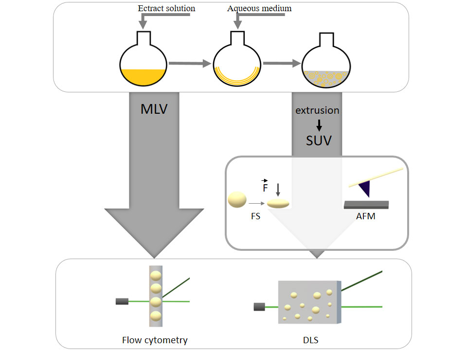
Liposomes were prepared by the thin film hydration (Bangham) method. The technique includes an organic phase for lipids dissolution followed by its removal under vacuum. Consequently, the thin, dry lipid film is formed. The film is further hydrated under agitation, which causes successive detachment of lipid lamellas and vesicles formation [12]. Each of the extracts used was dissolved in chloroform (20 mg/mL) and transferred to a round bottom flask. The chloroform was removed with the use of rotary evaporator until complete dryness to form thin lipid film. The film was hydrated with an aqueous solution (4 mL of 1% PBS buffer) at 42°C for 2 h in a water bath under continuous rotation. Obtained liposomal suspensions were divided into two parts. The first, containing multilamellar vesicles (MLV), was collected directly. The second part was extruded through a 100 nm ISOPORE® membrane filter (LiposoFast by Avestin, Germany) in 15 cycles which resulted in the formation of small unilamellar vesicles (SUV).
2.2.2 Zeta potential and size distribution
Zetasizer Nano ZS90 from Malvern Instruments (England) equipped with a 5 mW helium/neon laser was applied for the determination of both size and Zeta potential of the liposomes. The measurements were taken at 21°C for 200 s with a 90° detection angle. The final results were calculated using the cumulant technique with normal Gaussian distribution.
2.2.3 Flow cytometry
Flow cytometry is a multiparametric laser-based technique applied to evaluate the distribution of both the size and structure in populations of liposomes. The technique was initially applied for cell analysis [22]. However, there are a few reports in which flow cytometry was employed to the study of liposomes. Firstly, it was used to estimate the size and average membrane/internal volume with the use of fluorescent labelling [23]. The idea was further continued by Sato et al. who used double labelling to estimate membrane/internal volume as well as utilizing a fluorochrome with an affinity towards neutral lipids [24]. Alternatively, the size and structure distribution of positively charged liposomes in the size range of 100 to 1000 nm can be determined with the use of latex beads [25]. An idea of post-formation analysis of liposomes prepared from various lipids was presented recently [17]. The concept involved simultaneous labelling of vesicles with a hydrophilic dye that enters aqueous compartments and a lipophilic dye that binds to the phospholipid bilayer. Consequently, a method for fast and reliable analysis of liposomes without the need for problematic preparation or manipulation was developed [17]. The latter approach was applied in this research. The analysis was performed with the use of scattered laser light signals (irrespectively of detection of fluorescence signals from hydrophilic and lipophilic dyes) detected as the side and forward scatter, respectively abbreviated as SSC and FSC. The SSC collects signal at 90° angle to the incident laser light and represents the complexity of the assessed particles. The FSC collects signal in the same direction as the incident laser light and, in general, is proportional to the particles’ size [26]. The resolving power of a flow cytometer is determined by the performance of both the light detectors and digital signal processing. The instrument displays data in 262 144 channels per detector. Fluorescent staining and flow cytometry were used to compare the obtained populations of liposomes in terms of structure and size. Flow cytometric analysis was performed using BD FACS ARIA™III (Becton Dickinson, USA) flow cytometer (cell sorter) and was based on the protocol described previously [17]. Flow cytometric analyses were performed using logarithmic gains and specific detector settings (10 000 events were recorded per analysis). Each sample was analysed in triplicate. Data were acquired in a four-decade logarithmic scale as area signals (signal pulse sampling frequency was 10 MHz) and analysed with the FACS DIVA software (Becton Dickinson). Liposomes were characterised using two fluorescent parameters and two non-fluorescent parameters. Non-fluorescent parameters were forward and side scatter from FSC and SSC detectors, respectively, collected as area signals (FSC-A and SSC-A). FSC-A/SSC-A data provided the initial definition of particles’ size and structure. The complexity of liposomes was then evaluated using green and red fluorescence signals measurement from the combination of hydrophilic dye – thiazole orange (TO), collected using the FITC detector as area signals (FITC-A) and lipophilic dye – DiD, collected using the APC detector as area signals (APC-A), respectively. Distinct and representative (exhibiting low coefficient of variation values in the measured parameters) sub-populations of liposomes were discriminated.
2.2.4 Atomic force microscopy (AFM)
2.2.4.1 Preparation of the samples
AFM was originally designed to visualize nanoscopic and atomic surfaces of insulators [27]. Over the years, AFM began to offer much more than just topography imaging. Force spectroscopy (FS) is a powerful measurement mode which enables to determine nanomechanical parameters of investigated objects or surfaces, e.g.: the adhesion force, work of adhesion, elasticity (via Young’s modulus) [28]. These parameters can be determined, calculated and mapped in situ without damaging the sample with resolution in the pN range. This makes the method suitable for the analysis of demanding and extremely soft biological systems, e.g.: living cells, viruses, bacteria [29]. One of the practical and important applications of this technique is the ability to early distinguish between normal and cancer cells. Each aspect of an FS measurement can be controlled with negligible lateral displacements during the force curve cycle, with given indentation force or retraction rate for instance. Optical-lever feedback of AFM enables to perform experiments in a native biological buffer.
Mechanical properties of SUV were determined with the use of an Agilent 5500 (N9410S; Keysight, USA) atomic force microscope equipped with an environmental chamber and a liquid cell. The measurements were performed in force spectroscopy contact mode. A 2 cm2 sheets of freshly cleaved mica were mounted in a large liquid cell system (15 mm in diameter) and filled with 100 μL of 1% PBS that contained 100 μmoles of a selected type of SUV or MLV. Following a 30-minute incubation on top of the mica sheet, another 400 μL of 1% PBS was introduced to counter evaporation. The liquid cell was isolated from the influence of the external environment by a glass environmental chamber. The microscope was placed on an anti-vibration system and enclosed in an anti-acoustic chamber. The liquid cell was cleaned with ethanol before the introduction of subsequent samples.
2.2.4.2 Force spectroscopy (FS) measurements
Because of the progressive flattening processes of vesicles [17], each analysis of a sample was not longer than 40 min. During this time, it was possible to collect up to 7 FS force-curve grids. Each grid was obtained from a different part of the sample and consisted of 16 × 16 approach-retraction curves for a 16 μm2 surface area. 1792 approach-retraction curves were thus collected per sample. Each force curve was recorded with the same separation rate of 1 μm/s. The loading force did not exceed 20 nN in the grid measurements. However, to ensure full indentation of a vesicle, each sample was tested locally with a loading force up to 50 nN. The distance between each point at which a force curve was collected was 250 nm to ensure a single indentation event on a vesicle. Due to the very weak adhesion of the liposomes to the mica substrate, the measurements were done in the so-called blind mode in which the FS experiments were not preceded by a lateral topographical scan. Visualization of the vesicles suspended in the buffer by AFM prior to testing yielded unsatisfactory results. The thickness of lipid bilayer was assessed by indenting the bilayer patches that remained on the mica substrate. The exact procedure as applied here, was described elsewhere [30].
2.2.4.3 Cantilever details
The cantilever type was selected so as to enable the detection of very delicate vesicles (low force constant), avoid any perforation of the vesicle by a sharp and round tip (curvature of the end of the tip) and allow the collection of multiple force curves without degradation of the tip (material durability). To ensure the latter, two DNP B cantilevers were used in the experiment. Each cantilever tip was investigated under a Quanta 250 FEG (FEI, USA) scanning electron microscope before and after the measurement. Owing to the latter, the radius of the tip was determined (Figure 5f). To recalculate force units from the deflection signal for each force curve, it was necessary to find the deflection sensitivity and spring constant of the cantilever. The deflection sensitivity was determined with the use of a clean mica substrate in a buffer before each measurement. This was done for n=100 force curves at one point for each. The force constant of the cantilever was determined in air, according to an accurate reference method [31, 32]. The use of an interferometrically calibrated tipless reference cantilever, CLFC-NOBO C (with a spring constant of 0.112 N/m), enabled to perform the calibration with a total relative uncertainty lower than 5%. Detailed parameters that characterised the cantilever are presented in Table 2.
Mechanical parameters of cantilevers and tips used for AFM.
| Cantilever No. / Parameter | Type | Sensitivity | Force Constant | Tip [nm]*** Radius | Specimen |
|---|---|---|---|---|---|
| 1 | Bruker DNP B; (Silicon Nitride) | 168.1 ± 3.4 | 0.100 ± 0.004 (4.3%) | 21 ± 2 | H, CE |
| 2 | 80.6 ± 1.0 | 0.162 ± 0.004 (2.3%) | 19 ± 2 | EA, MA, HE |
*averaged from 80 measurements on mica in buffer, examined before each part of the experiment, uncertainty is the standard deviation of the mean; **data obtained using the reference method [33, 34] with Bruker CLFC-NOBO C (k=0.112 N/m), complex uncertainty, the relative uncertainty in percent is given in parentheses; ***tip radius examined before and after the experiment, without visible mechanical wear, uncertainty estimated.
2.2.4.4 Force curve selection procedure
Erroneous force curves or results without visible indentation of the vesicles were rejected before calculations. Only the results that showed proper indentation depth (close to the averaged maximum indentation depth) were taken into account. Only a small percentage of curves collected on the vesicles (no more than 6.7% per sample) were found valid. Because of the utilization of FS in blind mode, this step was necessary to make sure that the point of indentation was close to the geometric center of the tested vesicles.
2.2.4.5 Force curve analysis
Force curves were analysed quantitatively using AtomicJ 1.7.2 software [35]. The elasticity, in the form of Young’s modulus, was calculated by the Johnson-Kendall-Roberts (JKR) model [36]. This mathematical model is a popular and well-established modification of Hertz theory [37] in contact mechanics. It is suitable across a range of size scales – from nano- to microscale. The model assumes a finite contact area between the contacting surfaces at zero load and predicts an external, non-zero force, needed to separate them. It is suitable only for small loading forces and soft materials, without asperities that would create interfacial gaps, which strongly affect the attractive adhesion forces. In case of this work, the surface of the studied liposomes was very soft and smooth [17]. Thus, the JKR model was suitable and characterised the indentation of a vesicle well. Due to the nature of the measurement taken in a biological buffer, it was necessary to discuss the influence of the additional solvent hydration and electrical double layer forces acting at the interface between the vesicle and AFM tip. According to simulations by Korayem et al. that regard soft biological cells, the output from the JKR model for air and liquid environment is practically the same, differences are negligible [38]. Thus, the JKR model was applied. Only the linear region of the approach indentation curve was taken into account. The radii of both tips and the Poisson’s ratio, assumed to be 0.5 according to other papers [39, 40, 41], were included into calculations.
2.2.5 Statistical analysis
All experiments were performed in triplicates and the results were presented as means ± standard deviation (SD). The significance of the differences between mean values was determined by analysis of variance (ANOVA). Post-hoc analysis was done with the use of Tukey’s test. Principal Component Analysis (PCA) was applied in order to analyse the relations between membrane composition [13] and liposomes properties derived from AFM experiment. Pearson coefficient was calculated in order to analyse the relations between membrane composition and liposomes size, polydispersity, nanomechanics, Zeta potential. Results in all the tests were considered significant at α=0.05. The statistical analysis was performed using Statistica 10.0 software (StatSoft, Inc., Tulsa, USA).
Ethical approval: The conducted research is not related to either human or animal use.
3 Results and Discussion
3.1 General characteristics of liposomes
Populations of liposomes obtained using the tested extracts as membrane building material were analysed by DLS in terms of Z-average size [nm], polydispersity index PDI [-], and Zeta potential [mV] before MLV and after SUV the extrusion process (Figure 1abc). The Z-average size of MLV formed with EA, MA, H, and CE samples exceeded 1 μm. EA- and HE-derived vesicles were significantly distinct from the other liposomes as the biggest and the smallest, respectively. There were no significant differences in PDI values of the populations of MLV, thus they were comparable in terms of polydispersity. Differences in Z-average size and PDI values between MLV and SUV populations were clearly noticeable, which proved a successful extrusion process. The Z-average size of all the samples, after post-preparation processing, were below 240 nm. The effect of extrusion on the size of HE-derived liposomes was relatively small. It was likely a result of their small initial size. The EA-, MA-, CE-derived liposomes were characterised by very low PDI values. The vesicles obtained with MA extract showed the lowest PDI value that may indicate increased stability (Figure 1c).
![Figure 1 Dynamic light scattering and Zeta potential of liposomes measured directly after preparation: (a) Z-average size [nm]; (b) Zeta potential [mV]; (c) PDI. Bars bearing different superscripts are statistically, significantly different (α<0.05).](/document/doi/10.1515/chem-2019-0070/asset/graphic/j_chem-2019-0070_fig_001.jpg)
Dynamic light scattering and Zeta potential of liposomes measured directly after preparation: (a) Z-average size [nm]; (b) Zeta potential [mV]; (c) PDI. Bars bearing different superscripts are statistically, significantly different (α<0.05).
The Zeta potential is a parameter that reflects repulsion forces between colloidal particles, a measure of the stability of the system. Only slight differences of Zeta potential absolute values were determined between EA-, MA-, H-, and CE-derived MLV samples (Figure 1a). The HE-based vesicles were significantly different in terms of this parameter showing a potential almost two-fold higher than the other samples. This might have been related to the exceptionally high content of carotenoids in this extract in comparison to the other extracts (Table 1) [13]. Based on the similarity of Zeta potential obtained for MLV and SUV (Figure 1b), it can be stated that the extrusion process did not affect the Zeta potential of the analysed liposomes.
The intensity distribution [%] versus the size of the liposomes [nm] (Figure 2ab) is a 1st order result of dynamic light scattering (ISO 22412: 2008) used for reporting the size of each peak. Initially wide peaks (Figure 2a) became narrow after the processing and showed monomodal size distribution (Figure 2b). This confirmed the effect of extrusion on the mean diameter of the vesicles and corresponded with the data shown in the Figure 1 with the Z-average size of SUV in the range of 180-235 nm.
![Figure 2 Size distribution of MLV (a) and SUV (b) populations. Intensity [%] stands for intensity distribution - the 1st order result derived from dynamic light scattering measurement and represents the intensity distribution of particles sizes. The parameter is naturally weighted corresponding to the scattering intensity of each particle subpopulation/fraction.](/document/doi/10.1515/chem-2019-0070/asset/graphic/j_chem-2019-0070_fig_002.jpg)
Size distribution of MLV (a) and SUV (b) populations. Intensity [%] stands for intensity distribution - the 1st order result derived from dynamic light scattering measurement and represents the intensity distribution of particles sizes. The parameter is naturally weighted corresponding to the scattering intensity of each particle subpopulation/fraction.
3.2 Flow cytometry
The size and complexity distribution of both MLV and SUV was analyzed by studying FSC and SSC signals [42]. Moreover, a combination of TO and DiD fluorochromes was used to evaluate the feasibility of the system to detect the structural diversity of liposomes. It was assumed that TO would penetrate the liposome to its aqueous core and DiD would stain the membrane of the vesicle. The results are shown on FITC vs APC bivariate dot plots (Figure 3 , 4). FITC and APC axes of the dot plot present the intensity of green and red fluorescence signals, respectively. Background noise was discriminated by an analysis of the distribution of the bivariate FSC vs SSC dot plots.
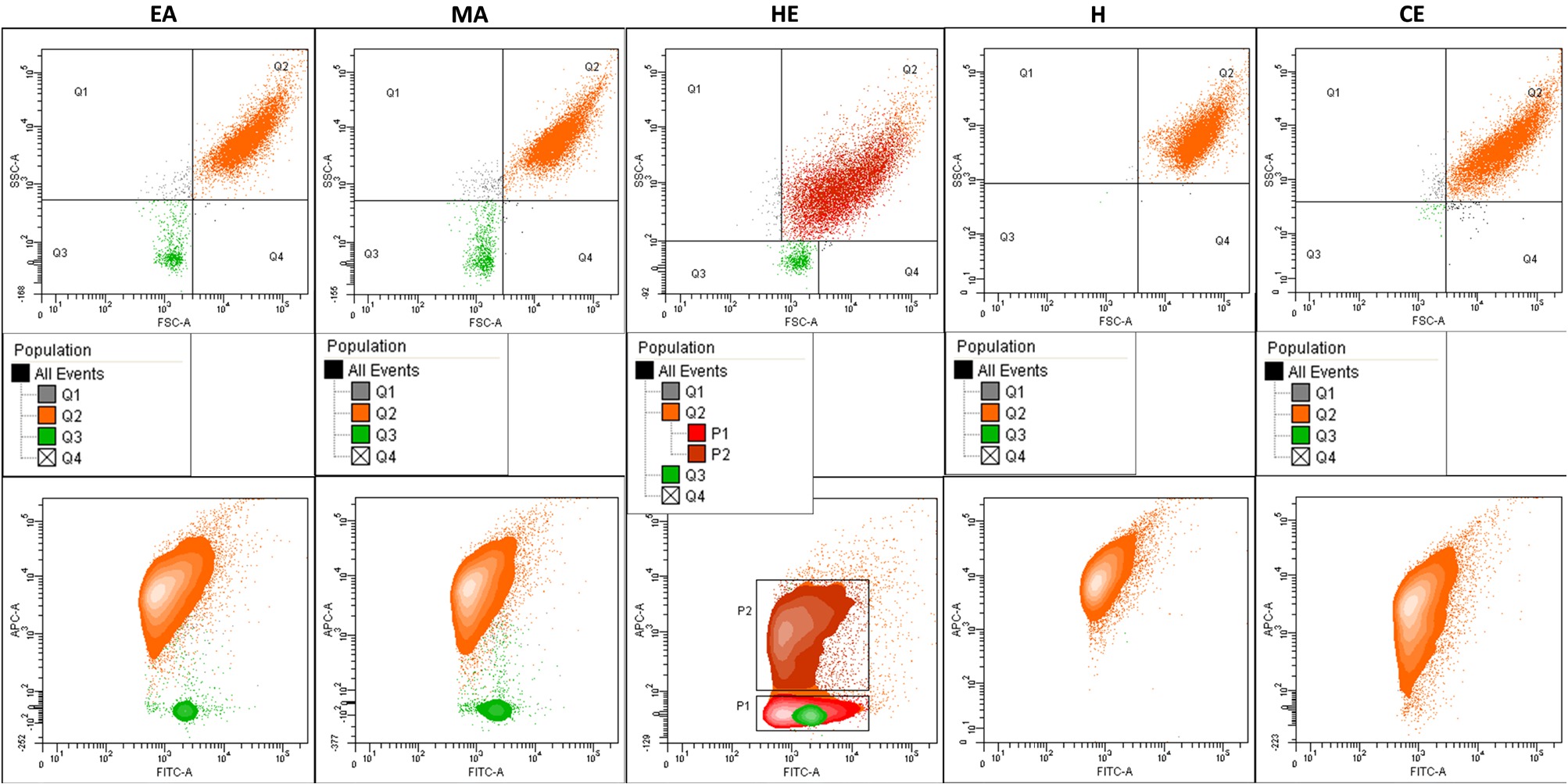
Diversity of MLV populations in terms of morphology and structure determined by flow cytometry. Bivariate contour plots in the upper row for each population FSC-A vs SSC-A, lower row – FITC-A vs APC-A. Diagrams indicate the gating strategy (structure of defined subpopulations). Sub-populations P1 and P2 (discriminated within the Q2 region using TO/DiD staining) indicate higher structural diversity of liposomes.
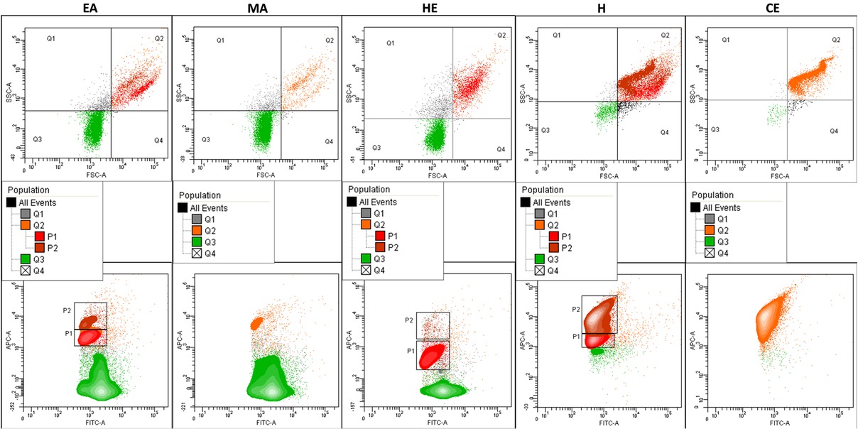
Diversity of SUV populations in terms of morphology and structure determined by flow cytometry. Bivariate contour plots in the upper row for each population FSC-A vs SSC-A, lower row – FITC-A vs APC-A. Diagrams indicate the gating strategy (structure of defined subpopulations). Sub-populations P1 and P2 (discriminated within the Q2 region using TO/DiD staining) indicate higher structural diversity of liposomes.
![Figure 5 Quantitative analysis of nanomechanical properties of SUV by FS: (a,b) illustrative scheme of the force spectroscopy experiment – force curve and indentation (I. – physical contact between AFM tip and surface of a vesicle, II. – indentation of a vesicle, III. – indentation of the mica substrate; symbols: z – scanner position, d – cantilever deflection, VD – cantilever deflection signal, h – height of vesicle, S – sensitivity, F – force); (c,d) typical approach (blue) – retraction (green) force curve recorded on a CE vesicle with calculated force [nN]; (d,e) the Johnson-Kendall-Roberts (JKR, red curve) model fitted to typical CE data region II. – force curve (d) and corresponding force-indentation profile (e); (f) scanning electron micrograph of Bruker DNP B tip after the experiment (cantilever no. 1, inset: radius R of the tip); (g,h) calculated Young’s modulus and adhesion force histograms for CE specimen (green striated curve – fitted Gauss distribution profile); (i) typical approach (blue) – retraction (green) force curve recorded on a H sample depicts indented lipid bilayer patch (region II.).](/document/doi/10.1515/chem-2019-0070/asset/graphic/j_chem-2019-0070_fig_005.jpg)
Quantitative analysis of nanomechanical properties of SUV by FS: (a,b) illustrative scheme of the force spectroscopy experiment – force curve and indentation (I. – physical contact between AFM tip and surface of a vesicle, II. – indentation of a vesicle, III. – indentation of the mica substrate; symbols: z – scanner position, d – cantilever deflection, VD – cantilever deflection signal, h – height of vesicle, S – sensitivity, F – force); (c,d) typical approach (blue) – retraction (green) force curve recorded on a CE vesicle with calculated force [nN]; (d,e) the Johnson-Kendall-Roberts (JKR, red curve) model fitted to typical CE data region II. – force curve (d) and corresponding force-indentation profile (e); (f) scanning electron micrograph of Bruker DNP B tip after the experiment (cantilever no. 1, inset: radius R of the tip); (g,h) calculated Young’s modulus and adhesion force histograms for CE specimen (green striated curve – fitted Gauss distribution profile); (i) typical approach (blue) – retraction (green) force curve recorded on a H sample depicts indented lipid bilayer patch (region II.).
According to FSC vs SSC dot plots (Figure 3), the majority of MLV liposomes represented a one disperse subpopulation Q2 (Q2 region that contained 92.3, 90, 93, 99.85, and 98.2% of particles of EA-, MA-, HE-, H-, and CE-derived liposomes, respectively). This was accompanied by a low structural diversity of MLV liposomes as indicated by the distribution of the population presented in FITC vs APC dot plots. The HE variant of MLV liposomes was an exception that manifested polydispersity in terms of both size and structure. In the case of this variant, two separate sub-populations, P1 and P2, were distinguishable when the TO/DiD-stained samples were analysed. This corresponded to the results of the DLS analysis (Figure 1), as the HE variant of MLV liposomes showed the smallest size and highest absolute value of Zeta potential. This could stem from the exceptionally high carotenoids content [13] (Table 1). Moreover, a sub-population Q3 (Q3 region), supposedly composed of small liposomes of a relatively simple complexity, was detected in the populations of EA-, MA-, and HE-based vesicles. This subpopulation amounted to 6.43, 8.27, and 6.13% of the total population of the respective samples.
FSC vs SSC bivariate dot plots revealed a transformation of the vesicles that resulted from extrusion (Figure 4). The analysis with flow cytometry demonstrated striking differences between MLV and SUV liposomes in terms of both size and structure. The observed differences were dependent on the extract variant used for the preparation of samples. EA-, MA-, and HE-derived vesicles showed striking distinction with the dominance of Q3 sub-population (Q3 region that contained 73.17, 85.47, and 68.3% of particles formed with EA, MA, and HE extracts, respectively). The liposomes prepared with H and CE extracts showed a dominance of sub-population Q2 (Q2 region). The analysis also revealed: (i) a more complex structure of Q2 sub-population, as presented for H extract, for which two separate sub-populations P1 and P2 were distinguishable after TO/DiD staining, and (ii) higher density of sub-population Q2, as indicated for the CE extract-based sample on FSC vs SSC and FITC vs APC dot plots. Thus, the majority of the produced vesicles underwent a reduction in size and lamellarity. In the case of H- and CE-derived samples, the dominant subpopulation did not change following the extrusion process. However, after the treatment, FSC vs SSC bivariate dot plots obtained for SUV showed S-like shapes that were found to be common in a previous report [17]. FITC vs APC bivariate dot plots demonstrated the presence of P1 and P2 sub-populations within the Q2 region in the samples based on EA, HE, and H extracts. An uneven distribution of SUV liposomes was thus determined. P1 and P2 sub-populations were oriented vertically (along the axis of ordinates of the dot plot) so the differences between these sub-populations were related to the red fluorescence signal generated by the DiD lipophilic dye. Nonetheless, the majority of EA-, MA-, and HE-derived liposomes were situated within sub-population Q3 (Q3 region) characterised by low lamellarity and complexity. The liposomes obtained with H and CE extracts showed different distribution of structural features with only 7.1 and 1.15% particles of the respective samples contained within sub-population Q3 (Q3 region). Moreover, the CE-derived population of vesicles became more uniform because of extrusion, especially in terms of structure. Compared to the other samples, this population was also found to show increased lamellarity and complexity of vesicles.
3.3 AFM and nanomechanical study
To analyse a scope of nanomechanical properties of the prepared SUV, FS, a powerful tool of the AFM technique, was employed. The FS measurements of the engineered egg yolk vesicles were done in the buffer, to ensure a quantitative output as close to native as possible. The vesicles were attached to the atomically flat muscovite mica in a soft manner via electrostatic interactions. To avoid any impurities, no functionalization of the substrate was employed. Separated vesicles were characterised in terms of force and work of adhesion, elasticity, bending modulus, and thickness of the lipid membrane. An attempt at correlating the mechanical properties with chemical composition was made. Only SUV were analysed (Figure 5) because they provide more approximate feedback on single lipid bilayer properties than MLV. The percentage of the SUV detected in relation to the introduced number of vesicles (the ratio of the number of curves on a vesicle to total curves obtained) was as follows: EA (8%), MA (93%), HE (1.4%), H (22%), and CE (66%). This suggests different adhesion properties of the outer lipid membrane (Table 1). As the mica is negatively charged, the deposition of vesicles is strongly related to both charge of liposomes and pH [33]. Differences in the deposition percentage could thus be expected.
The vesicles were indented by the AFM tip (regions I. and II.) until the significantly stiffer substrate was reached (region III.) (Figure 5a-c). The higher stiffness of the substrate is represented by the steeper slope of the approach indentation curve (blue). Moreover, the presence of a single jump-in event, marked as VD and Z2 in Figure 5b and visible also in CE force curve on Figure 5c, is a clear information on the elasticity limit of a vesicle which facilitates interpretation of the results. This particular event is related to the detection of a force gradient near the substrate. In this part of the curve (beginning of III.), the force gradient, due to the vertical proximity of the AFM tip and the substrate, exceeded the force constant of the AFM cantilever. The attractive forces brought the tip to the substrate through the vesicle. Such jump-in events were also described in other works related to liposomes that involved FS [39, 40]. During the indentation, the AFM tip was compressing the vesicle which underwent a change in shape – from almost round (region I., vesicle’s height h0, flattened by attractive electrostatic forces between the substrate and the vesicle) to planar (region II., vesicle’s height h1) and concave (beginning of region III., vesicle’s height h2) (Figure 5a) [30]. The retraction curve (green, Figure 5b-d) brings information about the adhesion force and its work. Adhesion peak height and its area with respect to the baseline are the measures of the respective parameters (Figure 5b-d). Elasticity was calculated from region II. of the approaching curve (Figure 5d). The red curve represents a typical JKR model fitted to the experimental data. The indentation fingerprint in form of force-indentation curve is also fitted with JKR (red curve, Figure 5e). The resulted histograms of Young’s modulus and adhesion force show Gaussian distribution (green dotted curve, Figure 5g,h). Such distribution is common for measurements of many isolated objects of the same type.
Table 3 presents parameters derived from the experimental data. The deformation of vesicles is an intermediate product obtained from the indentation procedure. Calculated elasticity was the highest for EA-based vesicles (28.53 MPa) which implied one order of magnitude higher stiffness compared to the other samples. In the case of these vesicles, the loading force (5.4 nN) caused a much smaller deformation (8.8 nm) in comparison to the other, softer vesicles. HE- and CE-derived vesicles had similar elasticity (within the margin of error) – 2.16 MPa and 2.21 MPa, respectively. The most delicate liposomes were obtained with MA (0.53 MPa) and H (1.42 MPa) extracts. The elasticity data were consistent with the values reported for different liposomes in the literature [39, 40]. Locally measured elasticity was related to the bending of the lipid bilayer. The bilayer, a building block of each vesicle, is more or less susceptible to bending [34]. This complex behaviour is described by the so-called bending modulus [43]. This parameter can be calculated if the elasticity, Poisson’s ratio and the thickness of the lipid bilayer of a vesicle are known [22, 41]. Bending modulus is temperature-dependent [39]. For the investigated samples, the bending modulus was closely correlated with elasticity represented by Young’s modulus (Table 3). The highest value was found for EA liposomes and equaled 1.1‧10-19 J. The values of bending modulus determined for the other vesicles were one order of magnitude smaller which indicated less rigid structure. The lowest found value was 0.09‧10-19 J for H vesicles. Noteworthy, the observed values of bending modulus data were consistent with the literature [39, 40, 43, 44].
Parameters obtained from the quantitative force curve analysis of SUV.
| Data Type / Sample | EA | MA | HE | H | CE |
|---|---|---|---|---|---|
| Loading Force [nN]* | 5.4 ± 0.3a | 2.4 ± 0.1b | 5.6 ± 0.3a | 6.8 ± 0.2c | 5.8 ± 0.1a |
| Deformation [nm]* | 8.8 ± 0.6a | 70.6 ± 1.8b | 67.1 ± 9.9b | 71.4 ± 1.5b | 49.7 ± 1.2c |
| Young’s Modulus [MPa]* | 28.53 ± 1.88a | 0.53 ± 0.01b | 2.16 ± 0.36c | 1.42 ± 0.03d | 2.21 ± 0.05c |
| Bending Modulus (10-19) [J]** | 1.10 ± 0.30 | 0.13 ± 0.04 | 0.18 ± 0.03 | 0.09 ± 0.02 | 0.17 ± 0.03 |
| Adhesion Force [nN]* | 0.93 ± 0.11a,d | 0.79 ± 0.02b | 1.14 ± 0.05a | 0.17 ± 0.01c | 0.99 ± 0.03d |
| Work of Adhesion [μJ]* | 273.3 ± 35.5a | 351.9 ± 22.7a,b | 398.3 ± 56.2b | 398.4 ± 20.1b | 359.5 ± 17.9b |
| Data Count*** | 21 | 96 | 16 | 97 | 117 |
| Thickness of the Lipid Double Layer [nm]* | 3.26 ± 0.24a | 5.98 ± 0.63b | 4.18 ± 0.23c | 3.78 ± 0.21a,c,d | 4.10 ± 0.23c,d |
| Data Count | 16 | 9 | 16 | 20 | 20 |
Values (means ± SD) with different superscripts are significantly different (α<0.05), *uncertainty is standard deviation of the mean; **complex uncertainty; ***data from separated vesicles (the thickness of the lipid double layer is given from a separate statistics).
Adhesion force recorded against the AFM tip was about 1 nN or less. The nonspecific adhesion force depends on the contact area between the sample and the AFM tip. Both utilized tips had comparable radii (Table 2). Thus, the data concerning adhesion force and work of adhesion are comparable for all the studied vesicle variants (Table 3). Notably low adhesion force of 0.17 nN was found for the H sample. These vesicles also showed one of the most delicate mechanical structures. The most rigid sample, EA, had relatively high adhesion force that equaled 0.93 nN and was comparable to HE and CE. However, the EA sample was characterised by an exceptionally low value of work of adhesion (273.3 μJ). Work of adhesion was comparable among the other tested samples. This parameter is related to the stiffness of the bilayer. Stiffer EA vesicles with their compact and rigid structure could have been easily separated during the retraction of the AFM tip. The structure of the vesicle was not susceptible to stretching.
The last assessed parameter was the thickness of the lipid bilayer (Figure 5i). The stiff EA vesicles had the thinnest lipid bilayer (3.26 nm). The bilayer of the most delicate MA vesicles was nearly twice as thick (5.98 nm). The bilayer of the delicate H vesicles, however, was similar to that of EA (3.78 nm). This suggests that the mechanical properties of the studied liposomes are not solely dependent on the vertical dimensions of the lipid bilayer.
3.4 Correlation analysis
As indicated in our previous work [13], the extracts used for obtaining the tested liposomes varied greatly in terms of chemical composition. Generally, phospholipids, acylglycerols and cholesterol were their main constituents. The phospholipid fraction of the extracts consisted of phosphatidylcholine (PC), lysophosphatidylcholine (LPC), phosphatidylethanolamine (PE), lysophosphatidylethanolamine (LPE) and sphingomyelin (SM). The extracts also contained tocopherols and carotenoids [13]. PCA analysis of the mechanical characteristics of the tested vesicles and the data on their chemical composition was performed to analyse the main factors determining the properties of the analysed liposomes. The two principal components (factor 1 and 2) explained 82.19% of the total variance, Figure 6.
![Figure 6 PCA loading plot (a) and score plot (b) of characteristics and nanomechanical properties of liposomes formed from different extracts (Table 3). The data on the composition of extracts were derived from our earlier work [13]. Abbreviations: PL – phospholipids; AG – acylglycerols; Chol – cholesterol; PC/PL – phosphatidylcholine share in total phospholipids; LPC/PL – lysophosphatidylcholine share in total phospholipids; PE/PL - phosphatidylethanolamine share in total phospholipids; LPE/PL – lysophosphatidylethanolamine share in total phospholipids; SM/PL – sphingomyelin share in total phospholipids; PF/AG - polar fraction share in total acylglycerols; NPF/AG – non-polar fraction share in total acylglycerols; α-T - alfa-tocopherol; γ-T - gamma-tocopherol; L – lutein; Z – zeaxanthin; Young – Young modulus; Bend – bending modulus; Af – adhesion force; Aw – work of adhesion; h – thickness of the lipid double layer.](/document/doi/10.1515/chem-2019-0070/asset/graphic/j_chem-2019-0070_fig_006.jpg)
PCA loading plot (a) and score plot (b) of characteristics and nanomechanical properties of liposomes formed from different extracts (Table 3). The data on the composition of extracts were derived from our earlier work [13]. Abbreviations: PL – phospholipids; AG – acylglycerols; Chol – cholesterol; PC/PL – phosphatidylcholine share in total phospholipids; LPC/PL – lysophosphatidylcholine share in total phospholipids; PE/PL - phosphatidylethanolamine share in total phospholipids; LPE/PL – lysophosphatidylethanolamine share in total phospholipids; SM/PL – sphingomyelin share in total phospholipids; PF/AG - polar fraction share in total acylglycerols; NPF/AG – non-polar fraction share in total acylglycerols; α-T - alfa-tocopherol; γ-T - gamma-tocopherol; L – lutein; Z – zeaxanthin; Young – Young modulus; Bend – bending modulus; Af – adhesion force; Aw – work of adhesion; h – thickness of the lipid double layer.
Projection of the cases on the factor plane (Figure 6b) pointed to a similarity of MA and EA extracts as well as CE and HE extracts. At the same time the H extract differed from the others. Similar results were found in our previous works describing in details extraction procedures and composition of extracts [13] as well as surface behaviour of monolayers they formed [21]. The H extract was obtained in simple one step procedure and was rich in acylglycerols but scant in carotenoids due to the use of nonpolar hexane as the extraction solvent. The CE and HE extracts were also obtained in a simple, one-step extraction procedure differing only in the temperature used. The liposomes obtained from the CE and HE extracts did not differ significantly in loading force, Young’s modulus, bending modulus, work of adhesion, thickness of lipid double layer values (Table 3). Nonetheless, these extracts differed in surface behaviour as the compression process of the HE monolayer is irreversible, whereas the CE monolayer revealed only small hysteresis [21]. This corresponds to the higher α-tocopherol content in CE extract [13]. We believe special attention should be paid to the HE extract, as this material enabled, even by simple Bangham procedure, to obtain relatively small vesicles of the highest absolute value of Zeta potential (Figure 1). This phenomenon is related to a distinctly high content of carotenoids [13]. Liposomes from EA and MA extracts of similar composition, obtained in the course of a few-step procedure and rich in phospholipids were characterised by significantly different mechanical properties excluding work of adhesion. Projection of the variables on the factor plane (Figure 6a) proved, however, that the phospholipid content had a positive but slight effect on Young and bending moduli, while the share of lysophosphatidylethanolamine in total phospholipids was of predominant influence. Cholesterol and minor constituents (tocopherols and carotenoids) were also relevant to the liposomes’ mechanical properties. Their effect on Young and bending moduli, in contrast to the impact of lysophosphatidylethanolamine, was negative. Cholesterol is a widely used additive in liposomal formulations as it improves membrane rigidity [40, 45, 46]. It interacts with the phospholipid molecules thus resulting in the modulation of membrane properties [47]. This explains the change in the mechanical strength of liposomes of varying cholesterol content. With regards to the shares of other phospholipids in total, the fraction with the biggest effect were phosphatidylcholine and lysophosphatidylcholine. However, their effects on the mechanical strength of liposomes were opposite, negative and positive respectively. Sphingomyelin also revealed small effect on vesicles mechanics while there was no effect of phosphatidylethanolamine share (Figure 6a). In general, the presence of the hydrolyzed forms of phospholipids, which are more polar than their non-hydrolyzed counterpart, stiffened the structure of the vesicle bilayer making it stronger mechanically.
Liposomes and cells membrane surface interactions are, in living organisms, responsible for fusion and absorption [48]. However, in liposomal systems they may influence stability of vesicles. Adhesion force is determined by the outer part of a vesicle, and was negatively correlated with the contents of the acylglycerol fraction and positively, but to a lesser extent, phospholipids. At the same time, the work of adhesion was mostly influenced by total tocopherols content. Both acylglycerols and phospholipids strongly determine the properties of the surface of liposomes as their polar heads form the superficial layer of the shell of the vesicles.
Comparison of the biomechanical analyses results (Table 3) with rheology data described in our previous work [21] indicated a correlation between the properties of mono- and bilayer vesicles. As evidenced by the Pearson correlation coefficients (Table 4), mechanical properties of liposomes described by Young and bending moduli did not show any significant correlation except for the one between bending and dilatational elastic moduli. In contrast, parameters relating to the stability of liposomal dispersion correlated with the values describing adhesion phenomena. The strongest and positive correlation was observed between the adhesion force and collapse surface pressure. It is worth to mention that H extract, rich in acylglycerols, formed monolayer of the lowest CSP value [21]. Moreover, the adhesion force correlated also with the dilatational elastic modulus and the Zeta potential, whereas the work of adhesion – with the dilatational elastic and the shear loss moduli as well as with the Z-average size. Lipid double layer thickness correlated with both loss moduli.
Pearson correlation coefficients for the interfacial rheological properties of monolayer [21] and nanomechanical properties of liposomes.
| Dilatational Elastic Modulus | Dilatational Loss Modulus | Shear Elastic Modulus | Shear Loss Modulus | Collapse Surface Pressure | Zeta Potential | Z-Average Size | |
|---|---|---|---|---|---|---|---|
| Young’s Modulus | 0.605* | -0.037* | -0.030* | 0.625* | 0.102 | 0.133 | -0.437 |
| Bending Modulus | 0.653 | -0.006 | -0.025* | 0.630* | 0.164 | 0.105 | -0.433 |
| Adhesion Force | 0.648 | 0.071 | 0.306 | 0.385 | 0.969 | -0.562 | 0.108 |
| Work of Adhesion | -0.761 | -0.394 | -0.133 | -0.703 | -0.256 | -0.348 | 0.705 |
| Thickness of the Lipid Double Layer | 0.212 | 0.731 | -0.219 | -0.512 | 0.300 | 0.003 | 0.046 |
*Significance at α=0.05.
4 Conclusions
As a result of this and previous work, it was proved that the chemical composition of a bilayer determined the mechanical properties, structure, and stability of liposomes. However, the effect of minor constituents was just as important as of main components. Moreover, the share of individual phospholipid fractions in total phospholipids or polar to non-polar fractions ratio of acylglycerols exerted a significant effect.In terms of its positive effect on the nanomechanical properties, described by Young and bending moduli, lysophosphatidylethanolamine share proved to be the most important of the phospholipids. Significantly weaker, but also a positive effect was observed for total phospholipids content as well as the share of lysophosphatidylcholine and sphingomyelin in the total phospholipids. Cholesterol, tocopherols, and carotenoids exerted the strongest negative effect on Young and bending moduli. The other negative, but significantly weaker, the effect on Young and bending moduli was a result of acylglycerol content as well as the share of phosphatidylcholine in total phospoholipds.
The share of individual phospholipid fractions in total phospholipids or polar to non-polar fractions ratio of acylglycerol affected not only nanomechanical properties of liposomes but also surface behaviour of analysed extracts. The components improving liposomes nanomechanical properties also increased dilatational elastic moduli.
Similarly, minor constituents affected nanomechanical properties of liposomes and surface behaviour of extracts. Tocopherols negatively influenced both interfacial shear moduli and dilatational elastic moduli. Contrarily, the presence of carotenoids positively affected interfacial shear elastic moduli. The presence of carotenoids was crucial for the stability of liposomes, represented by Zeta potential. The liposomes structure as well as the susceptibility to changes under the influence of the extrusion process also refleceted carotenoid content. Lipid double layer thickness depended mainly on the α-tocopherol content, which positively affected loss moduli of monolayer films too.
According to the presented results, MA and EA extracts as well as CE and HE extracts proved similar to each other, whereas H extract differed significantly. The H extract was rich in acylglycerols but deficient in carotenoids due to the use of nonpolar hexane as the extraction solvent. It formed liposomes of the lowest mechanical strength, stability, permeability, and heterogeneous in terms of structure.
The CE and HE extracts showed different surface properties but formed liposomes of approximate nanomechanical properties. The above corresponded to the higher α-tocopherol content in CE extract. HE extract, containing an exceptionally high amount of carotenoids, enabled, to obtain relatively small vesicles of the highest absolute value of Zeta potential, even by simple Bangham method. Mechanical strength of liposomes, made from MA and EA extracts, the richest in phospholipids, differed from each other due to differences in the share of lysophosphatidylethanolamine in the total phospholipid content. However, they did not show structural differences. Their structure was found to be of the lowest lamellarity and complexity.
Considering the potential application of developed extracts in technological practice it can be stated that, four of them i.e. MA, EA, CE, and HE could be used for the production of functional foods. The HE extract deserves special attention, as it was obtained by simple, one-step extraction and enabled the formation of small, stable liposomes by a simple procedure. At the same time, we would not recommend the H extract, as it resulted in liposomes of low stability and deficient permeability.
Acknowledgments
M.W. acknowledges the Ministry of Science and Higher Education in Poland for financial support within the project No 06/62/SBAD/1923 realized at the Faculty of Technical Physics, Poznan University of Technology.
Funding: This research was funded by the grant No. 508.771.01.5 funded by the Polish Ministry of Science and Higher Education for maintenance of research potential.
Conflict of Interest
Conflicts of Interest: The authors declare no conflict of interest.
Abbreviations
- Af
adhesion force
- ANOVA
analysis of variance
- Aw
work of adhesion
- AFM
atomic force microscopy
- AG
acylglycerols
- APC
allophycocyanine red fluorescence signal
- Chol
cholesterol
- CSP
collapse surface pressure
- DiD
1,1′-dioctadecyl-3,3,3′,3′-tetramethylindodicarbocyanine perchlorate
- DLS
dynamic light scattering
- DMSO
dimethylsulfoxide
- FITC
fluorescein 5(6)-isothiocyanate green fluorescence signal
- FS
force spectroscopy
- FSC
forward scatter
- h
thickness of the lipid double layer
- JKR
Johnson-Kendall-Roberts model
- L
lutein
- LPC/PL
lysophosphatidylcholine share in total phospholipids
- LPE/PL
lysophosphatidylethanolamine share in total phospholipids
- MLV
multilamellar vesicle
- NPF/AG
non-polar fraction share in total acylglycerols
- PBS
phosphate buffered saline
- PCA
principal component analysis
- PC/PL
phosphatidylcholine share in total phospholipids
- PDI
polydispersity index
- PE/PL
phosphatidylethanolamine share in total phospholipids
- PF/AG
polar fraction share in total acylglycerols
- PL
phospholipids
- SD
standard deviation
- SM/PL
sphingomyelin share in total phospholipids
- SSC
side scatter
- SUV
small unilamellar vesicle
- TO
thiazole orange (1-Methyl-4-[(3-methyl-2(3H)-benzothiazolylidene)methyl] quinolinium p-tosylate)
- Z
zeaxanthin
- γ-T
gamma-tocopherol
- α-T
alfa-tocopherol
References
[1] Bozzuto G., Molinari A., Liposomes as nanomedical devices. Int. J. Nanomedicine, 2015, 10, 975 – 999.10.2147/IJN.S68861Suche in Google Scholar PubMed PubMed Central
[2] Teixeira M. C., Severino P., Andreani T., Boonme P., Santini A., Silva A.M., Souto E.B., D-α-tocopherol nanoemulsions: Size properties, rheological behavior, surface tension, osmolarity and cytotoxicity. Saudi Pharm. J., 2017, 25, 2, 231 – 235.10.1016/j.jsps.2016.06.004Suche in Google Scholar PubMed PubMed Central
[3] Naseri N., Valizadeh H., and Zakeri-Milani P., Solid lipid nanoparticles and nanostructured lipid carriers: Structure preparation and application. Adv. Pharm. Bull., 2015, 5, 3, 305 – 313.10.15171/apb.2015.043Suche in Google Scholar PubMed PubMed Central
[4] Fathi M., Mozafari M.R., and Mohebbi M., Nanoencapsulation of food ingredients using lipid based delivery systems. Trends Food Sci. Technol., 2012, 23, 1, 13 – 27.10.1016/j.tifs.2011.08.003Suche in Google Scholar
[5] Teixeira M.C., Carbone C., and Souto E.B., Beyond liposomes: Recent advances on lipid based nanostructures for poorly soluble/poorly permeable drug delivery. Prog. Lipid Res., 2017, 68, 1 – 11.10.1016/j.plipres.2017.07.001Suche in Google Scholar PubMed
[6] Budai M., Chapela P.J., Grof P., Physicochemical characterization of stealth liposomes encapsulating an organophosphate hydrolyzing enzyme. J. Liposome Res., 2009, 19, 2, 163 – 168.10.1080/17482940902724044Suche in Google Scholar PubMed
[7] da Silva Malheiros P., Daroit D.J., and Brandelli A., Food applications of liposome-encapsulated antimicrobial peptides. Trends Food Sci. Technol., 2010, 21, 6, 284 – 292.10.1016/j.tifs.2010.03.003Suche in Google Scholar
[8] Sherry M., Charcosset C., Fessi H., and Greige-Gerges H., Essential oils encapsulated in liposomes: a review. J. Liposome Res., 2013, 23, 4, 268 – 75.10.3109/08982104.2013.819888Suche in Google Scholar PubMed
[9] Gonnet M., Lethuaut L., and Boury F., New trends in encapsulation of liposoluble vitamins. J. Control. Release, 2010, 146, 3, 276—290.10.1016/j.jconrel.2010.01.037Suche in Google Scholar PubMed
[10] Bryła A., Lewandowicz G., and Juzwa W., Encapsulation of elderberry extract into phospholipid nanoparticles. J. Food Eng., 2015, 167, 189 – 195.10.1016/j.jfoodeng.2015.07.025Suche in Google Scholar
[11] Emami S., Azadmard-Damirchi S., Peighambardoust S. H., Valizadeh H., and Hesari J., Liposomes as carrier vehicles for functional compounds in food sector. J. Exp. Nanosci., 2016, 11, 9, 737 – 759.10.1080/17458080.2016.1148273Suche in Google Scholar
[12] Meure L.A., Foster N.R., and Dehghani F., Conventional and dense gas techniques for the production of liposomes: a review. AAPS Pharm. Sci. Tech., 2008, 9, 3, 798 – 809.10.1208/s12249-008-9097-xSuche in Google Scholar PubMed PubMed Central
[13] Kondratowicz A., Neunert G., Niezgoda N., Bryś J., Siger A., Rudzińska M., Lewandowicz G., Egg yolk extracts as potential liposomes shell material: composition compared with vesicles characteristics. J. Food Sci., 2018, 83, 10, 2527-2535.10.1111/1750-3841.14341Suche in Google Scholar PubMed
[14] Bolean M., Borin I.A., Simão A.M.S., Bottini M., Bagatolli L. A., Hoylaerts M.F., Millán J.L., Ciancaglini P., Topographic analysis by atomic force microscopy of proteoliposomes matrix vesicle mimetics harboring TNAP and AnxA5. Biochim. Biophys. Acta, 2017, 1859, 10, 1911 – 1920.10.1016/j.bbamem.2017.05.010Suche in Google Scholar PubMed PubMed Central
[15] Kai-Chih C., Yu-Wei C., Chin-Hao Y., Je-Wen L., Atomic force microscopy in biology and biomedicine. Tzu Chi Medical Journal, 2012, 24, 162—169.10.1016/j.tcmj.2012.08.002Suche in Google Scholar
[16] Sebinelli H. G. , Borin I. A., Ciancaglini P. and Bolean M., Topographical and mechanical properties of liposomes surface harboring Na,K-ATPase by means of Atomic Force Microscopy. Soft Matter, 2019, DOI: 10.1039/C9SM00040B10.1039/C9SM00040BSuche in Google Scholar
[17] Bryła A., Juzwa W., Weiss M., and Lewandowicz G., Lipid nanoparticles assessment by flow cytometry. Int. J. Pharm., 2017, 520, 1 – 2, 149 – 157.10.1016/j.ijpharm.2017.01.047Suche in Google Scholar PubMed
[18] Palacios L. E. and Wang T. Egg yolk lecithin fractionation and characterization. J. Am. Oil Chem. Soc., 2005, 82, 8, 1 – 22.10.31274/rtd-20200817-18Suche in Google Scholar
[19] Roy B., Guha P., Bhattarai R., Nahak P., Karmakar G., Chettri P., Panda A.K., Influence of lipid composition, pH, and temperature on physicochemical properties of liposomes with curcumin as model drug. J. Oleo Sci., 2016, 65, 5, 399 – 411.10.5650/jos.ess15229Suche in Google Scholar PubMed
[20] Schnitzer E., Pinchuk I., Bor A.,. Leikin-Frenkel A, and Lichtenberg D., Oxidation of liposomal cholesterol and its effect on phospholipid peroxidation. Chem. Phys. Lipids, 2007, 146, 1, 43 – 53.10.1016/j.chemphyslip.2006.12.003Suche in Google Scholar PubMed
[21] Kondratowicz A. Dopierała K., Lewandowicz G., Interfacial Behaviour of Egg Yolk Extracts. Food Biophys., 2019, 42, 2, 205—213.10.1007/s11483-019-09572-4Suche in Google Scholar
[22] Childers N.K., Michalek S.M., Eldridge J.H., Denys F.R., Berry A.K., and McGhee J.R., Characterization of liposome suspensions by flow cytometry. J. Immunol. Methods, 1989, 119, 1, 135 – 143.10.1016/0022-1759(89)90390-6Suche in Google Scholar
[23] Oku N., Kendall D.A., and MacDonald R.C., A simple procedure for the determination of the trapped volume of liposomes. BBA - Biomembr., 1982, 691, 2, 332 – 340.10.1016/0005-2736(82)90422-9Suche in Google Scholar
[24] Sato K., Obinata K., Sugawara T., Urabe I., and Yomo T., Quantification of structural properties of cell-sized individual liposomes by flow cytometry. J. Biosci. Bioeng., 2006, 102, 3, 171 – 178.10.1263/jbb.102.171Suche in Google Scholar
[25] Vorauer-Uhl K., Wagner A., Borth N., and Katinger H., Determination of liposome size distribution by flow cytometry. Cytometry, 2000, 39, 2, 166 – 171.10.1002/(SICI)1097-0320(20000201)39:2<166::AID-CYTO10>3.0.CO;2-MSuche in Google Scholar
[26] Shaphiro H. Practical Flow Cytometry Fourth Edition. John Wiley and Sons Ltd., 2003, 41, 1.Suche in Google Scholar
[27] Binning G., Quate C. F., Gerber C., Atomic force microscope. Phys. Rev. Lett., 1986, 56, 9, 930 – 934.10.1103/PhysRevLett.56.930Suche in Google Scholar
[28] Butt H.-J., Capella B., and Kappl M., Force measurements with the atomic force microscope : Technique , interpretation and applications. Surf. Sci. Rep., 2005, 59, 1 – 152.10.1016/j.surfrep.2005.08.003Suche in Google Scholar
[29] Kasas S., Longo G., and Dietler G., Mechanical properties of biological specimens explored by atomic force. Journal of Physics D., 2013, 46, 13, 133001.10.1088/0022-3727/46/13/133001Suche in Google Scholar
[30] Mao G., Liang X., Ng K.Y.S., Direct force measurement of liposomes by atomic force microscopy, Dekker Encycl. Nanosci. Nanotechnol., 2004, 933 – 943.Suche in Google Scholar
[31] Ohler B., Practical advice on the determination of cantilever spring constants. Bruker Application Note# AN94, 1, 2007.Suche in Google Scholar
[32] Torii A., Sasaki M., and Hane K., A method for determining the spring constant of cantilevers for atomic force microscopy. Meas. Sci. Technol., 1996, 7, 179—184.10.1088/0957-0233/7/2/010Suche in Google Scholar
[33] Garcia-Manyes S., Oncins G., and Sanz F., Effect of pH and ionic strength on phospholipid nanomechanics and on deposition process onto hydrophilic surfaces measured by AFM. Electrochim. Acta, 2006, 51, 24, 5029 – 5036.10.1016/j.electacta.2006.03.062Suche in Google Scholar
[34] Fischer T.M., Bending stiffness of lipid bilayers. I. Bilayer couple or single-layer bending? Biophys. J., 1992, 63, 5, 1328 – 1335.10.1016/S0006-3495(92)81710-1Suche in Google Scholar
[35] Hermanowicz P., Sarna M., Burda K., Gabryś H., AtomicJ : An open source software for analysis of force curves. Rev. Sci. Instrum., 2014, 85, 6, 063703.10.1063/1.4881683Suche in Google Scholar
[36] Johnson K.L., Kendall K., Surface energy and the contact of elastic solids. Proc. R. Soc. Lond. A Math. Phys. Sci., 1971, 324, 1558, 301-313.10.1098/rspa.1971.0141Suche in Google Scholar
[37] Hertz H. Lenard P., Miscellaneous Papers, Macmillan. London, 1986.Suche in Google Scholar
[38] Korayem M.H., Rastegar Z., and Taheri M., Application of Johnson – Kendall – Roberts model in nanomanipulation of biological cell : air and liquid environment. Micro Nano Lett., 2012, 7, 6, 576 – 580.10.1049/mnl.2012.0292Suche in Google Scholar
[39] Liang X., Mao G., and Ng K.Y.S., Mechanical properties and stability measurement of cholesterol-containing liposome on mica by atomic force microscopy. J. Colloid Interface Sci. 2004, 278, 53 – 62.Suche in Google Scholar
[40] Laney D.E., Garcia R.A., Parsons S.M., and Hansma H.G., Changes in the Elastic Properties of Cholinergic Synaptic Vesicles as Measured by Atomic Force Microscopy. Biophys. J., 1997, 72, 806 – 813.10.1016/S0006-3495(97)78714-9Suche in Google Scholar
[41] Brochu H., Vermette P., Young ’ s Modulus of Surface-Bound Liposomes by Atomic Force Microscopy Force Measurements. Langmuir, 2008, 24, 5, 2009 – 2014.10.1021/la702382dSuche in Google Scholar
[42] Stoner S.A., Duggan E., Condello D., Guerrero A., Turk J.R., Narayanan P.K., Nolan J.P., High Sensitivity Flow Cytometry of Membrane Vesicles. Cytometry A, 2016, 89, 2, 196—206.10.1002/cyto.a.22787Suche in Google Scholar
[43] Rawicz W., Olbrich K.C., Mcintosh T., Needham D., and Evans E., Effect of Chain Length and Unsaturation on Elasticity of Lipid Bilayers. Biophys. J., 2000, 79, 1, 328 – 339.10.1016/S0006-3495(00)76295-3Suche in Google Scholar
[44] Meleard P., Gerbeaud C., Pott T., Fernandez-Puente L., Bivas I., Mitov M.D., Dufourcq J., Bothorel P., Bending Elasticities of Model Membranes : Influences of Temperature and Sterol Content. Biophys. J., 1997, 72, 2616 – 2629.10.1016/S0006-3495(97)78905-7Suche in Google Scholar
[45] Dynarowicz-Latka P., Hac-Wydro K., Interactions between phosphatidylcholines and cholesterol in monolayers at the air/water interface. Colloids Surf B, 2004, 37, 21 – 25.10.1016/j.colsurfb.2004.06.007Suche in Google Scholar PubMed
[46] Sulkowski W.W., Pentak D., Nowak K., Sulkowska A. The influence of temperature, cholesterol content and pH on liposome stability. J. Mol. Struct., 2005, 744, 737 – 747.10.1016/j.molstruc.2004.11.075Suche in Google Scholar
[47] Doxastakis M., Sum A.K., de Pablo J.J., Modulating membrane properties: the effect of trehalose and cholesterol on a phospholipid bilayer. J. Phys. Chem. B, 2005, 109, 24173 – 24181.10.1021/jp054843uSuche in Google Scholar PubMed
[48] Zheng T., Chen Y., Shi Y., Feng H., High efficiency liposome fusion induced by reducing undesired membrane peptides interaction. Open Chem. 2019, 17, 1, 31—42.10.1515/chem-2019-0004Suche in Google Scholar
© 2019 Anna Kondratowicz et al., published by De Gruyter
This work is licensed under the Creative Commons Attribution 4.0 Public License.
Artikel in diesem Heft
- Regular Articles
- Research on correlation of compositions with oestrogenic activity of Cistanche based on LC/Q-TOF-MS/MS technology
- Efficacy of Pyrus elaeagnifolia subsp. elaeagnifolia in acetic acid–induced colitis model
- Anti-inflammatory and antinociceptive features of Bryonia alba L.: As a possible alternative in treating rheumatism
- High efficiency liposome fusion induced by reducing undesired membrane peptides interaction
- Prediction of the Blood-Brain Barrier Permeability Using RP-18 Thin Layer Chromatography
- Phytic Acid Extracted from Rice Bran as a Growth Promoter for Euglena gracilis
- Development of a validated spectrofluorimetric method for assay of sotalol hydrochloride in tablets and human plasma: application for stability-indicating studies
- Topological Indices of Hyaluronic Acid-Paclitaxel Conjugates’ Molecular Structure in Cancer Treatment
- Thermodynamic properties of the bubble growth process in a pool boiling of water-ethanol mixture two-component system
- Critical Roles of the PI3K-Akt-mTOR Signaling Pathway in Apoptosis and Autophagy of Astrocytes Induced by Methamphetamine
- Characteristics of Stable Hydrogen and Oxygen Isotopes of Soil Moisture under Different Land Use in Dry Hot Valley of Yuanmou
- Specific, highly sensitive and simple spectrofluorimetric method for quantification of daclatasvir in HCV human plasma patients and in tablets dosage form
- Chromium-modified cobalt molybdenum nitrides as catalysts for ammonia synthesis
- Langerhans cell-like dendritic cells treated with ginsenoside Rh2 regulate the differentiation of Th1 and Th2 cells in vivo
- Identification of Powdery Mildew Blumeria graminis f. sp. tritici Resistance Genes in Selected Wheat Varieties and Development of Multiplex PCR
- Computational Analysis of new Degree-based descriptors of oxide networks
- The Use Of Chemical Composition And Additives To Classify Petrol And Diesel Using Gas Chromatography–Mass Spectrometry And Chemometric Analysis: A Uk Study
- Minimal Energy Tree with 4 Branched Vertices
- Jatropha seed oil derived poly(esteramide-urethane)/ fumed silica nanocomposite coatings for corrosion protection
- Calculating topological indices of certain OTIS interconnection networks
- Energy storage analysis of R125 in UIO-66 and MOF-5 nanoparticles: A molecular simulation study
- Velvet Antler compounds targeting major cell signaling pathways in osteosarcoma - a new insight into mediating the process of invasion and metastasis in OS
- Effects of Azadirachta Indica Leaf Extract, Capping Agents, on the Synthesis of Pure And Cu Doped ZnO-Nanoparticles: A Green Approach and Microbial Activity
- Aqueous Micro-hydration of Na+(H2O)n=1-7 Clusters: DFT Study
- A proposed image-based detection of methamidophos pesticide using peroxyoxalate chemiluminescence system
- Phytochemical screening and estrogenic activity of total glycosides of Cistanche deserticola
- Biological evaluation of a series of benzothiazole derivatives as mosquitocidal agents
- Chemical pretreatments of Trapa bispinosa's peel (TBP) biosorbent to enhance adsorption capacity for Pb(ll)
- Dynamic Changes in MMP1 and TIMP1 in the Antifibrotic Process of Dahuang Zhechong Pill in Rats with Liver Fibrosis
- The Optimization and Production of Ginkgolide B Lipid Microemulsion
- Photodynamic Therapy Enhanced the Antitumor Effects of Berberine on HeLa Cells
- Chiral and Achiral Enantiomeric Separation of (±)-Alprenolol
- Correlation of Water Fluoride with Body Fluids, Dental Fluorosis and FT4, FT3 –TSH Disruption among Children in an Endemic Fluorosis area in Pakistan
- A one-step incubation ELISA kit for rapid determination of dibutyl phthalate in water, beverage and liquor
- Free Radical Scavenging Activity of Essential Oil of Eugenia caryophylata from Amboina Island and Derivatives of Eugenol
- Effects of Blue and Red Light On Growth And Nitrate Metabolism In Pakchoi
- miRNA-199a-5p functions as a tumor suppressor in prolactinomas
- Solar photodegradation of carbamazepine from aqueous solutions using a compound parabolic concentrator equipped with a sun tracking system
- Influence of sub-inhibitory concentration of selected plant essential oils on the physical and biochemical properties of Pseudomonas orientalis
- Preparation and spectroscopic studies of Fe(II), Ru(II), Pd(II) and Zn(II) complexes of Schiff base containing terephthalaldehyde and their transfer hydrogenation and Suzuki-Miyaura coupling reaction
- Complex formation in a liquid-liquid extraction-chromogenic system for vanadium(IV)
- Synthesis, characterization (IR, 1H, 13C & 31P NMR), fungicidal, herbicidal and molecular docking evaluation of steroid phosphorus compounds
- Analysis and Biological Evaluation of Arisaema Amuremse Maxim Essential Oil
- A preliminary assessment of potential ecological risk and soil contamination by heavy metals around a cement factory, western Saudi Arabia
- Anti- inflammatory effect of Prunus tomentosa Thunb total flavones in LPS-induced RAW264.7 cells
- Collaborative Influence of Elevated CO2 Concentration and High Temperature on Potato Biomass Accumulation and Characteristics
- Methods of extraction, physicochemical properties of alginates and their applications in biomedical field – a review
- Characteristics of liposomes derived from egg yolk
- Preparation of ternary ZnO/Ag/cellulose and its enhanced photocatalytic degradation property on phenol and benzene in VOCs
- Influence of Human Serum Albumin Glycation on the Binding Affinities for Natural Flavonoids
- Synthesis and antioxidant activity of 2-methylthio-pyrido[3,2-e][1,2,4] triazolo[1,5-a]pyrimidines
- Comparative study on the antioxidant activities of ten common flower teas from China
- Molecular Properties of Symmetrical Networks Using Topological Polynomials
- Synthesis of Co3O4 Nano Aggregates by Co-precipitation Method and its Catalytic and Fuel Additive Applications
- Phytochemical analysis, Antioxidant and Antiprotoscolices potential of ethanol extracts of selected plants species against Echinococcus granulosus: In-vitro study
- Silver nanoparticles enhanced fluorescence for sensitive determination of fluoroquinolones in water solutions
- Simultaneous Quantification of the New Psychoactive Substances 3-FMC, 3-FPM, 4-CEC, and 4-BMC in Human Blood using GC-MS
- Biodiesel Production by Lipids From Indonesian strain of Microalgae Chlorella vulgaris
- Miscibility studies of polystyrene/polyvinyl chloride blend in presence of organoclay
- Antibacterial Activities of Transition Metal complexes of Mesocyclic Amidine 1,4-diazacycloheptane (DACH)
- Novel 1,8-Naphthyridine Derivatives: Design, Synthesis and in vitro screening of their cytotoxic activity against MCF7 cell line
- Investigation of Stress Corrosion Cracking Behaviour of Mg-Al-Zn Alloys in Different pH Environments by SSRT Method
- Various Combinations of Flame Retardants for Poly (vinyl chloride)
- Phenolic compounds and biological activities of rye (Secale cereale L.) grains
- Oxidative degradation of gentamicin present in water by an electro-Fenton process and biodegradability improvement
- Optimizing Suitable Conditions for the Removal of Ammonium Nitrogen by a Microbe Isolated from Chicken Manure
- Anti-inflammatory, antipyretic, analgesic, and antioxidant activities of Haloxylon salicornicum aqueous fraction
- The anti-corrosion behaviour of Satureja montana L. extract on iron in NaCl solution
- Interleukin-4, hemopexin, and lipoprotein-associated phospholipase A2 are significantly increased in patients with unstable carotid plaque
- A comparative study of the crystal structures of 2-(4-(2-(4-(3-chlorophenyl)pipera -zinyl)ethyl) benzyl)isoindoline-1,3-dione by synchrotron radiation X-ray powder diffraction and single-crystal X-ray diffraction
- Conceptual DFT as a Novel Chemoinformatics Tool for Studying the Chemical Reactivity Properties of the Amatoxin Family of Fungal Peptides
- Occurrence of Aflatoxin M1 in Milk-based Mithae samples from Pakistan
- Kinetics of Iron Removal From Ti-Extraction Blast Furnace Slag by Chlorination Calcination
- Increasing the activity of DNAzyme based on the telomeric sequence: 2’-OMe-RNA and LNA modifications
- Exploring the optoelectronic properties of a chromene-appended pyrimidone derivative for photovoltaic applications
- Effect of He Qi San on DNA Methylation in Type 2 Diabetes Mellitus Patients with Phlegm-blood Stasis Syndrome
- Cyclodextrin potentiometric sensors based on selective recognition sites for procainamide: Comparative and theoretical study
- Greener synthesis of dimethyl carbonate from carbon dioxide and methanol using a tunable ionic liquid catalyst
- Nonisothermal Cold Crystallization Kinetics of Poly(lactic acid)/Bacterial Poly(hydroxyoctanoate) (PHO)/Talc
- Enhanced adsorption of sulfonamide antibiotics in water by modified biochar derived from bagasse
- Study on the Mechanism of Shugan Xiaozhi Fang on Cells with Non-alcoholic Fatty Liver Disease
- Comparative Effects of Salt and Alkali Stress on Antioxidant System in Cotton (Gossypium Hirsutum L.) Leaves
- Optimization of chromatographic systems for analysis of selected psychotropic drugs and their metabolites in serum and saliva by HPLC in order to monitor therapeutic drugs
- Electrocatalytic Properties of Ni-Doped BaFe12O19 for Oxygen Evolution in Alkaline Solution
- Study on the removal of high contents of ammonium from piggery wastewater by clinoptilolite and the corresponding mechanisms
- Phytochemistry and toxicological assessment of Bryonia dioica roots used in north-African alternative medicine
- The essential oil composition of selected Hemerocallis cultivars and their biological activity
- Mechanical Properties of Carbon Fiber Reinforced Nanocrystalline Nickel Composite Electroforming Deposit
- Anti-c-myc efficacy block EGFL7 induced prolactinoma tumorigenesis
- Topical Issue on Applications of Mathematics in Chemistry
- Zagreb Connection Number Index of Nanotubes and Regular Hexagonal Lattice
- The Sanskruti index of trees and unicyclic graphs
-
Valency-based molecular descriptors of Bakelite network
- Computing Topological Indices for Para-Line Graphs of Anthracene
- Zagreb Polynomials and redefined Zagreb indices of Dendrimers and Polyomino Chains
- Topological Descriptor of 2-Dimensional Silicon Carbons and Their Applications
- Topological invariants for the line graphs of some classes of graphs
- Words for maximal Subgroups of Fi24‘
- Generators of Maximal Subgroups of Harada-Norton and some Linear Groups
- Special Issue on POKOCHA 2018
- Influence of Production Parameters on the Content of Polyphenolic Compounds in Extruded Porridge Enriched with Chokeberry Fruit (Aronia melanocarpa (Michx.) Elliott)
- Effects of Supercritical Carbon Dioxide Extraction (SC-CO2) on the content of tiliroside in the extracts from Tilia L. flowers
- Impact of xanthan gum addition on phenolic acids composition and selected properties of new gluten-free maize-field bean pasta
- Impact of storage temperature and time on Moldavian dragonhead oil – spectroscopic and chemometric analysis
- The effect of selected substances on the stability of standard solutions in voltammetric analysis of ascorbic acid in fruit juices
- Determination of the content of Pb, Cd, Cu, Zn in dairy products from various regions of Poland
- Special Issue on IC3PE 2018 Conference
- The Photocatalytic Activity of Zns-TiO2 on a Carbon Fiber Prepared by Chemical Bath Deposition
- N-octyl chitosan derivatives as amphiphilic carrier agents for herbicide formulations
- Kinetics and Mechanistic Study of Hydrolysis of Adenosine Monophosphate Disodium Salt (AMPNa2) in Acidic and Alkaline Media
- Antimalarial Activity of Andrographis Paniculata Ness‘s N-hexane Extract and Its Major Compounds
- Special Issue on ABB2018 Conference
- Special Issue on ICCESEN 2017
- Theoretical Diagnostics of Second and Third-order Hyperpolarizabilities of Several Acid Derivatives
- Determination of Gamma Rays Efficiency Against Rhizoctonia solani in Potatoes
- Studies On Compatibilization Of Recycled Polyethylene/Thermoplastic Starch Blends By Using Different Compatibilizer
- Liquid−Liquid Extraction of Linalool from Methyl Eugenol with 1-Ethyl-3-methylimidazolium Hydrogen Sulfate [EMIM][HSO4] Ionic Liquid
- Synthesis of Graphene Oxide Through Ultrasonic Assisted Electrochemical Exfoliation
- Special Issue on ISCMP 2018
- Synthesis and antiproliferative evaluation of some 1,4-naphthoquinone derivatives against human cervical cancer cells
- The influence of the grafted aryl groups on the solvation properties of the graphyne and graphdiyne - a MD study
- Electrochemical modification of platinum and glassy carbon surfaces with pyridine layers and their use as complexing agents for copper (II) ions
- Effect of Electrospinning Process on Total Antioxidant Activity of Electrospun Nanofibers Containing Grape Seed Extract
- Effect Of Thermal Treatment Of Trepel At Temperature Range 800-1200˚C
- Topical Issue on Agriculture
- The effect of Cladophora glomerata exudates on the amino acid composition of Cladophora fracta and Rhizoclonium sp.
- Influence of the Static Magnetic Field and Algal Extract on the Germination of Soybean Seeds
- The use of UV-induced fluorescence for the assessment of homogeneity of granular mixtures
- The use of microorganisms as bio-fertilizers in the cultivation of white lupine
- Lyophilized apples on flax oil and ethyl esters of flax oil - stability and antioxidant evaluation
- Production of phosphorus biofertilizer based on the renewable materials in large laboratory scale
- Human health risk assessment of potential toxic elements in paddy soil and rice (Oryza sativa) from Ugbawka fields, Enugu, Nigeria
- Recovery of phosphates(V) from wastewaters of different chemical composition
- Special Issue on the 4th Green Chemistry 2018
- Dead zone for hydrogenation of propylene reaction carried out on commercial catalyst pellets
- Improved thermally stable oligoetherols from 6-aminouracil, ethylene carbonate and boric acid
- The role of a chemical loop in removal of hazardous contaminants from coke oven wastewater during its treatment
- Combating paraben pollution in surface waters with a variety of photocatalyzed systems: Looking for the most efficient technology
- Special Issue on Chemistry Today for Tomorrow 2019
- Applying Discriminant and Cluster Analyses to Separate Allergenic from Non-allergenic Proteins
- Chemometric Expertise Of Clinical Monitoring Data Of Prolactinoma Patients
- Chemomertic Risk Assessment of Soil Pollution
- New composite sorbent for speciation analysis of soluble chromium in textiles
- Photocatalytic activity of NiFe2O4 and Zn0.5Ni0.5Fe2O4 modified by Eu(III) and Tb(III) for decomposition of Malachite Green
- Photophysical and antibacterial activity of light-activated quaternary eosin Y
- Spectral properties and biological activity of La(III) and Nd(III) Monensinates
- Special Issue on Monitoring, Risk Assessment and Sustainable Management for the Exposure to Environmental Toxins
- Soil organic carbon mineralization in relation to microbial dynamics in subtropical red soils dominated by differently sized aggregates
- A potential reusable fluorescent aptasensor based on magnetic nanoparticles for ochratoxin A analysis
- Special Issue on 13th JCC 2018
- Fluorescence study of 5-nitroisatin Schiff base immobilized on SBA-15 for sensing Fe3+
- Thermal and Morphology Properties of Cellulose Nanofiber from TEMPO-oxidized Lower part of Empty Fruit Bunches (LEFB)
- Encapsulation of Vitamin C in Sesame Liposomes: Computational and Experimental Studies
- A comparative study of the utilization of synthetic foaming agent and aluminum powder as pore-forming agents in lightweight geopolymer synthesis
- Synthesis of high surface area mesoporous silica SBA-15 by adjusting hydrothermal treatment time and the amount of polyvinyl alcohol
- Review of large-pore mesostructured cellular foam (MCF) silica and its applications
- Ion Exchange of Benzoate in Ni-Al-Benzoate Layered Double Hydroxide by Amoxicillin
- Synthesis And Characterization Of CoMo/Mordenite Catalyst For Hydrotreatment Of Lignin Compound Models
- Production of Biodiesel from Nyamplung (Calophyllum inophyllum L.) using Microwave with CaO Catalyst from Eggshell Waste: Optimization of Transesterification Process Parameters
- The Study of the Optical Properties of C60 Fullerene in Different Organic Solvents
- Composite Material Consisting of HKUST-1 and Indonesian Activated Natural Zeolite and its Application in CO2 Capture
- Topical Issue on Environmental Chemistry
- Ionic liquids modified cobalt/ZSM-5 as a highly efficient catalyst for enhancing the selectivity towards KA oil in the aerobic oxidation of cyclohexane
- Application of Thermal Resistant Gemini Surfactants in Highly Thixotropic Water-in-oil Drilling Fluid System
- Screening Study on Rheological Behavior and Phase Transition Point of Polymer-containing Fluids produced under the Oil Freezing Point Temperature
- The Chemical Softening Effect and Mechanism of Low Rank Coal Soaked in Alkaline Solution
- The Influence Of NO/O2 On The NOx Storage Properties Over A Pt-Ba-Ce/γ-Al2O3 Catalyst
- Special Issue on the International conference CosCI 2018
- Design of SiO2/TiO2 that Synergistically Increases The Hydrophobicity of Methyltrimethoxysilane Coated Glass
- Antidiabetes and Antioxidant agents from Clausena excavata root as medicinal plant of Myanmar
- Development of a Gold Immunochromatographic Assay Method Using Candida Biofilm Antigen as a Bioreceptor for Candidiasis in Rats
- Special Issue on Applied Biochemistry and Biotechnology 2019
- Adsorption of copper ions on Magnolia officinalis residues after solid-phase fermentation with Phanerochaete chrysosporium
- Erratum
- Erratum to: Sand Dune Characterization For Preparing Metallurgical Grade Silicon