Velvet Antler compounds targeting major cell signaling pathways in osteosarcoma - a new insight into mediating the process of invasion and metastasis in OS
-
Zhengyao Zhang


Abstract
Velvet antler is the only renewable bone tissue of mammalian animals, which consists of a variety of growth factors, amino acids and polypeptides. But the mechanism of high-speed proliferation without carcinogenesis is still mystifying. The previous study of this work found that the velvet antler peptides (VAP) could not only inhibit the proliferation and migration of osteosarcoma cell lines MG-63 and U2OS, but also induced U2OS apoptosis and inhibited MG-63 epithelial-mesenchymal transition (EMT) through TGF-β and Notch pathways. These results lead us to conclude that VAP has the potential ability to mediate osteosarcoma cells by regulating related signaling pathways and growth factors. Therefore, finding a new appropriate inhibitor for OS is a valuable research direction, which will give patients a better chance to receive proper therapy. From an applied perspective, this review summarized the effects of velvet antler, genes, growth factors and research progress of relative pathways and genes of osteosarcoma, which are poised to help link regenerative molecular biology and regenerative medicine in osteosarcoma pathogenesis.
1 Introduction
Osteosarcoma, derived from primitive bone-forming mesenchymal cells, is the most common primary bone malignancy that occurs most frequently in adolescents and elders over the age of 60 [1]. Osteosarcoma most frequently affects the growing ends of long bones and is often located adjacent to joints. Approximately one-half of all osteosarcomas affects the knee region, with the distal femur being the most commonly affected site [2]. Osteosarcoma is characterized by destruction of bone and soft tissue and is highly likely to be accompanied by metastasis of the cancer cells to the distal organ, where metastasis to the lung accounts for approximately 80%, with a poor prognosis. If clinical metastasis occurs, the patient’s five-year survival rate is between 20%-30% by the combination of conventional surgery and chemotherapy [3]. Deer velvet antler is one of the most important conventional Chinese medicines, the application of which was started two thousand years ago. It has been extensively used in traditional Chinese medicine (TCM) to treat a variety of diseases including degenerative disease (osteoarthritis), auto-immune or auto-inflammatory processes (rheumatoid arthritis and ankylosing spondylitis), infection (septic arthritis), idiopathic (juvenile idiopathic arthritis) and kidney diseases and so on. In a decade, researchers have extracted velvet antler peptides (VAP) by using ion exchange chromatography, gel filtration chromatography and high-performance liquid chromatography. Hence, the velvet antler contains a lot of growth factors such as insulin-like growth factor (IGF), nerve growth factor (NGF), epidermal growth factor (EGF) and transforming growth factor (TGF), which have different influences on osteosarcoma cells [4,5]. In addition, the progression of osteosarcoma is regulated by signaling pathways such as TGF-β, which can mediate the pro-apoptosis effects by doxorubicin [6] , and the inhibition of the Notch pathway can suppresses
Summary of the validated growth factors in preclinical experiments of osteosarcoma.
| Growth factors | Target | Functions in cancer |
|---|---|---|
| Sirtuin 6[17] | MMP9 ERK1/2 | Migration and invasion |
| Fractalkine[18] | CX3CR1 | Osteosarcoma metastasis |
| EZH2[19] | TUG1 | tumorigenesis |
| NKD2[20] | unclear | Tumor growth and metastasis |
| HIF1α[21] | COX2 | Carcinogenesis |
| VEGF | Metastasis | |
| Endocannabinoid/Endovanilloid [22] | CB1/2 | Anti-proliferative, pro-apoptotic |
| Anti-invasive | ||
| CCL5/CCR5[23] | VEGF | Tumor angiogenesis |
| BMP-9[24] | Smad | Cell apoptosis |
| Bcl-2 | Proliferation and metastasis | |
| MALAT1[25] | PI3K/AKT | Proliferation and metastasis |
osteosarcoma growth [7]. Wnt-β-catenin and PI3K/Akt can influence osteosarcoma cell proliferation and growth [8], and NF-κB can be used as a target to induce osteosarcoma cells apoptosis [9], thus the molecule-targeted treatment of tumors should be widely considered. Pathway research provides the basis for study of osteosarcoma diagnosis and drug targets. At the same time, during the progress of osteosarcoma formation, many genes have mutated expression and modified abnormally, including oncogene (Sema4d, Sema6d, ZNF217 and ZNF592) [10], tumor suppressor gene (Period2, Bax and P53) [11,12] and tumor migration gene (RANKL, CXCR4, RB1, MDM2) [13]. Identified and screened mutated genes and abnormally expressed genes are very important to treat and diagnose osteosarcoma. Chen et.al have found the extracts of velvet antler had a dose-response relationship for osteosarcoma cell-line UMR-106, when the concentration was higher than 0.972mg/L and lower than 97.2mg/L, samples inhibited the proliferation of the cells, when the concentration reached 97.2ml/L the role will change to promote, and the effects are increased with an increase in protein concentration [14]. However, its molecular mechanism is still unclear, which needs further study. Meanwhile, we have found that VAPs can inhibit the proliferation and migration of osteosarcoma cell lines MG-63 and U2OS. The VAP can also induce U2OS apoptosis and inhibit MG-63 epithelial-mesenchymal transition (EMT), while TGF-β and Notch pathways regulate these interactions. Thus, how far have we moved forward and what therapeutic strategy should we prefer for anti-pathway therapy? This review provides an overview of the most updated pathways and genes related in OS and discusses some clinical options in order to maintain or even improve progression-free survival.
2 Diagnosis and possible target therapies for osteosarcoma
In the recent years, the diagnosis and therapies of osteosarcoma have been improved a lot by the efforts of researchers. The conventional methods of diagnosis include performing biopsy of pathology tissue, magnetic resonance imaging (MRI), computed tomography (CT), positron emission tomography (PET) and so on. The new method which used to diagnose osteosarcoma aims at detecting biomarkers including microRNAs, long non-coding RNAs, circulating tumor cells, and circulating tumor DNA. There are a lot of advantages in the new method, called liquid biopsy, when compared to conventional methods, such as, low sample volume, greater accuracy, less expensive, and easy detection [1,15].
Several pieces of evidence strongly support the potential capability of new therapies such as cellular therapy and gene therapy to eradicate osteosarcoma. Thus, clinical human trials using peptides, cytokines and dendritic cells have been performed [16]. Investigators have found a variety of growth factors and microRNAs which have effective impact on osteosarcoma. These findings can be innovative therapies for osteosarcoma to improve survival and prognosis.
3 Gene mutated in Cancer Bone Disease
There are numerous gene mutations and changes in expression in the process of osteosarcoma compared with normal people. It plays a pivotal role to identify and screen mutated genes for diagnosing and treating osteosarcoma. It is widely believed that cell carcinogenesis and tumor metastasis are caused by changes in genetic information. It has been found that some mutations and aberrant expressions can occur during osteosarcoma formation including oncogenes (e.g.,HER2, c-myc, c-fos), tumor suppressor genes (e.g., TP53, Rb, and p16) and tumor migration genes (CD44, MMP-9, and nm23) [36,37]. In recent years, some researchers tend to screen the changes of massive genes in osteosarcoma to optimize its diagnosis and treatment. Wang’s group screened for mutations in 339 cancer-related genes from 10 osteosarcoma patients through high-throughput sequencing and observed novel 85 mutinied genes in at least one patient, 39 mutinied genes in at least five patients (Table 2) [38]. In addition, 12 osteosarcoma metastasis genes have been identified from 31 patients by cDNA subtraction experiment (Table 3) [39]. Also, more genes were found by other studies which are related to osteosarcoma. MET one of the oncogenes, was causally involved in the pathogenesis of osteosarcoma. Overexpression of MET could promote the conversion of primary human osteoblasts into osteosarcoma cells, displaying phenotype of tumor in vitro and the distinguishing features of human osteosarcomas in vivo [40]. ErbB2 is another important gene in osteosarcoma. It has been demonstrated that high levels of ErbB2 in osteosarcoma cells could increase event-free survival and overall survival significantly. Moreover, a decreased level of ErbB2 was associated with poor prognosis of osteosarcoma patients. Thus, ErbB2 might serve as a potential therapeutic biomarker target for predicting the chemotherapy progress as an illustrative example [41]. COX2 also known as PTGS encodes the inducible isozyme. It is required for tumoursphere formation, but tumourspheres increase invasiveness and tumourigenicity in osteosarcoma. Therefore, it could be a potential target to treat osteosarcoma. Furthermore, the expression of COX2 elevated 141-fold in a cancer stem cells (CSC) pool and plays a vital role in various aspects of carcinogenesis including the promotion of angiogenesis and the down-regulation of apoptosis [42]. Therefore, COX-2 could be a biomarker in human osteosarcoma and the inhibition might be a possible way to improve therapeutic outcome [43]. CUL4B gene is located on the X chromosome and largely expressed in the nucleus. It can promote proliferation and invasion and inhibit apoptosis of human osteosarcoma. In addition, CUL4B can not only influence H2AK119 monoubiquitination but also H3K9 tri-methylation and DNA methylation, thus suppressing the expression of relevant genes including the tumor-suppressor IGFBP3 [44]. Since the gene mutation is a major cause in tumor occurrence and invasion, the possible applications of which can be used as identification of new biomarkers for more accurate and efficient diagnosis.
Summary of the validated microRNA in preclinical experiments of osteosarcoma.
| microRNA | Target | Functions in cancer |
|---|---|---|
| miR-137[26] | FXYD6 | Cell growth |
| miR-154[27] | Wnt5a | Tumor suppressor |
| miR-646[28] | FGF2 | Osteosarcoma cells metastasis |
| miR-23a[29] | PTEN | Cell migration and invasion |
| miR-153[30] | TGF-β2 | Cell proliferation and invasion |
| miR-221[31] | PI3K | Osteosarcoma cells survival Cisplatin resistance |
| miR-29b[32] | CDK6 | Osteosarcoma cells proliferation Cells metastasis |
| miR-150[33] | ROCK1 | Osteosarcoma cells proliferation Invasion and migration |
| miR-543[34] | PRMT9 HIF-1α | Osteosarcoma cells proliferation and glycolysis |
| miR-16[35] | IGF1R | Cell proliferation |
Changes in the expression of mutated genes in bone cancer [45].
| Gene | Function | Gene | Function |
|---|---|---|---|
| ALK | oncogene | GLTSCR1 | tumor suppressor gene |
| ASPM | cell division | HSP90AA1 | encodes heat shock protein90AA1 |
| ATRX | transcriptional regulation and chromatin remodeling | ITGB3 | participate in cell adhesion and cell-surface mediated signaling |
| BCR | activate GTPase | KDR | mediate endothelial proliferation, survival, migration |
| BL0C1S2 | unknown | LATS2 | tumor suppressor gene |
| BRCA1 | tumor suppressor gene | MLH3 | DNA mismatch repair genes |
| BRCA2 | tumor suppressor gene | MMP14 | breakdown extracellular matrix |
| BRIP1 | a target of germline cancer-inducing mutations | RCBTB1 | induced cellular hypertrophy in vascular smooth muscle cells |
| CCNA2 | regulate cell cycle | RECQL4 | Maintain DNA stability of telomere and mitochondrial |
| CDKN2A | tumor suppressor gene | RNASEL | mediate tumor cell apoptosis |
| CHAT | catalyze the biosynthesis of the acetylcholine | RNU6-28P | Pseudogene and is affiliated with the snRNA class |
| CYP2D6 | drug metabolism | RPS6KB1 | promote protein synthesis, cell growth, and cell proliferation |
| DLC1 | tumor suppressor gene | SRC | proto-oncogene |
| DMBT1 | tumor suppressor gene | TMPRSS11A | regulate cell growth and cell cycle arrest |
| DPYD | pyrimidine catabolism | TNC | influence migration of neurons and axons, synaptic plasticity and neuronal regeneration |
| EGFR | induce receptor dimerization, tyrosine autophosphorylation and cell proliferation. | TP53 | tumor suppressor gene |
| EML4 | participate in microtubule formation | TRIM3 | participate in myosin V-mediated cargo transport |
| FANCA | participate in inter-strand DNA cross-link repair and maintain normal chromosome stability | XPA | DNA repair |
| FN1 | cell adhesion and migration | XPC | DNA repair |
| GATA3 | regulate cell proliferation |
4 Pathways in a Physiological and Pathological Context in the Osteosarcoma
4.1 TGF-β pathways
Latest research found that osteosarcoma can deregulate bone remolding and break the balance between bone formation and bone resorption. After deregulating bone remolding, it is induced to release TGF-β in bone matrix [47]. Antler polypeptides contain a variety of growth factors including TGF-β, they can become the source of exogenous TGF-β which can inhibit proliferation of osteosarcoma and slow down the disease and make the prognosis of patients better. It has been confirmed that high concentration of exogenous TGF-β can inhibit the proliferation of osteosarcoma MG-63 cells, however, the
Gene name, functions of 12 reference metastasis genes involved in Osteosarcoma [46].
| Gene | Functions | Gene | Functions |
|---|---|---|---|
| SMAD | signal transducers and transcriptional modulators | NSE2 | mediate the attachment of a SUMO protein to proteins, nuclear transport, transcription, chromosome segregation and DNA repair. |
| RANKL | regulate osteoclast differentiation, T cell-dependent immune response and cell apoptosis | RUNX2 | regulate osteoblastic differentiation, skeletal gene expression and skeletal morphogenesis and act as a scaffold for nucleic acids |
| Ezrin | participate in cell surface structure adhesion, migration and organization | TGF-β | regulate cell proliferation, differentiation and growth, and modulate expression and activation of other growth factors |
| IL-8 | mediate inflammatory response and induces chemotaxis and phagocytosis | MAPK | regulate cell proliferation, differentiation, signaling transcription and survival and apoptosis |
| β4 integrin | mediate cell-matrix or cell-cell adhesion and regulate gene expression | SPP1 | participate in biomineralization, bone remodeling and act as an anti-apoptotic factor |
| CLIC5 | participate in hair cell stereocilia formation, myoblast proliferation | TP53 | tumor suppressor gene |
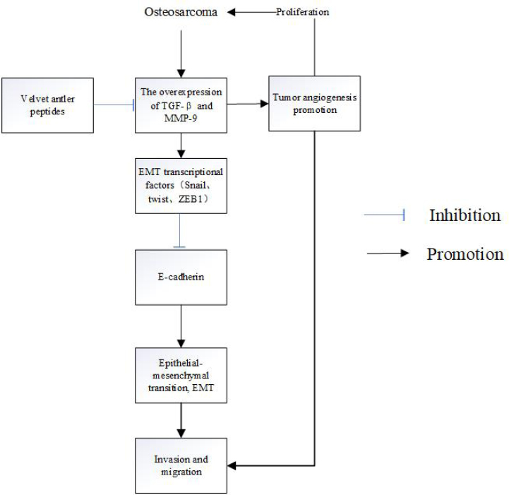
Velvet antler polypeptides regulate TGF-β in osteosarcoma cells. Note: The VAP inhibit the expression of EMT transcriptional factors and E-cadherin by down-regulating the TGF-β pathway in MG-63 cells, which interfere the invasion and migration afterwards.
lower concentration of TGF-β had no significant effect on it. Furthermore, exogenous TGF-β can inhibit the growth of MG- 63 cells cultured in vitro, which can increase the distribution of MG-63 cells in G1 phase and prevent the cells from entering S phase [48]. Exogenous TGF-β can make MG- 63 cells overexpressing a TGF-β inducible early gene (TIEG), and finally inhibit osteocalcin synthesis. While numerous studies have demonstrated that high expression of bone calcium in bone cells and osteosarcoma cells can inhibit the synthesis of osteocalcin then inhibit the proliferation of osteosarcoma cells [49]. Although researchers have confirmed the inhibitive role of TGF-β in tumorigenesis, it has on the contrary been shown to enhance metastasis of tumor cells and promote advanced tumors invasiveness [50]. TGF-β is a double-edged sword in cancer, on the one hand it can suppress tumor growth potently, on the other hand, it can also enhance invasion and metastasis of cancer by suppressing miR-143, inducing epithelial-mesenchymal transition (EMT) [51,52], which plays a vital role in tumor invasion and metastasis [53]. Furthermore, it has been reported that TGF-β can induce the expression of Snail which could repress the expression of E-cadherin, an important tumor suppressor [54]. Though TGF-β is involved in the EMT process, VAP has been shown to block the binding of TGF-β1 with its receptors, TGF-β receptor1 and 2, and inhibit the downstream activated pathway [55] (Figure1). It has also been confirmed that TGF-β1 not only stimulates the growth of osteosarcoma but also reduces the proliferative potential ability of osteosarcoma by inducing the expression of IGFBP-3 [56]. In addition to osteosarcoma, the antler polypeptide also affects other tumors. Tang et al. demonstrated that the top VAP can inhibit the migration of prostate cancer cells by downregulating the expression of its relevant gene, such as MMP-9 and VEGF [57].
4.2 Notch signaling pathways
The Notch signaling pathway includes Notch1, Notch2, Jag1, DLL1, DLL4, Hey1/Hey2 and CSL. It is implicated as a key mediator in a number of various cancers [58]. It is more like a double-edged sword, which can be used as an oncogene in one hand , while inhibiting tumor growth in some cases on the other hand [59]. In addition, regulating hes1 and DTX1 in notch signaling pathways can affect migration and invasion of osteosarcoma cells [60]. It has been confirmed that the expression of Notch and its target gene are up-regulated in osteosarcoma. Inhibiting Notch signaling by chemical and genetic ways can reduce nude mices’ tumor burden in vivo and decrease the proliferation of osteosarcoma cells in vitro [61]. Furthermore, it has been found that Notch pathways can regulate the osteosarcoma cell cycle by influencing the expression of cyclin E1, cyclin E2, c-Myc and so on. Notch signaling pathways play a very important role in osteosarcoma cell progression [62]. Besides these roles we have referred to, it also participates in EMT. The EMT progression can be arrested by the inhibition of notch signaling pathways [63]. Furthermore, notch pathways will become a mediator when some growth factors and drugs mitigate osteosarcoma. It has been found that BMP-9 can promote the growth of osteosarcoma, which is mediated by the notch signaling pathway. The study also demonstrated BMP-9 has effects on the receptors and ligands of notch signaling pathways including hey1, notch1, DLL1, JAG1 and JAG2 at the same time [64]. Moreover, some anticancer drugs exert anticancer effects by regulating notch signaling pathways. It has been shown that the anticancer drug, doxorubicin, can inhibit the proliferation of osteosarcoma cells by upregulating notch signaling pathways [65]. In addition, cinobufagin and curcumin can inhibit osteosarcoma growth in vivo, induce osteosarcoma cell apoptosis, cell growth inhibition, decrease cell survival, and improve mice survival by downregulating notch1 and its target gene [66,67]. Continuous medication can cause drug resistance in tumor cells, and the notch plays an important regulatory role in this process[68], which provides us a new way to cure osteosarcoma. In addition to growth factors and drugs, notch signaling pathways also participate in the process where microRNAs affect osteosarcoma. MicroRNA is a small non-coding RNA molecule found in plants, animals and some viruses, that functions in RNA silencing and post-transcriptional regulation of gene expression [69]. It is generally accepted that tumor recurrence often appears during the treatment of the tumor and the cancer stem cells play a key role in it. In recent years, researchers have found miR-135b can effect tumor metastasis and CSC-induced recurrence in osteosarcoma by regulating notch and Wnt/β-catenin signaling pathways, so they can be targeted to inhibit tumor metastasis and recurrence in osteosarcoma [70]. miR-34a-5p, miR-26a and miR-199b-5p also play an important role in osteosarcoma. The reduction of miR-26a can cause osteosarcoma metastasis and poor survival of osteosarcoma patients, miR26-a can also regulate cancer stem cells of osteosarcoma. But miR-26a only target Jagged1 which is a ligand in notch signaling pathways [71]. Moreover miR-34a-5p can promote multi-chemoresistance of osteosarcoma by down regulating DLL1 gene a ligand of notch signaling pathways and the inhibition of miRNA-199b-5p can change expression of notch pathway components [72,73]. These findings reveal that notch signaling pathways, and some microRNA can be novel targets for treating osteosarcoma and therapeutic options for osteosarcoma.
4.3 Wnt-β-catenin pathways
The Wnt signaling pathways are a group of signal transduction pathways made of proteins that pass signals into a cell through cell surface receptors. There are some proteins in them such as frizzled, disheveled, β-catenin, GSK3β, and axin scaffolding protein. They play a key role in cell cycle, apoptosis and tumorigenesis, and EMT [74]. It has been confirmed that it can decrease tumorigenicity, metastasis and EMT of osteosarcoma by downregulating LRP-5 which is a receptor of wnt in vivo and in nude mice experiments [75]. Thus LRP-5 can be a new target for inhibiting EMT. At the same time the activation of Wnt pathways can promote the tumorigenic phenotypes, and some receptors, ligands also up-regulated in osteosarcoma, therefore the treatment of osteosarcoma should aim at blocking Wnt pathways [76]. Rubin et al. provided Wnt Inhibitory Factor 1 can (WIF1) decreases tumorigenesis and metastasis in osteosarcoma cells line 143B cells and overexpression of WIF1 can inhibit lung metastasis in vivo in an orthotopic mouse model of osteosarcoma. Therefore, WIF1 is a potential target for treating osteosarcoma [77]. IWR-1, a tankyrase inhibitor which is a wnt/β-catenin signaling pathways inhibitor and a specifically cytotoxic for osteosarcoma cancer stem calls. It has been confirmed that IWR-1 not only can inhibit the growth of cancer stem cells in osteosarcoma in vivo and in vitro by targeting wnt signaling pathways but also can eradicate the aggressive osteosarcoma cancer stem cells and improve therapeutic outcomes [78] . DKK-3 is a Secreted Wnt Antagonist and involved in embryonic development through its interactions with the Wnt signaling pathway. Hoang et al. have confirmed that DDK-3 can decrease tumor growth and metastatic pulmonary nodules in nude mice. In addition, it can inhibit tumorigenic potential of osteosarcoma, decrease osteosarcoma cell motility in cells. So, we can make DDK3 as target to treat osteosarcoma [79]. Zhang et al. revealed that parathyroid hormone type 1 receptor (PTHR1) can promote malignancy for osteosarcoma through activation of wnt and angiogenesis signaling pathways. They have analyzed the microarray data extracted from the Gene Expression Omnibus (GEO) database and compared with PTHR1 knockdown samples [80]. However, a different opinion has been put forward that wnt signaling may act as a tumor repressor in osteosarcoma which is in contrast with its oncogenic role in other tumors [81]. Some medicines also exert their role by targeting wnt/β-catenin signaling pathways. Triptolide(TPL) is a diterpenoid epoxide which is produced by the thunder god vine. It can inhibit angiogenesis and induce cell apoptosis in osteosarcoma cells in a dose dependent manner through down-regulating Wnt/β-Catenin signaling [82]. Together, these findings have revealed that wnt/βcatenin could be a key pathway for treating osteosarcoma.
4.4 NF-κB pathway
NF-κB is a protein complex that controls transcription of DNA, cytokine production and cell survival. NF-κB plays a pivotal role in cell growth, apoptosis, migration and invasion of osteosarcoma [83]. Aspirin is a medication used to treat pain, fever, or inflammation. However, it has been confirmed that aspirin can influence osteosarcoma procession too. Kang et al. have demonstrated that aspirin can reduce cell viability and the more doses and time the better effects in osteosarcoma cells. Moreover, aspirin can also repress the migration and invasion of osteosarcoma cells and decrease osteosarcoma metastases to the lungs in nude mouse through regulating NF-κB pathway [84]. Thymoquinone is a phytochemical compound found in the plant Nigella sativa. It can inhibit cell growth and effectively induce tumor cell apoptosis and exert antiproliferative effects on several cancer cells in vitro. In addition, thymoquinone can inhibit tumor angiogenesis and tumor growth by downregulating NF-κB and its regulated molecules. Not only can thymoquinone inhibit osteosarcoma growth but also it can enhance sensitivity to chemotherapeutic agents [85]. Furthermore, NF-κB can contact with wnt pathway, wnt10b is a member of the wnt family which could upregulate interleukin-1α (IL-1α) and tumor necrosis factor-α (TNF-α), known inducers of NF-κB [86].
4.5 PI3K/Akt Pathway
PI3K-Akt pathway is a signal transduction pathway that promotes survival and growth in response to extracellular signals. Key proteins involved are PI3K (phosphatidylinositol 3-kinase) and Akt (Protein Kinase B). It is well known to be a major cell survival pathway and it can enhance resistance to apoptosis if activated [87]. Apo2L/TRAIL is a member of the tumor necrosis factor (TNF) family, which can induce apoptosis of cancer cells. It can induce osteosarcoma cell U2OS apoptosis by inhibiting akt expression and then reduce expression of Bcl-2 and activate caspase-9. Furthermore, it can also decrease osteosarcoma cell drug resistance by regulating Akt pathway [88,89]. Geraniin is an activated compound isolated from Geranium sibiricum. It has been found that Geraniin suppresses matrix metalloproteinase-9 (MMP-9) expression in a dose dependent manner and then inhibits the migration and invasion of osteosarcoma cells by suppressing the phosphorylation of the extracellular signal regulating kinase (ERK)1/2, phosphatidylinositide-3-kinase (PI3K), and Akt pathways [90]. miR-221 is an oncogenic microRNA and one of the most commonly upregulated miRNAs in cancer. It targets PTEN leading to activation of the Akt pathway, which is known as a major cell survival pathway in many cancers. It has been confirmed that knockdown of miR-221 in osteosarcoma cells can downregulate p-Akt expression, promote osteosarcoma cells apoptosis and lower cisplatin resistance through regulating PI3K/AKT pathway [91]. Some medicines can exert their roles through this pathway. Dryofragin is a phloroglucinol derivative extracted from Dryopteris fragrans. It has been found to be able to inhibit tumor proliferation and induce apoptosis. Moreover, in recent years it has been confirmed that dryofragin can suppress the migration and invasive ability of U2OS cells, downregulate the expression of MMP-2 and MMP-9 and upregulate the expression of TIMP-1 and TIMP-2 through PI3K/AKT and p38 MAPK signaling pathways [92]. Tricetin is a flavone, a type of flavonoid, that can also inhibit the metastasis of human osteosarcoma cells by transcriptionally repressing MMP-9 via p38 and Akt pathways [93]. All of them can be potentially used as anti-cancer agents for osteosarcoma treatment and supplement of osteosarcoma chemotherapy.
4.6 Other signaling pathways
Rheum palmatum L. is a common Chinese herb, also called Chinese rhubarb, ornamental rhubarb. Rheum palmatum L. has been used for anti-inflammatory and chronic liver diseases, and some cancer. In recent years, it has been indicated that Crude Extract of Rheum palmatum L. (CERP) can induce S phase arrest in osteosarcoma cells U2OS in a dose-dependent fashion, cause DNA damage and DNA condensation, up-regulate the expression of pro-apoptosis proteins such as Bax, Bak, p21, and p27 and activate caspase-3, -8, and -9 through mitochondrial-dependent pathways [94]. Aspidin PB is a phloroglucinol derivative isolated from Dryopteris fragrans (L.). Previous studies have found it can inhibit fibrogenesis [95] and induce apoptosis in human hepatocarcinoma HepG2 cells [96]. Moreover, it also has been confirmed that Aspidin PB can inhibit the proliferation of osteosarcoma cells in a dose-dependent and time-dependent manner and induce osteosarcoma cells apoptosis and cell cycle arrest through the p53/p21 and mitochondria-dependent pathways [97].
S-Adenosyl methionine (AdoMet) is a common co-substrate involved in methyl group transfers, transsulfuration, and aminopropylation. Naviglio’s group has confirmed that it can inhibit osteosarcoma cell proliferation by slowing-down cell cycle progression and by inducing apoptosis. Furthermore, it can downregulate ERK1/2 and STAT3 pathways to arrest cell cycle and induce osteosarcoma cell apoptosis [98]. Cucurbitacin B is class of tetracyclic triterpenoids and is extracted from Hemsleya endecaphylla (62 mg/72 g) and other plants [99]. It is the most abundant member of cucurbit family and has extensive pharmacological activities. For human osteosarcoma cells, it can downregulate MMP-2 and MMP-9 which contribute to the invasion and metastasis of tumor cells, upregulate the expression of pro-apoptotic proteins, and modulate JAK2/STAT3 signaling pathway [100].
5 Conclusion
Osteosarcoma is a complex, heterogenous, and interpatient, between different individuals and their living environment, disease. Therefore, successful treatment options are likely to arise from personalized precise treatment. There is paramount preclinical and clinical evidence for potent efficient activity of velvet antler and its extracts for use as therapeutics in bone fracture repair, osteoarthritis, osteoporosis and other bone diseases. Relating to TGF-β and Notch pathways, previous results suggested the involvement of VAP in regulation of proliferation and migration of osteosarcoma cells MG-63 as well as U2OS. Information about molecular mechanism and therapeutic targets of this reaction within the tumor cells needs to be elucidated. Furthermore, various growth factors such as sirtuin and NKD2 can regulate the osteosarcoma cell-cycle, and biomarkers will be essential to advance into clinical development to obtain meaningful and reliable answers on therapeutic ratios. Undoubtedly, further advances in our understanding of the pathology of osteosarcoma, and in the techniques for extracting the velvet antler, will strengthen our understanding of the ways in which the Chinese medicine counteracts cancer, and will aid in the development of anti-cancer therapeutic approaches.
Conflicts of interest The authors declared no potential conflicts of interest with respect to the research, authorship, and/or publication of this article.
Acknowledgements
This work was supported by the National Natural Science Foundation of China (grant no. 81503177 and 81573999) and Jilin science and technology development plan project (20160101158JC).
Reference
[1] Raimondi L., De A.L., Costa V., Amodio N., Carina V., Bellavia D., et al. Circulating biomarkers in osteosarcoma: new translational tools for diagnosis and treatment. Oncotarget., 2017, 8(59), 100831.10.18632/oncotarget.19852Search in Google Scholar PubMed PubMed Central
[2] Quan G.M., Slavin J.L., Schlicht S.M., Smith P.J., Powell G.J., Choong P.F. Osteosarcoma near joints: assessment and implications. J Surg Oncol., 2005, 91(3), 159.10.1002/jso.20268Search in Google Scholar PubMed
[3] Anderson M.E. Update on Survival in Osteosarcoma. Orthop Clin North Am., 2016, 47(1), 283-92.10.1016/j.ocl.2015.08.022Search in Google Scholar PubMed
[4] Sadighi M., Haines S.R., Skottner A., Harris A.J., Suttie J.M. Effects of insulin-like growth factor-I (IGF-I) and IGF-II on the growth of antler cells in vitro. J Endocrinol., 1994, 143(3), 461-9.10.1677/joe.0.1430461Search in Google Scholar PubMed
[5] Deng H.M., Ding Q.N., Wang C.M., Geng D., Dai J.D., Dong L. The Contents of the Growth Factors in Different Sections of Sika Deer (Cervus nippon) Velvet Antler Treated by Traditional Hot Processing. Special Wild Economic Animal & Plant Research., 2018.Search in Google Scholar
[6] Sun Y., Xia P., Zhang H., Liu B., Shi Y. P53 is required for Doxorubicin-induced apoptosis via the TGF-beta signaling pathway in osteosarcoma-derived cells. Am J Cancer Res., 2016, 6(1), 114-25.Search in Google Scholar
[7] Tanaka M., Setoguchi T., Hirotsu M., Gao H., Sasaki H., Matsunoshita Y., et al. Inhibition of Notch pathway prevents osteosarcoma growth by cell cycle regulation. Br J Cancer., 2009, 100(12), 1957-65.10.1038/sj.bjc.6605060Search in Google Scholar PubMed PubMed Central
[8] Keremu A., Maimaiti X., Aimaiti A., Yushan M., Alike Y., Yilihamu Y., et al. NRSN2 promotes osteosarcoma cell proliferation and growth through PI3K/Akt/MTOR and Wnt/β-catenin signaling. Am J Cancer Res., 2017, 7(3), 565-73.Search in Google Scholar
[9] Hafeez B.B., Ahmed S., Wang N., Gupta S., Zhang A., Haqqi T.M. Green tea polyphenols-induced apoptosis in human osteosarcoma SAOS-2 cells involves a caspase-dependent mechanism with downregulation of nuclear factor-kappaB. Toxicol Appl Pharmacol., 2006, 216(1), 11-9.10.1016/j.taap.2006.03.013Search in Google Scholar PubMed
[10] Moriarity B.S., Otto G.M., Rahrmann E.P., Rathe S.K., Wolf N.K., Weg M.T., et al. A Sleeping Beauty forward genetic screen identifies new genes and pathways driving osteosarcoma development and metastasis. Nat Genet., 2015, 47(6), 615-24.10.1038/ng.3293Search in Google Scholar PubMed PubMed Central
[11] Cheng A.Y., Zhang Y., Mei H.J., Fang S., Ji P., Yang J., et al. Construction of a plasmid for overexpression of human circadian gene period2 and its biological activity in osteosarcoma cells. Tumour Biol., 2015, 36(5), 3735-43.10.1007/s13277-014-3013-7Search in Google Scholar PubMed
[12] Kaseta M.K., Khaldi L., Gomatos I.P., Tzagarakis G.P., Alevizos L., Leandros E., et al. Prognostic value of bax, bcl-2, and p53 staining in primary osteosarcoma. J Surg Oncol., 2010, 97(3), 259-66.10.1002/jso.20913Search in Google Scholar PubMed
[13] Salinas-Souza C., De O.R., Alves M.T., Garcia Filho R.J., Petrilli A.S., Toledo S.R. The metastatic behavior of osteosarcoma by gene expression and cytogenetic analyses. Hum Pathol., 2013, 44(10), 2188-98.10.1016/j.humpath.2013.04.013Search in Google Scholar PubMed
[14] Chen X.C., Ke L.J., Chen G.R., Liu S.T., Huo Y.S., Rao P.F. The modulation of pilose antler extract (PAE) on the proliferation of rat osteogenic cells UMR-106. China Journal of Chinese Materia Medica., 2004, 29(1), 74-77.Search in Google Scholar
[15] Taupin T., Decouvelaere A.V., Vaz G., Thiesse P. Accuracy of core needle biopsy for the diagnosis of osteosarcoma: A retrospective analysis of 73 patients. Diagn Interv Imaging., 2015, 97(3), 327-31.10.1016/j.diii.2015.09.013Search in Google Scholar PubMed
[16] Mori K., Rédini F., Gouin F., Cherrier B., Heymann D. Osteosarcoma: current status of immunotherapy and future trends (Review). Oncol Rep., 2006, 15(3), 693-700.10.3892/or.15.3.693Search in Google Scholar
[17] Lin H., Hao Y., Zhao Z., Tong Y. Sirtuin 6 contributes to migration and invasion of osteosarcoma cells via the ERK1/2/MMP9 pathway. FEBS open bio., 2017, 7(9), 1291-301.10.1002/2211-5463.12265Search in Google Scholar PubMed PubMed Central
[18] Liu J.F., Tsao Y.T., Hou C.H. Fractalkine/CX3CL1 induced intercellular adhesion molecule-1-dependent tumor metastasis through the CX3CR1/PI3K/Akt/NF-κB pathway in human osteosarcoma. Oncotarget., 2017, 8(33), 54136-48.10.18632/oncotarget.11250Search in Google Scholar PubMed PubMed Central
[19] Cao J., Han X., Qi X., Jin X., Li X. TUG1 promotes osteosarcoma tumorigenesis by upregulating EZH2 expression via miR-144-3p. Int J Oncol., 2017, 51(4), 1115-23.10.3892/ijo.2017.4110Search in Google Scholar PubMed PubMed Central
[20] Zhao S., Kurenbekova L., Gao Y., Roos A., Creighton C.J., Rao P., et al. NKD2, a negative regulator of Wnt signaling, suppresses tumor growth and metastasis in osteosarcoma. Oncogene., 2015, 34(39), 5069-79.10.1038/onc.2014.429Search in Google Scholar PubMed PubMed Central
[21] Nagaraju G.P., Bramhachari P.V., Raghu G., El-Rayes B.F. Hypoxia inducible factor-1α: Its role in colorectal carcinogenesis and metastasis. Cancer Lett., 2015, 366(1), 11-8.10.1016/j.canlet.2015.06.005Search in Google Scholar PubMed
[22] Punzo F., Tortora C., Di P.D., Manzo I., Bellini G., Casale F., et al. Anti-proliferative, pro-apoptotic and anti-invasive effect of EC/EV system in human osteosarcoma. Oncotarget., 2017, 54459-71.10.18632/oncotarget.17089Search in Google Scholar PubMed PubMed Central
[23] Wang S.W., Liu S.C., Sun H.L., Huang T.Y., Chan C.H., Yang C.Y., et al. CCL5/CCR5 axis induces vascular endothelial growth factor-mediated tumor angiogenesis in human osteosarcoma microenvironment. Carcinogenesis., 2015, 36(1), 104-14.10.1093/carcin/bgu218Search in Google Scholar PubMed
[24] Zhang J., Liang J.H., Huang J.G. Bone morphogenetic protein 9 facilitates osteocarcinoma cell apoptosis and inhibits in vivo tumor growth. Genet Mol Res., 2016, 15(3).10.4238/gmr.15038036Search in Google Scholar PubMed
[25] Dong Y., Liang G., Yuan B., Yang C., Gao R., Zhou X. MALAT1 promotes the proliferation and metastasis of osteosarcoma cells by activating the PI3K/Akt pathway. Tumour Biol., 2015, 36(3), 1477-86.10.1007/s13277-014-2631-4Search in Google Scholar PubMed
[26] Li Z.M., Zhang H.Y., Wang Y.X., Wang W.B. MicroRNA-137 is downregulated in human osteosarcoma and regulates cell proliferation and migration through targeting FXYD6. J Drug Target., 2016, 24(2), 1-9.10.3109/1061186X.2015.1057149Search in Google Scholar PubMed
[27] Zhou H., Zhang M., Yuan H., Zheng W., Meng C., Zhao D. MicroRNA-154 functions as a tumor suppressor in osteosarcoma by targeting Wnt5a. Oncol Rep., 2016, 35(3), 1851-8.10.3892/or.2015.4495Search in Google Scholar PubMed
[28] Sun X.H., Geng X.L., Zhang J., Zhang C. miRNA-646 suppresses osteosarcoma cell metastasis by downregulating fibroblast growth factor 2 (FGF2). Tumour Biol., 2015, 36(3), 2127-34.10.1007/s13277-014-2822-zSearch in Google Scholar PubMed
[29] Tian K., Di R., Wang L. MicroRNA-23a enhances migration and invasion through PTEN in osteosarcoma. Cancer Gene Ther., 2015, 22(7), 351-9.10.1038/cgt.2015.27Search in Google Scholar PubMed
[30] Niu G., Li B., Sun L., An C. MicroRNA-153 Inhibits Osteosarcoma Cells Proliferation and Invasion by Targeting TGF-β2. PLoS One., 2015, 10(3), e0119225.10.1371/journal.pone.0119225Search in Google Scholar PubMed PubMed Central
[31] Zhen Y., Yu Z., Xian Z., Mingyu Z., Hongliang L., Shaobo Z., et al. Serum microRNA-221 functions as a potential diagnostic and prognostic marker for patients with osteosarcoma. Biomed Pharmacother., 2015, 75153-8.Search in Google Scholar
[32] Zhu K., Liu L., Zhang J., Wang Y., Liang H., Fan G., et al. MiR-29b suppresses the proliferation and migration of osteosarcoma cells by targeting CDK6. Protein Cell., 2016, 7(6), 434-44.10.1007/s13238-016-0277-2Search in Google Scholar PubMed PubMed Central
[33] Li C.H., Yu T.B., Qiu H.W., Zhao X., Zhou C.L., Qi C. miR-150 is downregulated in osteosarcoma and suppresses cell proliferation, migration and invasion by targeting ROCK1. Oncol Lett., 2017, 13(4), 2191-97.10.3892/ol.2017.5709Search in Google Scholar PubMed PubMed Central
[34] Zhang H., Xing F., Wang T., Hu Z., Que X., Tian Q., et al. MiRNA-543 promotes osteosarcoma cell proliferation and glycolysis by partially suppressing PRMT9 and stabilizing HIF-1α protein. Oncotarget., 2017, 8(2), 2342-55.10.18632/oncotarget.13672Search in Google Scholar PubMed PubMed Central
[35] Chen L., Wang Q., Wang G.D., Wang H.S., Huang Y., Liu X.M., et al. miR-16 inhibits cell proliferation by targeting IGF1R and the Raf1-MEK1/2-ERK1/2 pathway in osteosarcoma. FEBS Lett., 2013, 587(9), 1366-72.10.1016/j.febslet.2013.03.007Search in Google Scholar PubMed
[36] Wang D.G., Fan J.B., Siao C.J., Berno A., Young P., Sapolsky R., et al. Large-scale identification, mapping, and genotyping of single-nucleotide polymorphisms in the human genome. Science., 1998, 280(5366), 1077.10.1126/science.280.5366.1077Search in Google Scholar PubMed
[37] Tanaka T., Yui Y., Naka N., Wakamatsu T., Yoshioka K., Araki N., et al. Dynamic analysis of lung metastasis by mouse osteosarcoma LM8: VEGF is a candidate for anti-metastasis therapy. Clin Exp Metastasis., 2013, 30(4), 369-79.10.1007/s10585-012-9543-8Search in Google Scholar PubMed PubMed Central
[38] Xiao X., Wang W., Zhang H., Gao P., Fan B., Huang C., et al. Individualized chemotherapy for osteosarcoma and identification of gene mutations in osteosarcoma. Tumour Biol., 2015, 36(4), 2427-35.10.1007/s13277-014-2853-5Search in Google Scholar PubMed
[39] Shi R., Li J., Tang F., Luo Y.I., Tu C.Q. Identification and functional study of osteosarcoma metastasis marker genes. Oncol Lett., 2015, 10(3), 1848-52.10.3892/ol.2015.3444Search in Google Scholar PubMed PubMed Central
[40] Patanè S., Avnet S., Coltella N., Costa B., Sponza S., Olivero M., et al. MET overexpression turns human primary osteoblasts into osteosarcomas. Cancer Res., 2006, 66(9), 4750-7.10.1158/0008-5472.CAN-05-4422Search in Google Scholar PubMed
[41] Akatsuka T., Wada T., Kokai Y., Kawaguchi S., Isu K., Yamashiro K., et al. ErbB2 expression is correlated with increased survival of patients with osteosarcoma. Cancer., 2002, 94(5), 1397-404.10.1002/cncr.10360Search in Google Scholar PubMed
[42] Pang L.Y., Gatenby E.L., Kamida A., Whitelaw B.A., Hupp T.R., Argyle D.J. Global Gene Expression Analysis of Canine Osteosarcoma Stem Cells Reveals a Novel Role for COX-2 in Tumour Initiation. PLoS One., 2014, 9(1), e83144.10.1371/journal.pone.0083144Search in Google Scholar PubMed PubMed Central
[43] Duan D.P., Dang X.Q., Wang K.Z., Wang Y.P., Zhang H., You W.L. The cyclooxygenase-2 inhibitor NS-398 inhibits proliferation and induces apoptosis in human osteosarcoma cells via downregulation of the survivin pathway. Oncol Rep., 2012, 28(5), 1693-700.10.3892/or.2012.1992Search in Google Scholar PubMed
[44] Chen Z., Shen B.L., Fu Q.G., Wang F., Tang Y.X., Hou C.L., et al. CUL4B promotes proliferation and inhibits apoptosis of human osteosarcoma cells. Oncol Rep., 2014, 32(5), 2047-53.10.3892/or.2014.3465Search in Google Scholar PubMed
[45] Xiao X., Wang W., Zhang H., Gao P., Fan B., Huang C., et al. Individualized chemotherapy for osteosarcoma and identification of gene mutations in osteosarcoma. Tumour Biol., 2015, 36(4), 2427-35.10.1007/s13277-014-2853-5Search in Google Scholar
[46] Kansara M., Teng M.W., Smyth M.J., Thomas D.M. Translational biology of osteosarcoma. Nat Rev Cancer., 2014, 14(11), 722-35.10.1038/nrc3838Search in Google Scholar PubMed
[47] Lamora A., Talbot J., Mullard M., Royer B.L., Redini F., Verrecchia F. TGF-β Signaling in Bone Remodeling and Osteosarcoma Progression. J Clin Med., 2016, 5(11), 96.10.3390/jcm5110096Search in Google Scholar PubMed PubMed Central
[48] Kewei J.I., Shao Z., Xiong X. Study of MG-63 cell growth regulated by exogenous TGF-β1 in vitro. Chinese Journal of Bone Tumor & Bone Disease., 2005.Search in Google Scholar
[49] Hefferan T.E., Reinholz G.G., Rickard D.J., Johnsen S.A., Waters K.M., Subramaniam M., et al. Overexpression of a nuclear protein, TIEG, mimics transforming growth factor-beta action in human osteoblast cells. J Biol Chem., 2000, 275(27), 20255-9.10.1074/jbc.C000135200Search in Google Scholar PubMed
[50] Pardali K., Moustakas A. Actions of TGF-beta as tumor suppressor and pro-metastatic factor in human cancer. Biochim Biophys Acta., 2007, 1775(1), 21-62.10.1016/j.bbcan.2006.06.004Search in Google Scholar PubMed
[51] Li F., Li S., Cheng T. TGF-β1 Promotes Osteosarcoma Cell Migration and Invasion Through the miR-143-Versican Pathway. Cell Physiol Biochem., 2014, 34(6), 2169-79.10.1159/000369660Search in Google Scholar PubMed
[52] Chen Y., Zhang K., Li Y., He Q. Estrogen-related receptor alpha participates transforming growth factor-beta (TGF-beta) induced epithelial-mesenchymal transition of osteosarcoma cells. Cell Adh Migr., 2017, 11(4), 338-46.10.1080/19336918.2016.1221567Search in Google Scholar PubMed PubMed Central
[53] Jing Y., Han Z., Zhang S., Yan L., Wei L. Epithelial-Mesenchymal Transition in tumor microenvironment. Cell Biosci., 2011, 1(1), 29.10.1186/2045-3701-1-29Search in Google Scholar PubMed PubMed Central
[54] Yu H., Shen Y., Hong J., Xia Q., Zhou F., Liu X. The contribution of TGF-β in Epithelial-Mesenchymal Transition (EMT): Down-regulation of E-cadherin via snail. Neoplasma., 2015, 62(1), 1-15.10.4149/neo_2015_002Search in Google Scholar PubMed
[55] Zhao L., Mi Y., Guan H., Xu Y., Mei Y. Velvet antler peptide prevents pressure overload-induced cardiac fibrosis via transforming growth factor (TGF)-β1 pathway inhibition. Eur J Pharmacol., 2016, 78333-46.10.1016/j.ejphar.2016.04.039Search in Google Scholar PubMed
[56] Schedlich L.J., Yenson V.M., Baxter R.C. TGF-β-induced expression of IGFBP-3 regulates IGF1R signaling in human osteosarcoma cells. Mol Cell Endocrinol., 2013, 377(1–2), 56-64.10.1016/j.mce.2013.06.033Search in Google Scholar PubMed
[57] Tang Y., Jeon B., Wang Y., Choi E., Kim Y., Hwang J., et al. First evidence that Sika Deer (Cervus nippon) velvet antler extract suppresses migration of human prostate cancer cells. Korean J Food Sci An., 2015, 35(4), 507-14.10.5851/kosfa.2015.35.4.507Search in Google Scholar PubMed PubMed Central
[58] McManus M.M., Weiss K.R., Hughes D.P. Understanding the role of Notch in osteosarcoma. Adv Exp Med Biol., 2014, 80467-92.10.1007/978-3-319-04843-7_4Search in Google Scholar PubMed
[59] Bray S. Notch signalling: a simple pathway becomes complex. Nat Rev Mol Cell Biol., 2006, 7(9), 678-89.10.1038/nrm2009Search in Google Scholar PubMed
[60] Zhang P., Yang Y., Zweidlermckay P., Hughes D. Notch/HES1/DTX1 signaling controls osteosarcoma invasiveness and metastasis in vivo. Cancer Res., 2008, 68.Search in Google Scholar
[61] Engin F., Bertin T., Ma O., Jiang M., Wang L., Sutton R., et al. Notch signaling contributes to the pathogenesis of human osteosarcomas. Hum Mol Genet., 2009, 18(8), 1464-70.10.1093/hmg/ddp057Search in Google Scholar PubMed PubMed Central
[62] Tanaka M., Setoguchi T., Hirotsu M., Gao H., Sasaki H., Matsunoshita Y., et al. Inhibition of Notch pathway prevents osteosarcoma growth by cell cycle regulation. Br J Cancer., 2009, 100(12), 1957-65.10.1038/sj.bjc.6605060Search in Google Scholar PubMed PubMed Central
[63] Yang J., Guo W., Wang L., Yu L., Mei H., Fang S., et al. Notch signaling is important for epithelial-mesenchymal transition induced by low concentrations of doxorubicin in osteosarcoma cell lines. Oncol Lett., 2017, 13(4), 2260-68.10.3892/ol.2017.5708Search in Google Scholar PubMed PubMed Central
[64] Liu P., Man Y., Wang Y., Bao Y. Mechanism of BMP9 promotes growth of osteosarcoma mediated by the Notch signaling pathway. Oncol Lett., 2016, 11(2), 1367-70.10.3892/ol.2015.4067Search in Google Scholar PubMed PubMed Central
[65] Ji P., Yu L., Guo W.C., Mei H.J., Wang X.J., Chen H., et al. Doxorubicin Inhibits Proliferation of Osteosarcoma Cells Through Upregulation of the Notch Signaling Pathway. Oncol Res., 2014, 22(4), 185-91.10.3727/096504015X14343704124340Search in Google Scholar PubMed PubMed Central
[66] Cao Y., Yu L., Dai G., Zhang S., Zhang Z., Gao T., et al. Cinobufagin induces apoptosis of osteosarcoma cells through inactivation of Notch signaling. Eur J Pharmacol., 2017, 79477-84.10.1016/j.ejphar.2016.11.016Search in Google Scholar PubMed
[67] Li Y., Zhang J., Ma D., Zhang L., Meng S., Han Y., et al. Curcumin inhibits proliferation and invasion of osteosarcoma cells through inactivation of Notch‐1 signaling. FEBS J., 2012, 279(12), 2247-59.10.1111/j.1742-4658.2012.08607.xSearch in Google Scholar PubMed
[68] Li C., Guo D., Tang B., Zhang Y., Zhang K., Nie L. Notch1 is associated with the multidrug resistance of hypoxic osteosarcoma by regulating MRP1 gene expression. Neoplasma., 2016, 63(5), 734-42.10.4149/neo_2016_510Search in Google Scholar PubMed
[69] Ambros V. The functions of animal microRNAs. Nature., 2004, 431(7006), 350-5.10.1038/nature02871Search in Google Scholar PubMed
[70] Jin H., Luo S., Wang Y., Liu C., Piao Z., Xu M., et al. miR-135b Stimulates Osteosarcoma Recurrence and Lung Metastasis via Notch and Wnt/β-Catenin Signaling. Molecular Therapy Nucleic Acids., 2017, 8111-22.10.1016/j.omtn.2017.06.008Search in Google Scholar PubMed PubMed Central
[71] Lu J., Song G., Tang Q., Yin J., Zou C., Zhao Z., et al. MiR-26a inhibits stem cell-like phenotype and tumor growth of osteosarcoma by targeting Jagged1. Oncogene., 2017, 36(2), 231-41.10.1038/onc.2016.194Search in Google Scholar PubMed
[72] Pu Y., Zhao F., Wang H., Cai S. MiR-34a-5p promotes multi-chemoresistance of osteosarcoma through down-regulation of the DLL1 gene. Sci Rep., 2017, 744218.10.1038/srep44218Search in Google Scholar PubMed PubMed Central
[73] Won K.Y., Kim Y.W., Kim H.S., Lee S.K., Jung W.W., Park Y.K. MicroRNA-199b-5p is involved in the Notch signaling pathway in osteosarcoma. Hum Pathol., 2013, 44(8), 1648-55.10.1016/j.humpath.2013.01.016Search in Google Scholar PubMed
[74] Lustig B., Behrens J. The Wnt signaling pathway and its role in tumor development. J Cancer Res Clin Oncol., 2003, 129(4), 199-221.10.1007/s00432-003-0431-0Search in Google Scholar PubMed
[75] Guo Y., Zi X., Koontz Z., Kim A., Xie J., Gorlick R., et al. Blocking Wnt/LRP5 signaling by a soluble receptor modulates the epithelial to mesenchymal transition and suppresses met and metalloproteinases in osteosarcoma Saos cells. J Orthop Res., 2010, 25(7), 964-71.10.1002/jor.20356Search in Google Scholar PubMed
[76] Hoang B.H. Wnt, osteosarcoma, and future therapy. J Am Acad Orthop Surg., 2012, 20(1), 58-9.10.5435/00124635-201201000-00007Search in Google Scholar
[77] Rubin E.M., Yi G., Tu K., Xie J., Zi X., Bang H.H. Wnt Inhibitory Factor 1 Decreases Tumorigenesis and Metastasis in Osteosarcoma. Mol Cancer Ther., 2010, 9(3), 731-41.10.1158/1535-7163.MCT-09-0147Search in Google Scholar PubMed PubMed Central
[78] Martins-Neves S.R., Paiva-Oliveira D.I., Fontes-Ribeiro C., Bovee J., Cleton-Jansen A.M., Gomes C.M.F. IWR-1, a tankyrase inhibitor, attenuates Wnt/beta-catenin signaling in cancer stem-like cells and inhibits in vivo the growth of a subcutaneous human osteosarcoma xenograft. Cancer Lett., 2018, 4141-15.10.1016/j.canlet.2017.11.004Search in Google Scholar PubMed
[79] Lin C.H., Guo Y., Ghaffar S., Mcqueen P., Pourmorady J., Christ A., et al. Dkk-3, a secreted wnt antagonist, suppresses tumorigenic potential and pulmonary metastasis in osteosarcoma. Sarcoma., 2013,2013, 147541.10.1155/2013/147541Search in Google Scholar PubMed PubMed Central
[80] Li S., Dong Y., Wang K., Wang Z., Zhang X. Transcriptomic analyses reveal the underlying pro-malignant functions of PTHR1 for osteosarcoma via activation of Wnt and angiogenesis pathways. J Orthop Surg Res., 2017, 12(1), 168.10.1186/s13018-017-0664-2Search in Google Scholar PubMed PubMed Central
[81] Cai Y., Mohseny A.B., Karperien M., Hogendoorn P.C., Zhou G., Cleton-Jansen A.M. Inactive Wnt/β‐catenin pathway in conventional high-grade osteosarcoma. J Pathol., 2010, 220(1), 24-33.10.1002/path.2628Search in Google Scholar PubMed
[82] Li X., Lu Q., Xie W., Wang Y., Wang G. Anti-tumor effects of triptolide on angiogenesis and cell apoptosis in osteosarcoma cells by inducing autophagy via repressing Wnt/beta-Catenin signaling. Biochem Biophys Res Commun., 2018, 496(2), 443-49.10.1016/j.bbrc.2018.01.052Search in Google Scholar PubMed
[83] Oeckinghaus A., Hayden M.S., Ghosh S. Crosstalk in NF-κB signaling pathways. Nat Immunol., 2011, 12(8), 695-708.10.1038/ni.2065Search in Google Scholar PubMed
[84] Liao D., Zhong L., Duan T., Zhang R.H., Wang X., Wang G., et al. Aspirin Suppresses the Growth and Metastasis of Osteosarcoma through the NF-κB Pathway. Clin Cancer Res., 2016, 21(23), 5349-59.10.1158/1078-0432.CCR-15-0198Search in Google Scholar PubMed
[85] Peng L., Liu A., Shen Y., Xu H.Z., Yang S.Z., Ying X.Z., et al. Antitumor and anti-angiogenesis effects of thymoquinone on osteosarcoma through the NF-κB pathway. Oncol Rep., 2013, 29(2), 571-78.10.3892/or.2012.2165Search in Google Scholar PubMed
[86] Mödder U.I., Oursler M.J., Khosla S., Monroe D.G. Wnt10b activates the Wnt, notch, and NFκB pathways in U2OS osteosarcoma cells. J Cell Biochem., 2011, 112(5), 1392-402.10.1002/jcb.23048Search in Google Scholar PubMed PubMed Central
[87] Dasari V.R., Kaur K., Velpula K.K., Gujrati M., Fassett D., Klopfenstein J.D., et al. Upregulation of PTEN in glioma cells by cord blood mesenchymal stem cells inhibits migration via downregulation of the PI3K/Akt pathway. PLoS One., 2010, 5(4), e10350.10.1371/journal.pone.0010350Search in Google Scholar PubMed PubMed Central
[88] Cenni V., Maraldi N.M., Ruggeri A., Secchiero P., Del C.R., De P.A., et al. Sensitization of multidrug resistant human ostesarcoma cells to Apo2 Ligand/TRAIL-induced apoptosis by inhibition of the Akt/PKB kinase. Int J Oncol., 2004, 25(6), 1599-608.10.3892/ijo.25.6.1599Search in Google Scholar
[89] Zhang G., Li M., Zhu X., Bai Y., Yang C. Knockdown of Akt Sensitizes Osteosarcoma Cells to Apoptosis Induced by Cisplatin Treatment. Int J Mol Sci., 2011, 12(5), 2994-3005.10.3390/ijms12052994Search in Google Scholar PubMed PubMed Central
[90] Wang Y., Wan D., Zhou R., Zhong W., Lu S., Chai Y. Geraniin inhibits migration and invasion of human osteosarcoma cancer cells through regulation of PI3K/Akt and ERK1/2 signaling pathways. Anticancer Drugs., 2017, 28(9), 959-66.10.1097/CAD.0000000000000535Search in Google Scholar PubMed
[91] Zhao G., Cai C., Yang T., Qiu X., Liao B., Li W., et al. MicroRNA-221 Induces Cell Survival and Cisplatin Resistance through PI3K/Akt Pathway in Human Osteosarcoma. PLoS One., 2013, 8(1), e53906.10.1371/journal.pone.0053906Search in Google Scholar PubMed PubMed Central
[92] Su Y., Wan D., Song W. Dryofragin inhibits the migration and invasion of human osteosarcoma U2OS cells by suppressing MMP-2/9 and elevating TIMP-1/2 through PI3K/AKT and p38 MAPK signaling pathways. Anticancer Drugs., 2016, 27(7), 660-8.10.1097/CAD.0000000000000381Search in Google Scholar PubMed
[93] Chang P.Y., Hsieh M.J., Hsieh Y.S., Chen P.N., Yang J.S., Lo F.C., et al. Tricetin inhibits human osteosarcoma cells metastasis by transcriptionally repressing MMP-9 via p38 and Akt pathways. Environ Toxicol., 2017, 32(8), 2032-40.10.1002/tox.22380Search in Google Scholar PubMed
[94] Lin C.C., Lee M.H., Lin J.H., Lin M.L., Chueh F.S., Yu C.C., et al. Crude extract of Rheum palmatum L. Induces cell cycle arrest S phase and apoptosis through mitochondrial‐dependent pathways in U2OS human osteosarcoma cells. Environ Toxicol., 2016, 31(8), 957-69.10.1002/tox.22105Search in Google Scholar PubMed
[95] Song R., Li G., Li S. Aspidin PB, a novel natural anti-fibrotic compound, inhibited fibrogenesis in TGF-β1-stimulated keloid fibroblasts via PI-3K/Akt and Smad signaling pathways. Chem Biol Interact., 2015, 23866-73.10.1016/j.cbi.2015.06.005Search in Google Scholar PubMed
[96] Sun Y., Gao C., Luo M., Wang W., Gu C.B., Zu Y.G., et al. Aspidin PB, a phloroglucinol derivative, induces apoptosis in human hepatocarcinoma HepG2 cells by modulating PI3K/Akt/GSK3 beta pathway. Chem Biol Interact., 2013, 201(1-3), 1-8.10.1016/j.cbi.2012.11.005Search in Google Scholar PubMed
[97] Wan D., Jiang C., Hua X., Wang T., Chai Y. Cell cycle arrest and apoptosis induced by aspidin PB through the p53/p21 and mitochondria-dependent pathways in human osteosarcoma cells. Anticancer Drugs., 2015, 26(9), 931-41.10.1097/CAD.0000000000000269Search in Google Scholar PubMed
[98] Ilisso C.P., Sapio L., Delle Cave D., Illiano M., Spina A., Cacciapuoti G., et al. S‐Adenosylmethionine Affects ERK1/2 and Stat3 Pathways and Induces Apotosis in Osteosarcoma Cells. J Cell Physiol., 2016, 231(2), 428-35.10.1002/jcp.25089Search in Google Scholar PubMed
[99] Chen J.C., Xu Z.Z., Yang L.X., He X.Y., Chen C., Zhang G.H., et al. A new N -containing cucurbitacin from Hemsleya endecaphylla. Chem Nat Compd., 2012, 48(4), 591-93.10.1007/s10600-012-0319-9Search in Google Scholar
[100] Zhang Z.R., Gao M.X., Yang K. Cucurbitacin B inhibits cell proliferation and induces apoptosis in human osteosarcoma cells via modulation of the JAK2/STAT3 and MAPK pathways. Exp Ther Med., 2017, 14(1), 805-12.10.3892/etm.2017.4547Search in Google Scholar PubMed PubMed Central
© 2019 Zhengyao Zhang et al., published by De Gruyter
This work is licensed under the Creative Commons Attribution 4.0 Public License.
Articles in the same Issue
- Regular Articles
- Research on correlation of compositions with oestrogenic activity of Cistanche based on LC/Q-TOF-MS/MS technology
- Efficacy of Pyrus elaeagnifolia subsp. elaeagnifolia in acetic acid–induced colitis model
- Anti-inflammatory and antinociceptive features of Bryonia alba L.: As a possible alternative in treating rheumatism
- High efficiency liposome fusion induced by reducing undesired membrane peptides interaction
- Prediction of the Blood-Brain Barrier Permeability Using RP-18 Thin Layer Chromatography
- Phytic Acid Extracted from Rice Bran as a Growth Promoter for Euglena gracilis
- Development of a validated spectrofluorimetric method for assay of sotalol hydrochloride in tablets and human plasma: application for stability-indicating studies
- Topological Indices of Hyaluronic Acid-Paclitaxel Conjugates’ Molecular Structure in Cancer Treatment
- Thermodynamic properties of the bubble growth process in a pool boiling of water-ethanol mixture two-component system
- Critical Roles of the PI3K-Akt-mTOR Signaling Pathway in Apoptosis and Autophagy of Astrocytes Induced by Methamphetamine
- Characteristics of Stable Hydrogen and Oxygen Isotopes of Soil Moisture under Different Land Use in Dry Hot Valley of Yuanmou
- Specific, highly sensitive and simple spectrofluorimetric method for quantification of daclatasvir in HCV human plasma patients and in tablets dosage form
- Chromium-modified cobalt molybdenum nitrides as catalysts for ammonia synthesis
- Langerhans cell-like dendritic cells treated with ginsenoside Rh2 regulate the differentiation of Th1 and Th2 cells in vivo
- Identification of Powdery Mildew Blumeria graminis f. sp. tritici Resistance Genes in Selected Wheat Varieties and Development of Multiplex PCR
- Computational Analysis of new Degree-based descriptors of oxide networks
- The Use Of Chemical Composition And Additives To Classify Petrol And Diesel Using Gas Chromatography–Mass Spectrometry And Chemometric Analysis: A Uk Study
- Minimal Energy Tree with 4 Branched Vertices
- Jatropha seed oil derived poly(esteramide-urethane)/ fumed silica nanocomposite coatings for corrosion protection
- Calculating topological indices of certain OTIS interconnection networks
- Energy storage analysis of R125 in UIO-66 and MOF-5 nanoparticles: A molecular simulation study
- Velvet Antler compounds targeting major cell signaling pathways in osteosarcoma - a new insight into mediating the process of invasion and metastasis in OS
- Effects of Azadirachta Indica Leaf Extract, Capping Agents, on the Synthesis of Pure And Cu Doped ZnO-Nanoparticles: A Green Approach and Microbial Activity
- Aqueous Micro-hydration of Na+(H2O)n=1-7 Clusters: DFT Study
- A proposed image-based detection of methamidophos pesticide using peroxyoxalate chemiluminescence system
- Phytochemical screening and estrogenic activity of total glycosides of Cistanche deserticola
- Biological evaluation of a series of benzothiazole derivatives as mosquitocidal agents
- Chemical pretreatments of Trapa bispinosa's peel (TBP) biosorbent to enhance adsorption capacity for Pb(ll)
- Dynamic Changes in MMP1 and TIMP1 in the Antifibrotic Process of Dahuang Zhechong Pill in Rats with Liver Fibrosis
- The Optimization and Production of Ginkgolide B Lipid Microemulsion
- Photodynamic Therapy Enhanced the Antitumor Effects of Berberine on HeLa Cells
- Chiral and Achiral Enantiomeric Separation of (±)-Alprenolol
- Correlation of Water Fluoride with Body Fluids, Dental Fluorosis and FT4, FT3 –TSH Disruption among Children in an Endemic Fluorosis area in Pakistan
- A one-step incubation ELISA kit for rapid determination of dibutyl phthalate in water, beverage and liquor
- Free Radical Scavenging Activity of Essential Oil of Eugenia caryophylata from Amboina Island and Derivatives of Eugenol
- Effects of Blue and Red Light On Growth And Nitrate Metabolism In Pakchoi
- miRNA-199a-5p functions as a tumor suppressor in prolactinomas
- Solar photodegradation of carbamazepine from aqueous solutions using a compound parabolic concentrator equipped with a sun tracking system
- Influence of sub-inhibitory concentration of selected plant essential oils on the physical and biochemical properties of Pseudomonas orientalis
- Preparation and spectroscopic studies of Fe(II), Ru(II), Pd(II) and Zn(II) complexes of Schiff base containing terephthalaldehyde and their transfer hydrogenation and Suzuki-Miyaura coupling reaction
- Complex formation in a liquid-liquid extraction-chromogenic system for vanadium(IV)
- Synthesis, characterization (IR, 1H, 13C & 31P NMR), fungicidal, herbicidal and molecular docking evaluation of steroid phosphorus compounds
- Analysis and Biological Evaluation of Arisaema Amuremse Maxim Essential Oil
- A preliminary assessment of potential ecological risk and soil contamination by heavy metals around a cement factory, western Saudi Arabia
- Anti- inflammatory effect of Prunus tomentosa Thunb total flavones in LPS-induced RAW264.7 cells
- Collaborative Influence of Elevated CO2 Concentration and High Temperature on Potato Biomass Accumulation and Characteristics
- Methods of extraction, physicochemical properties of alginates and their applications in biomedical field – a review
- Characteristics of liposomes derived from egg yolk
- Preparation of ternary ZnO/Ag/cellulose and its enhanced photocatalytic degradation property on phenol and benzene in VOCs
- Influence of Human Serum Albumin Glycation on the Binding Affinities for Natural Flavonoids
- Synthesis and antioxidant activity of 2-methylthio-pyrido[3,2-e][1,2,4] triazolo[1,5-a]pyrimidines
- Comparative study on the antioxidant activities of ten common flower teas from China
- Molecular Properties of Symmetrical Networks Using Topological Polynomials
- Synthesis of Co3O4 Nano Aggregates by Co-precipitation Method and its Catalytic and Fuel Additive Applications
- Phytochemical analysis, Antioxidant and Antiprotoscolices potential of ethanol extracts of selected plants species against Echinococcus granulosus: In-vitro study
- Silver nanoparticles enhanced fluorescence for sensitive determination of fluoroquinolones in water solutions
- Simultaneous Quantification of the New Psychoactive Substances 3-FMC, 3-FPM, 4-CEC, and 4-BMC in Human Blood using GC-MS
- Biodiesel Production by Lipids From Indonesian strain of Microalgae Chlorella vulgaris
- Miscibility studies of polystyrene/polyvinyl chloride blend in presence of organoclay
- Antibacterial Activities of Transition Metal complexes of Mesocyclic Amidine 1,4-diazacycloheptane (DACH)
- Novel 1,8-Naphthyridine Derivatives: Design, Synthesis and in vitro screening of their cytotoxic activity against MCF7 cell line
- Investigation of Stress Corrosion Cracking Behaviour of Mg-Al-Zn Alloys in Different pH Environments by SSRT Method
- Various Combinations of Flame Retardants for Poly (vinyl chloride)
- Phenolic compounds and biological activities of rye (Secale cereale L.) grains
- Oxidative degradation of gentamicin present in water by an electro-Fenton process and biodegradability improvement
- Optimizing Suitable Conditions for the Removal of Ammonium Nitrogen by a Microbe Isolated from Chicken Manure
- Anti-inflammatory, antipyretic, analgesic, and antioxidant activities of Haloxylon salicornicum aqueous fraction
- The anti-corrosion behaviour of Satureja montana L. extract on iron in NaCl solution
- Interleukin-4, hemopexin, and lipoprotein-associated phospholipase A2 are significantly increased in patients with unstable carotid plaque
- A comparative study of the crystal structures of 2-(4-(2-(4-(3-chlorophenyl)pipera -zinyl)ethyl) benzyl)isoindoline-1,3-dione by synchrotron radiation X-ray powder diffraction and single-crystal X-ray diffraction
- Conceptual DFT as a Novel Chemoinformatics Tool for Studying the Chemical Reactivity Properties of the Amatoxin Family of Fungal Peptides
- Occurrence of Aflatoxin M1 in Milk-based Mithae samples from Pakistan
- Kinetics of Iron Removal From Ti-Extraction Blast Furnace Slag by Chlorination Calcination
- Increasing the activity of DNAzyme based on the telomeric sequence: 2’-OMe-RNA and LNA modifications
- Exploring the optoelectronic properties of a chromene-appended pyrimidone derivative for photovoltaic applications
- Effect of He Qi San on DNA Methylation in Type 2 Diabetes Mellitus Patients with Phlegm-blood Stasis Syndrome
- Cyclodextrin potentiometric sensors based on selective recognition sites for procainamide: Comparative and theoretical study
- Greener synthesis of dimethyl carbonate from carbon dioxide and methanol using a tunable ionic liquid catalyst
- Nonisothermal Cold Crystallization Kinetics of Poly(lactic acid)/Bacterial Poly(hydroxyoctanoate) (PHO)/Talc
- Enhanced adsorption of sulfonamide antibiotics in water by modified biochar derived from bagasse
- Study on the Mechanism of Shugan Xiaozhi Fang on Cells with Non-alcoholic Fatty Liver Disease
- Comparative Effects of Salt and Alkali Stress on Antioxidant System in Cotton (Gossypium Hirsutum L.) Leaves
- Optimization of chromatographic systems for analysis of selected psychotropic drugs and their metabolites in serum and saliva by HPLC in order to monitor therapeutic drugs
- Electrocatalytic Properties of Ni-Doped BaFe12O19 for Oxygen Evolution in Alkaline Solution
- Study on the removal of high contents of ammonium from piggery wastewater by clinoptilolite and the corresponding mechanisms
- Phytochemistry and toxicological assessment of Bryonia dioica roots used in north-African alternative medicine
- The essential oil composition of selected Hemerocallis cultivars and their biological activity
- Mechanical Properties of Carbon Fiber Reinforced Nanocrystalline Nickel Composite Electroforming Deposit
- Anti-c-myc efficacy block EGFL7 induced prolactinoma tumorigenesis
- Topical Issue on Applications of Mathematics in Chemistry
- Zagreb Connection Number Index of Nanotubes and Regular Hexagonal Lattice
- The Sanskruti index of trees and unicyclic graphs
-
Valency-based molecular descriptors of Bakelite network
- Computing Topological Indices for Para-Line Graphs of Anthracene
- Zagreb Polynomials and redefined Zagreb indices of Dendrimers and Polyomino Chains
- Topological Descriptor of 2-Dimensional Silicon Carbons and Their Applications
- Topological invariants for the line graphs of some classes of graphs
- Words for maximal Subgroups of Fi24‘
- Generators of Maximal Subgroups of Harada-Norton and some Linear Groups
- Special Issue on POKOCHA 2018
- Influence of Production Parameters on the Content of Polyphenolic Compounds in Extruded Porridge Enriched with Chokeberry Fruit (Aronia melanocarpa (Michx.) Elliott)
- Effects of Supercritical Carbon Dioxide Extraction (SC-CO2) on the content of tiliroside in the extracts from Tilia L. flowers
- Impact of xanthan gum addition on phenolic acids composition and selected properties of new gluten-free maize-field bean pasta
- Impact of storage temperature and time on Moldavian dragonhead oil – spectroscopic and chemometric analysis
- The effect of selected substances on the stability of standard solutions in voltammetric analysis of ascorbic acid in fruit juices
- Determination of the content of Pb, Cd, Cu, Zn in dairy products from various regions of Poland
- Special Issue on IC3PE 2018 Conference
- The Photocatalytic Activity of Zns-TiO2 on a Carbon Fiber Prepared by Chemical Bath Deposition
- N-octyl chitosan derivatives as amphiphilic carrier agents for herbicide formulations
- Kinetics and Mechanistic Study of Hydrolysis of Adenosine Monophosphate Disodium Salt (AMPNa2) in Acidic and Alkaline Media
- Antimalarial Activity of Andrographis Paniculata Ness‘s N-hexane Extract and Its Major Compounds
- Special Issue on ABB2018 Conference
- Special Issue on ICCESEN 2017
- Theoretical Diagnostics of Second and Third-order Hyperpolarizabilities of Several Acid Derivatives
- Determination of Gamma Rays Efficiency Against Rhizoctonia solani in Potatoes
- Studies On Compatibilization Of Recycled Polyethylene/Thermoplastic Starch Blends By Using Different Compatibilizer
- Liquid−Liquid Extraction of Linalool from Methyl Eugenol with 1-Ethyl-3-methylimidazolium Hydrogen Sulfate [EMIM][HSO4] Ionic Liquid
- Synthesis of Graphene Oxide Through Ultrasonic Assisted Electrochemical Exfoliation
- Special Issue on ISCMP 2018
- Synthesis and antiproliferative evaluation of some 1,4-naphthoquinone derivatives against human cervical cancer cells
- The influence of the grafted aryl groups on the solvation properties of the graphyne and graphdiyne - a MD study
- Electrochemical modification of platinum and glassy carbon surfaces with pyridine layers and their use as complexing agents for copper (II) ions
- Effect of Electrospinning Process on Total Antioxidant Activity of Electrospun Nanofibers Containing Grape Seed Extract
- Effect Of Thermal Treatment Of Trepel At Temperature Range 800-1200˚C
- Topical Issue on Agriculture
- The effect of Cladophora glomerata exudates on the amino acid composition of Cladophora fracta and Rhizoclonium sp.
- Influence of the Static Magnetic Field and Algal Extract on the Germination of Soybean Seeds
- The use of UV-induced fluorescence for the assessment of homogeneity of granular mixtures
- The use of microorganisms as bio-fertilizers in the cultivation of white lupine
- Lyophilized apples on flax oil and ethyl esters of flax oil - stability and antioxidant evaluation
- Production of phosphorus biofertilizer based on the renewable materials in large laboratory scale
- Human health risk assessment of potential toxic elements in paddy soil and rice (Oryza sativa) from Ugbawka fields, Enugu, Nigeria
- Recovery of phosphates(V) from wastewaters of different chemical composition
- Special Issue on the 4th Green Chemistry 2018
- Dead zone for hydrogenation of propylene reaction carried out on commercial catalyst pellets
- Improved thermally stable oligoetherols from 6-aminouracil, ethylene carbonate and boric acid
- The role of a chemical loop in removal of hazardous contaminants from coke oven wastewater during its treatment
- Combating paraben pollution in surface waters with a variety of photocatalyzed systems: Looking for the most efficient technology
- Special Issue on Chemistry Today for Tomorrow 2019
- Applying Discriminant and Cluster Analyses to Separate Allergenic from Non-allergenic Proteins
- Chemometric Expertise Of Clinical Monitoring Data Of Prolactinoma Patients
- Chemomertic Risk Assessment of Soil Pollution
- New composite sorbent for speciation analysis of soluble chromium in textiles
- Photocatalytic activity of NiFe2O4 and Zn0.5Ni0.5Fe2O4 modified by Eu(III) and Tb(III) for decomposition of Malachite Green
- Photophysical and antibacterial activity of light-activated quaternary eosin Y
- Spectral properties and biological activity of La(III) and Nd(III) Monensinates
- Special Issue on Monitoring, Risk Assessment and Sustainable Management for the Exposure to Environmental Toxins
- Soil organic carbon mineralization in relation to microbial dynamics in subtropical red soils dominated by differently sized aggregates
- A potential reusable fluorescent aptasensor based on magnetic nanoparticles for ochratoxin A analysis
- Special Issue on 13th JCC 2018
- Fluorescence study of 5-nitroisatin Schiff base immobilized on SBA-15 for sensing Fe3+
- Thermal and Morphology Properties of Cellulose Nanofiber from TEMPO-oxidized Lower part of Empty Fruit Bunches (LEFB)
- Encapsulation of Vitamin C in Sesame Liposomes: Computational and Experimental Studies
- A comparative study of the utilization of synthetic foaming agent and aluminum powder as pore-forming agents in lightweight geopolymer synthesis
- Synthesis of high surface area mesoporous silica SBA-15 by adjusting hydrothermal treatment time and the amount of polyvinyl alcohol
- Review of large-pore mesostructured cellular foam (MCF) silica and its applications
- Ion Exchange of Benzoate in Ni-Al-Benzoate Layered Double Hydroxide by Amoxicillin
- Synthesis And Characterization Of CoMo/Mordenite Catalyst For Hydrotreatment Of Lignin Compound Models
- Production of Biodiesel from Nyamplung (Calophyllum inophyllum L.) using Microwave with CaO Catalyst from Eggshell Waste: Optimization of Transesterification Process Parameters
- The Study of the Optical Properties of C60 Fullerene in Different Organic Solvents
- Composite Material Consisting of HKUST-1 and Indonesian Activated Natural Zeolite and its Application in CO2 Capture
- Topical Issue on Environmental Chemistry
- Ionic liquids modified cobalt/ZSM-5 as a highly efficient catalyst for enhancing the selectivity towards KA oil in the aerobic oxidation of cyclohexane
- Application of Thermal Resistant Gemini Surfactants in Highly Thixotropic Water-in-oil Drilling Fluid System
- Screening Study on Rheological Behavior and Phase Transition Point of Polymer-containing Fluids produced under the Oil Freezing Point Temperature
- The Chemical Softening Effect and Mechanism of Low Rank Coal Soaked in Alkaline Solution
- The Influence Of NO/O2 On The NOx Storage Properties Over A Pt-Ba-Ce/γ-Al2O3 Catalyst
- Special Issue on the International conference CosCI 2018
- Design of SiO2/TiO2 that Synergistically Increases The Hydrophobicity of Methyltrimethoxysilane Coated Glass
- Antidiabetes and Antioxidant agents from Clausena excavata root as medicinal plant of Myanmar
- Development of a Gold Immunochromatographic Assay Method Using Candida Biofilm Antigen as a Bioreceptor for Candidiasis in Rats
- Special Issue on Applied Biochemistry and Biotechnology 2019
- Adsorption of copper ions on Magnolia officinalis residues after solid-phase fermentation with Phanerochaete chrysosporium
- Erratum
- Erratum to: Sand Dune Characterization For Preparing Metallurgical Grade Silicon