Facile, polyherbal drug-mediated green synthesis of CuO nanoparticles and their potent biological applications
-
Mohammad Azam Ansari
 ,
Hassan Nassr Al Dhneem
,
Hassan Nassr Al Dhneem

Abstract
Copper oxide nanoparticles (CuO NPs) were synthesized using ayurvedic medicine septilin. The septilin-mediated CuO NPs were characterized using UV–Vis, fourier-transform infrared spectroscopy, X-ray diffraction (XRD), scanning electron microscope (SEM), and transmission electron microscope (TEM). The average particle size of CuO NPs was 8 nm as evident from TEM. Minimum inhibitory concentration of CuO NPs against Escherichia coli, Pseudomonas aeruginosa, methicillin-resistant Staphylococcus aureus (MRSA), and Candida albicans was found in the range of 1–2.5 mg·mL−1. CuO NPs dose-dependently decreased the biofilm formation from 0.0315 to 2 mg·mL−1, at the highest dose of 2 mg·mL−1 of CuO NPs; 92.91%, 79.84%, and 71.57% decrease in biofilm was observed for P. aeruginosa, MRSA, and C. albicans, respectively. Down-regulation of biofilm upon treatment with nanoparticles (NPs) was also observed by SEM analysis. SEM analysis also showed the change in morphological structure, and deformities in bacterial and fungal cells upon treatment of NPs. Furthermore, the anticancer efficacy of NPs was assessed using colon cancer (HCT-116). The 3-(4,5-dimethylthiazol-2-yl)-2,5-diphenyltetrazolium bromide assay clearly showed the anticancer potential of NPs, as the concentration of CuO NPs increased, the number of viable cells decreased. The produced CuO NPs have promise for future investigations in many biological and therapeutic domains, including the treatment of microbial biofilm infections, as well as the inhibition of cancer cell growth.
1 Introduction
The multi-drug resistance (MDR) can be defined in a simpler form as the resistance developed by the microorganism against the lethal doses of antimicrobials. The MDR developed by microorganisms has become a health concern, specifically against pathogenic diseases [1]. The ineffectiveness of antimicrobials against MDR has risen many folds during the last few years, several reports have warned that MDR is a considerable risk to human health in European countries [2]. According to data from the United States, the additional societal and healthcare costs associated with MDR diseases are estimated to 55 billion dollars per year [3]. The delay in developing new antimicrobial agents further worsens the situation [4]. The data collected from the different hospitals in the United States reports 40–60% of Staphylococcus aureus strains to be methicillin resistant and, in some cases, even vancomycin and carbapenem resistant [5].
Biofilms are the communities of microorganism that are complex and attach to the surface or substratum. These complex communities are enclosed in a self-polymer matrix which is composed of polysaccharide, proteins, and DNA [6]. The formation of biofilm by microorganisms is one of the adaptive features to survive in harsh environmental conditions [7]. The biofilm provides a better adaptive environment for survival compared to the planktonic cells, which includes a stronger ability to grow in an oligotrophic environment [8], and provides better nutrition [9] and improved organism productivity and their interactions [10].
Colon cancer, also called colorectal cancer (HCT-116), is one of the most common cancers in developing and developed nations [11,12]. Colorectal cancer is the third most cancer in the category of malignant tumors occurring in humans, and the mortality rate is increasing globally with a decrease in survival rate [13]. Treatment of colorectal cancer is still an unsolved issue, but for the sake of treatment, the only option left is chemotherapy [14].
Nanotechnology in today’s world is not only confined to the electronics but also has shown promising results in biomedical, pharmaceutical, and environmental sciences [15]. Due to the involvement of nanotechnology in different aspects, it is believed that it can also play a key role in the improvement of human health [16]. Nanoparticles (NPs) can be produced via physical, chemical, or biological processes. A few drawbacks of physical and chemical processes are that they need high temperatures, while chemical methods need hazardous chemicals [17]. Biological method is the most preferred method since it is safer, cheaper, and nonhazardous and does not involve any toxic product [18,19]. Green route of NP synthesis has been used for different NPs, including silver [20], gold [21], platinum [22], and zinc [23]. The results from gold, silver, and platinum in terms of antimicrobial and antibiofilm are promising, but when it is compared with copper, it is cheaper and more effective than gold and silver [24]. Due to the easy availability of copper and being antimicrobial in nature, the copper oxide nanoparticles (CuO NPs) are gaining attention [25].
The use of nanomaterials in medicine is not new; it has been in practice since ancient times. Ayurveda is a traditional system of medicine that has been practiced in India for seven centuries. In ancient times, people used metal ash, which is also called Bhasma, to treat different diseases [26]. Septilin is one of the ayurvedic drugs manufactured by Himalayan Drug Company (India), which contains extracts of several ayurvedic plants. Septilin drug has been extensively used in severe acute and chronic infections due to its antibacterial, anti-inflammatory, immunomodulatory, and immunopotentiating effects [27,28]. Keeping in view the importance of green synthesis of NPs, we have synthesized CuO NPs from the ayurvedic drug septilin and tested them for their potential pharmaceutical and medical applications, such as antimicrobial, antibiofilm, and anticancer.
2 Materials and methods
2.1 Preparation of septilin aqueous extract
Ayurvedic herbal medicine septilin (manufactured by Himalaya Company) was purchased from the local market. Few tablets (drugs) were taken and ground into the fine powder using mortar and pestle and passed through the muslin cloth. A rotary shaker was used for mixing 10 g of powdered septilin with 90 mL of sterile water for 30 min at 60 rpm. After that, the extract was filtered and preserved for subsequent use at 4°C.
2.2 Septilin-mediated biosynthesis of CuO NPs
Ten milliliters of septilin extract was added to 90 mL of copper nitrate solution. The solution was subsequently left on the stirrer for 24 h, after which an apparent change in color was seen.
2.3 Characterization techniques
The septilin-mediated NPs were scanned using UV-Vis Spectrophotometer (UV-Vis) at the wavelength of 230–330 nm. Fourier-transform infrared spectroscopy (FTIR) was performed in the range of 4,000–500 cm−1 at room temperature. FTIR has been used to identify the functional groups that are present in the synthesized CuO NPs. It utilizes the energy absorption bands associated with each chemical bond to determine the structural and bonding information of the complex. This allows for the identification of the bonding type and strength of the bond. To know the morphology and size of NPs, scanning electron microscope (SEM) and transmission electron microscope (TEM) were performed by the method previously described [29]. X-ray diffraction (XRD) (Rigaku, Pittsburg, PA, USA) analysis was performed to know the nature of NPs, whether amorphous or crystalline at 2θ ranges from 20° to 70° at 40 keV.
2.4 Determination of minimum inhibitory concentration (MIC)
MIC of green-synthesized CuO NPs was determined using the microbroth dilution method previously described [30]. The bacterial strains, i.e., Escherichia coli, MDR-Pseudomonas aeruginosa (MDR-PA), methicillin-resistant S. aureus (MRSA), and Candida albicans cultures, were treated to twofold serial dilutions of CuO NPs and then incubated at a 37°C for 24 h. The MIC was tested using the brain heart infusion broth. The MIC value refers to the initial concentration of NPs at which no observable growth is detected [30].
2.5 Antibiofilm using crystal violet assay
The antibiofilm potential of NPs was evaluated using the method previously described [30]. Briefly, 100 µL of mid-exponential culture of bacteria and fungus were incubated in a 96-well culture plates at 37 and 28°C for 24 h in a shaking incubator with or without CuO NPs. The wells were washed and 0.1% w/v crystal violet was added in each well for 30 min. After further washing, wells were filled with ethanol, and optical density was recorded at 595 nm.
2.6 SEM analysis of interaction of CuO NPs with bacterial/fungal cells
The interaction of bacterial and fungal cells was observed using SEM as previously described [31]. Briefly, the overnight-grown bacterial and fungal cells were treated with CuO NPs and then incubated for 16 h at 37°C. After the period of incubation, the samples underwent centrifugation for a duration of 15 min. The resulting pellets were subsequently subjected to four rounds of washing using PBS. Following a fixation step using 2.5% glutaraldehyde and 1% osmium tetroxide, it was dehydrated with 20, 30, 40, 50, 60, 70, 80, 90, and 100% ethanol. Subsequently, the samples were placed onto the aluminum stubs and then coated with gold. The impact of NPs on the structure of tested pathogens was examined using an SEM operating at 20 kV [30].
3 Cytotoxicity analysis
3.1 Anticancer activity
Cell proliferation test was used to examine the impact of different concentrations of NPs on colon cancer cell viability using 3-(4,5-dimethylthiazol-2-yl)-2,5-diphenyltetrazolium bromide (MTT) assay as the method described in our previous study [30]. In brief, the cells were cultured at 37°C in dulbecco’s modified eagle medium with 1% penicillin–streptomycin, 1% l-glutamine, and 10% fetal bovine serum. The culture was maintained in a humid room with a 5% CO2 atmosphere. The cells were subsequently treated to a concentration range of 11–118 µg·mL−1 of NPs for a duration of 48 h, after which they were prepared for the cell viability experiment. No NPs were added to the untreated control. Following 4 h of MTT (5.0 mg·mL−1) incubation, cell viability was calculated at 570 nm using the below formula:
3.2 Microscopic analysis
Under an inverted microscope, the structural morphology of HCT-116 cells was examined. In brief, HCT-116 cells were exposed to different doses of CuO NPs and then incubated for a duration of 48 h. Following a 48-hour treatment period, the cells were rinsed and the impact of NPs on the morphology and cellular proliferation of HCT-116 was assessed by observing them under an inverted microscope.
4 Result
4.1 Synthesis and characterization of CuO NPs
The schematic illustration (Figure 1) shows the formation of CuO NPs from the drug septilin. UV–Vis spectra show the intense peak at 285 nm, which is due to the surface plasmon resonance (Figure 2). FTIR spectra showed various peaks at 3,243, 2,925, 1,697, 1,576, 1,325, 1,019, 730, and 517, which corresponds to OH stretching, C–H asymmetric, C═C stretching, C═C stretching, OH bending, C–OH bending, C═C bending, and halo compounds, respectively (Figure 3). The XRD peaks of CuO NPs were located at 2θ = 32.2°, 35.2°, 38.8°, 48.6°, 53.2°, 57.8°, 61.3°, and 67.8° mainly attributed to the (110), (002), (111), (202), (020), (202), (113), and (220) planes, respectively, that represent the monoclinic structure of CuO NPs (Figure 4) The crystalline size calculated was 10.88 nm. SEM represents the surface morphology (Figure 5a), and it shows that the NPs formed are segregated not clumped, although the better picture is represented by TEM (Figure 5b), where the individual NPs can be more clearly seen and average size was found 8 nm (Figure 5c).
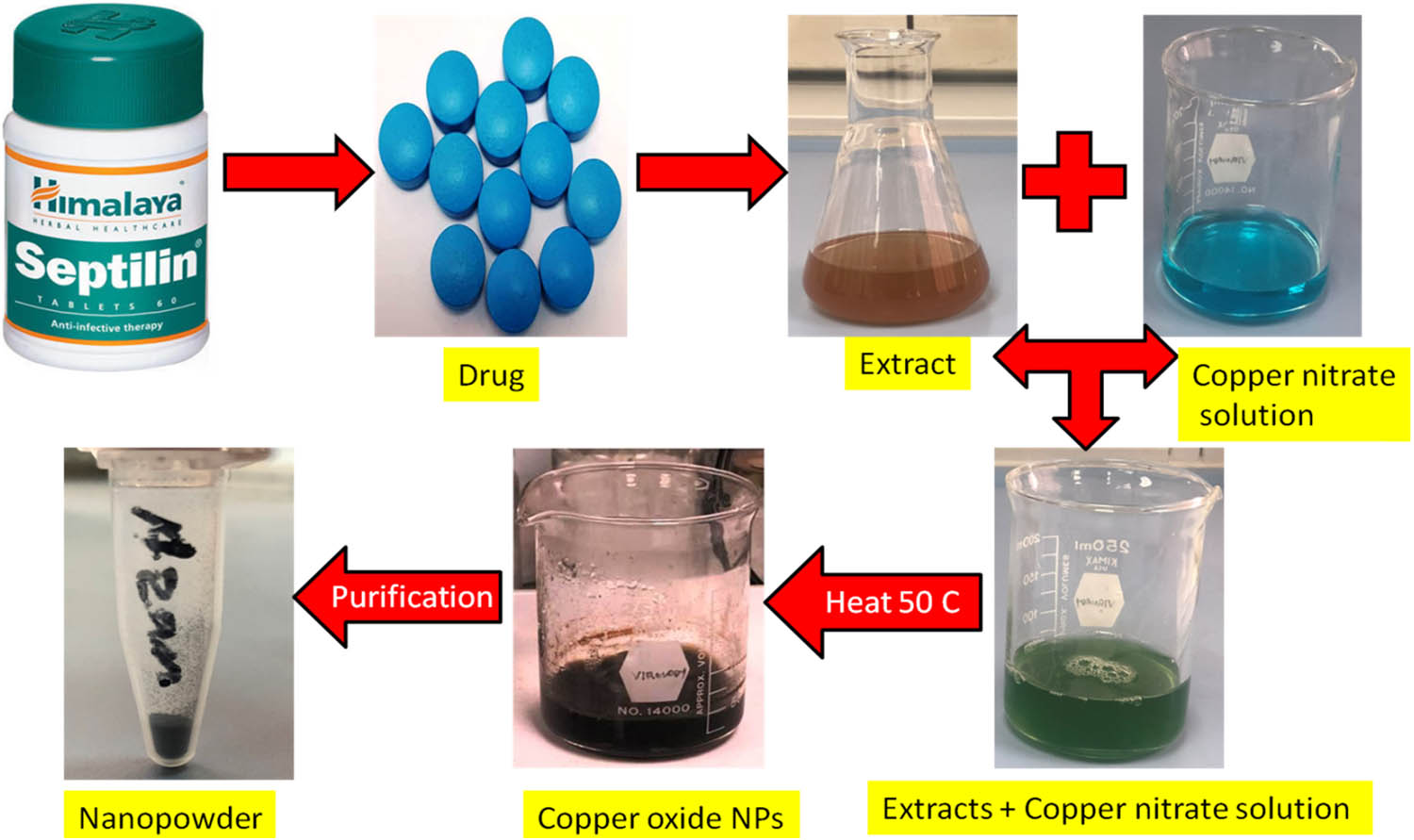
Scheme of green synthesis of CuO NPs.

UV–Vis spectra of green-synthesized CuO NPs.

FTIR spectra of herbal drug extract (a) and green-synthesized CuO NPs (b).

XRD pattern of as-prepared CuO NPs.

SEM (a), TEM, (b), and histogram (c) analysis.
5 Antimicrobial activity
5.1 MIC
MIC for gram-negative E. coli and MDR-PA accounts for 2.5 mg·mL−1, whereas the MIC for gram-positive MRSA was 1 mg·mL−1 and it was 2.5 mg·mL−1 for C. albicans.
5.2 Antibiofilm activity
Inhibition of biofilm was observed at all the tested concentrations. The decrease in biofilm for P. aeruginosa ranged from 28.47% to 92.91%. At the lowest dose, i.e., 0.0315 mg·mL−1, a 28.47% decrease in biofilm was observed, whereas at the highest dose, i.e., 2 mg·mL−1, the maximum reduction of 92.91% in biofilm was observed. Similarly, a 24.8–79.84% decrease in biofilm was observed for MRSA. The lowest concentration of CuO NPs (0.0315 mg·mL−1) decreased the biofilm by 24.8% and, at 2 mg·mL−1 of CuO NPs, the maximum decrease of 79.84% was observed. For C. albicans decrease in biofilm ranged from 16.45% to 71.57%. At the lowest concentration of CuO NPs (0.0315 mg·mL−1), a 16.45% decrease was observed, whereas a 71.5% decrease in biofilm was observed at 2 mg·mL−1 of CuO NPs (Figure 6).

Effects of CuO NPs on biofilm-forming abilities of tested pathogens.
5.3 Interaction of bacterial/fungal cells
SEM analysis revealed the microscopic changes in P. aeruginosa, MRSA, and C. albicans upon treatment with CuO NPs. SEM images are indicative of structural changes in cells. Figure 7(a–c) shows the normal morphology of P. aeruginosa, MRSA, and C. albicans. In contrast, treated cells Figure 7(a1–c1) shows the destruction in cell morphology, shrinkage in cell size, and distorted cell structures.

SEM analysis. (a) MRSA control and (a1) treated MRSA; (b) P. aeruginosa control and (b1) treated; and (c) C. albicans control and (c1) treated.
5.4 Anticancer potential of CuO NPs
A statistical decrease in cell viability (HCT-116 cell line) was observed upon treatment with CuO NPs at all tested concentrations as revealed by MTT (Figure 8). At 11.80 µg·mL−1 of NPs, 80.62% of cells were viable, whereas at 23.60 µg·mL−1, only 60% of cells were viable. Furthermore, an increase in CuO NPs to 59.0 µg·mL−1 down-regulated the viability, and only 37.03% of cells remain viable, and at the highest dose, i.e., 118 µg·mL−1, the maximum decrease in viability was observed and only 10.15% of cells remain viable (Figure 9).

Cell viability analysis of HCT-116 cells by MTT assay.

Microscopic analysis of the HCT-116 cells. (a) Control and (b–e) treated with 11.8, 23.6, 59, and 118 µg·mL−1 of NPs, respectively.
6 Discussion
The color change of septilin extract + copper nitrate solution was the initial sign of NP formation, which was further confirmed by the UV–Vis (Figure 1). The strong peaks detected at a wavelength of 285 nm are similar to the ultraviolet range of CuO NPs (Figure 2). An identical absorbance peak at a wavelength of 290 nm was observed for the CuO NPs that were produced through green method [32]. The UV spectra results align with prior studies that have reported absorption peaks at around 250 and 272 nm for CuO NPs produced from Caesalpinia bonducella [33] and Ephedra alata [34] extracts, respectively. The highest absorbance peak of CuO NPs is influenced by various factors, including temperature, type precursor salts and plant extract, and the synthesis method used. However, values as low as 219–500 nm have been documented for CuO NPs in the literature [35]. The medicinal extract from the septlin drug may function as a capping, reducer, and stabilizer agent, reducing copper nitrate into CuO NPs. FTIR analysis was conducted to locate the stretching and vibrating bonds in biosynthesized NPs and to locate the biomolecules present in the septilin extract that may act as reducing and capping agents (Figure 3). The peak at 3,284 cm−1 in extract (Figure 3a) and 3,243 cm−1 (Figure 3b) of green-synthesized CuO NPs correlates to the vibrational frequency of O–H stretching [35], whereas the peak in the herbal extract at 1,634 cm−1 indicates the C═O stretching of acids or ketones [33]. The spectral peaks seen at 2,925 cm−1 correspond to the stretching of C–H bonds [34]. The prominent peaks observed at 1,325, 1,576, and 1,697 cm−1 correspond to the bending of phenolic O–H bonds and the stretching of C═O bonds in ketones [35,36]. The peaks at 1,019 and 730 cm−1 corresponds C–O and C–H bonds, respectively [37]. The peaks at 517 cm−1 suggest the formation of a CuO nanostructure and the existence of Cu–O stretching [35,37]. The FTIR spectrum provides evidence that the plant extract contained carboxylic acid, carbohydrates, flavonoids, phenols, and alkaloids. These compounds facilitated the synthesis of NPs and functioned as capping and stabilizing agents [34,36].
The purity, crystalline nature, and size of NPs were assessed using XRD analysis. The XRD analysis revealed that the CuO NPs exhibited diffraction peaks at 2θ, namely 32.2°, 35.2°, 38.8°, 48.6°, 53.2°, 57.8°, 61.3°, and 67.8°. These peaks corresponded to the crystallographic planes (110), (002), (111), (202), (020), (202), (113) and (220), respectively (Figure 4). These findings confirm the successful green synthesis of pure and monoclinic structure of CuO NPs. The present findings align with prior studies on CuO NPs synthesized via environmentally friendly techniques [34,37,38]. The average crystallite size, as determined by the Debye–Scherer formula, was 10.88 nm. The current investigation is consistent with earlier research, which found that Seriphidium oliverianum produced CuO NPs with a size of 12.44 nm [38].
The shape and size of the synthesized CuO NPs were assessed using SEM and TEM (Figure 5). The CuO NPs that were synthesized exhibit a distinct spherical shape, as depicted in the SEM and TEM images presented in Figure 5(a). It was observed that the biological synthesis of CuO NPs results in the production of small spherical particles with uniform dimensions. The presence of biological ingredients in the extract demonstrates the relatively small aggregation. The CuO NPs produced from the herbal drug extract exhibited a spherical morphology, aligning with earlier research findings [37,39,40,41]. Furthermore, the size and shape of the manufactured CuO NPs were also observed by TEM (Figure 5b). The TEM micrographs demonstrated that the NPs had an almost spherical shape and were in the ranged of 4–20 nm (average size ∼8 nm). This is similar to earlier investigations [32,40,42].
Standard broth dilution techniques were used to assess the antibacterial and antifungal activity (MIC) that were found in the range of 1–2.5 mg·mL−1 against tested pathogens. The study demonstrated that CuO NPs had better antibacterial efficacy against MRSA in comparison to MDR-PA and C. albicans. The results of MIC values align with the findings reported by Javadhesari et al. [43] in their earlier research. In a previous study an MIC value of 2.5 to 3.5 mg·mL−1 has been reported for E. coli and S. aureus, respectively [43]. Amiri et al. also observed a comparable trend in the MIC of CuO NPs against Candida species. The MIC50 of CuO NPs against Candida species was reported as 1,000 µg·mL−1 [3,44]. The CuO NPs produced by Solanum tuberosum extract have been shown to have the antimicrobial activity of 0.2–1 mg·mL−1 [41]. Ren et al. previously documented that CuO NPs had antibacterial efficacy within the concentration range of 2,500–5,000 µg·mL−1 against several bacterial strains [45].
In addition, the impact of CuO NPs on the physical characteristics of MRSA, MDR-PA, and C. albicans was also examined by SEM (Figure 7). The control without treatment MRSA cells exhibited a smooth and intact cell surface, displaying typical, regular, and spherical morphological characteristics (Figure 7a). Nevertheless, the application of CuO NPs to MRSA cells demonstrated substantial impairment of the microorganisms and a significant decrease in their cell population. The cell wall and membrane exhibited distortions, irregularities, roughness, and lack of integrity, suggesting the loss of membrane integrity that ultimately results in cell death (Figure 7a1). Similarly, the MDR-PA cells that were not treated exhibited a healthy, normal, and rod-like architecture with a smooth cellular membrane (Figure 7b). However, when MDR-PA cells were exposed to CuO NPs, the cells exhibited severe damage. The cell membrane and wall were found to be disrupted, deformed, inconsistent, and coarse indicating a lack of cellular membrane integrity (Figure 7b1). The C. albicans cells that were not treated exhibited a sleek cell architecture characterized by an undamaged oval-shaped appearance (Figure 7c). The C. albicans cells that were subjected to CuO NPs exhibited an irregular and arbitrary cell surface, resulting in significant damage to the cells. In addition, the cells that were severely damaged were no longer in their original state, which could ultimately result in cell death (Figure 7c1). It was suggested that the Cu2+ ions have the potential to be easily released from CuO NPs, enabling them to interact more efficiently with membrane lipids and potentially trigger oxidation [35,36]. These interaction causes the membrane to collapse, resulting in the release of intracellular substances and enabling the interaction of CuO NPs with bacterial DNA, enzymes, and proteins [34]. Furthermore, it has been stated that the production of reactive oxygen species is responsible for CuO’s antibacterial action [35,36]. These processes can diminish the potential for survival of the cells and result in cellular death.
Furthermore, inhibition of biofilm formation by CuO NPs was investigated by microtiter crystal violet assay (Figure 6). Microorganisms adhere irreversibly to surfaces to form biofilms, which are the sources of subsequent infections. CuO NPs dose-dependently decreased the biofilm formation in tested pathogens. As the concentration of CuO NPs increased, the decrease in biofilm was observed. The highest concentration of 2 mg·mL−1 decreased the biofilm by 92.91%, 79.84%, and 71.57% for P. aeruginosa, MRSA, and C. albicans, respectively (Figure 6). The findings of our study align well with prior research, which has demonstrated the suppression of biofilm formation through the utilization of CuO NPs. Bai et al. [46] observed a significant reduction of 90% in S. aureus biofilm when exposed to 1,000 µg·mL−1 of CuO NPs. CuO NPs were found to suppress C. albicans biofilm development in a dose-dependent manner and inhibit it by 75% at 500 µg·mL−1 [47]. LewisOscar et al. [48] also reported a 90% decrease in biofilm by copper NPs. We are also of the opinion that the reduction in cell numbers in treated samples may be due to the absence of exopolysaccharides (EPS) secretions that did not allow the microorganism to adhere to the surface. It has been reported that EPS is crucial for the persistent adhesion of microbes to a surface [49]. Additionally, the EPS layers serve as a barrier to protect the microorganisms from adverse environmental conditions [50]. Furthermore, research has indicated that biofilm inhibition by CuO NPs is might be due to the suppression of enzymes, EPS, and virulence factors [46].
To predict the probable side effects of a drug, toxicity analysis is a critical component of toxicity studies. The cytotoxicity of CuO NPs has been further examined using MTT (Figure 8) and microscopic (Figure 9) techniques. MTT assay is associated with measuring material cytotoxicity dose dependently [51]. The assay relies on the concept that viable cells possess the ability to convert the MTT dye into a purple crystalline formazan product by the action of the NAD(P)H-dependent oxidoreductase enzyme, resulting in a color change from yellow to insoluble purple. The findings demonstrated a reduction in cell viability that was directly proportional to the dosage when treated with CuO NPs. It was found that there was an 89.85% decline in cell viability at the highest concentration of NPs i.e., 118 µg·mL−1 (Figure 8). Furthermore, in comparison to the control, the microscopic images revealed a decrease in the number of viable cells (Figure 9). After being exposed to NPs, HCT-116 cells may experience morphological changes that are obvious signs of the NPs’ cytotoxicity. The microscopic images shown in Figure 9 illustrate the apparent loss of the original shape and size of cells under the experimental treatments. The cells treated with 11.8, 23.6, 59, and 118 µg·mL−1 of CuO NPs notably show a considerable loss in plasma membrane and integrity of structure when compared to the control group without treatment (Figure 9). This was most likely the result of effective contact between the CuO NPs and HCT-116 cells, which increased stress and may cause cell death. It is clear that the internalization of CuO NPs via cell membrane penetration served as the primary mechanism for cell death in HCT-116 cells due to a substantial damage of cytoplasm [52]. Our findings align with the prior research conducted by Tabrez et al. [53], which demonstrated the suppression of the HCT-116 cell line through the use of CuO NPs and found that when exposed to a concentration of 35 µg·mL−1, only 21% of the cells were able to survive. In addition, Gnanavel et al. [54] showed that when 100 µg·mL−1 of CuO NPs are applied to the HCT-116, 22% of the cells remain viable. It has already been shown that CuO NPs in the range of 100–5,000 µg·mL−1 did not show any cytotoxic effect on human cells [55]. In addition, CuO NPs have been utilized since the nineteenth century as a secure and efficient antibacterial agent [56]. From a cytotoxic perspective, the US Environmental Protection Agency has found that CuO NPs are safe for human usage because they are low in toxicity and environmentally safe [57,58].
7 Conclusion
NPs can also be synthesized from herbal drugs since previous reports are only regarding synthesizing CuO NPs from plants or microorganisms. The production of CuO NPs from the herbal drug septilin is described for the first time in this report. Green-synthesized CuO NPs from the drug septilin can be effectively used as antimicrobial against gram-positive, gram-negative, as well as fungus. CuO NPs also possess antibiofilm efficacy as they downregulated the biofilm formation at all concentrations tested. Furthermore, the CuO NPs also showed anticancer activity as tested on human colorectal cancer (HCT-116). The number of viable cells (HCT-116) decreased upon treatment with NPs. The present study emphasizes a new and innovative approach for generating NPs with significant potential in the field of biotechnology. This method offers a viable avenue for large-scale production of NPs using environmentally sustainable and cost-effective procedures. Hence, the produced CuO NPs have promise for future investigations in many biological and therapeutic domains, including the treatment of microbial biofilm infections, as well as the inhibition of cancer cell growth.
Acknowledgments
The authors extend their appreciation to the Deanship of Scientific Research at King Khalid University for funding this work through small Group research project under grant number (RGP1/433/44).
-
Funding information: This research was supported by the Deanship of Scientific Research at King Khalid University under grant number (RGP1/433/44).
-
Author contributions: Mohammad Azam Ansari: writing – original draft, writing – review and editing, visualization, concept, project administration; Hassan Nassr Al Dhneem, Sarah Asiri, and Firdos Alam Khan: methodology, experiments; Syed Ghazanfar Ali: writing – original draft; Yahya Fahad Jamous, Mohammad Nasser Alomary, Banan Atwah, Maryam Saleh Alhumaidi, Umme Hani, and Nazima Haider: writing – review and editing, formal analysis, visualization, revision of manuscript, resources.
-
Conflict of interest: Authors state no conflict of interest.
-
Data availability statement: The datasets generated during and/or analyzed during the current study are available from the corresponding author on reasonable request.
References
[1] Desselberger U. Emerging and re-emerging infectious diseases. J Infect. 2000;40(1):3–15.10.1053/jinf.1999.0624Suche in Google Scholar PubMed
[2] Llor C, Bjerrum L. Antimicrobial resistance: Risk associated with antibiotic overuse and initiatives to reduce the problem. Ther Adv Drug Saf. 2014;5(6):229–41.10.1177/2042098614554919Suche in Google Scholar PubMed PubMed Central
[3] Prestinaci F, Pezzotti P, Pantosti A. Antimicrobial resistance: a global multifaceted phenomenon. Pathog Glob health. 2015;109(7):309–18.10.1179/2047773215Y.0000000030Suche in Google Scholar PubMed PubMed Central
[4] Li B, Webster TJ. Bacteria antibiotic resistance: New challenges and opportunities for implant‐associated orthopedic infections. J Orthop Res. 2018;36(1):22–32.10.1002/jor.23656Suche in Google Scholar PubMed PubMed Central
[5] Ventola CL. The antibiotic resistance crisis: Part 1: Causes and threats. Pharm Therapeut. 2015;40(4):277.Suche in Google Scholar
[6] Tremblay YD, Lévesque C, Segers RP, Jacques M. Method to grow Actinobacillus pleuropneumoniaebiofilm on a biotic surface. BMC Vet Res. 2013;9(1):1–8.10.1186/1746-6148-9-213Suche in Google Scholar PubMed PubMed Central
[7] Koczan JM, Lenneman BR, McGrath MJ, Sundin GW. Cell surface attachment structures contribute to biofilm formation and xylem colonization by Erwinia amylovora. Appl Environ Microbiol. 2011;77(19):7031–9.10.1128/AEM.05138-11Suche in Google Scholar PubMed PubMed Central
[8] Bowden GH, Li YH. Nutritional influences on biofilm development. Adv Dental Res. 1997;11(1):81–99.10.1177/08959374970110012101Suche in Google Scholar PubMed
[9] Dang H, Lovell CR. Microbial surface colonization and biofilm development in marine environments. Microbiol Mol Biol Rev. 2016;80(1):91–138.10.1128/MMBR.00037-15Suche in Google Scholar PubMed PubMed Central
[10] Røder HL, Herschend J, Russel J, Andersen MF, Madsen JS, Sørensen SJ, et al. Enhanced bacterial mutualism through an evolved biofilm phenotype. ISME J. 2018 Nov;12(11):2608–18.10.1038/s41396-018-0165-2Suche in Google Scholar PubMed PubMed Central
[11] Juul JS, Hornung N, Andersen B, Laurberg S, Olesen F, Vedsted P. The value of using the faecal immunochemical test in general practice on patients presenting with non-alarm symptoms of colorectal cancer. Br J Cancer. 2018 Aug;119(4):471–9.10.1038/s41416-018-0178-7Suche in Google Scholar PubMed PubMed Central
[12] Astin M, Griffin T, Neal RD, Rose P, Hamilton W. The diagnostic value of symptoms for colorectal cancer in primary care: a systematic review. Br J Gen Pract. 2011 May;61(586):e231–43.10.3399/bjgp11X572427Suche in Google Scholar PubMed PubMed Central
[13] Xi Y, Xu P. Global colorectal cancer burden in 2020 and projections to 2040. Transl Oncol. 2021 Oct;14(10):101174.10.1016/j.tranon.2021.101174Suche in Google Scholar PubMed PubMed Central
[14] Zocche DM, Ramirez C, Fontao FM, Costa LD, Redal MA. Global impact of KRAS mutation patterns in FOLFOX treated metastatic colorectal cancer. Front Genet. 2015 Mar;6:116.10.3389/fgene.2015.00116Suche in Google Scholar PubMed PubMed Central
[15] Tuutijärvi T, Lu J, Sillanpää M, Chen G. As (V) adsorption on maghemite nanoparticles. J Hazard Mater. 2009 Jul;166(2–3):1415–20.10.1016/j.jhazmat.2008.12.069Suche in Google Scholar PubMed
[16] Nalwa HS. Handbook of nanostructured materials and nanotechnology Vol 1, Synthesis and processing. United states: Academic Press; 2000.Suche in Google Scholar
[17] Buazar F, Sweidi S, Badri M, Kroushawi F. Biofabrication of highly pure copper oxide nanoparticles using wheat seed extract and their catalytic activity: A mechanistic approach. Green Process Synth. 2019 Jan;8(1):691–702.10.1515/gps-2019-0040Suche in Google Scholar
[18] Sukhwal A, Jain D, Joshi A, Rawal P, Kushwaha HS. Biosynthesised silver nanoparticles using aqueous leaf extract of Tagetes patula L. and evaluation of their antifungal activity against phytopathogenic fungi. IET Nanobiotechnol. 2017 Aug;11(5):531–7.10.1049/iet-nbt.2016.0175Suche in Google Scholar PubMed PubMed Central
[19] Jain D, Kour R, Bhojiya AA, Meena RH, Singh A, Mohanty SR, et al. Zinc tolerant plant growth promoting bacteria alleviates phytotoxic effects of zinc on maize through zinc immobilization. Sci Rep. 2020 Aug;10(1):13865.10.1038/s41598-020-70846-wSuche in Google Scholar PubMed PubMed Central
[20] Ali SG, Jalal M, Ahmad H, Sharma D, Ahmad A, Umar K, et al. Green synthesis of silver nanoparticles from Camellia sinensis and its antimicrobial and antibiofilm effect against clinical isolates. Materials. 2022 Oct;15(19):6978.10.3390/ma15196978Suche in Google Scholar PubMed PubMed Central
[21] Ali SG, Ansari MA, Alzohairy MA, Alomary MN, AlYahya S, Jalal M, et al. Biogenic gold nanoparticles as potent antibacterial and antibiofilm nano-antibiotics against Pseudomonas aeruginosa. Antibiotics. 2020 Feb 27;9(3):100.10.3390/antibiotics9030100Suche in Google Scholar PubMed PubMed Central
[22] Fahmy SA, Preis E, Bakowsky U, Azzazy HM. Platinum nanoparticles: Green synthesis and biomedical applications. Molecules. 2020 Oct;25(21):4981.10.3390/molecules25214981Suche in Google Scholar PubMed PubMed Central
[23] Ali SG, Ansari MA, Jamal QM, Almatroudi A, Alzohairy MA, Alomary MN, et al. Butea monosperma seed extract mediated biosynthesis of ZnO NPs and their antibacterial, antibiofilm and anti-quorum sensing potentialities. Arab J Chem. 2021 Apr;14(4):103044.10.1016/j.arabjc.2021.103044Suche in Google Scholar
[24] Jia B, Mei Y, Cheng L, Zhou J, Zhang L. Preparation of copper nanoparticles coated cellulose films with antibacterial properties through one-step reduction. ACS Appl Mater Interfaces. 2012 Jun;4(6):2897–902.10.1021/am3007609Suche in Google Scholar PubMed
[25] Sankar R, Manikandan P, Malarvizhi V, Fathima T, Shivashangari KS, Ravikumar V. Green synthesis of colloidal copper oxide nanoparticles using Carica papaya and its application in photocatalytic dye degradation. Spectrochim Acta Part A: Mol Biomol Spectrosc. 2014 Mar;121:746–50.10.1016/j.saa.2013.12.020Suche in Google Scholar PubMed
[26] Balkrishna A, Solleti SK, Singh H, Tomer M, Sharma N, Varshney A. Calcio-herbal formulation, Divya-Swasari-Ras, alleviates chronic inflammation and suppresses airway remodelling in mouse model of allergic asthma by modulating pro-inflammatory cytokine response. Biomed Pharmacother. 2020 Jun;126:110063.10.1016/j.biopha.2020.110063Suche in Google Scholar PubMed
[27] Khanna N, Sharma SB. Anti-inflammatory and analgesic effect of herbal preparation: Septilin. Indian J Med Sci. 2001 Apr;55(4):195–202.Suche in Google Scholar
[28] Naguib NI, Abd El Maguid A, Fahmy N. Radioprotective effect of septilin on radiation-induced histological and biochemical changes of rat eyes. Isotope Radiat Res. 2006;39(1):137–51.Suche in Google Scholar
[29] Ali SG, Ansari MA, Alzohairy MA, Alomary MN, Jalal M, AlYahya S, et al. Effect of biosynthesized ZnO nanoparticles on multi-drug resistant Pseudomonas aeruginosa. Antibiotics. 2020 May;9(5):260.10.3390/antibiotics9050260Suche in Google Scholar PubMed PubMed Central
[30] Ansari MA, Govindasamy R, Begum MY, Ghazwani M, Alqahtani A, Alomary MN, et al. Bioinspired ferromagnetic CoFe2O4 nanoparticles: Potential pharmaceutical and medical applications. Nanotechnol Rev. 2023 Jul;12(1):20230575.10.1515/ntrev-2023-0575Suche in Google Scholar
[31] Jalal M, Ansari MA, Shukla AK, Ali SG, Khan HM, Pal R, et al. Green synthesis and antifungal activity of Al2O3 NPs against fluconazole-resistant Candida spp. isolated from a tertiary care hospital. RSC Adv. 2016;109:107577–90.10.1039/C6RA23365ASuche in Google Scholar
[32] Garcia-Marin LE, Juarez-Moreno K, Vilchis-Nestor AR, Castro-Longoria E. Highly antifungal activity of biosynthesized copper oxide nanoparticles against Candida albicans. Nanomaterials. 2022 Nov;12(21):3856.10.3390/nano12213856Suche in Google Scholar PubMed PubMed Central
[33] Sukumar S, Rudrasenan A, Padmanabhan Nambiar D. Green-synthesized rice-shaped copper oxide nanoparticles using Caesalpinia bonducella seed extract and their applications. ACS Omega. 2020 Jan;5(2):1040–51.10.1021/acsomega.9b02857Suche in Google Scholar PubMed PubMed Central
[34] Atri A, Echabaane M, Bouzidi A, Harabi I, Soucase BM, Chaâbane RB. Green synthesis of copper oxide nanoparticles using Ephedra Alata plant extract and a study of their antifungal, antibacterial activity and photocatalytic performance under sunlight. Heliyon. 2023 Feb;9(2):e13484.10.1016/j.heliyon.2023.e13484Suche in Google Scholar PubMed PubMed Central
[35] Weldegebrieal GK. Photocatalytic and antibacterial activityof CuO nanoparticles biosynthesized using Verbascum thapsus leaves extract. Optik. 2020 Feb;204:164230.10.1016/j.ijleo.2020.164230Suche in Google Scholar
[36] Ramasubbu K, Padmanabhan S, Al-Ghanim KA, Nicoletti M, Govindarajan M, Sachivkina N, et al. Green synthesis of copper oxide nanoparticles using sesbania grandiflora leaf extract and their evaluation of anti-diabetic, cytotoxic, anti-microbial, and anti-inflammatory properties in an in-vitro approach. Fermentation. 2023 Mar;9(4):332.10.3390/fermentation9040332Suche in Google Scholar
[37] Amin F, Khattak B, Alotaibi A, Qasim M, Ahmad I, Ullah R, et al. Green synthesis of copper oxide nanoparticles using Aerva javanica leaf extract and their characterization and investigation of in vitro antimicrobial potential and cytotoxic activities. Evid-Based Complement Altern Med. 2021 Jun;2021:5589703.10.1155/2021/5589703Suche in Google Scholar PubMed PubMed Central
[38] Aroob S, Carabineiro SA, Taj MB, Bibi I, Raheel A, Javed T, et al. Green synthesis and photocatalytic dye degradation activity of CuO nanoparticles. Catalysts. 2023 Feb;13(3):502.10.3390/catal13030502Suche in Google Scholar
[39] Ijaz F, Shahid S, Khan SA, Ahmad W, Zaman S. Green synthesis of copper oxide nanoparticles using Abutilon indicum leaf extract: Antimicrobial, antioxidant and photocatalytic dye degradation activitie. Trop J Pharm Res. 2017 May;16(4):743–53.10.4314/tjpr.v16i4.2Suche in Google Scholar
[40] Nzilu DM, Madivoli ES, Makhanu DS, Wanakai SI, Kiprono GK, Kareru PG. Green synthesis of copper oxide nanoparticles and its efficiency in degradation of rifampicin antibiotic. Sci Rep. 2023 Aug;13(1):14030.10.1038/s41598-023-41119-zSuche in Google Scholar PubMed PubMed Central
[41] Alishah H, Pourseyedi S, Ebrahimipour SY, Mahani SE, Rafiei N. Green synthesis of starch-mediated CuO nanoparticles: preparation, characterization, antimicrobial activities and in vitro MTT assay against MCF-7 cell line. Rend Lincei. 2017 Mar;28:65–71.10.1007/s12210-016-0574-ySuche in Google Scholar
[42] Mali SC, Raj S, Trivedi RJ. Biosynthesis of copper oxide nanoparticles using Enicostemma axillare (Lam.) leaf extract. Biochem Biophys Rep. 2019;20:100699.10.1016/j.bbrep.2019.100699Suche in Google Scholar PubMed PubMed Central
[43] Javadhesari SM, Alipour S, Mohammadnejad S, Akbarpour MR. Antibacterial activity of ultra-small copper oxide (II) nanoparticles synthesized by mechanochemical processing against S. aureus and E. coli. Mater Sci Eng: C. 2019 Dec;105:110011.10.1016/j.msec.2019.110011Suche in Google Scholar PubMed
[44] Amiri M, Etemadifar Z, Daneshkazemi A, Nateghi M. Antimicrobial effect of copper oxide nanoparticles on some oral bacteria and candida species. J Dental Biomater. 2017 Mar;4(1):347.Suche in Google Scholar
[45] Ren G, Hu D, Cheng EW, Vargas-Reus MA, Reip P, Allaker RP. Characterisation of copper oxide nanoparticles for antimicrobial applications. Int J Antimicrob Agents. 2009 Jun;33(6):587–90.10.1016/j.ijantimicag.2008.12.004Suche in Google Scholar PubMed
[46] Bai B, Saranya S, Dheepaasri V, Muniasamy S, Alharbi NS, Selvaraj B, et al. Biosynthesized copper oxide nanoparticles (CuO NPs) enhances the anti-biofilm efficacy against K. pneumoniae and S. aureus. J King Saud Univ-Sci. 2022 Aug;34(6):102120.10.1016/j.jksus.2022.102120Suche in Google Scholar
[47] Ansarifard E, Zareshahrabadi Z, Sarafraz N, Zomorodian K. Evaluation of antimicrobial and antibiofilm activities of copper oxide nanoparticles within soft denture liners against oral pathogens. Bioinorg Chem Appl. 2021 Jun;2021:1–7.10.1155/2021/9939275Suche in Google Scholar PubMed PubMed Central
[48] LewisOscar F, MubarakAli D, Nithya C, Priyanka R, Gopinath V, Alharbi NS, et al. One pot synthesis and anti-biofilm potential of copper nanoparticles (CuNPs) against clinical strains of Pseudomonas aeruginosa. Biofouling. 2015 Apr;31(4):379–91.10.1080/08927014.2015.1048686Suche in Google Scholar PubMed
[49] Stoodley P, Sauer K, Davies DG, Costerton JW. Biofilms as complex differentiated communities. Annu Rev Microbiology. 2002 Oct;56(1):187–209.10.1146/annurev.micro.56.012302.160705Suche in Google Scholar PubMed
[50] Flemming HC, Wingerder J. The biofilm matrix. Nat Rev Microbiol. 2010;8:623–33.10.1038/nrmicro2415Suche in Google Scholar PubMed
[51] Gai X, Liu C, Wang G, Qin Y, Fan C, Liu J, et al. A novel method for evaluating the dynamic biocompatibility of degradable biomaterials based on real-time cell analysis. Regen Biomater. 2020;7:321–9.10.1093/rb/rbaa017Suche in Google Scholar PubMed PubMed Central
[52] Almutairi HH, Parveen N, Ansari SA. Hydrothermal synthesis of multifunctional bimetallic Ag-CuO nanohybrids and their antimicrobial, antibiofilm and antiproliferative potential. Nanomaterials. 2022 Nov;12(23):4167.10.3390/nano12234167Suche in Google Scholar PubMed PubMed Central
[53] Tabrez S, Khan AU, Mirza AA, Suhail M, Jabir NR, Zughaibi TA, et al. Biosynthesis of copper oxide nanoparticles and its therapeutic efficacy against colon cancer. Nanotechnol Rev. 2022;11:1322–31.10.1515/ntrev-2022-0081Suche in Google Scholar
[54] Gnanavel V, Palanichamy V, Roopan SM. Biosynthesis and characterization of copper oxide nanoparticles and its anticancer activity on human colon cancer cell lines (HCT-116). J Photochem Photobiol B: Biol. 2017;171:133–8.10.1016/j.jphotobiol.2017.05.001Suche in Google Scholar PubMed
[55] Allaker RP. The use of nanoparticles to control oral biofilm formation. J Dent Res. 2010;89:1175–86.10.1177/0022034510377794Suche in Google Scholar PubMed
[56] Dollwet HHA, Sorenson JRJ. Historic uses of copper compounds in medicine. Trace Elem Med. 1985;2(2):80–7.Suche in Google Scholar
[57] Sankar R, Maheswari R, Karthik S, Shivashangari KS, Ravikumar V. Anticancer activitiy of Ficus religiosa engineered copper oxide nanoparticles. Mater Sci Eng C. 2014;44:234–9.10.1016/j.msec.2014.08.030Suche in Google Scholar PubMed
[58] Elemike EE, Onwudiwe DC, Nundkumar N, Singh M. CuO and Au-CuO nanoparticles mediated by Stigmaphyllon ovatum leaf extract and their anticancer potential. Inorg Chem Commun. 2019 Jun;104:93–7.10.1016/j.inoche.2019.03.039Suche in Google Scholar
© 2024 the author(s), published by De Gruyter
This work is licensed under the Creative Commons Attribution 4.0 International License.
Artikel in diesem Heft
- Research Articles
- Green polymer electrolyte and activated charcoal-based supercapacitor for energy harvesting application: Electrochemical characteristics
- Research on the adsorption of Co2+ ions using halloysite clay and the ability to recover them by electrodeposition method
- Simultaneous estimation of ibuprofen, caffeine, and paracetamol in commercial products using a green reverse-phase HPTLC method
- Isolation, screening and optimization of alkaliphilic cellulolytic fungi for production of cellulase
- Functionalized gold nanoparticles coated with bacterial alginate and their antibacterial and anticancer activities
- Comparative analysis of bio-based amino acid surfactants obtained via Diels–Alder reaction of cyclic anhydrides
- Biosynthesis of silver nanoparticles on yellow phosphorus slag and its application in organic coatings
- Exploring antioxidant potential and phenolic compound extraction from Vitis vinifera L. using ultrasound-assisted extraction
- Manganese and copper-coated nickel oxide nanoparticles synthesized from Carica papaya leaf extract induce antimicrobial activity and breast cancer cell death by triggering mitochondrial caspases and p53
- Insight into heating method and Mozafari method as green processing techniques for the synthesis of micro- and nano-drug carriers
- Silicotungstic acid supported on Bi-based MOF-derived metal oxide for photodegradation of organic dyes
- Synthesis and characterization of capsaicin nanoparticles: An attempt to enhance its bioavailability and pharmacological actions
- Synthesis of Lawsonia inermis-encased silver–copper bimetallic nanoparticles with antioxidant, antibacterial, and cytotoxic activity
- Facile, polyherbal drug-mediated green synthesis of CuO nanoparticles and their potent biological applications
- Zinc oxide-manganese oxide/carboxymethyl cellulose-folic acid-sesamol hybrid nanomaterials: A molecularly targeted strategy for advanced triple-negative breast cancer therapy
- Exploring the antimicrobial potential of biogenically synthesized graphene oxide nanoparticles against targeted bacterial and fungal pathogens
- Biofabrication of silver nanoparticles using Uncaria tomentosa L.: Insight into characterization, antibacterial activities combined with antibiotics, and effect on Triticum aestivum germination
- Membrane distillation of synthetic urine for use in space structural habitat systems
- Investigation on mechanical properties of the green synthesis bamboo fiber/eggshell/coconut shell powder-based hybrid biocomposites under NaOH conditions
- Green synthesis of magnesium oxide nanoparticles using endophytic fungal strain to improve the growth, metabolic activities, yield traits, and phenolic compounds content of Nigella sativa L.
- Estimation of greenhouse gas emissions from rice and annual upland crops in Red River Delta of Vietnam using the denitrification–decomposition model
- Synthesis of humic acid with the obtaining of potassium humate based on coal waste from the Lenger deposit, Kazakhstan
- Ascorbic acid-mediated selenium nanoparticles as potential antihyperuricemic, antioxidant, anticoagulant, and thrombolytic agents
- Green synthesis of silver nanoparticles using Illicium verum extract: Optimization and characterization for biomedical applications
- Antibacterial and dynamical behaviour of silicon nanoparticles influenced sustainable waste flax fibre-reinforced epoxy composite for biomedical application
- Optimising coagulation/flocculation using response surface methodology and application of floc in biofertilisation
- Green synthesis and multifaceted characterization of iron oxide nanoparticles derived from Senna bicapsularis for enhanced in vitro and in vivo biological investigation
- Potent antibacterial nanocomposites from okra mucilage/chitosan/silver nanoparticles for multidrug-resistant Salmonella Typhimurium eradication
- Trachyspermum copticum aqueous seed extract-derived silver nanoparticles: Exploration of their structural characterization and comparative antibacterial performance against gram-positive and gram-negative bacteria
- Microwave-assisted ultrafine silver nanoparticle synthesis using Mitragyna speciosa for antimalarial applications
- Green synthesis and characterisation of spherical structure Ag/Fe2O3/TiO2 nanocomposite using acacia in the presence of neem and tulsi oils
- Green quantitative methods for linagliptin and empagliflozin in dosage forms
- Enhancement efficacy of omeprazole by conjugation with silver nanoparticles as a urease inhibitor
- Residual, sequential extraction, and ecological risk assessment of some metals in ash from municipal solid waste incineration, Vietnam
- Green synthesis of ZnO nanoparticles using the mangosteen (Garcinia mangostana L.) leaf extract: Comparative preliminary in vitro antibacterial study
- Simultaneous determination of lesinurad and febuxostat in commercial fixed-dose combinations using a greener normal-phase HPTLC method
- A greener RP-HPLC method for quaternary estimation of caffeine, paracetamol, levocetirizine, and phenylephrine acquiring AQbD with stability studies
- Optimization of biomass durian peel as a heterogeneous catalyst in biodiesel production using microwave irradiation
- Thermal treatment impact on the evolution of active phases in layered double hydroxide-based ZnCr photocatalysts: Photodegradation and antibacterial performance
- Preparation of silymarin-loaded zein polysaccharide core–shell nanostructures and evaluation of their biological potentials
- Preparation and characterization of composite-modified PA6 fiber for spectral heating and heat storage applications
- Preparation and electrocatalytic oxygen evolution of bimetallic phosphates (NiFe)2P/NF
- Rod-shaped Mo(vi) trichalcogenide–Mo(vi) oxide decorated on poly(1-H pyrrole) as a promising nanocomposite photoelectrode for green hydrogen generation from sewage water with high efficiency
- Green synthesis and studies on citrus medica leaf extract-mediated Au–ZnO nanocomposites: A sustainable approach for efficient photocatalytic degradation of rhodamine B dye in aqueous media
- Cellulosic materials for the removal of ciprofloxacin from aqueous environments
- The analytical assessment of metal contamination in industrial soils of Saudi Arabia using the inductively coupled plasma technology
- The effect of modified oily sludge on the slurry ability and combustion performance of coal water slurry
- Eggshell waste transformation to calcium chloride anhydride as food-grade additive and eggshell membranes as enzyme immobilization carrier
- Synthesis of EPAN and applications in the encapsulation of potassium humate
- Biosynthesis and characterization of silver nanoparticles from Cedrela toona leaf extracts: An exploration into their antibacterial, anticancer, and antioxidant potential
- Enhancing mechanical and rheological properties of HDPE films through annealing for eco-friendly agricultural applications
- Immobilisation of catalase purified from mushroom (Hydnum repandum) onto glutaraldehyde-activated chitosan and characterisation: Its application for the removal of hydrogen peroxide from artificial wastewater
- Sodium titanium oxide/zinc oxide (STO/ZnO) photocomposites for efficient dye degradation applications
- Effect of ex situ, eco-friendly ZnONPs incorporating green synthesised Moringa oleifera leaf extract in enhancing biochemical and molecular aspects of Vicia faba L. under salt stress
- Biosynthesis and characterization of selenium and silver nanoparticles using Trichoderma viride filtrate and their impact on Culex pipiens
- Photocatalytic degradation of organic dyes and biological potentials of biogenic zinc oxide nanoparticles synthesized using the polar extract of Cyperus scariosus R.Br. (Cyperaceae)
- Assessment of antiproliferative activity of green-synthesized nickel oxide nanoparticles against glioblastoma cells using Terminalia chebula
- Chlorine-free synthesis of phosphinic derivatives by change in the P-function
- Anticancer, antioxidant, and antimicrobial activities of nanoemulsions based on water-in-olive oil and loaded on biogenic silver nanoparticles
- Study and mechanism of formation of phosphorus production waste in Kazakhstan
- Synthesis and stabilization of anatase form of biomimetic TiO2 nanoparticles for enhancing anti-tumor potential
- Microwave-supported one-pot reaction for the synthesis of 5-alkyl/arylidene-2-(morpholin/thiomorpholin-4-yl)-1,3-thiazol-4(5H)-one derivatives over MgO solid base
- Screening the phytochemicals in Perilla leaves and phytosynthesis of bioactive silver nanoparticles for potential antioxidant and wound-healing application
- Graphene oxide/chitosan/manganese/folic acid-brucine functionalized nanocomposites show anticancer activity against liver cancer cells
- Nature of serpentinite interactions with low-concentration sulfuric acid solutions
- Multi-objective statistical optimisation utilising response surface methodology to predict engine performance using biofuels from waste plastic oil in CRDi engines
- Microwave-assisted extraction of acetosolv lignin from sugarcane bagasse and electrospinning of lignin/PEO nanofibres for carbon fibre production
- Biosynthesis, characterization, and investigation of cytotoxic activities of selenium nanoparticles utilizing Limosilactobacillus fermentum
- Highly photocatalytic materials based on the decoration of poly(O-chloroaniline) with molybdenum trichalcogenide oxide for green hydrogen generation from Red Sea water
- Highly efficient oil–water separation using superhydrophobic cellulose aerogels derived from corn straw
- Beta-cyclodextrin–Phyllanthus emblica emulsion for zinc oxide nanoparticles: Characteristics and photocatalysis
- Assessment of antimicrobial activity and methyl orange dye removal by Klebsiella pneumoniae-mediated silver nanoparticles
- Influential eradication of resistant Salmonella Typhimurium using bioactive nanocomposites from chitosan and radish seed-synthesized nanoselenium
- Antimicrobial activities and neuroprotective potential for Alzheimer’s disease of pure, Mn, Co, and Al-doped ZnO ultra-small nanoparticles
- Green synthesis of silver nanoparticles from Bauhinia variegata and their biological applications
- Synthesis and optimization of long-chain fatty acids via the oxidation of long-chain fatty alcohols
- Eminent Red Sea water hydrogen generation via a Pb(ii)-iodide/poly(1H-pyrrole) nanocomposite photocathode
- Green synthesis and effective genistein production by fungal β-glucosidase immobilized on Al2O3 nanocrystals synthesized in Cajanus cajan L. (Millsp.) leaf extracts
- Green stability-indicating RP-HPTLC technique for determining croconazole hydrochloride
- Green synthesis of La2O3–LaPO4 nanocomposites using Charybdis natator for DNA binding, cytotoxic, catalytic, and luminescence applications
- Eco-friendly drugs induce cellular changes in colistin-resistant bacteria
- Tangerine fruit peel extract mediated biogenic synthesized silver nanoparticles and their potential antimicrobial, antioxidant, and cytotoxic assessments
- Green synthesis on performance characteristics of a direct injection diesel engine using sandbox seed oil
- A highly sensitive β-AKBA-Ag-based fluorescent “turn off” chemosensor for rapid detection of abamectin in tomatoes
- Green synthesis and physical characterization of zinc oxide nanoparticles (ZnO NPs) derived from the methanol extract of Euphorbia dracunculoides Lam. (Euphorbiaceae) with enhanced biosafe applications
- Detection of morphine and data processing using surface plasmon resonance imaging sensor
- Effects of nanoparticles on the anaerobic digestion properties of sulfamethoxazole-containing chicken manure and analysis of bio-enzymes
- Bromic acid-thiourea synergistic leaching of sulfide gold ore
- Green chemistry approach to synthesize titanium dioxide nanoparticles using Fagonia Cretica extract, novel strategy for developing antimicrobial and antidiabetic therapies
- Green synthesis and effective utilization of biogenic Al2O3-nanocoupled fungal lipase in the resolution of active homochiral 2-octanol and its immobilization via aluminium oxide nanoparticles
- Eco-friendly RP-HPLC approach for simultaneously estimating the promising combination of pentoxifylline and simvastatin in therapeutic potential for breast cancer: Appraisal of greenness, whiteness, and Box–Behnken design
- Use of a humidity adsorbent derived from cockleshell waste in Thai fried fish crackers (Keropok)
- One-pot green synthesis, biological evaluation, and in silico study of pyrazole derivatives obtained from chalcones
- Bio-sorption of methylene blue and production of biofuel by brown alga Cystoseira sp. collected from Neom region, Kingdom of Saudi Arabia
- Synthesis of motexafin gadolinium: A promising radiosensitizer and imaging agent for cancer therapy
- The impact of varying sizes of silver nanoparticles on the induction of cellular damage in Klebsiella pneumoniae involving diverse mechanisms
- Microwave-assisted green synthesis, characterization, and in vitro antibacterial activity of NiO nanoparticles obtained from lemon peel extract
- Rhus microphylla-mediated biosynthesis of copper oxide nanoparticles for enhanced antibacterial and antibiofilm efficacy
- Harnessing trichalcogenide–molybdenum(vi) sulfide and molybdenum(vi) oxide within poly(1-amino-2-mercaptobenzene) frameworks as a photocathode for sustainable green hydrogen production from seawater without sacrificial agents
- Magnetically recyclable Fe3O4@SiO2 supported phosphonium ionic liquids for efficient and sustainable transformation of CO2 into oxazolidinones
- A comparative study of Fagonia arabica fabricated silver sulfide nanoparticles (Ag2S) and silver nanoparticles (AgNPs) with distinct antimicrobial, anticancer, and antioxidant properties
- Visible light photocatalytic degradation and biological activities of Aegle marmelos-mediated cerium oxide nanoparticles
- Physical intrinsic characteristics of spheroidal particles in coal gasification fine slag
- Exploring the effect of tea dust magnetic biochar on agricultural crops grown in polycyclic aromatic hydrocarbon contaminated soil
- Crosslinked chitosan-modified ultrafiltration membranes for efficient surface water treatment and enhanced anti-fouling performances
- Study on adsorption characteristics of biochars and their modified biochars for removal of organic dyes from aqueous solution
- Zein polymer nanocarrier for Ocimum basilicum var. purpurascens extract: Potential biomedical use
- Green synthesis, characterization, and in vitro and in vivo biological screening of iron oxide nanoparticles (Fe3O4) generated with hydroalcoholic extract of aerial parts of Euphorbia milii
- Novel microwave-based green approach for the synthesis of dual-loaded cyclodextrin nanosponges: Characterization, pharmacodynamics, and pharmacokinetics evaluation
- Bi2O3–BiOCl/poly-m-methyl aniline nanocomposite thin film for broad-spectrum light-sensing
- Green synthesis and characterization of CuO/ZnO nanocomposite using Musa acuminata leaf extract for cytotoxic studies on colorectal cancer cells (HCC2998)
- Review Articles
- Materials-based drug delivery approaches: Recent advances and future perspectives
- A review of thermal treatment for bamboo and its composites
- An overview of the role of nanoherbicides in tackling challenges of weed management in wheat: A novel approach
- An updated review on carbon nanomaterials: Types, synthesis, functionalization and applications, degradation and toxicity
- Special Issue: Emerging green nanomaterials for sustainable waste management and biomedical applications
- Green synthesis of silver nanoparticles using mature-pseudostem extracts of Alpinia nigra and their bioactivities
- Special Issue: New insights into nanopythotechnology: current trends and future prospects
- Green synthesis of FeO nanoparticles from coffee and its application for antibacterial, antifungal, and anti-oxidation activity
- Dye degradation activity of biogenically synthesized Cu/Fe/Ag trimetallic nanoparticles
- Special Issue: Composites and green composites
- Recent trends and advancements in the utilization of green composites and polymeric nanocarriers for enhancing food quality and sustainable processing
- Retraction
- Retraction of “Biosynthesis and characterization of silver nanoparticles from Cedrela toona leaf extracts: An exploration into their antibacterial, anticancer, and antioxidant potential”
- Retraction of “Photocatalytic degradation of organic dyes and biological potentials of biogenic zinc oxide nanoparticles synthesized using the polar extract of Cyperus scariosus R.Br. (Cyperaceae)”
- Retraction to “Green synthesis on performance characteristics of a direct injection diesel engine using sandbox seed oil”