Green synthesis of silver nanoparticles using mature-pseudostem extracts of Alpinia nigra and their bioactivities
-
Suree Tongwanichniyom


Abstract
Green synthesis of silver nanoparticles (AgNPs) employing agricultural wastes as plant extracts to improve environmental benignity and also economic value added is the highlight of this research. The mature pseudostem of Alpinia nigra is an unbeneficial raw material discarded from several food ingredients and medicinal formulas. Therefore, this research focused on condition optimization for AgNP synthesis with ecofriendly techniques using A. nigra mature-pseudostem extracts and evaluation of their antioxidant, antibacterial activities, and toxicity with brine shrimp lethality assay (BSLA). The optimal reaction conditions were achieved by using 5 mM silver nitrate (AgNO3) solution with a volume ratio of 2:8 for the extract to AgNO3 at pH 12 under room temperature. The morphology and crystalline phase of the generated AgNPs were characterized using UV–visible spectrophotometry, field emission-scanning electron microscope (FE-SEM), energy dispersive X-ray, X-ray diffraction, and Fourier transform-infrared (FTIR) techniques. The FE-SEM analysis exposed spherical shapes with an average diameter of approximately 49 nm. The XRD analysis indicated their face center cubic structure, and the FTIR spectra confirmed that phytochemicals from A. nigra extract promoted the synthesis of AgNPs. In particular, the biosynthesized AgNPs presented potential antibacterial activity against both Staphylococcus aureus and Escherichia coli and effective antioxidant capacity using the DPPH radical scavenging assay. Additionally, non-toxic desired AgNPs were confirmed with BSLA.
1 Introduction
Owing to the outstanding properties of silver nanoparticles (AgNPs), which offer several medical benefits such as antimicrobial [1,2,3,4,5], antioxidant [3,4,5,6], anticancer [7,8], anti-acetylcholinesterase [9], and anti-glycation effects [10] along with low toxicity advantages [11], they were underscored as significantly interesting nanomaterials. These nanoparticles were synthesized by using physical or chemical methods with a wide range of raw materials, often involving the use of numerous toxic chemical reagents. Most recently, environmentally friendly processes and waste material management have gained significant recognition, and biosynthetic processes were presented to compensate for the formerly conventional procedures. Greener approaches using plant-mediated synthesis to overcome the physicochemical disadvantages were popular. Specifically, the use of agricultural waste as a substitute for raw materials in the biosynthesis process of AgNPs was one of the selectively intriguing methodologies. In extensive literature reviews, huge plant materials were used as starting materials in the initial extraction process for AgNP preparation [1,2,3,4,6,10,11]. Alpinia nigra, one of the aromatic medicinal plants in the ginger family (Zingiberaceae), also known as Noh kala in Thai name [12], is found abundantly in Koh Kret, Nonthaburi, Thailand. It is well-known for being a favorite food ingredient in several Thai traditional dishes including fried coconut shoots, sour soup made of tamarind paste, chicken in coconut milk soup, and sour prawn soup. In addition, it was also used in traditional medicine to treat dyspepsia, bronchitis, gastric ulcers, intestinal parasitic infections, and insect bites [13,14,15]. However, only young pseudostem, shoot, leaf, seed, and rhizome could be selected as ingredients in all of the food recipes and therapeutic medications [15,16,17], while a mature-pseudostem portion was typically considered agricultural waste. Furthermore, no research has been done on A. nigra mature-pseudostem in our best prior search of previous literature reviews. Therefore, in the present work, the mature pseudostem of A. nigra was first studied to be used as the plant material in the extraction process of the biosynthesized AgNPs preparation in order to promote value-added agricultural waste to beneficial nanoparticles for pharmaceutical applications in both antibacterial and antioxidant activities. Furthermore, the in vivo brine shrimp lethality test was used for preliminary evaluation of the plant-mediated AgNPs toxicity.
2 Materials and methods
All of the chemicals and reagents employed in this research were of analytical grade. Silver nitrate (AgNO3) was purchased from TC Sathaporn Group, Co., Ltd., while plate count agar (PCA), 1,1-diphenyl-2-picrylhydrazyl (DPPH), and other reagents were supplied by Italmar (Thailand) Co., Ltd. Dry mature pseudostems of A. nigra were collected from Koh Kret island, Nonthaburi province, Thailand. Staphylococcus aureus ATCC 25923 and Escherichia coli ATCC 25922 were obtained from the Department of Medical Sciences Culture Collection. Their cultures in nutrient broth (NB) were incubated at 37°C for 24 h and inoculated into 0.85% (w/v) sterile NaCl. They were standardized by using McFarland standard No. 0.5 in order to adjust a bacterial density of approximately 1 × 10⁸ colony forming units per milliliter (CFU·mL−1) [18].
2.1 Extraction of A. nigra mature-pseudostem
The dried and ground mature pseudostems of A. nigra were collected from Koh Kret island in Nonthaburi province, Thailand. The extraction in deionized (DI) water at a ratio of 20 g of the plant materials to 200 mL of DI water was performed by employing ultrasound-assisted extraction techniques at 30°C for 1 h. Then, the extracts were filtered under vacuum conditions and stored at 4°C until further utilization.
2.2 Phytochemical screening
The qualitative analysis of secondary metabolites was determined for the freshly prepared mature-pseudostem extract of A. nigra. Phytochemicals including phenolics, flavonoids, alkaloids, terpenoids, steroids, saponins, glycosides, and carbohydrates were screened through the established methodologies [12,19].
2.3 Biosynthesis of AgNPs from mature-pseudostem extract of A. nigra
The synthesis of plant-mediated AgNPs using the mature-pseudostem extract of A. nigra was studied to obtain the optimum biosynthetic conditions by determination of four factors: reaction time (1–50 h), concentration of silver nitrate (AgNO3) solution (1, 3, 5, and 10 mM), volume ratio of the extract to AgNO3 solution (1:9, 2:8, and 5:5), and pH of the extract (5, 8, 10, and 12). The AgNPs were prepared by adding the A. nigra mature-pseudostem extract into AgNO3 solution in a 125 mL-erlenmeyer flask that was stirred with a stirrer at room temperature. The reaction progress of AgNP formation was primarily investigated based on the reaction-mixture color changing from pale yellow to reddish brown. Additionally, a double-beam UV–Vis spectrophotometer (Biochrom, Libra S80) was employed to examine the formation of nanoparticle by determining the absorbance values within the range of 300–700 nm at various reaction times. According to a number of earlier studies, electromagnetic radiation absorption of AgNPs increased between 400 and 500 nm in the visible spectrum [20,21,22,23].
2.3.1 Reaction time
To determine the optimal conditions for plant-mediated AgNP synthesis using the mature-pseudostem extract of A. nigra, the reaction time was first examined under the initial pH value of the extract (pH 5) using 1 mM AgNO3 solution and maintaining the extract to AgNO3 solution at a volume ratio of 1:9, following the previously reported methodology [7,8,24]. The reaction mixture was prepared by adding 5 mL of the A. nigra extract to 45 mL of AgNO3 solution and stirred at room temperature. The reaction color change was observed, and the absorbance values of the reaction mixture were monitored from 1 h of reaction time until the maximum absorbance value was achieved in order to determine the appropriate reaction conditions.
2.3.2 Concentration of AgNO3 solution
The concentration of AgNO3 solution was examined at several values, 1, 3, 5, and 10 mM, at the initial pH value of the extract (pH 5) and under the normal conditions of the volume ratio 1:9 of the extract to AgNO3 solution. The reactions were examined after the mixture was stirred for 1 h using UV–Vis spectrophotometry as described above until the maximum value of absorbance was reached.
2.3.3 Volume ratio of A. nigra extract to AgNO3 solution
Following the completion of the investigation on the effect of AgNO3 concentration on the efficiency of nanoparticle synthesis, an appropriate concentration of the AgNO3 solution was prepared for use in the evaluation of volume ratio of reaction mixtures. The examined ratios of the A. nigra extract to AgNO3 solution were 1:9, 2:8, and 5:5 by volume. According to the analytical protocol for the previous study of AgNO3 concentration, the absorbance of the reactions was determined until the highest absorbance values were reached.
2.3.4 pH value of the A. nigra extract
With the appropriate concentration of AgNO3 solution and the volume ratio of the extract to AgNO3 were determined, the pH value of the A. nigra extract was evaluated. The pH values in the range of acidic to basic conditions including 5, 8, 10, and 12 were studied. The experiments began with the synthesis of the A. nigra extracts at their pH values before mixing with AgNO3 solution at the optimum AgNO3 concentration and the extract to AgNO3 volume ratio. Similarly, the reaction mixture absorbances were monitored until the maximum absorbance values were reached. After the optimum reaction conditions were obtained, the reaction mixtures were centrifuged at 5,000 rpm for 10 min × 3 replicates to decant the supernatant from the biosynthesized AgNPs. The AgNP suspension was redispersed in DI water and repeated in the centrifugation process five times to purify the plant-mediated AgNPs from free biomass residue. Then, the expected nanoparticles were dried at 80°C for 2 h and kept in dry bottles for further characterization and bioactivity test.
2.4 Characterization
The morphology and topography of the biosynthesized AgNPs were identified by using a JSM-7600F Schottky field emission-scanning electron microscope (FE-SEM), and the presence of silver was confirmed by energy dispersive X-ray (EDX). The crystalline structure of the nanoparticles was determined by employing a Bruker X-ray diffractometer (model: D2 Phaser). The functional groups in A. nigra mature-pseudostem extracts and A. nigra-AgNPs were analyzed by using a Bruker Fourier transform-infrared spectrometer (FTIR) with ATR technique between the range of 4,000–500 cm−1.
2.5 In vitro antioxidative activity
DPPH radical scavenging assay was chosen to predict the antioxidative activity of the synthesized AgNPs in comparison to the A. nigra mature-pseudostem extracts. The solution of the generated AgNPs and extracts were prepared at concentrations of 62.5, 125, 250, 500, and 1,000 µg·mL−1 in methanol. Also, several concentrations of positive control, gallic acid, were provided including 2.5, 5, 10, and 15 µg·mL−1 in methanol. The methodology of He et al. [22] with some modifications for DPPH radical inhibitory was used. Briefly, 1 mL of 0.1 mM DPPH solution in methanol was mixed with 1 mL of the settled concentrations of the test samples using a vortex mixer, and then they were kept in dark conditions at room temperature for 30 min. After that, their absorbances were measured at 517 nm compared with a control consisting of 1 mL of 0.1 mM DPPH solution in 1 mL of methanol. The DPPH inhibitory effect was calculated by the following equation:
where A 0 is the absorbance of the control and A t is the absorbance of the sample solution.
2.6 Antibacterial activity
Gram-positive bacteria, Staphylococcus aureus, and gram-negative bacteria, Escherichia coli, obtained from the Department of Medical Science Culture Collection, were chosen for growth inhibition testing. Both organisms were inoculated in NB – culture media at 37°C for 24 h incubation time. Furthermore, they were adjusted to contain a bacterial density of 1 × 10⁸ colony forming units per milliliter (CFU·mL−1) by employing McFarland standard No. 0.5. Both the disc diffusion assay and the well diffusion method were used to confirm the antibacterial activity of the produced AgNPs. These approaches were performed in accordance with the procedures of Tongwanichniyom et al. [25].
2.6.1 Disc diffusion assay
About 0.1 mL of each bacteria cell suspension was spread thoroughly on the Mueller Hinton Agar (MHA) surface within the plates. Then paper discs (0.6 mm diameter) were submerged in 0.1 mL of the biosynthesized AgNPs solution at different concentrations (250, 500, and 1,000 µg·mL−1) and were placed on the bacteria culture plates in comparison to a positive control, ciprofloxacin, and a negative control, sterile distilled water. All experiments were triplicate for reliably accurate results. All experiments were incubated at 37°C for 18–24 h before their inhibition zones were noticed and then were calculated by the following equation:
where D C is the clear-zone diameter (mm) and D S is the diameter of the paper disc (mm).
2.6.2 Well diffusion assay
The bacteria cell suspensions were prepared following the disc diffusion method. MHA was drilled by using a cork borer with a diameter of approximately 6 mm in order to fill 0.1 mL of AgNP solution at concentrations of 250, 500, and 1,000 µg·mL−1. Experiments were conducted in triplicate for each concentration with ciprofloxacin being the positive control and sterile distilled water being the negative control. They were incubated for 18–24 h at 37°C, and their inhibitory zones were measured using a vernier caliper.
2.6.3 Minimum inhibitory concentration (MIC)
For quantitative determination of MIC, colony counting technique was performed following the ASTM E2149-01 standard method on PCA with some modifications [25]. The prepared bacterial suspensions (1 × 10⁸ CFU·mL−1) were diluted to 1 × 10⁴ CFU·mL−1. Subsequently, 0.1 mL of the biosynthesized AgNPs suspended in sterile distilled water at various concentrations was introduced into 0.9 mL of the diluted bacterial cell suspension to yield final solution concentrations of 7.9, 15.7, 31.3, 62.5, 125, 250, 500, and 1,000 µg·mL−1, which were attained by twofold serial dilutions. Then, the suspensions were incubated for 18 h at 37°C before being spread onto the PCA. Thereafter, they underwent another round of incubation at 37°C for an additional 18 h, followed by the measurement of the inhibition zones.
2.7 In vivo toxicity on brine shrimp lethality assay (BSLA)
The plant-mediated AgNPs were evaluated in toxicity by employing BSLA following the methodology of Kumutanat et al. [26]. Briefly, hatching of brine shrimp cysts (Artemia sp., 0.15 g) was performed in a 1 L-Erlenmeyer flask containing artificial seawater prepared by using 13.15 g of NaCl, 0.37 g of KCl, 0.50 g of CaCl2, 1.43 g of MgCl2, and 1.97 g of MgSO4·7H2O in 500 mL of DI water with pH adjusted to 7.8. The cysts were incubated under lighted conditions with well-aeration for 48 h. After that, varied concentrations of the biosynthesized AgNPs including 62.5, 125, 250, 500, and 1,000 µg·mL−1 were prepared in artificial seawater. Concurrently, transference of Artemia larvae into each concentration of the AgNP suspensions was rendered during the preparation in order to maintain the sample concentrations. Similarly, a positive control, potassium dichromate (K2Cr2O7) solution, was prepared with the same approach in 10, 20, 25, 30, and 50 μg·mL−1 concentrations. Moreover, the nauplii in artificial seawater were determined as blank. All experiments were operated in triplicate with 10 nauplii/sample tubes. Then, all samples were incubated under light conditions for 24 h at room temperature. The percentage of lethality was calculated from the counted alive nauplii as follows:
Probit analysis program was utilized to calculate the lethal concentration of half of the test organisms (LC50).
2.8 Statistical analysis
The antibacterial results were represented as mean ± standard deviation (SD). The analysis of variance was used to evaluate the antibacterial activity of the biosynthesized AgNPs at various concentrations. Duncan’s new multiple range test was performed for comparison of mean differences in the SPSS 26.0 statistical package at the 0.05 significance level.
3 Results
3.1 Phytochemical screening
Preliminary phytoconstituent screening of the mature-pseudostem extract of A. nigra presented various types of phytoconstituents, as summarized in Table 1. The biochemicals in the aqueous extract of the mature pseudostem consisted of phenolics, flavonoids, alkaloids, terpenoids, steroids, saponins, and carbohydrates. Likewise, these constituents were described as the bio-composition in several parts of A. nigra, as reported in the previous literature (Table 2). Versatile functional groups in these phytochemicals indicated that electron-donating groups act as the electron distributors to support the plant-mediated AgNP formation. Additionally, they encapsulated the synthesized AgNPs against agglomeration and also stabilized its colloidal solution [8,27].
Phytochemical screening results of mature-pseudostem extract of A. nigra
| No. | Chemical constituents | Mature-pseudostem extract of A. nigra |
|---|---|---|
| 1 | Phenolics | + |
| 2 | Flavonoids | + |
| 3 | Alkaloids | + |
| 4 | Terpenoids | + |
| 5 | Steroids | + |
| 6 | Saponins | + |
| 7 | Glycosides | − |
| 8 | Carbohydrates | + |
(+) Presence, (−) Absence.
Phytoconstituents of several parts of A. nigra
| Parts of A. nigra | Phytoconstituents | Reference |
|---|---|---|
| Leaves | Phenolics, flavonoids, alkaloids, terpenoids | [12] |
| Leaves | Phenolics, flavonoids | [13] |
| Leaves | Flavonoids, alkaloids, steroids, saponins, glycosides, cardiac glycosides, tannin, anthraquinone | [28] |
| Fruits | Phenolics, flavonoids, alkaloids, steroids, saponins, glycosides | [14] |
| Shoot | Phenolics, flavonoids, alkaloids | [29] |
| Shoot | Phenolics, flavonoids, alkaloids, saponins, glycosides, carbohydrates | [30] |
3.2 Biosynthesized AgNPs
The biosynthesized AgNPs were generated due to the reduction of Ag+ in AgNO3 solution to be Ag0 of AgNPs by reducing power of the secondary metabolites in mature-pseudostem extract of A. nigra. First, the formation of AgNPs was observed by the color of the reaction mixture, reddish brown color, which indicated the appearance of the generated AgNPs from A. nigra mature-pseudostem extract. The high absorbance value at 400–500 nm of wavelength revealed AgNP occurrence according to surface plasmon resonance theory [22,23]. The reaction time had an impact on the reaction progress and the quantity of the generated AgNPs [23,31]. Thus, it was first monitored to determine the appropriate duration for further investigation of other influential factors in achieving the desired AgNPs synthesis. Subsequently, due to the pivotal role of AgNO3 in initiating AgNPs biosynthesis, its concentration became a crucial aspect under study. The volume ratios of the extract to AgNO3 solution also affected the quantity of secondary metabolites to promote and stabilize the biosynthesized AgNPs generation [32]. Furthermore, the pH values of the extracts importantly influenced the efficacy of the plant-mediated AgNPs preparation, because the pH conditions impacted on dispersion and size of the expected AgNPs [22,23,31,33,34].
3.2.1 Effect of reaction time
When biosynthesized AgNPs were incipiently generated, the change of the reaction color from pale yellow to clearly brown was observed gradually until 4 h of the reaction time (Figure 1). Concurrently, its absorbance value slowly increased. Subsequently, the reddish-brown color of the synthesized AgNPs occurred distinctively at 24 h of reaction time, corresponding with the significantly intensified absorbance (Figures 1 and 2). It was in accordance with the previous report mentioning that increasing reaction time afforded a sharper absorbance peak [35]. Its absorbance was further monitored until 50 h, because the observed results at 48 h of the contacted time revealed the maximum absorbance of 0.203 at 478 nm, and then it decreased to 0.188 at 50 h (Figure 2). These results showed that 48 h of the synthesized time exhibited as an optimum condition for AgNPs synthesis employing A. nigra mature-pseudostem extract as plant media, because it provided the highest absorbance value. Also, increasing the intensity of surface plasmon absorbance peak also afforded higher production of the biosynthesized AgNPs [31].
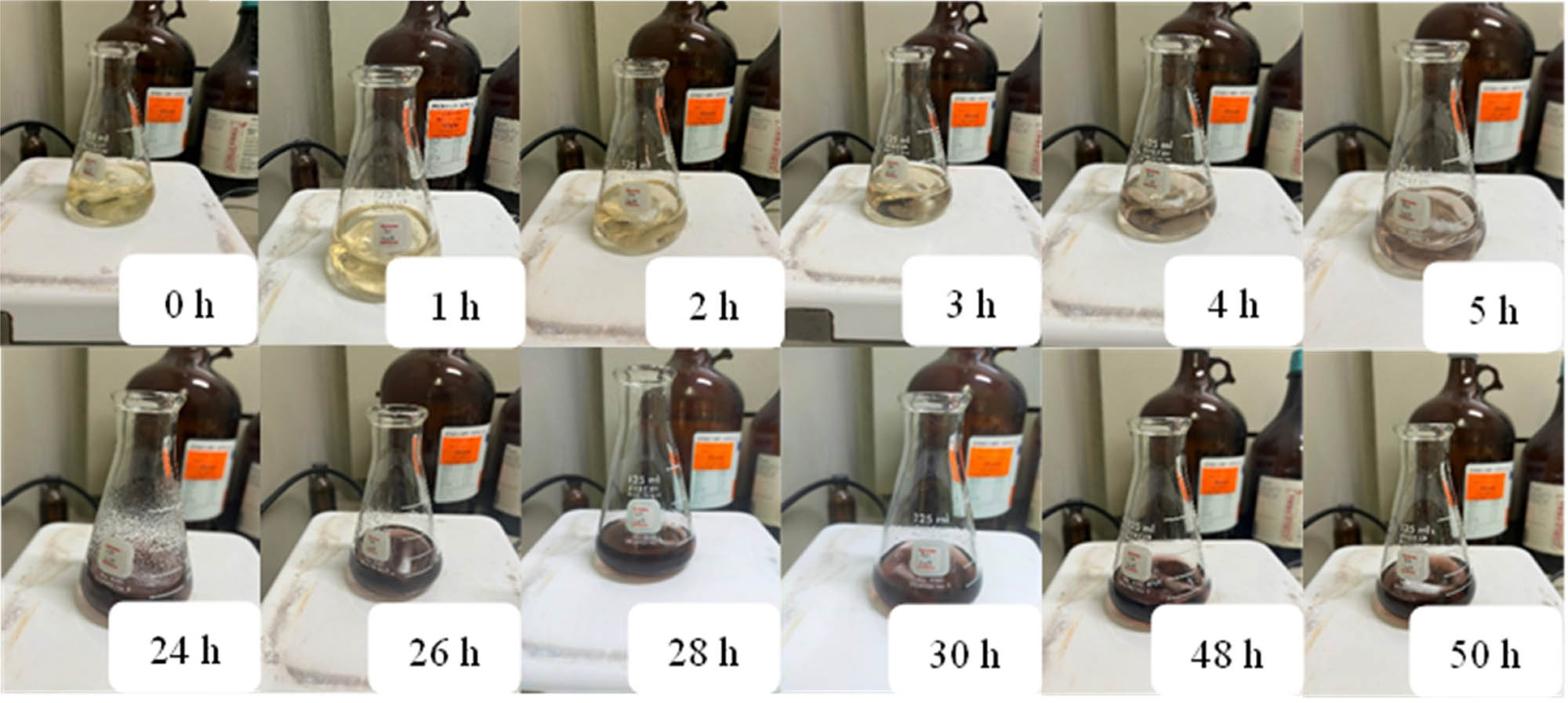
Color of the AgNPs synthesized reaction at various reaction times.

Absorbance value of AgNPs synthesis monitoring of reaction times.
3.2.2 Effect of AgNO3 concentration
Silver ion (Ag+) from AgNO3 solution played an important role as one of the vital starting materials for AgNP preparation. Hence, the concentration of AgNO3 solution exhibited a significant impact on the synthetic efficiency [2,36]. From the reaction monitoring, the reaction progress was first observed from color changing of the reaction mixture. Significantly darker reddish-brown solution has occurred after only 2 h of the reaction time when using 3 and 5 mM of AgNO3 solution. On the other hand, the reaction mixtures employing 1 and 10 mM of Ag+ solution displayed only pale yellow-brown color until 24 h, as shown in Figure 3. Up to 48 h of reaction time, the intensity of surface plasmon resonance increased directly with the concentration of Ag+, and the maximum absorbance was accomplished by employing 5 mM AgNO3 solution (Figure 4). However, a decrease in the absorbance at the highest concentration, 10 mM, indicated that stable nanoparticle formation required an optimum Ag+ concentration [2,35]. The AgNO3 concentration of 5 mM provided not only a sharper absorbance peak but also gave a blue shift of surface plasmon resonance from 478 nm of 1 mM to 447 nm, as shown in Figure 4. According to previous reports, these peak characteristics explained that 5 mM AgNO3 solution afforded larger amounts and smaller particle sizes of the generated AgNPs [9,35]. Therefore, the 5 mM AgNO3 solution was utilized for further investigations.

Color of the reaction mixtures at varied concentrations of AgNO3 solution: (a) 1 mM at 2 h, (b) 1 mM at 24 h, (c) 3 mM at 2 h, (d) 5 mM at 2 h, (e) 10 mM at 2 h, and (f) 10 mM at 24 h.

Effect of AgNO3 concentrations on absorbance value of AgNP reaction mixtures.
3.2.3 Effect of volume ratio of the extract to AgNO3 solution
The volume ratio of the extract to AgNO3 solution also provided a substantial effect on the efficacy of the biosynthesized AgNPs formation. Because diverse types and quantities of secondary metabolites in different plant extracts could have influences on AgNP size, aggregation, and stability [2,8]. The experiments for evaluating the effect of A. nigra extract volume on AgNPs generation were investigated by using the results of the earlier study with 5 mM AgNO3 solution and initial pH 5 of the extract. The maximum wavelength (λ max) for absorption of the reactions at the volume ratio of 1:9 and 2:8 were obtained at 447 nm, whereas 5:5 ratio provided only reducing absorbance on that wavelength as shown in Figure 5. At higher concentration of the extract, 2:8 v/v of extract:AgNO3, increasing intensity of the absorption peak was observed. While the highest extract concentration with equal volume of extract to AgNO3 (5:5) afforded low absorbance at AgNP wavelength during 400–500 nm. The results indicated that the optimum volume of the extract provided superior absorbance affecting the increasing AgNP formation [1,21,35]. Hence, the volume ratio of 2:8 was the proper ratio for providing the bio-reduced AgNPs formation. It was noted that increasing concentration of A. nigra extract could accelerate the reduction of Ag+ to achieve the plant-mediated AgNPs [21,35]. However, excess volume of the extract gave reverse results, reducing absorbance during 400–500 nm was obtained by the reaction mixture of volume ratio 5:5. Therefore, AgNP synthetic efficiency was affected by optimal volume of the extract [1]. And the appropriate volume ratio, 2:8, was employed for further evaluation. Nevertheless, all experiments exhibited the maximum absorbance value at 48 h indicating relatively long reaction time.

Ratio of the extract to AgNO3 volume effected on absorbance value of AgNP reaction mixtures.
3.2.4 Effect of pH value
According to earlier findings, basic conditions influenced the rapid reduction of Ag+ and could resolve the previously mentioned time-consuming drawback [22]. Hence, the pH of the A. nigra extract was investigated within the range from the initial acidic pH 5 to basic pH 12 under previously determined optimal conditions, 5 mM AgNO3 concentration and volume ratio of the extract:AgNO3 as 2:8. The absorbance intensities of the reaction mixtures at different pH values (5, 8, 10, and 12) at 24 h of reaction time are displayed in Figure 6. As the results are shown in Figure 6, only 24 h reaction time of the reaction at pH 12 revealed a similar absorbance intensity to the pH 5 reaction conducted at 48 h in our previous experiment (Figure 5). It was found that more basic conditions provided higher efficiency for plant-mediated AgNPs generation. Additionally, the λ max of the reaction at pH 5 was 438 nm, whereas the conditions of the chosen basic pH at 8, 10, and 12 revealed a blue shift to 413–415 nm (Figure 6). In extensive literature reviews, larger-sized biosynthesized AgNPs presented higher λ max for absorbance. Thus, the acidic conditions of the extract induced nanoparticle aggregation to form large-sized AgNPs [21,31,33,34]. It is worth noting that the highest investigated pH of 12 exhibited a distinctly high intensity of absorbance value (1.146) in comparison to the earlier results, along with blue-shift surface plasmon resonance. This result supported that the basic reaction medium at pH 12 afforded appropriate conditions for numerous amounts of AgNPs formation from A. nigra extract with small and highly dispersed nanoparticles.

The effect of the pH value of the A. nigra extract on the absorbance value of AgNP reaction mixtures at 24 h of reaction time.
Furthermore, the absorbance value of the pure 5 mM AgNO3 solution at pH 12 was also measured; however, the results were unable to yield an absorbance value at wavelength between 400 and 500 nm . It was confirmed that the AgNO3 solution alone was unable to produce the expected AgNPs (Figure 7).

The absorbance of 5 mM AgNO3 solution at the optimal conditions.
3.3 Characterization of AgNPs
The morphology, particle size, and crystalline structure of the biosynthesized AgNPs were evaluated by using FE-SEM, EDX, and XRD techniques. FTIR analysis was used to confirm the presence of the same functional groups in A. nigra mature-pseudostem extracts as in A. nigra AgNPs.
3.3.1 FE-SEM
The morphology and particle size of the prepared nanoparticles were analyzed with the help of FE-SEM. The SEM image shown in Figure 8, clearly identified spherical-shaped nanoparticles with an average diameter size of 49.36 nm calculated by the image J program.

SEM analysis of the AgNP formulated from mature-pseudostem extract of A. nigra.
3.3.2 EDX
Elemental components of the synthesized AgNPs were determined by EDX, as shown in Figure 9. The strong absorption peak of the silver element in the nanoparticles was clearly presented at 3 keV in the EDX spectra. Also, other peaks observed for carbon, oxygen, and chlorine indicated successful capping of extracellular bioactive constituents from A. nigra mature-pseudostem extract on the surface of the prepared AgNPs.

EDX spectrum of plant-mediated AgNPs synthesized from the A. nigra extract.
3.3.3 XRD
XRD diffractrogram of the generated AgNPs presented Bragg’s peaks at 2θ degrees of 38.3°, 44.4°, 64.6°, and 77.5° confirming the (111), (200), (220), and (311) planes of face-centered cubic (fcc) crystalline structure of metallic silver particles, respectively (Figure 10). The Bragg plane of the reflection (111) revealed an outstandingly sharp peak compared to (200), (220), and (311) reflections. This observation clearly indicated the presence of generated AgNPs [22,37,38,39].

XRD pattern of the synthesized AgNPs.
3.3.4 FTIR
FTIR spectra as shown in Figure 11 demonstrated major functional groups of the bio-organic compounds containing in mature-pseudostem extract of A. nigra at absorption peaks 3,344, 1,632, 1,369, and 1,315 cm−1 attributed to O–H, C═C, C–O, and C–N group [8,39,40,41]. The peak intensities defined functional groups of phytochemicals within A. nigra extract according to the earlier results in Table 1. Similarly, the vibration of the A. nigra-mediated AgNPs was observed at wavenumbers corresponding to those of the A. nigra extract. The reduction of peak intensity in the spectrum of the AgNPs at 3,340 and 1,632 cm−1, corresponding to O–H and C═C groups, and also the absence of peaks at 1,369 and 1,315 cm−1, attributed to C–O and C–N groups, respectively, suggested that the functional groups in various phytochemicals within the mature-pseudostem extract of A. nigra acting as reducing agents and were responsible for bioreduction of Ag+ to the expected AgNPs [23,39,41].

FTIR spectra of A. nigra extract (a) and the synthesized A. nigra – AgNPs (b).
3.4 Bioactivities of the biosynthesized AgNPs
3.4.1 Antioxidant activity
DPPH radical scavenging assay was examined for antioxidant activity of the synthesized AgNPs in comparison to A. nigra extract by employing gallic acid as a positive control, and % DPPH inhibition results are shown in Figure 12. From antioxidative percentage results, 50% inhibitory concentration (IC50) was calculated utilizing a calibration curve. Gallic acid provided an IC50 value of 8.10 ± 0.15 μg·mL−1. The IC50 value for stable DPPH radical inhibition of A. nigra – AgNPs was 282.13 ± 10.19 μg·mL−1, which was lower than that of the A. nigra extract (IC50 615.25 ± 5.61 μg·mL−1). Consequently, the fabricated AgNPs showed stronger promising antioxidant activity against DPPH radical scavenging than A. nigra extracts. Simultaneously, the phytochemicals in A. nigra mature-pseudostem extract help to encourage antioxidative efficacy of the generated AgNPs [24]. It is worth mentioning that the biosynthesized AgNPs might be further developed to be employed in pharmaceuticals for anticancer drugs or in cosmetics for anti-aging products.

DPPH inhibition activity of the biosynthesized AgNPs and the A. nigra extract at various concentrations.
3.4.2 Antibacterial activity
The biologically synthesized AgNPs from the A. nigra extracts were analyzed for antibacterial activity of representative human pathogenic bacteria, Staphylococcus aureus and Escherichia coli [42,43,44]. The generated nanoparticles were prepared in concentrations of 250, 500, and 1,000 μg·mL−1 in sterile water to evaluate the clear zone of the solution-soaked disc in the disc diffusion method and the solution-filled well in the well diffusion methodology. The results, as shown in Figure 13 and Table 3, indicated that the prepared AgNPs could inhibit both pathogenic bacteria. The AgNP solution showed more effective inhibition of gram-positive bacteria, S. aureus, than gram-negative one, E. coli, which were according to several previous reports [4,45,46]. In addition, Epand et al. explained that the presence of double protection on gram-negative bacteria with the outer membrane and peptidoglycan layer provided more resistance to antibacterial agents than gram-positive bacteria, which had only peptidoglycan as a cell wall [47].

Inhibition zone against S. aureus and E. coli of the generated AgNPs at different concentrations by employing disc diffusion method (a) and (b), and well diffusion assay (c) and (d).
Zone of inhibition against S. aureus and E. coli of the plant-mediated AgNPs at different concentrations
| Concentration of AgNPs (µg·mL−1) | Inhibition zone (mean ± SD, mm) | |||
|---|---|---|---|---|
| Disc diffusion method | Well diffusion method | |||
| S. aureus | E. coli | S. aureus | E. coli | |
| 250 | 1.10 ± 0.66a | 0.74 ± 0.40a | 1.52 ± 0.32a | 0.82 ± 0.36a |
| 500 | 2.80 ± 0.89b | 1.58 ± 0.57b | 1.98 ± 0.30b | 1.33 ± 0.35a |
| 1,000 | 3.84 ± 0.31c | 3.09 ± 0.29c | 3.19 ± 0.33c | 2.18 ± 0.83b |
Mean values with different superscript letters within the same column are significantly different (p < 0.05).
Additionally, quantitative analysis of antibacterial activity determined with the MIC value was investigated and the percentage of the number of decreasing bacteria are shown in Table 4. Owing to their qualitative antibacterial data, the phytosynthesized AgNPs were interesting to determine the minimum concentration for inhibition of both bacteria of which twofold reducing concentrations. The results revealed distinctively high efficiency of the biogenic nanoparticles to inhibit S. aureus at the lowest concentration of 7.9 µg·mL−1 with approximately 78%; hence, it was shown that the synthesized AgNPs had an MIC value less than 7.9 µg·mL−1. Although E. coli bacteria were suppressed by the AgNPs in lower percentage than that of S. aureus, the minimum concentration (7.9 µg·mL−1) also showed reduction in the viable bacteria. MIC results were in accordance with the clear zone inhibition, S. aureus was more sensitive to be inhibited by the biosynthesized AgNPs than E. coli.
The MIC values of the synthesized AgNPs against S. aureus and E. coli
| Concentration of AgNPs (µg·mL−1) | Percentage of reduction of the number of bacteria (mean ± SD, %) | |
|---|---|---|
| S. aureus | E. coli | |
| 0 | 0.21 ± 0.02a | 0.41 ± 0.02a |
| 7.9 | 78.21 ± 13.95b | 14.84 ± 7.98a,b |
| 15.7 | 79.17 ± 11.29b | 20.63 ± 8.49b,c |
| 31.3 | 81.03 ± 15.37b | 21.48 ± 11.93b,c |
| 62.5 | 82.30 ± 3.63b,c | 25.37 ± 20.32b,c |
| 125 | 82.92 ± 1.12b,c | 36.05 ± 11.25c,d |
| 250 | 84.78 ± 5.79b,c | 44.49 ± 8.84d,e |
| 500 | 85.79 ± 5.16b,c | 51.70 ± 14.68d,e |
| 1,000 | 92.86 ± 2.47c | 62.01 ± 28.20e |
Different superscripts within the same column indicate significant differences (p < 0.05).
3.4.3 In vivo toxicity using BSLA
Investigation of in vivo toxicity of the plant-mediated AgNPs using BSLA. The brine shrimp were treated with varied concentrations of A. nigra – AgNPs to determine their 50% lethal concentration (LC50). The results as shown in Figure 14 were examined by using Probit analysis to provide LC50 of 1,728.82 µg·mL−1 of the biosynthesized AgNPs. It was mentioned by several previous literature reviews that LC50 > 500 µg·mL−1 indicating non-toxic of the desired products [48,49,50]. Therefore, it is worth noting that the expected synthesized AgNPs were preliminary safety for biomedical applications.

% Mortality of brine shrimp of the biosynthesized AgNPs at various concentrations.
4 Conclusions
Our finding is the first work using mature pseudostem of A. nigra as raw material for the extraction procedure to improve its beneficial applications. The plant-mediated AgNPs synthesized from mature-pseudostem extract of A. nigra were not only environmentally friendly preparation methodology, but they also promoted value-added agricultural waste. The A. nigra-mediated AgNPs were synthesized effectively in the optimal basic conditions (pH 12) with a volume ratio of 2:8 of the extract to AgNO3 solution by employing 5 mM AgNO3 solution. The spherical-shaped particle size was 49.36 nm with a face-center cubic structure confirmed by FE-SEM, EDX, and XRD techniques. Additionally, the desired AgNPs provided high efficiency for antibacterial activity on both S. aureus and E. coli bacteria with an MIC value of less than 7.9 µg·mL−1, and also effective DPPH radical scavenging capability with an IC50 of 282.13 ± 10.19 μg·mL−1. In particular, the brine shrimp lethality test presented significant non-toxicity of the bio-reduced nanoparticles with an LC50 value of 1,728.82 μg·mL−1. Consequently, the phytosynthesized AgNPs from A. nigra mature-pseudostem extracts might be further developed to utilize in pharmaceutical, cosmetic, and household products.
Acknowledgement
The authors are thankful to the Faculty of Science at Sriracha, Kasetsart University, Sriracha Campus, Thailand, for providing all necessary facilities for this work. The authors would also like to express their sincere appreciation to Asst. Prof. Anwaraporn Niltharach for supporting of FE-SEM analysis and Asst. Prof. Somkiat Phornphisutthimas for statistical analysis.
-
Funding information: This research was funded by the Faculty of Science at Sriracha, Kasetsart University, Sriracha Campus, Thailand.
-
Author contributions: Napasawan Chumnanvej: writing – original draft, writing – review and editing, methodology, formal analysis, project administration, conceptualization, funding acquisition; Suree Tongwanichniyom: resources, supervision, methodology, investigation, formal analysis; Nuttapong Phewrat: methodology, investigation, data collection; Nattacha Rangsarikorn: methodology, investigation, data collection; Suthisa Leasen: resources, investigation; Suwaporn Luangkamin: funding acquisition, investigation, writing – review and editing.
-
Conflict of interest: Authors state no conflict of interest.
-
Data availability statement: The datasets generated during and/or analyzed during the current study are available from the corresponding author on reasonable request.
References
[1] Jalab J, Abdelwahed W, Kitaz A, Al-Kayali R. Green synthesis of silver nanoparticles using aqueous extract of Acacia cyanophylla and its antibacterial activity. Heliyon. 2021;7(9):e08033. 10.1016/j.heliyon.2021.e08033.Search in Google Scholar PubMed PubMed Central
[2] Amaladhas TP, Sivagami S, Devi TA, Ananthi N, Velammal SP. Biogenic synthesis of silver nanoparticles by leaf extract of Cassia angustifolia. Adv Nat Sci: Nanosci Nanotechnol. 2012;3(4):045006. 10.1088/2043-6262/3/4/045006.Search in Google Scholar
[3] Moteriya P, Chanda S. Biosynthesis of silver nanoparticles formation from Caesalpinia pulcherrima stem metabolites and their broad spectrum biological activities. J Genet Eng Biotechnol. 2018;16(1):105–13. 10.1016/j.jgeb.2017.12.003.Search in Google Scholar PubMed PubMed Central
[4] Laib I, Ali BD, Boudebia O. Green synthesis of silver nanoparticles using Helianthemum lippii extracts (Hl-NPs): characterization, antioxidant and antibacterial activities, and study of interaction with DNA. J Organomet Chem. 2023;986:122619. 10.1016/j.jorganchem.2023.122619.Search in Google Scholar
[5] Sivasubramanian K, Sabarinathan S, Muruganandham M, Velmurugan P, Arumugam N, Almansour AI, et al. Antioxidant, antibacterial, and cytotoxicity potential of synthesized silver nanoparticles from the Cassia alata leaf aqueous extract. Green Process Synth. 2023;12(1):20230018. 10.1515/gps-2023-0018.Search in Google Scholar
[6] Dipankar C, Murugan S. The green synthesis, characterization and evaluation of the biological activities of silver nanoparticles synthesized from Iresine herbstii leaf aqueous extracts. Colloids Surf B. 2012;98:112–9. 10.1016/j.colsurfb.2012.04.006.Search in Google Scholar PubMed
[7] Bhusari S, Sah PM, Lakkakula J, Roy A, Raut R, Chondekar R, et al. Green synthesis of silver nanoparticles via Taxus wallichiana Zucc. plant-derived Taxol: novel utilization as anticancer, antioxidation, anti-inflammation, and antiurolithic potential. Green Process Synth. 2023;12(1):20230051. 10.1515/gps-2023-0051.Search in Google Scholar
[8] Nguyen LAT, Mai BV, Nguyen DV, Nguyen NQT, Pham VV, Pham TLM, et al. Green synthesis of silver nanoparticles using Callisia fragrans leaf extract and its anticancer activity against MCF-7, HepG2, KB, LU-1, and MKN-7 cell lines. Green Process Synth. 2023;12(1):20230024. 10.1515/gps-2023-0024.Search in Google Scholar
[9] Huda NU, Ghneim HK, Fozia F, Ahmed M, Mushtaq N, Sher N, et al. Green synthesis of Kickxia elatine-induced silver nanoparticles and their role as anti-acetylcholinesterase in the treatment of Alzheimer’s disease. Green Process Synth. 2023;12(1):20230060. 10.1515/gps-2023-0060.Search in Google Scholar
[10] Anjum S, Chaudhary R, Khan AK, Hashim M, Anjum I, Hano C, et al. Light-emitting diode (LED)-directed green synthesis of silver nanoparticles and evaluation of their multifaceted clinical and biological activities. RSC Adv. 2022;12:22266–84. 10.1039/d2ra03503k.Search in Google Scholar PubMed PubMed Central
[11] Alam M. Analyses of biosynthesized silver nanoparticles produced from strawberry fruit pomace extracts in terms of biocompatibility, cytotoxicity, antioxidant ability, photodegradation, and in-silico studies. J King Saud Univ Sci. 2022;34(8):102327. 10.1016/j.jksus.2022.102327.Search in Google Scholar
[12] Gupta MK, Singh R, Rangan L. Phytochemical screening, antibacterial, anti-biofilm and quorum sensing inhibiting activity of Alpinia nigra leaf extract against infectious pathogen Pseudomonas aeruginosa PAO1. Food Control. 2023;143:109327. 10.1016/j.foodcont.2022.109327.Search in Google Scholar
[13] Sahoo S, Ghosh G, Das D, Nayak S. Phytochemical investigation and in vitro antioxidant activity of an indigenous medicinal plant Alpinia nigra B.L. Burtt. Asian Pac J Trop Biomed. 2013;3(11):871–6. 10.1016/S2221-1691(13)60171-9.Search in Google Scholar
[14] Baruah D, Yadav RNS, Yadav A, Das AM. Alpinia nigra fruits mediated synthesis of silver nanoparticles and their antimicrobial and photocatalytic activities. J Photochem Photobiol B Biol. 2019;201:111649. 10.1016/j.jphotobiol.2019.111649.Search in Google Scholar PubMed
[15] Saikia J, Washmin N, Borah T, Sarmah P, Konwar P, Siga A, et al. Physicochemical properties, chemical composition and sensory attributes of Alpinia nigra (Gaertn.) B.L. Burtt rhizome: an underutilized spice source. Eur Food Res Technol. 2023;249:1097–112. 10.1007/s00217-023-04200-5.Search in Google Scholar
[16] Baruah R, Goswami M, Das AM, Nath D, Talukdar K. Multifunctional ZnO bionanocomposites in the treatment of polluted water and controlling of multi-drug resistant bacteria. J Mol Struct. 2023;1283:135251. 10.1016/j.molstruc.2023.135251.Search in Google Scholar
[17] Roy B, Swargiary A, Giri BR. Alpinia nigra (family Zingiberaceae): an anthelmintic medicinal plant of North-East India. Adv Life Sci. 2012;2(3):39–51. 10.5923/j.als.20120203.01.Search in Google Scholar
[18] Roy A, Gauri SS, Bhattacharya M, Bhattacharya J. Antimicrobial activity of CaO nanoparticles. J Biomed Nanotechnol. 2013;9:1570–8. 10.1166/jbn.2013.1681.Search in Google Scholar PubMed
[19] Kancherla N, Dhakshinamoothi A, Chitra K, Komaram RB. Preliminary analysis of phytoconstituents and evaluation of anthelminthic property of Cayratia auriculata (in vitro). Maedica J Clin Med. 2019;14(4):350–6. 10.26574/maedica.2019.14.4.350.Search in Google Scholar PubMed PubMed Central
[20] Tarannum N, Divya, Gautam YK. Facile green synthesis and applications of silver nanoparticles: a state-of-the-art review. RSC Adv. 2019;9:34926–48. 10.1039/c9ra04164h.Search in Google Scholar PubMed PubMed Central
[21] Kumar R, Ghoshal G, Jain A, Goyal M. Rapid green synthesis of silver nanoparticles (AgNPs) using (Prunus persica) plants extract: exploring its antimicrobial and catalytic activities. J Nanomed Nanotechnol. 2017;8(4):1000452. 10.4172/2157-7439.1000452.Search in Google Scholar
[22] He Y, Wei F, Ma Z, Zhang H, Yang Q, Yao B, et al. Green synthesis of silver nanoparticles using seed extract of Alpinia katsumadai, and their antioxidant, cytotoxicity, and antibacterial activities. RSC Adv. 2017;7:39842–51. 10.1039/c7ra05286c.Search in Google Scholar
[23] Tripathy A, Raichur AM, Chandrasekaran N, Prathna TC, Mukherjee A. Process variables in biomimetic synthesis of silver nanoparticles by aqueous extract of Azadirachta indica (Neem) leaves. J Nanopart Res. 2010;12:237–46. 10.1007/s11051-009-9602-5.Search in Google Scholar
[24] Haq SI, Nisar M, Zahoor M, Ikram M, Islam NU, Ullah R, et al. Green fabrication of silver nanoparticles using Melia azedarach ripened fruit extract, their characterization, and biological properties. Green Process Synth. 2023;12(1):20230029. 10.1515/gps-2023-0029.Search in Google Scholar
[25] Tongwanichniyom S, Kitjaruwankul S, Phornphisutthimas S. Production of biomaterials from seafood waste for application as vegetable wash disinfectant. Heliyon. 2022;8(5):e09357. 10.1016/j.heliyon.2022.e09357.Search in Google Scholar PubMed PubMed Central
[26] Kumutanat W, Hongthong S, Thanasansurapong S, Kongkum N, Chumnanvej N. GC-MS and bioassay-guided isolation of xanthones from Mammea siamensis. Indones J Chem. 2023;23(3):716–26. 10.22146/ijc.79987.Search in Google Scholar
[27] Ahmad N, Sharma S, Alam MK, Singh VN, Shamsi SF, Mehta BR, et al. Rapid synthesis of silver nanoparticles using dried medicinal plant of basil. Colloids Surf B. 2010;81(1):81–6. 10.1016/j.colsurfb.2010.06.029.Search in Google Scholar PubMed
[28] Ahmed AMA, Sharmen F, Mannan A, Rahman MA. Phytochemical, analgesic, antibacterial, and cytotoxic effects of Alpinia nigra (Gaertn.) Burtt leaf extract. J Tradit Complement Med. 2015;5(4):248–52. 10.1016/j.jtcme.2014.11.012.Search in Google Scholar PubMed PubMed Central
[29] Nishat FS, Nilima N, Saikia BM. Phytochemical analysis of Lasia spinosa and Alpinia nigra, potential medicinal plants of Assam. Med Plants – Int J Phytomed Relat Ind. 2012;4(3):170–3. 10.5958/j.0975-4261.4.3.021.Search in Google Scholar
[30] Ananta S, Bishnupada R. Phytochemical screening and antioxidant property of shoot extracts of Alpinia nigra (Gaertn.) Burtt (Family - Zingiberaceae). Med Plants – Int J Phytomed Relat Ind. 2015;7(1):48–54. 10.5958/0975-6892.2015.00007.6.Search in Google Scholar
[31] Sathishkumar M, Sneha K, Yun YS. Immobilization of silver nanoparticles synthesized using Curcuma longa tuber powder and extract on cotton cloth for bactericidal activity. Bioresour Technol. 2010;101:7958–65. 10.1016/j.biortech.2010.05.051.Search in Google Scholar PubMed
[32] Ibrahim MH, Xue Z, Abdu HI, Shinger MI, Idris AM, Edris MM, et al. Sensitive and selective colorimetric nitrite ion assay using silver nanoparticles easily synthesized and stabilized by AHNDMS and functionalized with PABA. Nanoscale Adv. 2019;1:1207–14. 10.1039/c8na00146d.Search in Google Scholar PubMed PubMed Central
[33] Iravani S, Zolfaghari B. Green synthesis of silver nanoparticles using Pinus eldarica bark extract. Biomed Res Int. 2013;2013:639725. 10.1155/2013/639725.Search in Google Scholar PubMed PubMed Central
[34] Veerasamy R, Xin TZ, Gunasagaran S, Xiang TFW, Yang EFC, Jeyakumar N, et al. Biosynthesis of silver nanoparticles using mangosteen leaf extract and evaluation of their antimicrobial activities. J Saudi Chem Soc. 2011;15(2):113–20. 10.1016/j.jscs.2010.06.004.Search in Google Scholar
[35] Dubey SP, Lahtinen M, Särkkä H, Sillanpää M. Bioprospective of Sorbus aucuparia leaf extract in development of silver and gold nanocolloids. Colloids Surf B. 2010;80(1):26–33. 10.1016/j.colsurfb.2010.05.024.Search in Google Scholar PubMed
[36] Htwe YZN, Chow WS, Suda Y, Mariatti M. Effect of silver nitrate concentration on the production of silver nanoparticles by green method. Mater Today: Proc. 2019;17(3):568–73. 10.1016/j.matpr.2019.06.336.Search in Google Scholar
[37] Datta S, Kanjilal B, Sarkar P. Silver nanoparticles decorated eggshell membrane as an effective platform for interference free sensing of dopamine. J Env Sci Health A. 2018;53(12):1048–55. 10.1080/10934529.2018.1474574.Search in Google Scholar PubMed
[38] Kaur B, Pandiyan T, Satpati B, Srivastava R. Simultaneous and sensitive determination of ascorbic acid, dopamine, uric acid, and tryptophan with silver nanoparticles-decorated reduced graphene oxide modified electrode. Colloids Surf B. 2013;111:97–106. 10.1016/j.colsurfb.2013.05.023.Search in Google Scholar PubMed
[39] Kumar B, Smita K, Seqqat R, Benalcazar K, Grijalva M, Cumbal L. In vitro evaluation of silver nanoparticles cytotoxicity on Hepatic cancer (Hep-G2) cell line and their antioxidant activity: green approach for fabrication and application. J Photochem Photobiol B, Biol. 2016;159:8–13. 10.1016/j.jphotobiol.2016.03.011.Search in Google Scholar PubMed
[40] Chakrabartty I, Kalita NK, Boruah P, Katiyar V, Hakeem KR, Rangan L. Physico-rheological characterization of organically derived seed samples from Alpinia nigra (Gaertn.) B.L. Burtt, an ethnic medicinal plant of Northeast India. Ind Crop Prod. 2020;152:112560. 10.1016/j.indcrop.2020.112560.Search in Google Scholar
[41] Lin L, Wang W, Huang J, Li Q, Sun D, Yang X, et al. Nature factory of silver nanowires: plant-mediated synthesis using broth of Cassia fistula leaf. Chem Eng J. 2010;162:852–8. 10.1016/j.cej.2010.06.023.Search in Google Scholar
[42] Enan G, Al-Mohammadi AR, Mahgoub S, Abdel-Shafi S, Askar E, Ghaly MF, et al. Inhibition of Staphylococcus aureus LC 554891 by Moringa oleifera seed extract either singly or in combination with antibiotics. Molecules. 2020;25(19):4583. 10.3390/molecules25194583.Search in Google Scholar PubMed PubMed Central
[43] Akinduti PA, Emoh-Robinson V, Obamoh-Triumphant HF, Obafemi YD, Banjo TT. Antibacterial activities of plant leaf extracts against multi-antibiotic resistant Staphylococcus aureus associated with skin and soft tissue infections. BMC Complement Med Ther. 2022;22:47. 10.1186/s12906-022-03527-y.Search in Google Scholar PubMed PubMed Central
[44] Noormandi A, Dabaghzadeh F. Effects of green tea on Escherichia coli as a uropathogen. J Tradit Complement Med. 2015;5:15–20. 10.1016/j.jtcme.2014.10.005.Search in Google Scholar PubMed PubMed Central
[45] Lakshmanan G, Sathiyaseelan A, Kalaichelvan PT, Murugesan K. Plant-mediated synthesis of silver nanoparticles using fruit extract of Cleome viscosa L.: assessment of their antibacterial and anticancer activity. Karbala Int J Mod Sci. 2018;4(1):61–8. 10.1016/j.kijoms.2017.10.007.Search in Google Scholar
[46] Jyoti K, Baunthiyal M, Singh A. Characterization of silver nanoparticles synthesized using Urtica dioica Linn. leaves and their synergistic effects with antibiotics. J Radiat Res Appl Sci. 2016;9(3):217–27. 10.1016/j.jrras.2015.10.002.Search in Google Scholar
[47] Epand RM, Walker C, Epand RF, Magarvey NA. Molecular mechanisms of membrane targeting antibiotics. Biochim Biophys Acta. 2016;1858(5):980–7. 10.1016/j.bbamem.2015.10.018.Search in Google Scholar PubMed
[48] Clemen-Pascual LM, Macahig RAS, Rojas NRL. Comparative toxicity, phytochemistry, and use of 53 Philippine medicinal plants. Toxicol Rep. 2022;9:22–35. 10.1016/j.toxrep.2021.12.002.Search in Google Scholar PubMed PubMed Central
[49] Moshi MJ, Innocent E, Magadula JJ, Otieno DF, Weisheit A, Mbabazi PK, et al. Brine shrimp toxicity of some plants used as traditional medicines in Kagera Region, north western Tanzania. Tanzan J Health Res. 2010;12(1):63–7. 10.4314/thrb.v12i1.56287.Search in Google Scholar PubMed
[50] Mkangara M, Mpenda FN. Antimicrobial and cytotoxicity activities of medicinal plants against Salmonella gallinarum isolated from chickens. Vet Med Int. 2022;2022:2294120. 10.1155/2022/2294120.Search in Google Scholar PubMed PubMed Central
© 2024 the author(s), published by De Gruyter
This work is licensed under the Creative Commons Attribution 4.0 International License.
Articles in the same Issue
- Research Articles
- Green polymer electrolyte and activated charcoal-based supercapacitor for energy harvesting application: Electrochemical characteristics
- Research on the adsorption of Co2+ ions using halloysite clay and the ability to recover them by electrodeposition method
- Simultaneous estimation of ibuprofen, caffeine, and paracetamol in commercial products using a green reverse-phase HPTLC method
- Isolation, screening and optimization of alkaliphilic cellulolytic fungi for production of cellulase
- Functionalized gold nanoparticles coated with bacterial alginate and their antibacterial and anticancer activities
- Comparative analysis of bio-based amino acid surfactants obtained via Diels–Alder reaction of cyclic anhydrides
- Biosynthesis of silver nanoparticles on yellow phosphorus slag and its application in organic coatings
- Exploring antioxidant potential and phenolic compound extraction from Vitis vinifera L. using ultrasound-assisted extraction
- Manganese and copper-coated nickel oxide nanoparticles synthesized from Carica papaya leaf extract induce antimicrobial activity and breast cancer cell death by triggering mitochondrial caspases and p53
- Insight into heating method and Mozafari method as green processing techniques for the synthesis of micro- and nano-drug carriers
- Silicotungstic acid supported on Bi-based MOF-derived metal oxide for photodegradation of organic dyes
- Synthesis and characterization of capsaicin nanoparticles: An attempt to enhance its bioavailability and pharmacological actions
- Synthesis of Lawsonia inermis-encased silver–copper bimetallic nanoparticles with antioxidant, antibacterial, and cytotoxic activity
- Facile, polyherbal drug-mediated green synthesis of CuO nanoparticles and their potent biological applications
- Zinc oxide-manganese oxide/carboxymethyl cellulose-folic acid-sesamol hybrid nanomaterials: A molecularly targeted strategy for advanced triple-negative breast cancer therapy
- Exploring the antimicrobial potential of biogenically synthesized graphene oxide nanoparticles against targeted bacterial and fungal pathogens
- Biofabrication of silver nanoparticles using Uncaria tomentosa L.: Insight into characterization, antibacterial activities combined with antibiotics, and effect on Triticum aestivum germination
- Membrane distillation of synthetic urine for use in space structural habitat systems
- Investigation on mechanical properties of the green synthesis bamboo fiber/eggshell/coconut shell powder-based hybrid biocomposites under NaOH conditions
- Green synthesis of magnesium oxide nanoparticles using endophytic fungal strain to improve the growth, metabolic activities, yield traits, and phenolic compounds content of Nigella sativa L.
- Estimation of greenhouse gas emissions from rice and annual upland crops in Red River Delta of Vietnam using the denitrification–decomposition model
- Synthesis of humic acid with the obtaining of potassium humate based on coal waste from the Lenger deposit, Kazakhstan
- Ascorbic acid-mediated selenium nanoparticles as potential antihyperuricemic, antioxidant, anticoagulant, and thrombolytic agents
- Green synthesis of silver nanoparticles using Illicium verum extract: Optimization and characterization for biomedical applications
- Antibacterial and dynamical behaviour of silicon nanoparticles influenced sustainable waste flax fibre-reinforced epoxy composite for biomedical application
- Optimising coagulation/flocculation using response surface methodology and application of floc in biofertilisation
- Green synthesis and multifaceted characterization of iron oxide nanoparticles derived from Senna bicapsularis for enhanced in vitro and in vivo biological investigation
- Potent antibacterial nanocomposites from okra mucilage/chitosan/silver nanoparticles for multidrug-resistant Salmonella Typhimurium eradication
- Trachyspermum copticum aqueous seed extract-derived silver nanoparticles: Exploration of their structural characterization and comparative antibacterial performance against gram-positive and gram-negative bacteria
- Microwave-assisted ultrafine silver nanoparticle synthesis using Mitragyna speciosa for antimalarial applications
- Green synthesis and characterisation of spherical structure Ag/Fe2O3/TiO2 nanocomposite using acacia in the presence of neem and tulsi oils
- Green quantitative methods for linagliptin and empagliflozin in dosage forms
- Enhancement efficacy of omeprazole by conjugation with silver nanoparticles as a urease inhibitor
- Residual, sequential extraction, and ecological risk assessment of some metals in ash from municipal solid waste incineration, Vietnam
- Green synthesis of ZnO nanoparticles using the mangosteen (Garcinia mangostana L.) leaf extract: Comparative preliminary in vitro antibacterial study
- Simultaneous determination of lesinurad and febuxostat in commercial fixed-dose combinations using a greener normal-phase HPTLC method
- A greener RP-HPLC method for quaternary estimation of caffeine, paracetamol, levocetirizine, and phenylephrine acquiring AQbD with stability studies
- Optimization of biomass durian peel as a heterogeneous catalyst in biodiesel production using microwave irradiation
- Thermal treatment impact on the evolution of active phases in layered double hydroxide-based ZnCr photocatalysts: Photodegradation and antibacterial performance
- Preparation of silymarin-loaded zein polysaccharide core–shell nanostructures and evaluation of their biological potentials
- Preparation and characterization of composite-modified PA6 fiber for spectral heating and heat storage applications
- Preparation and electrocatalytic oxygen evolution of bimetallic phosphates (NiFe)2P/NF
- Rod-shaped Mo(vi) trichalcogenide–Mo(vi) oxide decorated on poly(1-H pyrrole) as a promising nanocomposite photoelectrode for green hydrogen generation from sewage water with high efficiency
- Green synthesis and studies on citrus medica leaf extract-mediated Au–ZnO nanocomposites: A sustainable approach for efficient photocatalytic degradation of rhodamine B dye in aqueous media
- Cellulosic materials for the removal of ciprofloxacin from aqueous environments
- The analytical assessment of metal contamination in industrial soils of Saudi Arabia using the inductively coupled plasma technology
- The effect of modified oily sludge on the slurry ability and combustion performance of coal water slurry
- Eggshell waste transformation to calcium chloride anhydride as food-grade additive and eggshell membranes as enzyme immobilization carrier
- Synthesis of EPAN and applications in the encapsulation of potassium humate
- Biosynthesis and characterization of silver nanoparticles from Cedrela toona leaf extracts: An exploration into their antibacterial, anticancer, and antioxidant potential
- Enhancing mechanical and rheological properties of HDPE films through annealing for eco-friendly agricultural applications
- Immobilisation of catalase purified from mushroom (Hydnum repandum) onto glutaraldehyde-activated chitosan and characterisation: Its application for the removal of hydrogen peroxide from artificial wastewater
- Sodium titanium oxide/zinc oxide (STO/ZnO) photocomposites for efficient dye degradation applications
- Effect of ex situ, eco-friendly ZnONPs incorporating green synthesised Moringa oleifera leaf extract in enhancing biochemical and molecular aspects of Vicia faba L. under salt stress
- Biosynthesis and characterization of selenium and silver nanoparticles using Trichoderma viride filtrate and their impact on Culex pipiens
- Photocatalytic degradation of organic dyes and biological potentials of biogenic zinc oxide nanoparticles synthesized using the polar extract of Cyperus scariosus R.Br. (Cyperaceae)
- Assessment of antiproliferative activity of green-synthesized nickel oxide nanoparticles against glioblastoma cells using Terminalia chebula
- Chlorine-free synthesis of phosphinic derivatives by change in the P-function
- Anticancer, antioxidant, and antimicrobial activities of nanoemulsions based on water-in-olive oil and loaded on biogenic silver nanoparticles
- Study and mechanism of formation of phosphorus production waste in Kazakhstan
- Synthesis and stabilization of anatase form of biomimetic TiO2 nanoparticles for enhancing anti-tumor potential
- Microwave-supported one-pot reaction for the synthesis of 5-alkyl/arylidene-2-(morpholin/thiomorpholin-4-yl)-1,3-thiazol-4(5H)-one derivatives over MgO solid base
- Screening the phytochemicals in Perilla leaves and phytosynthesis of bioactive silver nanoparticles for potential antioxidant and wound-healing application
- Graphene oxide/chitosan/manganese/folic acid-brucine functionalized nanocomposites show anticancer activity against liver cancer cells
- Nature of serpentinite interactions with low-concentration sulfuric acid solutions
- Multi-objective statistical optimisation utilising response surface methodology to predict engine performance using biofuels from waste plastic oil in CRDi engines
- Microwave-assisted extraction of acetosolv lignin from sugarcane bagasse and electrospinning of lignin/PEO nanofibres for carbon fibre production
- Biosynthesis, characterization, and investigation of cytotoxic activities of selenium nanoparticles utilizing Limosilactobacillus fermentum
- Highly photocatalytic materials based on the decoration of poly(O-chloroaniline) with molybdenum trichalcogenide oxide for green hydrogen generation from Red Sea water
- Highly efficient oil–water separation using superhydrophobic cellulose aerogels derived from corn straw
- Beta-cyclodextrin–Phyllanthus emblica emulsion for zinc oxide nanoparticles: Characteristics and photocatalysis
- Assessment of antimicrobial activity and methyl orange dye removal by Klebsiella pneumoniae-mediated silver nanoparticles
- Influential eradication of resistant Salmonella Typhimurium using bioactive nanocomposites from chitosan and radish seed-synthesized nanoselenium
- Antimicrobial activities and neuroprotective potential for Alzheimer’s disease of pure, Mn, Co, and Al-doped ZnO ultra-small nanoparticles
- Green synthesis of silver nanoparticles from Bauhinia variegata and their biological applications
- Synthesis and optimization of long-chain fatty acids via the oxidation of long-chain fatty alcohols
- Eminent Red Sea water hydrogen generation via a Pb(ii)-iodide/poly(1H-pyrrole) nanocomposite photocathode
- Green synthesis and effective genistein production by fungal β-glucosidase immobilized on Al2O3 nanocrystals synthesized in Cajanus cajan L. (Millsp.) leaf extracts
- Green stability-indicating RP-HPTLC technique for determining croconazole hydrochloride
- Green synthesis of La2O3–LaPO4 nanocomposites using Charybdis natator for DNA binding, cytotoxic, catalytic, and luminescence applications
- Eco-friendly drugs induce cellular changes in colistin-resistant bacteria
- Tangerine fruit peel extract mediated biogenic synthesized silver nanoparticles and their potential antimicrobial, antioxidant, and cytotoxic assessments
- Green synthesis on performance characteristics of a direct injection diesel engine using sandbox seed oil
- A highly sensitive β-AKBA-Ag-based fluorescent “turn off” chemosensor for rapid detection of abamectin in tomatoes
- Green synthesis and physical characterization of zinc oxide nanoparticles (ZnO NPs) derived from the methanol extract of Euphorbia dracunculoides Lam. (Euphorbiaceae) with enhanced biosafe applications
- Detection of morphine and data processing using surface plasmon resonance imaging sensor
- Effects of nanoparticles on the anaerobic digestion properties of sulfamethoxazole-containing chicken manure and analysis of bio-enzymes
- Bromic acid-thiourea synergistic leaching of sulfide gold ore
- Green chemistry approach to synthesize titanium dioxide nanoparticles using Fagonia Cretica extract, novel strategy for developing antimicrobial and antidiabetic therapies
- Green synthesis and effective utilization of biogenic Al2O3-nanocoupled fungal lipase in the resolution of active homochiral 2-octanol and its immobilization via aluminium oxide nanoparticles
- Eco-friendly RP-HPLC approach for simultaneously estimating the promising combination of pentoxifylline and simvastatin in therapeutic potential for breast cancer: Appraisal of greenness, whiteness, and Box–Behnken design
- Use of a humidity adsorbent derived from cockleshell waste in Thai fried fish crackers (Keropok)
- One-pot green synthesis, biological evaluation, and in silico study of pyrazole derivatives obtained from chalcones
- Bio-sorption of methylene blue and production of biofuel by brown alga Cystoseira sp. collected from Neom region, Kingdom of Saudi Arabia
- Synthesis of motexafin gadolinium: A promising radiosensitizer and imaging agent for cancer therapy
- The impact of varying sizes of silver nanoparticles on the induction of cellular damage in Klebsiella pneumoniae involving diverse mechanisms
- Microwave-assisted green synthesis, characterization, and in vitro antibacterial activity of NiO nanoparticles obtained from lemon peel extract
- Rhus microphylla-mediated biosynthesis of copper oxide nanoparticles for enhanced antibacterial and antibiofilm efficacy
- Harnessing trichalcogenide–molybdenum(vi) sulfide and molybdenum(vi) oxide within poly(1-amino-2-mercaptobenzene) frameworks as a photocathode for sustainable green hydrogen production from seawater without sacrificial agents
- Magnetically recyclable Fe3O4@SiO2 supported phosphonium ionic liquids for efficient and sustainable transformation of CO2 into oxazolidinones
- A comparative study of Fagonia arabica fabricated silver sulfide nanoparticles (Ag2S) and silver nanoparticles (AgNPs) with distinct antimicrobial, anticancer, and antioxidant properties
- Visible light photocatalytic degradation and biological activities of Aegle marmelos-mediated cerium oxide nanoparticles
- Physical intrinsic characteristics of spheroidal particles in coal gasification fine slag
- Exploring the effect of tea dust magnetic biochar on agricultural crops grown in polycyclic aromatic hydrocarbon contaminated soil
- Crosslinked chitosan-modified ultrafiltration membranes for efficient surface water treatment and enhanced anti-fouling performances
- Study on adsorption characteristics of biochars and their modified biochars for removal of organic dyes from aqueous solution
- Zein polymer nanocarrier for Ocimum basilicum var. purpurascens extract: Potential biomedical use
- Green synthesis, characterization, and in vitro and in vivo biological screening of iron oxide nanoparticles (Fe3O4) generated with hydroalcoholic extract of aerial parts of Euphorbia milii
- Novel microwave-based green approach for the synthesis of dual-loaded cyclodextrin nanosponges: Characterization, pharmacodynamics, and pharmacokinetics evaluation
- Bi2O3–BiOCl/poly-m-methyl aniline nanocomposite thin film for broad-spectrum light-sensing
- Green synthesis and characterization of CuO/ZnO nanocomposite using Musa acuminata leaf extract for cytotoxic studies on colorectal cancer cells (HCC2998)
- Review Articles
- Materials-based drug delivery approaches: Recent advances and future perspectives
- A review of thermal treatment for bamboo and its composites
- An overview of the role of nanoherbicides in tackling challenges of weed management in wheat: A novel approach
- An updated review on carbon nanomaterials: Types, synthesis, functionalization and applications, degradation and toxicity
- Special Issue: Emerging green nanomaterials for sustainable waste management and biomedical applications
- Green synthesis of silver nanoparticles using mature-pseudostem extracts of Alpinia nigra and their bioactivities
- Special Issue: New insights into nanopythotechnology: current trends and future prospects
- Green synthesis of FeO nanoparticles from coffee and its application for antibacterial, antifungal, and anti-oxidation activity
- Dye degradation activity of biogenically synthesized Cu/Fe/Ag trimetallic nanoparticles
- Special Issue: Composites and green composites
- Recent trends and advancements in the utilization of green composites and polymeric nanocarriers for enhancing food quality and sustainable processing
- Retraction
- Retraction of “Biosynthesis and characterization of silver nanoparticles from Cedrela toona leaf extracts: An exploration into their antibacterial, anticancer, and antioxidant potential”
- Retraction of “Photocatalytic degradation of organic dyes and biological potentials of biogenic zinc oxide nanoparticles synthesized using the polar extract of Cyperus scariosus R.Br. (Cyperaceae)”
- Retraction to “Green synthesis on performance characteristics of a direct injection diesel engine using sandbox seed oil”