Green synthesis of silver nanoparticles from Bauhinia variegata and their biological applications
-
Mohamed K. Y. Soliman


Abstract
The production of silver nanoparticles (Ag NPs) utilizing biological means with renewable resources is thought to be risk-free, environmentally benign, and safe. In this work, the capacity of Bauhinia variegata to produce Ag NPs was measured. Numerous methods, including UV–Vis spectroscopy, TEM, FTIR spectroscopy, and XRD, were employed for the analysis of the produced Ag NPs. Ag NP antimicrobial capacity has been examined through microtitreplate as well as agar well diffusion techniques. Ag NPs’ ability to scavenge free radicals at varying concentrations was assessed using the DPPH technique. The MICs were 1,000 µg·mL−1 against pathogenic microbes including Staphylococcus aureus, Pseudomonas aeruginosa, and Candida albicans, while 500 and 250 µg·mL−1 were versus Bacillus subtilis and Escherichia coli, respectively. Silver showed an intriguing antioxidant capacity, achieving IC50 of 46.23 μg·mL−1. Additionally, Ag NPs demonstrated possible anticancer action when applied to the carcinoma cell lines Caco-2, with IC50 of 396.2 μg·mL−1 and cytotoxicity toward normal Vero cell lines with IC50 of 609.45 μg·mL−1. Furthermore, Ag NPs demonstrated a range of antibiofilm activities toward S. aureus (MRSA). In conclusion, Ag NPs biosynthesized via B. variegata show promise for a variety of safe biological applications.
1 Introduction
Natural ingredients have a wide range of biological functions, which makes them an important component throughout the pharmaceutical development [1,2]. Nanotechnology uses atoms as well as molecules of materials to fabricate new materials at the nanoscale (from one to hundred nm in diameter) [3]. In the past decade, nanotechnology has been gaining popularity due to its significant contribution to advancements in engineering, technology, and healthcare [4]. The areas of medicine as well as biology have shown promise since the development of nanotechnology [5,6,7]. Nanoparticles’ tiny size, high surface-area-to-volume proportions, and variety of mechanical, optical, and magnetic characteristics make them promising candidates for use as anticancer, antimicrobial, and antioxidants agents [8,9,10,11,12,13]. Because of its numerous diagnostic uses, the noble metal nanoparticles, which may be formed of silver, gold, etc., have drawn significant interest in biological fields [14,15]. These novel active ingredients are often made utilizing a variety of techniques, such as chemical, physical, as well as biological approaches [3,15,16,17]. However, those produced via chemical and physical procedures are not environmentally friendly, possess poor yields, need extreme conditions, and create hazardous byproducts [18,19]. All of these disadvantages make it unfavorable to employ NPs made by chemical and physical means in the field of medicine. As a result, there exists a pressing demand for low-cost, ecologically safe, as well as not hazardous NP synthesis techniques. The synthesis of nanomaterials through metabolic materials produced by plants or microorganisms is easy to use and environmentally friendly [20,21,22,23,24]. Plant-mediated formation of nanomaterials is becoming more and more popular because of their minimal toxicity, effectiveness, environmental friendliness, and quick time to processing [25].
Investigating a wide variety of plants and showcasing their distinct phytochemical components and characteristics, this study on a plant-based nano-synthesis. Utilizing the stems, roots, and leaves of plants for medicinal purposes as well as agricultural fruit and vegetable seeds, an environmentally friendly basis for nanomaterial production is created. Because of their biocompatibility, readily adjustable physicochemical characteristics, and sustainable source, these green-synthesized nanoparticles exhibit promise in a variety of applications [26]. A wealth of bioactive substances found in plants have the ability to decrease, stabilize, along with cap metabolites, turning metallic ions into nanomaterials. This may result in the creation of nanomaterials containing proteins, aldehydes, terpenoids, polyphenols, tannins, flaviolin, and polysaccharides, among other predetermined properties [27].
A specific type of plant known for its antioxidant ability is the Bauhinia variegata orchid plant. Its vibrant blooms and extensive pharmacological background have made it a popular choice for decades in traditional therapy as a natural antioxidant. The plant has physiologically active compounds that lessen the effects of oxidative stress and protect cells from damaging free radical damage [28]. By adding B. variegata into food or incorporating it into meals, customers may reap the benefits of enhanced general health and strengthened defenses of the immune system against naturally occurring antioxidants [29].
Over the past 20 years, Ag NPs have become a popular kind of generated metal nanoparticles because of its unique chemical, mechanical, and biological characteristics [30]. Research studies showed that at lower amounts of AgNO3, there is an improvement in biocompatibility, catalytic activity, and therapeutic potential, despite the fact that AgNO3 may be harmful in elevated concentrations [31]. There is ample evidence supporting the antibacterial as well as anticancer properties of Ag NPs [19,32,33]. Additionally, there is currently little research involving natural synthesis employing native and naturally occurring plants with potential uses in biomedicine, despite the fact that several studies have demonstrated the green synthesis of Ag NPs utilizing leaves from plants [34,35,36]. Reactive oxygen species (ROS) counteraction by Ag NPs has demonstrated significant promise, suggesting a possible role in lowering levels of oxidative stress [37]. Their tailored cytotoxicity towards cancer cells ultimately sparks research into their antitumor mechanisms, resulting in the creation of innovative, focused cancer therapies [38]. Furthermore, Ag NPs’ wide-spectrum antibacterial efficacy against a range of pathogens highlights how crucial they are for combating resistance to antibiotics and diseases that are infectious [39].
The aim of this research is to provide a contribution to the growing corpus of information regarding green nanotechnology as well as its role in the preservation of the environment along with healthcare. Then, B. variegata generated the Ag NPs, which were characterized utilizing a wide variety of analytical methods, such as UV, XRD pattern, FTIR spectroscopy, and TEM analysis. Ag NPs’ antibacterial action against harmful microorganisms, antibiofilm, antioxidant agent, and their cytotoxicity on human cells in addition to anticancer activity.
2 Materials and methods
2.1 Materials
Sigma-Aldrich provided all analytical-grade research chemicals (Darmstadt, Germany). Mueller–Hinton (M–H) agar, Luria–Bertani broth, methanol, glacial acetic acid, dimethyl sulfoxide (DMSO), silver nitrate (AgNO3), crystal violet, NaOH, and HCl were among them.
2.2 Plant-mediated synthesize Ag nanoparticles
The Ag NPs were prepared as a previous study with minor modification [40] as follows: the dried leaves of the B. variegata plant were gathered, cleaned under running tap water, allowed to dry, and converted to powder. The resulting powder was about 10 g mixed with 150 mL of distilled H2O and heated for 60 min at 45°C at 140 rpm of agitation. After centrifuging the mixture for 7 minutes at 10,000 rpm, the resulting liquid was extracted to facilitate the Ag NPs manufacturing process. The biogenesis process involved mixing 16.9 mg of AgNO3 with eighty milliliters of dH2O and then adding a total of 20 mL of B. variegata extract to get an ultimate 1 mM of Ag nanoparticles. The prior mixture had been stirred for 60 minutes at 140 rpm, and then NaOH (1 M) was added until the mixture’s pH reached 8. The resulting mixture was then left to stand at ambient temperature throughout the whole night in order to verify whether the starter of metal had completely reduced in order to create a nanostructure, where could be seen by a shift in color to give yellowish brown color from a colorless appearance.
2.3 Characterization
The biosynthesized Ag NPs were characterized by identifying a strong absorption peak linked to surface Plasmon excitation. The optical UV–vis absorption characteristics were obtained using the JASCO-730 double-beam spectrophotometer at 200–800 nm wavelengths. Using TEM-JEOL 1010, the NPs’ dimensions and shape were evaluated. The NP mixture was added to the carbon/copper TEM grid until complete adsorbed took place. To get rid of any extra solution, the filled grids were gently dabbed with paper towels before examination. The average radius and standard deviation of NPs were determined to express the poly-dispersity of the NPs in terms of PDI according to Rudrappa et al. [41]. Further analysis of various functional groups present in the bio-fabricated nanoparticles was done with FTIR spectroscopy (JASCO, FTIR 6100). After mixing by KBr, specimens of silver nanoparticles were securely smashed into discs. The discs in question were subjected to scanning within 400–4,000 cm−1 to acquire FTIR spectra. The crystalline composition of the synthetic Ag NPs which developed was examined using an X-ray diffract pattern. Having 40 kV voltage, 30 mA electricity, and a Cu–Kα source of radiation with λ = 1.54 Å, the measured 2θ frequencies of the XRD structure fell between 5 and 80°.
2.4 Antimicrobial activity
2.4.1 Agar well method
Escherichia coli, Staphylococcus aureus, Pseudomonas aeruginosa, Bacillus subtilis, and Candida albicans (unicellular fungi) were among the numerous human microbial pathogens used as pathogenic microorganisms. On sterilized Petri dishes, the studied microorganisms were uniformly dispersed via M–H agar. A sterile 8 mm diameter well was made in the plates employing a cork-borer. The well was filled with 100 μL of silver nanoparticles to assess the antibacterial activity. Subsequent to the plates being incubated at 37°C and 30°C for 24 and 72 h, respectively, the inhibition zones were measured [42].
2.4.2 Determination of MIC
Applying the broth-based microdilution method, the MIC values for Ag NPs were investigated against previously mentioned microbes. Different concentrations of Ag NPs (1,000 to 15.75 μg·mL−1) have been generated. Sterilized microtiter plate wells were filled with 100 μL of M–H broth and then supplied with Ag NPs (100 μL) at different concentrations. Twenty microliter (20 μL) of bacterial cell suspension matching to OD similar to 0.5 McFarland standard was added to all except the negative control well. Positive control wells were filled with bacterial solution to see if M–H broth would maintain bacterial growth. At 37°C, these plates were left to incubate for 24 h. Following that, 30 µL of HiMedia’s resazurin solution (0.02% wt/v) was then added into every well inside the plate, and it was then incubated once again for 6 h to look for growth of bacteria. The growth control wells’ color changed from blue to red provided the strains were grown correctly, while the negative control well’s color remained unchanged when there was no contamination. The investigation was run three times [33].
2.5 Biofilm inhibition assay
To assess the silver nanoparticles’ potential to prevent or reduce the formation of bacterial biofilms, the microtitreplate (MTP) approach was used versus S. aureus MRSA, a clinical isolate known to be a strong biofilm-forming agent. We made various modifications to the biofilm experiment based on prior work [43]. Simply, MTP incorporating TSB media enriched with 1% glucose was subjected to graded concentrations of the silver nanoparticles. The test microorganism was inoculated into MTP for 48 h at 37°C before being diluted (1:100) with TSB. After incubation removal of the well materials without disrupting the formed biofilms, the resulting biofilm was fixed over 10 min with 200 μL of 95% methanol and three times washing with phosphate-buffered saline (PBS) at 7.4 pH. The 200 μL wells were filled with 0.3% w/v crystal violet, then allowed to incubate for 15 min at 37°C. Lastly, enabling the quantitative measurement of biofilm development, 30% acetic acid was put into the wells of the plate after they had been cleaned with distilled water. The microplate reader (STATFAX-USA) assessed the absorbance at O.D 540 nm. the outcomes were verified by contrasting the treated and untreated control wells [19].
2.6 Antioxidant assay
The antioxidant capacity exhibited by the generated Ag NPs was assessed utilizing the DPPH (2,2-diphenyl-1-picrylhydrazyl) method [33]. This specific procedure was used to silver NPs within a range (from 1,000 to 15.62 µg·mL−1). Then, 10 µL of DPPH solution was added to all well, and the mixture was placed in an incubator which was set at 37°C for 30 min while being shaken at 120 rpm under a dark environment. Ascorbic acid was the positive control under the same situations and concentrations. A negative control was a DPPH and tris-buffer without any treatment (ascorbic acid or Ag NPs) in the exact same setting as the experiment. After the incubation period, the resulting color’s absorbance near 517 nm was measured. We used the following formulas to get the proportions of scavenging free radicals [44]:
A 1 is the absorbance of control and A 2 is the absorbance of treatment.
2.7 Cytotoxic and anticancer activity
The American Type Culture Collection provided two distinct cell cultures (Caco-2): adenocarcinoma of the colorectal epithelia and normal Vero cells. To generate a full monolayer sheet, 1 × 10⁵ cells mL−1 (100 µL well−1) were added into 96-well tissue culture plates and cultured for 24 h at 37°C. The growth material was taken out of the 96-well microtiter plates after a confluent sheet of cells had developed, and the cell monolayer was twice washed using washing fluid. The test specimen was divided into two halves and diluted twice in RPMI medium containing 2% serum (maintenance medium). In many wells, each dilution was assessed in increments of 0.1 mL, while three wells functioned as controls and were just supplied with maintenance medium. After being incubated at 37°C, the plate was examined. Physical indicators of toxic effects, such as shrinkage, trimming, the formation of gran or either a partial or whole loss of the monolayer, were examined within the cells. A 5 mg·mL−1 solution of MTT was generated in PBS. Formazan, an MTT metabolic final product, maybe redissolved in 200 µL DMSO. To completely combine the formazan and solvent, put on a shaker for 5 min at 150 rpm. Eliminate background and measure optical density at 560 nm. The number of cells and optical density should be directly correlated [45,46].
The percentage of cell survival was calculated using the formula below:
A 1 is the absorbance of the tested sample (Ag nanoparticles) and A 2 is the absorbance of control.
2.8 Statistical analysis
All of the findings were derived using the means of the three replications and the standard error. The Sigma plot 12.5 program was used to analyze the data for means of variance.
3 Results and discussion
3.1 Bio-fabricated silver NPs via B. variegata
The plant and its extracts have recently piqued the interest of scientists studying nanotechnology, particularly with regard to the production of Ag NPs given their advantageous characteristics in a variety of applications [47,48,49]. Through the use of plant extracts, our environmentally friendly technique transforms silver ions into stable, biodegradable nanoparticles. This ecologically friendly method of producing NPs makes use of B. variegata water extract, a substance with a number of benefits over conventional chemical methods. The method’s little environmental effect is one of its most significant benefits. As opposed to chemical procedures, which sometimes need the use of hazardous substances and produce undesirable byproducts. Ag ions are more easily reduced to silver nanoparticles attributable to the bioactive chemicals found in B. variegata. Furthermore, the phytochemicals serve as capping agents, preventing NP aggregation and maintaining NP stability. This green synthesis approach yields Ag NPs with enhanced biocompatibility that are both ecologically benign and helpful to the atmosphere as well. Similar research used the lowering activity of active metabolites found in the plant leaves’ aqueous extract to create a variety of NPs [4]. The hue shift to a deep dark brown is linked to surface plasmon resonance. Wavelength is measured between 200 and 800 nm by the UV–Vis spectrophotometer. A broadband was observed at 415 nm in the wavelength where the Ag NPs formed, as seen in Figure 1.
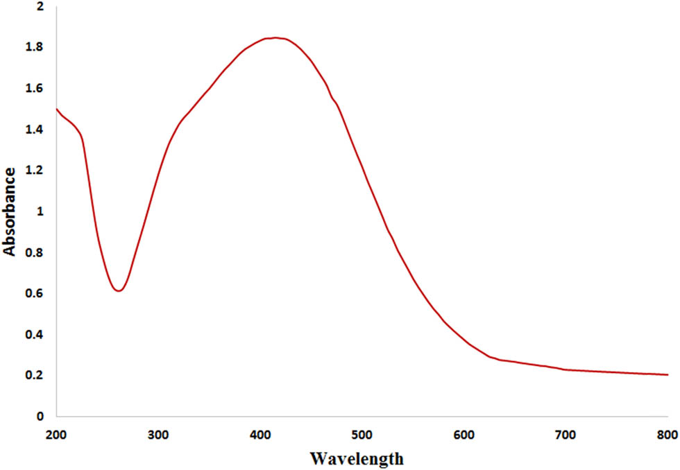
The UV–Vis spectrum for Ag NPs produced by B. variegate extract.
XRD is utilized to determine the distinctive diffraction of each crystalline material and to identify its crystalline form. Figure 2 displays the outcome. The XRD representation of biosynthesized Ag NPs is presented with four primary peak patterns at 2-theta angles of 38, 44, 64, and 77°, respectively. These peaks are associated with reflection planes of 111, 200, 220, and 311. This research correlates with other research [50].
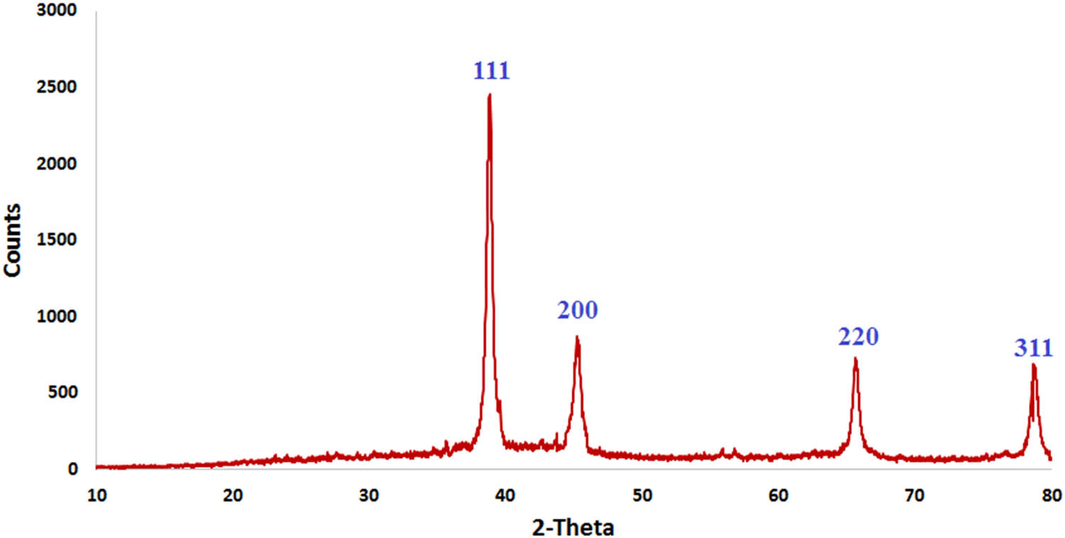
XRD analysis for Ag NPs produced by B. variegate extract.
TEM was used to assess the mean size and shape. Ag NPs are polydisperse, spherical, and have a PDI value of 0.43. Their diameters range from 10 to 55 nm, as seen in Figure 3 from a TEM picture. Ag NPs were found in a range of 37–87 nm in the prior article [50]. The aforementioned work demonstrates how extract may provide stable, homogenous, spherical, and polycrystalline Ag NPs. These investigations support the previously described bio-reduced Ag NPs [48,49,50,51].
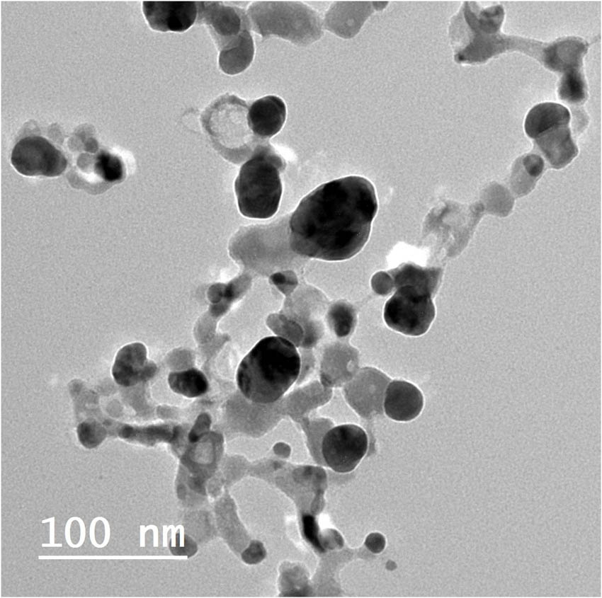
TEM image of Ag NPs produced by B. variegate extract.
To verify that groups with functions were present on the outer layer of biosynthetic Ag NPs, FTIR was employed Figure 4. The characteristic peaks of the amide I band (C═O stretch/hydrogen bond coupled with COO–) are assigned to a frequency of 1,639 and 1,556 cm−1, as the spectra show. Conversely, the amide-II band (NH bending coupled with CN stretching) is assigned to frequencies of 1,389, and 1,331 cm−1 (COO– symmetrical stretching). The C–O stretching frequency at 983 cm−1 is linked to the amide III band. They identify OH and –COO stretching as the cause of the peaks at 3,065 and 2,915 cm−1, respectively. The Ag NPs are represented by the peak positions at 872, 510, and 425 cm−1 frequencies.
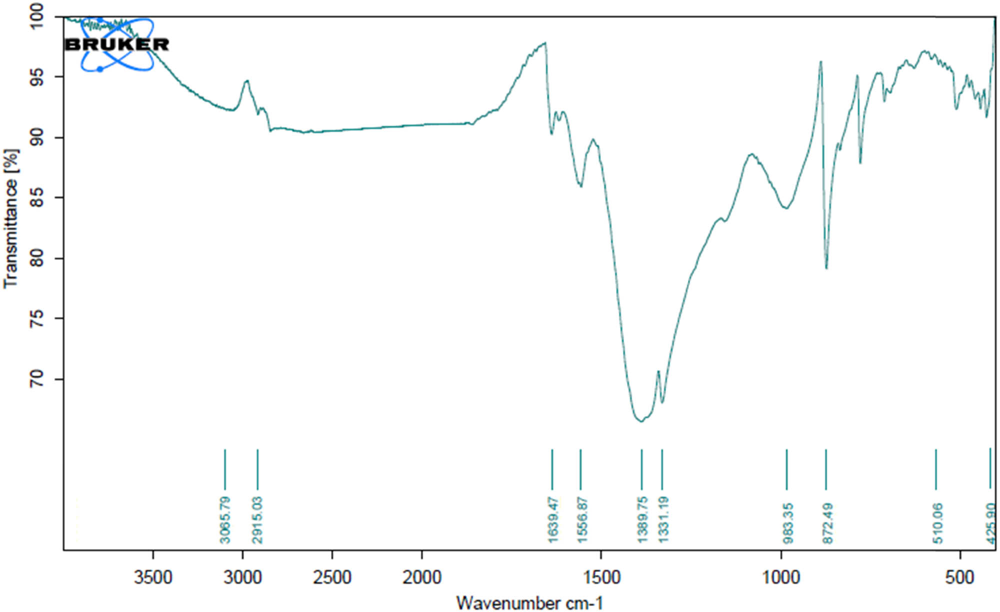
FTIR analysis of Ag NPs.
3.2 Antimicrobial assay
The antibacterial efficacy of biologically produced Ag NPs was assessed in this work against five microbial pathogens via the diffusion technique of agar wells. Regarding each microbe’s zone of inhibition, Ag NPs significantly inhibited C. albicans, E. coli, B. subtilis, S. aureus, and P. aeruginosa by 16.8 ± 0.4, 15.2 ± 0.3, 13.8 ± 0.26, 12.7 ± 0.3, and 12.66 ± 0.15 mm, respectively (Figure 5). The results are consistent with a prior study on the antibacterial efficacy of Ag NPs produced via extracts of L. stoechas, which found that of the tested strains, C. albicans was the most susceptible microbial species, while Gram-positive bacteria S. aureus less sensitive than C. albicans [52]. The negatively charged surface bacteria cell wall attracts the Ag ions that come from nanoparticles. They move and connect to the cell wall of bacteria because they experience magnetic attraction, changing the permeability and composition of that wall [53]. Microorganisms’ DNA becomes incapable of reproducing itself and of making cellular proteins like ribosome subunits. When exposed to Ag+ ions, the majority of the enzymes required for ATP synthesis inactivate [54].
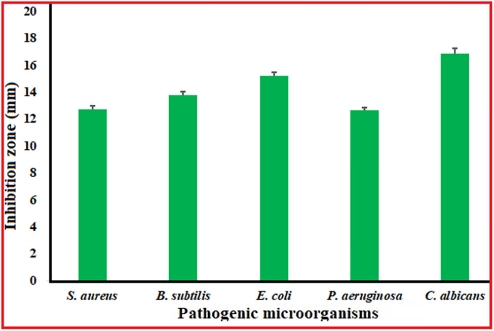
Antimicrobial efficacy of silver NPs via agar well procedure.
The investigation focused on the inhibitory effects of Ag nanoparticles at different dosages (16.62–1,000 µg·mL−1). The outcomes demonstrated that Ag NPs’ MIC against P. aeruginosa, S. aureus, and C. albicans were 1,000 µg·mL−1. B. subtilis and E. coli came in second and third, at 500 and 250 µg·mL−1, respectively (Figure 6). Another research claims that nanoparticles prevent the growth of harmful microorganisms by engaging with DNA’s phosphorous moiety. This stops the replication of DNA, which reduces the effectiveness of enzymes [51,55,56]. It also has the power to inhibit the enzymes in bacteria that produce oxygen, which stops the manufacture of ATP and eventually kills the cells. Additionally, the negative-charged bacterial surface and the positively charged NPs created electrostatic interactions which resulted in a variety of changes, such as separation of the membranes, cytoplasm deformation, and ultimately membrane rupture [57,58]. Increased interaction with bacterial cells due to the larger surface area boosts the effectiveness of antimicrobial agents. Silver nanoparticles also naturally possess antimicrobial properties. They have the capacity to release silver ions, which harm essential components and interfere with biological processes, making them toxic to bacteria. The plant extract’s properties combined with those of the Ag NPs formed a more potent antibacterial action against both types of bacteria (Gram positive and Gram negative) [59]. The increased efficiency might be attributed to the unique properties of Ag NPs, including their increased area of surface in comparison to the volume ratio [60]. Antimicrobial activity was tested versus C. albicans, E. coli, and S. aureus [61].
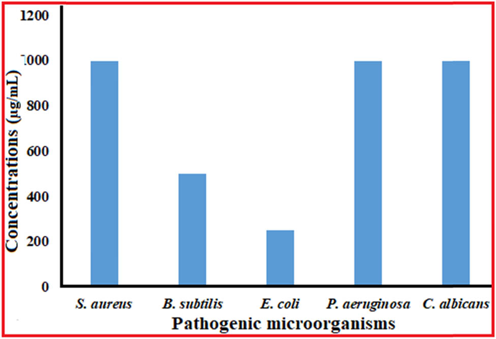
MIC of Ag NPs versus different microbial strains.
3.3 Antibiofilm
The antibiofilm efficacy of nanoparticles against S. aureus MRSA showed variable results in this experiment. Consequently, when used at double-fold doses from, Ag NPs had the greatest efficacy against the development of biofilms generated by MRSA. The amount of biofilm formed was decreased by 67.05%, 58.04%, 44.65%, 36.93%, 27.9%, 27.14%, 17.3%, and 12.97% at doses varying from 7.8 to 1,000 μg·mL−1 (Figure 7). Depending on the quantitative and qualitative assessments of each individual, Ag NPs prevented the early phases of MRSA biofilm development (Figure 8). The biofilm inhibition results from our crystal violet approach mirrored those reported by Khan et al. [62]. Because biofilms contribute to bacterial pathogenesis and are difficult to completely eradicate with antibiotics, they are important in medical settings [63]. Bacterial cell-to-cell communication tightly controls the extremely ordered sequence of events that results in the generation of biofilms [64]. About 80% of infections were linked to the formation of biofilms, and the matrix’s composition – which is mostly made up of EPS, proteins, lipids, and eDNA – has a significant impact on how long these biofilms survive [65]. The investigation’s findings are in line with other studies, which showed that S. aureus biofilms were decreased by over 40% at half sub-inhibitory dosage when Ag NPs generated via the green approach [66]. The channels of water that assist with nutrition transfer inside the biofilm matrix additionally allow nanoparticle diffusion and anti-biofilm action. Moreover, it has been shown that nanoparticles may adhere to and pass through the membranes of bacteria, build up inside bacterial cells, and eventually cause bacterial death [67].
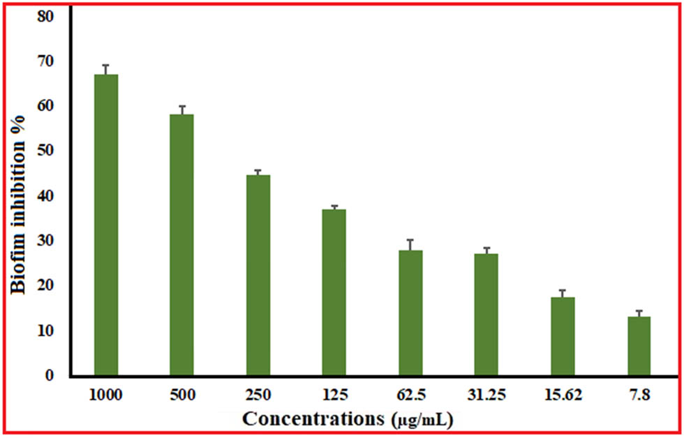
Antibiofilm activity of Ag NPs versus MRSA.
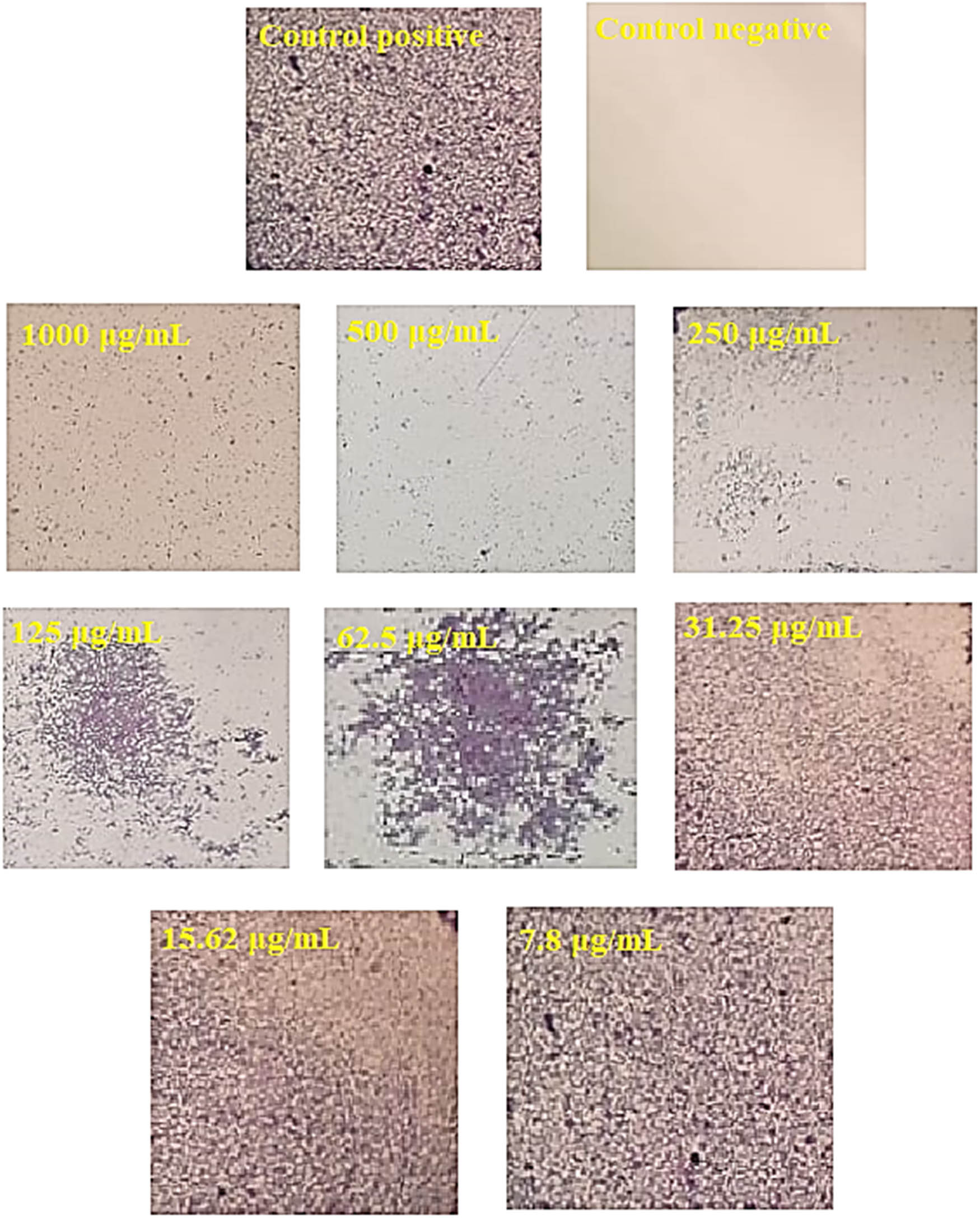
Microscopically light-inverted photos of MRSA biofilms developed with different conc. of Ag NPs.
3.4 Antioxidant
ROS, which are created by biological processes and oxidatively damage biological components, are typically the cause of cell death [68]. Following that, antioxidant chemicals were utilized to reduce the damaging impacts of ROS. Moreover, antioxidants are widely employed as medical treatments due to their anti-atherosclerotic, anti-mutagenic, anti-inflammatory, anticancer, and antibacterial properties [69,70]. The antioxidant capacity of green-produced Ag NPs was evaluated in this work using the DPPH free radical test. Silver nanoparticles’ antioxidant activity at different concentrations is displayed in Figure 9, with ascorbic acid (positive control). Considering the outcomes, silver nanoparticles had an IC50 of 46.23 μg·mL−1. When DPPH was used to measure the antioxidant assay of Ag NPs generated from Psidium guajava, there were no discernible changes between concentrations ranging from 100 to 120 µg·mL−1 relative to the positive control [71]. Researchers concluded that ascorbic acid showed scavenging percentages of 90% and 89.0% at 100 and 120 µg·mL−1 of Ag NPs, respectively, whereas scavenging ratios were 83.6% and 89.0% at those points. The scientists observed that Ag NPs derived from Atrocarpus altilis leaf extract exhibited 79.79% radical scavenging capabilities for DPPH [72]. According to the DPPH technique, the IC50 level of Ag NPs produced by Erythrina suberosa was 30 µg·mL−1 [73]. Silver NPs as a substitute antioxidant for treating diseases brought on by free radical damage. Silver NPs are derived via plant extracts have been demonstrated shown in multiple investigations to possess significant antioxidant properties [74,75]. Ag NPs’ capacity to scavenge or destroy free radicals via giving or receiving electrons explains their ability to act as antioxidants. This is because silver could be present in two distinct oxidation states Ag2+ and Ag+, based on the conditions of the process [76]. All things considered; the greenly produced Ag NPs are capable of being employed as a type of antioxidant to safely shield human health versus degenerative diseases brought on by different oxidative stressors.
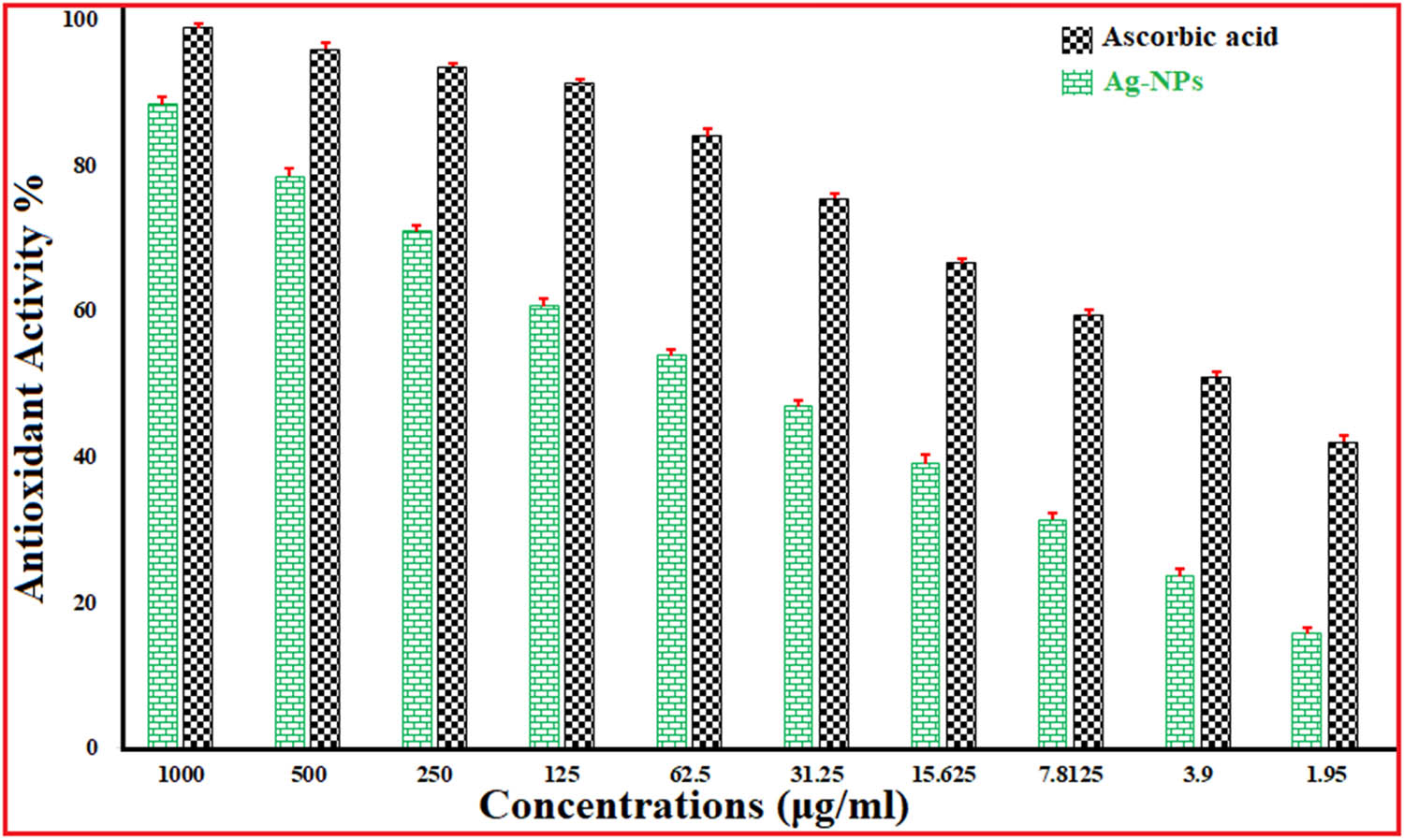
Antioxidant activity of Ag NPs.
3.5 The cytotoxic effect
In vitro cytotoxicity assessments on normal cell lines are the first step in evaluating the safety of naturally occurring materials [77]. The cytotoxicity of green-synthesized Ag NPs on the Vero cell line is demonstrated in Figure 10. The interquartile range (IC50) for nano-sized silver nanoparticles (NPs) was determined to be between 1,000 and 31.25 μg·mL−1. The results showed that the Ag nanoparticles’ IC50 was 609.45 μg·mL−1. If the IC50 value of a chemical is greater than 90 μg·mL−1, it is generally regarded as non-cytotoxic [78].
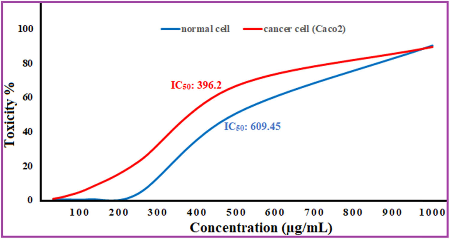
Cytotoxicity and anti-tumor activity of silver NPs.
3.6 Anticancer activity
According to a number of recent research, metallic nanoparticles may hinder some cancer kinds of cells from growing. Ag NPs induce harm by the breakdown of lipoteichoic acid, phosphatidyle thanol amine, and peptidoglycan which releases reactive oxygen radicals. This is the mechanism behind Ag NPs poisoning. As well as the deadly creation of ROS such hydroxyl as well as superoxide. Ag NPs harm cells and induce apoptosis by generating Ag+ in the nucleus and mitochondria. It has been observed that in normal human cells, Ag NPs have very little toxicity [79,80]. At dosages ranging from 1,000 to 31.25 μg·mL−1, we evaluated the anticancer effects of plant-synthesized silver nanoparticles against cancer cells. The human colorectal adenocarcinoma epithelium (Caco-2) is shown in Figure 11. The results indicated that the IC50 of silver nanoparticles is 396.2 μg·mL−1. The harmful impact of silver nanoparticles was found to depend on their size, concentration, as well as exterior composition [81]. Furthermore, the tiny size along with the charge on the surface improved transportation and distribution within the malignant matrix [82]. Further studies have shown that Ag NPs made with Talaromyces purpurogenus extracellular pigment significantly cytotoxically affected a liver tumor cell line [83]. At the cells of MCF-7, silver nanoparticles caused cytotoxicity according to a dose–response fashion [84]. In another study Ag nanoparticles evaluated versus Caco-2 in a dosage ranging from 1.953 to 4,000 µg·mL−1, the activity against cancer had an IC50 threshold of 252.83 µg·mL−1 following a 24-hour exposure period [85]. Ag NP-containing polyelectrolyte complexes also showed cytotoxic effects on Caco-2 cells, but not on normal African green monkey cells (Vero cells), at levels higher than 100 µg·mL−1 [86].
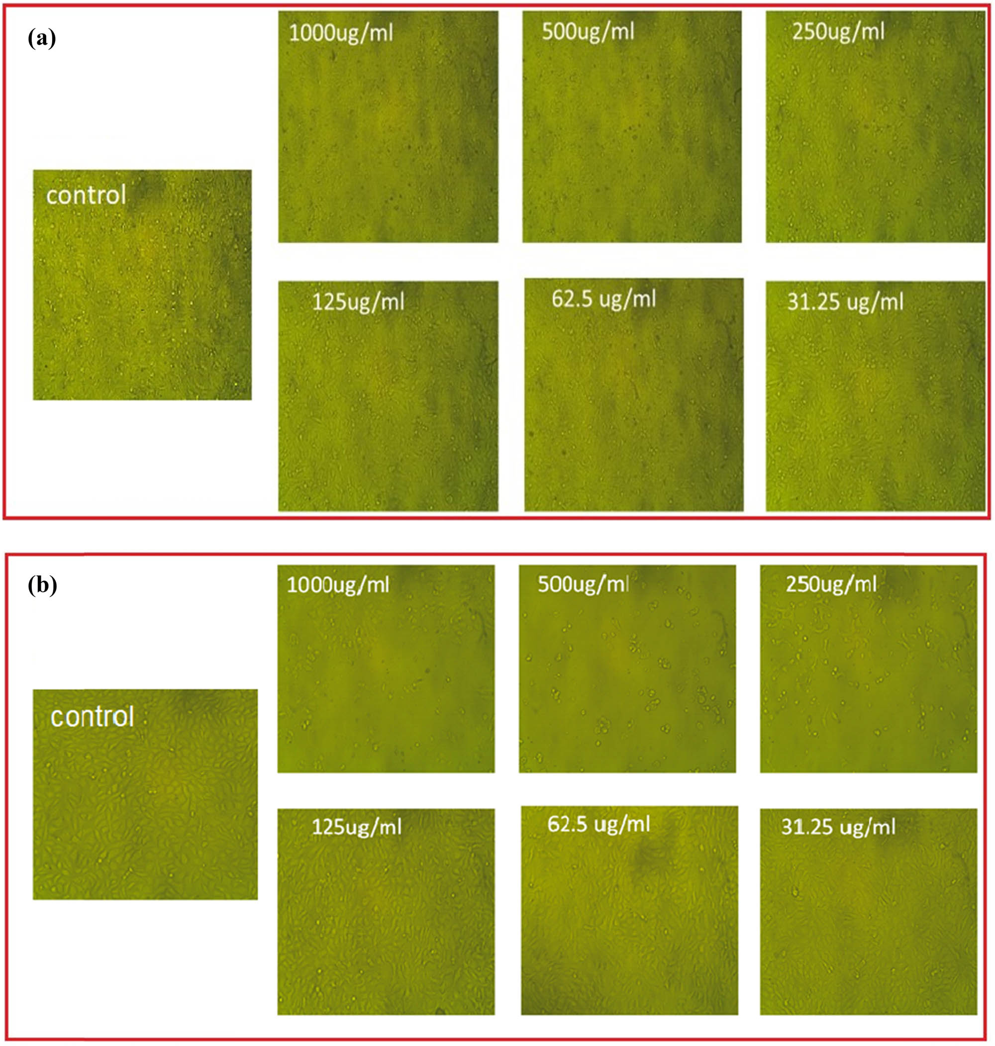
Silver NP effects on Vero cell (a) and Caco-2 tumor cell (b) at numerous concentrations.
4 Conclusion
A safe and effective procedure for synthesizing NPs without harming the environment is provided by the “green synthesis” of Ag NPs from the aqueous extract of B. variegata. The formed Ag NPs are particularly effective against pathogenic microorganisms. Ag NPs were shown an antibiofim activity versus biofilm producer S. aureus (MRSA), Additionally, Ag NPs’ antibacterial and antibiofilm properties depend on their concentration. They also exhibit antioxidant properties and the ability to scavenge free radicals. Similarly, they demonstrated potent anticancer action towards the Caco-2 colorectal adenocarcinoma epithelial cell line while having no impact on the Vero normal cell line. The preliminary results of the investigation suggest that Ag NPs generated by B. variegata could serve as an alternative to traditional antibiotics. Scientists can develop novel drugs to cure a variety of bacterial, fungal, and cancerous disorders by comprehending the mechanisms of action of these nanosilver composite Ag NPs.
Acknowledgments
The authors express their sincere thanks to the Faculty of Science (Boyes), Al-Azhar University, Cairo, Egypt, for providing the necessary research facilities. The authors extend their appreciation to the researchers supporting project number (RSP2024R505), King Saud University, Riyadh, Saudi Arabia.
-
Funding information: The authors extend their appreciation to the researchers supporting project number (RSP2024R505), King Saud University, Riyadh, Saudi Arabia.
-
Author contributions: Mohamed K. Y. Soliman and Salem S Salem: conceptualization, methodology, validation, formal analysis, investigation, data curation, writing – original draft preparation, and writing – review and editing. Amr H. Hashem: methodology, writing – original draft preparation, and writing – review and editing. Abdulaziz A. Al-Askar and Gehad AbdElgayed: writing – review and editing and funding acquisition.
-
Conflict of interest: Authors state no conflict of interest.
-
Data availability statement: The datasets generated during and/or analyzed during the current study are available from the corresponding author on reasonable request.
References
[1] Jin Y, Lin J, Sathiyaseelan A, Zhang X, Wang M-H. Comparative studies on antibacterial, antibiofilm, antioxidant, and cytotoxicity properties of chemically and Paeonia lactiflora extract-assisted synthesized silver nitroprusside nanoparticles. J Drug Delivery Sci Technol. 2024;92:105269.10.1016/j.jddst.2023.105269Suche in Google Scholar
[2] Thirumoorthy G, Balasubramanian B, George JA, Nizam A, Nagella P, Srinatha N, et al. Phytofabricated bimetallic synthesis of silver-copper nanoparticles using Aerva lanata extract to evaluate their potential cytotoxic and antimicrobial activities. Sci Rep. 2024;14:1270.10.1038/s41598-024-51647-xSuche in Google Scholar PubMed PubMed Central
[3] Salem SS, Hammad EN, Mohamed AA, El-Dougdoug W. A comprehensive review of nanomaterials: Types, synthesis, characterization, and applications. Biointerface Res Appl Chem. 2023;13(1):41.10.33263/BRIAC131.041Suche in Google Scholar
[4] Karan T, Erenler R, Gonulalan Z, Kolemen U. Biogenic synthesis of silver nanoparticles using Sambucus nigra leaves: Elucidation, antimicrobial, antioxidant activities and quantification of phenolics. Chem Pap. 2024;78:473–81.10.1007/s11696-023-03103-9Suche in Google Scholar
[5] Sahu T, Ratre YK, Chauhan S, Bhaskar LV, Nair MP, Verma HK. Nanotechnology based drug delivery system: Current strategies and emerging therapeutic potential for medical science. J Drug Delivery Sci Technol. 2021;63:102487.10.1016/j.jddst.2021.102487Suche in Google Scholar
[6] Dezfuli AAZ, Abu-Elghait M, Salem SS. Recent Insights into Nanotechnology in Colorectal Cancer. Appl Biochem Biotechnol. 2024;196:4457–71.10.1007/s12010-023-04696-3Suche in Google Scholar PubMed
[7] Hussein AS, Hashem AH, Salem SS. Mitigation of the hyperglycemic effect of streptozotocin-induced diabetes albino rats using biosynthesized copper oxide nanoparticles. Biomol Concepts. 2023;14:20220037.10.1515/bmc-2022-0037Suche in Google Scholar PubMed
[8] Tanase C, Berta L, Coman NA, Roșca I, Man A, Toma F, et al. Investigation of in vitro antioxidant and antibacterial potential of silver nanoparticles obtained by biosynthesis using beech bark extract. Antioxidants. 2019;8:459.10.3390/antiox8100459Suche in Google Scholar PubMed PubMed Central
[9] Elkady FM, Hashem AH, Salem SS, El-Sayyad GS, Tawab AA, Alkherkhisy MM. Unveiling biological activities of biosynthesized starch/silver-selenium nanocomposite using Cladosporium cladosporioides CBS 174.62. BMC Microbiol. 2024;24:78.10.1186/s12866-024-03228-1Suche in Google Scholar PubMed PubMed Central
[10] Salem SS. Bio-fabrication of Selenium Nanoparticles Using Baker’s Yeast Extract and Its Antimicrobial Efficacy on Food Borne Pathogens. Appl Biochem Biotechnol. 2022;194:1898–910.10.1007/s12010-022-03809-8Suche in Google Scholar PubMed
[11] Alabssawy AN, Abu-Elghait M, Azab AM, Khalaf-Allah HM, Ashry AS, Ali AO, et al. Hindering the biofilm of microbial pathogens and cancer cell lines development using silver nanoparticles synthesized by epidermal mucus proteins from Clarias gariepinus. BMC Biotechnol. 2024;24:28.10.1186/s12896-024-00852-7Suche in Google Scholar PubMed PubMed Central
[12] Ibraheem DR, Hussein NN, Sulaiman GM, Mohammed HA, Khan RA, Al Rugaie O. Ciprofloxacin-Loaded Silver Nanoparticles as Potent Nano-Antibiotics against Resistant Pathogenic Bacteria. Nanomaterials. 2022;12:2808.10.3390/nano12162808Suche in Google Scholar PubMed PubMed Central
[13] Johnson P, Krishnan V, Loganathan C, Govindhan K, Raji V, Sakayanathan P, et al. Rapid biosynthesis of Bauhinia variegata flower extract-mediated silver nanoparticles: an effective antioxidant scavenger and α-amylase inhibitor. Artif Cells, Nanomed Biotechnol. 2018;46:1488–94.10.1080/21691401.2017.1374283Suche in Google Scholar PubMed
[14] Vijayaram S, Razafindralambo H, Sun Y-Z, Vasantharaj S, Ghafarifarsani H, Hoseinifar SH, et al. Applications of green synthesized metal nanoparticles—a review. Biol Trace Elem Res. 2024;202:360–86.10.1007/s12011-023-03645-9Suche in Google Scholar PubMed PubMed Central
[15] Salem SS. A mini review on green nanotechnology and its development in biological effects. Arch Microbiol. 2023;205:128.10.1007/s00203-023-03467-2Suche in Google Scholar PubMed PubMed Central
[16] Abu-Elghait M, Soliman MKY, Azab MS, Salem SS. Response surface methodology: Optimization of myco-synthesized gold and silver nanoparticles by Trichoderma saturnisporum. Biomass Convers Biorefin. 2023;1–14.10.1007/s13399-023-05188-4Suche in Google Scholar
[17] Salem SS, Soliman MKY, Azab MS, Abu-Elghait M. Optimization growth conditions of fusarium pseudonygamai for myco-synthesized gold and silver nanoparticles using response surface methodology. BioNanoScience. 2024. 10.1007/s12668-024-01349-5.Suche in Google Scholar
[18] Kulkarni AG, De Britto S, Jogaiah S. Economic considerations and limitations of green synthesis vs chemical synthesis of nanomaterials. Advances in Nano-fertilizers and Nano-pesticides in agriculture. Sawston, Cambridge: Elsevier; 2021. p. 459–68.10.1016/B978-0-12-820092-6.00018-5Suche in Google Scholar
[19] Soliman MK, Salem SS, Abu-Elghait M, Azab MS. Biosynthesis of silver and gold nanoparticles and their efficacy towards antibacterial, antibiofilm, cytotoxicity, and antioxidant activities. Appl Biochem Biotechnol. 2023;195:1158–83.10.1007/s12010-022-04199-7Suche in Google Scholar PubMed PubMed Central
[20] Giri AK, Jena B, Biswal B, Pradhan AK, Arakha M, Acharya S, et al. Green synthesis and characterization of silver nanoparticles using Eugenia roxburghii DC. extract and activity against biofilm-producing bacteria. Sci Rep. 2022;12:8383.10.1038/s41598-022-12484-ySuche in Google Scholar PubMed PubMed Central
[21] Singh J, Dutta T, Kim K-H, Rawat M, Samddar P, Kumar P. ‘Green’synthesis of metals and their oxide nanoparticles: applications for environmental remediation. J nanobiotechnol. 2018;16:1–24.10.1186/s12951-018-0408-4Suche in Google Scholar PubMed PubMed Central
[22] Abd-Elkhalek HF, Badawy AA, Al-Askar AA, Elgawad HA, Hashem AH, Salem SS. Biosynthesis and characterization of selenium and silver nanoparticles using Trichoderma viride filtrate and their impact on Culex pipiens. Green Process Synth. 2024;13:20240025.10.1515/gps-2024-0025Suche in Google Scholar
[23] Al-Zahrani FAM, Salem SS, Al-Ghamdi HA, Nhari LM, Lin L, El-Shishtawy RM. Green synthesis and antibacterial activity of Ag/Fe2O3 nanocomposite using Buddleja lindleyana extract. Bioengineering. 2022;9:452.10.3390/bioengineering9090452Suche in Google Scholar PubMed PubMed Central
[24] Abdelfattah NAH, Yousef MA, Badawy AA, Salem SS. Influence of biosynthesized magnesium oxide nanoparticles on growth and physiological aspects of cowpea (Vigna unguiculata L.) plant, cowpea beetle, and cytotoxicity. Biotechnol J. 2023;18:2300301.10.1002/biot.202300301Suche in Google Scholar PubMed
[25] Dheyab MA, Oladzadabbasabadi N, Aziz AA, Khaniabadi PM, Al-ouqaili MTS, Jameel MS, et al. Recent advances of plant-mediated metal nanoparticles: synthesis, properties, and emerging applications for wastewater treatment. J Environ Chem Eng. 2024;12(2):112345.10.1016/j.jece.2024.112345Suche in Google Scholar
[26] Adeyemi JO, Oriola AO, Onwudiwe DC, Oyedeji AO. Plant extracts mediated metal-based nanoparticles: synthesis and biological applications. Biomolecules. 2022;12:627.10.3390/biom12050627Suche in Google Scholar PubMed PubMed Central
[27] Rajan R, Chandran K, Harper SL, Yun S-I, Kalaichelvan PT. Plant extract synthesized silver nanoparticles: An ongoing source of novel biocompatible materials. Ind Crop Prod. 2015;70:356–73.10.1016/j.indcrop.2015.03.015Suche in Google Scholar
[28] Abbasi MA, Kausar A, Rehman A, Hina S, Jahangir SM, Siddiqui SZ, et al. Preparation of new formulations of anti-acne creams and their efficacy. Afr J Pharm Pharmacol. 2010;4:298–303.Suche in Google Scholar
[29] Abdel-Tawab M, Werz O, Schubert-Zsilavecz M. Boswellia serrata: an overall assessment of in vitro, preclinical, pharmacokinetic and clinical data. Clin Pharmacokinet. 2011;50:349–69.10.2165/11586800-000000000-00000Suche in Google Scholar PubMed
[30] Ahn E-Y, Jin H, Park Y. Assessing the antioxidant, cytotoxic, apoptotic and wound healing properties of silver nanoparticles green-synthesized by plant extracts. Mater Sci Eng: C. 2019;101:204–16.10.1016/j.msec.2019.03.095Suche in Google Scholar PubMed
[31] Fahimirad S, Ajalloueian F, Ghorbanpour M. Synthesis and therapeutic potential of silver nanomaterials derived from plant extracts. Ecotoxicol Environ Saf. 2019;168:260–78.10.1016/j.ecoenv.2018.10.017Suche in Google Scholar PubMed
[32] Haggag EG, Elshamy AM, Rabeh MA, Gabr NM, Salem M, Youssif KA, et al. Antiviral potential of green synthesized silver nanoparticles of Lampranthus coccineus and Malephora lutea. Int J Nanomed. 2019;14:6217–29.10.2147/IJN.S214171Suche in Google Scholar PubMed PubMed Central
[33] Soliman MK, Abu-Elghait M, Salem SS, Azab MS. Multifunctional properties of silver and gold nanoparticles synthesis by Fusarium pseudonygamai. Biomass Convers Biorefin. 2022;1–18.10.1007/s13399-022-03507-9Suche in Google Scholar
[34] Waghchaure RH, Adole VA. Biosynthesis of metal and metal oxide nanoparticles using various parts of plants for antibacterial, antifungal and anticancer activity: A review. J Indian Chem Soc. 2023;5:100987.10.1016/j.jics.2023.100987Suche in Google Scholar
[35] Chandrasekharan S, Chinnasamy G, Bhatnagar S. Sustainable phyto-fabrication of silver nanoparticles using Gmelina arborea exhibit antimicrobial and biofilm inhibition activity. Sci Rep. 2022;12:156.10.1038/s41598-021-04025-wSuche in Google Scholar PubMed PubMed Central
[36] Shreyash N, Bajpai S, Khan MA, Vijay Y, Tiwary SK, Sonker M. Green synthesis of nanoparticles and their biomedical applications: a review. ACS Appl Nano Mater. 2021;4:11428–57.10.1021/acsanm.1c02946Suche in Google Scholar
[37] Villalobos Gutiérrez PT, Muñoz Carrillo JL, Sandoval Salazar C, Viveros Paredes JM, Gutiérrez Coronado O. Functionalized metal nanoparticles in cancer therapy. Pharmaceutics. 2023;15:1932.10.3390/pharmaceutics15071932Suche in Google Scholar PubMed PubMed Central
[38] Kashyap BK, Singh VV, Solanki MK, Kumar A, Ruokolainen J, Kesari KK. Smart nanomaterials in cancer theranostics: challenges and opportunities. ACS Omega. 2023;8:14290–320.10.1021/acsomega.2c07840Suche in Google Scholar PubMed PubMed Central
[39] Summer M, Ali S, Tahir HM, Abaidullah R, Fiaz U, Mumtaz S, et al. Mode of action of biogenic silver, zinc, copper, titanium and cobalt nanoparticles against antibiotics resistant pathogens. J Inorg Organomet Polym Mater. 2024;34(4):1417–51.10.1007/s10904-023-02935-ySuche in Google Scholar
[40] Sundar M, Rajagopal G, Nivetha A, Prabu Kumar S, Muthukumar S. Phyto-mediated green synthesis of silver nanoparticles using an aqueous leaf extract of Momordica cymbalaria: Antioxidant, cytotoxic, antibacterial, and photocatalytic properties. Separations. 2024;11:61.10.3390/separations11020061Suche in Google Scholar
[41] Rudrappa M, Rudayni HA, Assiri RA, Bepari A, Basavarajappa DS, Nagaraja SK, et al. Plumeria alba-mediated green synthesis of silver nanoparticles exhibits antimicrobial effect and anti-oncogenic activity against glioblastoma U118 MG cancer cell line. Nanomaterials. 2022;12:493.10.3390/nano12030493Suche in Google Scholar PubMed PubMed Central
[42] Perez C. Antibiotic assay by agar-well diffusion method. Acta Biol Med Exp. 1990;15:113–5.Suche in Google Scholar
[43] Khattab AM, Abo-Taleb HA, Abdelaziz AM, El-Tabakh MA, El-Feky MMM, Abu-Elghait M. Daphnia magna and Gammarus pulex, novel promising agents for biomedical and agricultural applications. Sci Rep. 2022;12:13690.10.1038/s41598-022-17790-zSuche in Google Scholar PubMed PubMed Central
[44] Munteanu IG, Apetrei C. Analytical methods used in determining antioxidant activity: A review. Int J Mol Sci. 2021;22:3380.10.3390/ijms22073380Suche in Google Scholar PubMed PubMed Central
[45] Tolosa L, Donato MT, Gómez-Lechón MJ. General cytotoxicity assessment by means of the MTT assay. In: Vinken M, Rogiers V, editors. Protocols in in vitro hepatocyte research. Methods in molecular biology, Vol. 1250. New York: Humana Press; 2015. p. 333–48.10.1007/978-1-4939-2074-7_26Suche in Google Scholar PubMed
[46] Supino R. MTT assays. In vitro toxicity testing protocols. Methods in molecular biology, Vol. 43. Totowa: Humana Press; 1995. p. 137–49.10.1385/0-89603-282-5:137Suche in Google Scholar PubMed
[47] Salem SS, Ali OM, Reyad AM, Abd-Elsalam KA, Hashem AH. Pseudomonas indica-mediated silver nanoparticles: Antifungal and antioxidant biogenic tool for suppressing mucormycosis fungi. J Fungi. 2022;8:126.10.3390/jof8020126Suche in Google Scholar PubMed PubMed Central
[48] Al-Rajhi AMH, Salem SS, Alharbi AA, Abdelghany TM. Ecofriendly synthesis of silver nanoparticles using Kei-apple (Dovyalis caffra) fruit and their efficacy against cancer cells and clinical pathogenic microorganisms. Arab J Chem. 2022;15:103927.10.1016/j.arabjc.2022.103927Suche in Google Scholar
[49] Salem SS, EL-Belely EF, Niedbała G, Alnoman MM, Hassan SE-D, Eid AM, et al. Bactericidal and in-vitro cytotoxic efficacy of silver nanoparticles (Ag-NPs) fabricated by endophytic actinomycetes and their use as coating for the textile fabrics. Nanomaterials. 2020;10:2082.10.3390/nano10102082Suche in Google Scholar PubMed PubMed Central
[50] Mane PC, Sayyed SAR, Kadam DD, D. Shinde M, Fatehmulla A, Aldhafiri AM, et al. Terrestrial snail-mucus mediated green synthesis of silver nanoparticles and in vitro investigations on their antimicrobial and anticancer activities. Sci Rep. 2021;11:13068.10.1038/s41598-021-92478-4Suche in Google Scholar PubMed PubMed Central
[51] Salem SS. Baker’s yeast-mediated silver nanoparticles: Characterisation and antimicrobial biogenic tool for suppressing pathogenic microbes. BioNanoScience. 2022;12(4):1220–9.10.1007/s12668-022-01026-5Suche in Google Scholar
[52] Lin P, Yan Z-F, Li C-T. Biosynthesis of silver nanoparticles using Lavandula stoechas and an enhancement of its antibacterial activity with antibiotics. Biotechnol Bioprocess Eng. 2021;26:650–9.10.1007/s12257-020-0379-9Suche in Google Scholar
[53] Ahmed S, Ahmad M, Swami BL, Ikram S. Green synthesis of silver nanoparticles using Azadirachta indica aqueous leaf extract. J Radiat Res Appl Sci. 2016;9:1–7.10.1016/j.jrras.2015.06.006Suche in Google Scholar
[54] Franci G, Falanga A, Galdiero S, Palomba L, Rai M, Morelli G, et al. Silver nanoparticles as potential antibacterial agents. Molecules. 2015;20:8856–74.10.3390/molecules20058856Suche in Google Scholar PubMed PubMed Central
[55] Joshi AS, Singh P, Mijakovic I. Interactions of gold and silver nanoparticles with bacterial biofilms: Molecular interactions behind inhibition and resistance. Int J Mol Sci. 2020;21:7658.10.3390/ijms21207658Suche in Google Scholar PubMed PubMed Central
[56] Aref MS, Salem SS. Bio-callus synthesis of silver nanoparticles, characterization, and antibacterial activities via Cinnamomum camphora callus culture. Biocatal Agric Biotechnol. 2020;27:101689.10.1016/j.bcab.2020.101689Suche in Google Scholar
[57] Patil MP, Kim GD. Eco-friendly approach for nanoparticles synthesis and mechanism behind antibacterial activity of silver and anticancer activity of gold nanoparticles. Appl Microbiol Biotechnol. 2017;101:79–92.10.1007/s00253-016-8012-8Suche in Google Scholar PubMed
[58] Nayem SMA, Sultana N, Haque MA, Miah B, Hasan M, Islam T, et al. Green synthesis of gold and silver nanoparticles by using amorphophallus paeoniifolius tuber extract and evaluation of their antibacterial activity. Molecules. 2020;25:4773.10.3390/molecules25204773Suche in Google Scholar PubMed PubMed Central
[59] Hamad A, Khashan KS, Hadi A. Silver nanoparticles and silver ions as potential antibacterial agents. J Inorg Organomet Polym Mater. 2020;30:4811–28.10.1007/s10904-020-01744-xSuche in Google Scholar
[60] Tripathi D, Pandey-Rai S. Impacts of green synthesized silver nanoparticles with natural bioactive compounds on plant’s developmental behavior. Natural bioactive compounds. London, United Kingdom: Elsevier; 2021. p. 435–52.10.1016/B978-0-12-820655-3.00022-7Suche in Google Scholar
[61] Miranda A, Akpobolokemi T, Chung E, Ren G, Raimi-Abraham BT. pH alteration in plant-mediated green synthesis and its resultant impact on antimicrobial properties of silver nanoparticles (AgNPs). Antibiotics. 2022;11:1592.10.3390/antibiotics11111592Suche in Google Scholar PubMed PubMed Central
[62] Khan F, Manivasagan P, Lee J-W, Pham DT, Oh J, Kim Y-M, Mar Drugs. 2019Suche in Google Scholar
[63] Hall CW, Mah T-F. Molecular mechanisms of biofilm-based antibiotic resistance and tolerance in pathogenic bacteria. FEMS Microbiol Rev. 2017;41:276–301.10.1093/femsre/fux010Suche in Google Scholar PubMed
[64] Qin N, Tan X, Jiao Y, Liu L, Zhao W, Yang S, et al. RNA-Seq-based transcriptome analysis of methicillin-resistant Staphylococcus aureus biofilm inhibition by ursolic acid and resveratrol. Sci Rep. 2014;4:5467.10.1038/srep05467Suche in Google Scholar PubMed PubMed Central
[65] Flemming H-C, Wingender J. The biofilm matrix. Nat Rev Microbiol. 2010;8:623–33.10.1038/nrmicro2415Suche in Google Scholar PubMed
[66] Qais FA, Ahmad I. Broad‐spectrum inhibitory effect of green synthesised silver nanoparticles from Withania somnifera (L.) on microbial growth, biofilm and respiration: a putative mechanistic approach. Iet Nanobiotechnol. 2018;12:325.10.1049/iet-nbt.2017.0193Suche in Google Scholar
[67] Ansari MA, Khan HM, Khan AA, Cameotra SS, Saquib Q, Musarrat J. Gum arabic capped‐silver nanoparticles inhibit biofilm formation by multi‐drug resistant strains of Pseudomonas aeruginosa. J Basic Microbiol. 2014;54:688–99.10.1002/jobm.201300748Suche in Google Scholar PubMed
[68] Cui J-L, Guo T-T, Ren Z-X, Zhang N-S, Wang M-L. Diversity and antioxidant activity of culturable endophytic fungi from alpine plants of Rhodiola crenulata, R. angusta, and R. sachalinensis. PLoS One. 2015;10:e0118204.10.1371/journal.pone.0118204Suche in Google Scholar PubMed PubMed Central
[69] Li Y, Li X, Wong Y-S, Chen T, Zhang H, Liu C, et al. The reversal of cisplatin-induced nephrotoxicity by selenium nanoparticles functionalized with 11-mercapto-1-undecanol by inhibition of ROS-mediated apoptosis. Biomaterials. 2011;32:9068–76.10.1016/j.biomaterials.2011.08.001Suche in Google Scholar PubMed
[70] Tinggi U. Selenium: its role as antioxidant in human health. Environ Health Prev Med. 2008;13:102–8.10.1007/s12199-007-0019-4Suche in Google Scholar PubMed PubMed Central
[71] Wang L, Wu Y, Xie J, Wu S, Wu Z. Characterization, antioxidant and antimicrobial activities of green synthesized silver nanoparticles from Psidium guajava L. leaf aqueous extracts. Mater Sci Eng: C. 2018;86:1–8.10.1016/j.msec.2018.01.003Suche in Google Scholar PubMed
[72] Ravichandran V, Vasanthi S, Shalini S, Shah SAA, Harish R. Green synthesis of silver nanoparticles using Atrocarpus altilis leaf extract and the study of their antimicrobial and antioxidant activity. Mater Lett. 2016;180:264–7.10.1016/j.matlet.2016.05.172Suche in Google Scholar
[73] Mohanta YK, Panda SK, Jayabalan R, Sharma N, Bastia AK, Mohanta TK. Antimicrobial, antioxidant and cytotoxic activity of silver nanoparticles synthesized by leaf extract of Erythrina suberosa (Roxb.). Front Mol Biosci. 2017;4:14.10.3389/fmolb.2017.00014Suche in Google Scholar PubMed PubMed Central
[74] Sarwer Q, Amjad MS, Mehmood A, Binish Z, Mustafa G, Farooq A, et al. Green synthesis and characterization of silver nanoparticles using Myrsine africana leaf extract for their antibacterial, antioxidant and phytotoxic activities. Molecules. 2022;27:7612.10.3390/molecules27217612Suche in Google Scholar PubMed PubMed Central
[75] Nagaich U, Gulati N, Chauhan S. Antioxidant and antibacterial potential of silver nanoparticles: biogenic synthesis utilizing apple extract. J Pharm. 2016;2016:7141523.10.1155/2016/7141523Suche in Google Scholar PubMed PubMed Central
[76] Bedlovičová Z, Strapáč I, Baláž M, Salayová A. A brief overview on antioxidant activity determination of silver nanoparticles. Molecules. 2020;25:3191.10.3390/molecules25143191Suche in Google Scholar PubMed PubMed Central
[77] Salem SS, Fouda A. Green synthesis of metallic nanoparticles and their prospective biotechnological applications: an overview. Biol Trace Elem Res. 2021;199:344–70.10.1007/s12011-020-02138-3Suche in Google Scholar PubMed
[78] Ioset J-R, Brun R, Wenzler T, Kaiser M, Yardley V. Drug screening for kinetoplastids diseases. A training manual for screening in neglected diseases. 2009;74:1–74.Suche in Google Scholar
[79] Rama P, Mariselvi P, Sundaram R, Muthu K. Eco-friendly green synthesis of silver nanoparticles from Aegle marmelos leaf extract and their antimicrobial, antioxidant, anticancer and photocatalytic degradation activity. Heliyon. 2023;9(6):e16277.10.1016/j.heliyon.2023.e16277Suche in Google Scholar PubMed PubMed Central
[80] Abu‐Dief AM, Abdel‐Rahman LH, Abd‐El Sayed M, Zikry MM, Nafady A. Green synthesis of AgNPs () ultilizing Delonix Regia extract as anticancer and antimicrobial agents. ChemistrySelect. 2020;5:13263–8.10.1002/slct.202003218Suche in Google Scholar
[81] Huerta-Aguilar CA, Ramírez-Guzmán B, Thangarasu P, Narayanan J, Singh N. Simultaneous recognition of cysteine and cytosine using thiophene-based organic nanoparticles decorated with Au NPs and bio-imaging of cells. Photochem Photobiol Sci. 2019;18:1761–72.10.1039/c9pp00060gSuche in Google Scholar PubMed
[82] Netala VR, Bethu MS, Pushpalatha B, Baki VB, Aishwarya S, Rao JV, et al. Biogenesis of silver nanoparticles using endophytic fungus Pestalotiopsis microspora and evaluation of their antioxidant and anticancer activities. Int J Nanomed. 2016;11:5683–96.10.2147/IJN.S112857Suche in Google Scholar PubMed PubMed Central
[83] Bhatnagar S, Kobori T, Ganesh D, Ogawa K, Aoyagi H. Biosynthesis of silver nanoparticles mediated by extracellular pigment from Talaromyces purpurogenus and their biomedical applications. Nanomaterials. 2019;9:1042.10.3390/nano9071042Suche in Google Scholar PubMed PubMed Central
[84] Ranjitham AM, Suja R, Caroling G, Tiwari S. In vitro evaluation of antioxidant, antimicrobial, anticancer activities and characterisation of Brassica oleracea. var. Bortrytis. L synthesized silver nanoparticles. Int J Pharm Pharm Sci. 2013;5:239–51.Suche in Google Scholar
[85] El-Sheekh MM, Hassan LH, Morsi HH. Assessment of the in vitro anticancer activities of cyanobacteria mediated silver oxide and gold nanoparticles in human colon CaCo-2 and cervical HeLa cells. Environ Nanotechnol Monit Manag. 2021;16:100556.10.1016/j.enmm.2021.100556Suche in Google Scholar
[86] Martins AF, Follmann HD, Monteiro JP, Bonafé EG, Nocchi S, Silva CTP, et al. Polyelectrolyte complex containing silver nanoparticles with antitumor property on Caco-2 colon cancer cells. Int J Biol Macromol. 2015;79:748–55.10.1016/j.ijbiomac.2015.05.036Suche in Google Scholar PubMed
© 2024 the author(s), published by De Gruyter
This work is licensed under the Creative Commons Attribution 4.0 International License.
Artikel in diesem Heft
- Research Articles
- Green polymer electrolyte and activated charcoal-based supercapacitor for energy harvesting application: Electrochemical characteristics
- Research on the adsorption of Co2+ ions using halloysite clay and the ability to recover them by electrodeposition method
- Simultaneous estimation of ibuprofen, caffeine, and paracetamol in commercial products using a green reverse-phase HPTLC method
- Isolation, screening and optimization of alkaliphilic cellulolytic fungi for production of cellulase
- Functionalized gold nanoparticles coated with bacterial alginate and their antibacterial and anticancer activities
- Comparative analysis of bio-based amino acid surfactants obtained via Diels–Alder reaction of cyclic anhydrides
- Biosynthesis of silver nanoparticles on yellow phosphorus slag and its application in organic coatings
- Exploring antioxidant potential and phenolic compound extraction from Vitis vinifera L. using ultrasound-assisted extraction
- Manganese and copper-coated nickel oxide nanoparticles synthesized from Carica papaya leaf extract induce antimicrobial activity and breast cancer cell death by triggering mitochondrial caspases and p53
- Insight into heating method and Mozafari method as green processing techniques for the synthesis of micro- and nano-drug carriers
- Silicotungstic acid supported on Bi-based MOF-derived metal oxide for photodegradation of organic dyes
- Synthesis and characterization of capsaicin nanoparticles: An attempt to enhance its bioavailability and pharmacological actions
- Synthesis of Lawsonia inermis-encased silver–copper bimetallic nanoparticles with antioxidant, antibacterial, and cytotoxic activity
- Facile, polyherbal drug-mediated green synthesis of CuO nanoparticles and their potent biological applications
- Zinc oxide-manganese oxide/carboxymethyl cellulose-folic acid-sesamol hybrid nanomaterials: A molecularly targeted strategy for advanced triple-negative breast cancer therapy
- Exploring the antimicrobial potential of biogenically synthesized graphene oxide nanoparticles against targeted bacterial and fungal pathogens
- Biofabrication of silver nanoparticles using Uncaria tomentosa L.: Insight into characterization, antibacterial activities combined with antibiotics, and effect on Triticum aestivum germination
- Membrane distillation of synthetic urine for use in space structural habitat systems
- Investigation on mechanical properties of the green synthesis bamboo fiber/eggshell/coconut shell powder-based hybrid biocomposites under NaOH conditions
- Green synthesis of magnesium oxide nanoparticles using endophytic fungal strain to improve the growth, metabolic activities, yield traits, and phenolic compounds content of Nigella sativa L.
- Estimation of greenhouse gas emissions from rice and annual upland crops in Red River Delta of Vietnam using the denitrification–decomposition model
- Synthesis of humic acid with the obtaining of potassium humate based on coal waste from the Lenger deposit, Kazakhstan
- Ascorbic acid-mediated selenium nanoparticles as potential antihyperuricemic, antioxidant, anticoagulant, and thrombolytic agents
- Green synthesis of silver nanoparticles using Illicium verum extract: Optimization and characterization for biomedical applications
- Antibacterial and dynamical behaviour of silicon nanoparticles influenced sustainable waste flax fibre-reinforced epoxy composite for biomedical application
- Optimising coagulation/flocculation using response surface methodology and application of floc in biofertilisation
- Green synthesis and multifaceted characterization of iron oxide nanoparticles derived from Senna bicapsularis for enhanced in vitro and in vivo biological investigation
- Potent antibacterial nanocomposites from okra mucilage/chitosan/silver nanoparticles for multidrug-resistant Salmonella Typhimurium eradication
- Trachyspermum copticum aqueous seed extract-derived silver nanoparticles: Exploration of their structural characterization and comparative antibacterial performance against gram-positive and gram-negative bacteria
- Microwave-assisted ultrafine silver nanoparticle synthesis using Mitragyna speciosa for antimalarial applications
- Green synthesis and characterisation of spherical structure Ag/Fe2O3/TiO2 nanocomposite using acacia in the presence of neem and tulsi oils
- Green quantitative methods for linagliptin and empagliflozin in dosage forms
- Enhancement efficacy of omeprazole by conjugation with silver nanoparticles as a urease inhibitor
- Residual, sequential extraction, and ecological risk assessment of some metals in ash from municipal solid waste incineration, Vietnam
- Green synthesis of ZnO nanoparticles using the mangosteen (Garcinia mangostana L.) leaf extract: Comparative preliminary in vitro antibacterial study
- Simultaneous determination of lesinurad and febuxostat in commercial fixed-dose combinations using a greener normal-phase HPTLC method
- A greener RP-HPLC method for quaternary estimation of caffeine, paracetamol, levocetirizine, and phenylephrine acquiring AQbD with stability studies
- Optimization of biomass durian peel as a heterogeneous catalyst in biodiesel production using microwave irradiation
- Thermal treatment impact on the evolution of active phases in layered double hydroxide-based ZnCr photocatalysts: Photodegradation and antibacterial performance
- Preparation of silymarin-loaded zein polysaccharide core–shell nanostructures and evaluation of their biological potentials
- Preparation and characterization of composite-modified PA6 fiber for spectral heating and heat storage applications
- Preparation and electrocatalytic oxygen evolution of bimetallic phosphates (NiFe)2P/NF
- Rod-shaped Mo(vi) trichalcogenide–Mo(vi) oxide decorated on poly(1-H pyrrole) as a promising nanocomposite photoelectrode for green hydrogen generation from sewage water with high efficiency
- Green synthesis and studies on citrus medica leaf extract-mediated Au–ZnO nanocomposites: A sustainable approach for efficient photocatalytic degradation of rhodamine B dye in aqueous media
- Cellulosic materials for the removal of ciprofloxacin from aqueous environments
- The analytical assessment of metal contamination in industrial soils of Saudi Arabia using the inductively coupled plasma technology
- The effect of modified oily sludge on the slurry ability and combustion performance of coal water slurry
- Eggshell waste transformation to calcium chloride anhydride as food-grade additive and eggshell membranes as enzyme immobilization carrier
- Synthesis of EPAN and applications in the encapsulation of potassium humate
- Biosynthesis and characterization of silver nanoparticles from Cedrela toona leaf extracts: An exploration into their antibacterial, anticancer, and antioxidant potential
- Enhancing mechanical and rheological properties of HDPE films through annealing for eco-friendly agricultural applications
- Immobilisation of catalase purified from mushroom (Hydnum repandum) onto glutaraldehyde-activated chitosan and characterisation: Its application for the removal of hydrogen peroxide from artificial wastewater
- Sodium titanium oxide/zinc oxide (STO/ZnO) photocomposites for efficient dye degradation applications
- Effect of ex situ, eco-friendly ZnONPs incorporating green synthesised Moringa oleifera leaf extract in enhancing biochemical and molecular aspects of Vicia faba L. under salt stress
- Biosynthesis and characterization of selenium and silver nanoparticles using Trichoderma viride filtrate and their impact on Culex pipiens
- Photocatalytic degradation of organic dyes and biological potentials of biogenic zinc oxide nanoparticles synthesized using the polar extract of Cyperus scariosus R.Br. (Cyperaceae)
- Assessment of antiproliferative activity of green-synthesized nickel oxide nanoparticles against glioblastoma cells using Terminalia chebula
- Chlorine-free synthesis of phosphinic derivatives by change in the P-function
- Anticancer, antioxidant, and antimicrobial activities of nanoemulsions based on water-in-olive oil and loaded on biogenic silver nanoparticles
- Study and mechanism of formation of phosphorus production waste in Kazakhstan
- Synthesis and stabilization of anatase form of biomimetic TiO2 nanoparticles for enhancing anti-tumor potential
- Microwave-supported one-pot reaction for the synthesis of 5-alkyl/arylidene-2-(morpholin/thiomorpholin-4-yl)-1,3-thiazol-4(5H)-one derivatives over MgO solid base
- Screening the phytochemicals in Perilla leaves and phytosynthesis of bioactive silver nanoparticles for potential antioxidant and wound-healing application
- Graphene oxide/chitosan/manganese/folic acid-brucine functionalized nanocomposites show anticancer activity against liver cancer cells
- Nature of serpentinite interactions with low-concentration sulfuric acid solutions
- Multi-objective statistical optimisation utilising response surface methodology to predict engine performance using biofuels from waste plastic oil in CRDi engines
- Microwave-assisted extraction of acetosolv lignin from sugarcane bagasse and electrospinning of lignin/PEO nanofibres for carbon fibre production
- Biosynthesis, characterization, and investigation of cytotoxic activities of selenium nanoparticles utilizing Limosilactobacillus fermentum
- Highly photocatalytic materials based on the decoration of poly(O-chloroaniline) with molybdenum trichalcogenide oxide for green hydrogen generation from Red Sea water
- Highly efficient oil–water separation using superhydrophobic cellulose aerogels derived from corn straw
- Beta-cyclodextrin–Phyllanthus emblica emulsion for zinc oxide nanoparticles: Characteristics and photocatalysis
- Assessment of antimicrobial activity and methyl orange dye removal by Klebsiella pneumoniae-mediated silver nanoparticles
- Influential eradication of resistant Salmonella Typhimurium using bioactive nanocomposites from chitosan and radish seed-synthesized nanoselenium
- Antimicrobial activities and neuroprotective potential for Alzheimer’s disease of pure, Mn, Co, and Al-doped ZnO ultra-small nanoparticles
- Green synthesis of silver nanoparticles from Bauhinia variegata and their biological applications
- Synthesis and optimization of long-chain fatty acids via the oxidation of long-chain fatty alcohols
- Eminent Red Sea water hydrogen generation via a Pb(ii)-iodide/poly(1H-pyrrole) nanocomposite photocathode
- Green synthesis and effective genistein production by fungal β-glucosidase immobilized on Al2O3 nanocrystals synthesized in Cajanus cajan L. (Millsp.) leaf extracts
- Green stability-indicating RP-HPTLC technique for determining croconazole hydrochloride
- Green synthesis of La2O3–LaPO4 nanocomposites using Charybdis natator for DNA binding, cytotoxic, catalytic, and luminescence applications
- Eco-friendly drugs induce cellular changes in colistin-resistant bacteria
- Tangerine fruit peel extract mediated biogenic synthesized silver nanoparticles and their potential antimicrobial, antioxidant, and cytotoxic assessments
- Green synthesis on performance characteristics of a direct injection diesel engine using sandbox seed oil
- A highly sensitive β-AKBA-Ag-based fluorescent “turn off” chemosensor for rapid detection of abamectin in tomatoes
- Green synthesis and physical characterization of zinc oxide nanoparticles (ZnO NPs) derived from the methanol extract of Euphorbia dracunculoides Lam. (Euphorbiaceae) with enhanced biosafe applications
- Detection of morphine and data processing using surface plasmon resonance imaging sensor
- Effects of nanoparticles on the anaerobic digestion properties of sulfamethoxazole-containing chicken manure and analysis of bio-enzymes
- Bromic acid-thiourea synergistic leaching of sulfide gold ore
- Green chemistry approach to synthesize titanium dioxide nanoparticles using Fagonia Cretica extract, novel strategy for developing antimicrobial and antidiabetic therapies
- Green synthesis and effective utilization of biogenic Al2O3-nanocoupled fungal lipase in the resolution of active homochiral 2-octanol and its immobilization via aluminium oxide nanoparticles
- Eco-friendly RP-HPLC approach for simultaneously estimating the promising combination of pentoxifylline and simvastatin in therapeutic potential for breast cancer: Appraisal of greenness, whiteness, and Box–Behnken design
- Use of a humidity adsorbent derived from cockleshell waste in Thai fried fish crackers (Keropok)
- One-pot green synthesis, biological evaluation, and in silico study of pyrazole derivatives obtained from chalcones
- Bio-sorption of methylene blue and production of biofuel by brown alga Cystoseira sp. collected from Neom region, Kingdom of Saudi Arabia
- Synthesis of motexafin gadolinium: A promising radiosensitizer and imaging agent for cancer therapy
- The impact of varying sizes of silver nanoparticles on the induction of cellular damage in Klebsiella pneumoniae involving diverse mechanisms
- Microwave-assisted green synthesis, characterization, and in vitro antibacterial activity of NiO nanoparticles obtained from lemon peel extract
- Rhus microphylla-mediated biosynthesis of copper oxide nanoparticles for enhanced antibacterial and antibiofilm efficacy
- Harnessing trichalcogenide–molybdenum(vi) sulfide and molybdenum(vi) oxide within poly(1-amino-2-mercaptobenzene) frameworks as a photocathode for sustainable green hydrogen production from seawater without sacrificial agents
- Magnetically recyclable Fe3O4@SiO2 supported phosphonium ionic liquids for efficient and sustainable transformation of CO2 into oxazolidinones
- A comparative study of Fagonia arabica fabricated silver sulfide nanoparticles (Ag2S) and silver nanoparticles (AgNPs) with distinct antimicrobial, anticancer, and antioxidant properties
- Visible light photocatalytic degradation and biological activities of Aegle marmelos-mediated cerium oxide nanoparticles
- Physical intrinsic characteristics of spheroidal particles in coal gasification fine slag
- Exploring the effect of tea dust magnetic biochar on agricultural crops grown in polycyclic aromatic hydrocarbon contaminated soil
- Crosslinked chitosan-modified ultrafiltration membranes for efficient surface water treatment and enhanced anti-fouling performances
- Study on adsorption characteristics of biochars and their modified biochars for removal of organic dyes from aqueous solution
- Zein polymer nanocarrier for Ocimum basilicum var. purpurascens extract: Potential biomedical use
- Green synthesis, characterization, and in vitro and in vivo biological screening of iron oxide nanoparticles (Fe3O4) generated with hydroalcoholic extract of aerial parts of Euphorbia milii
- Novel microwave-based green approach for the synthesis of dual-loaded cyclodextrin nanosponges: Characterization, pharmacodynamics, and pharmacokinetics evaluation
- Bi2O3–BiOCl/poly-m-methyl aniline nanocomposite thin film for broad-spectrum light-sensing
- Green synthesis and characterization of CuO/ZnO nanocomposite using Musa acuminata leaf extract for cytotoxic studies on colorectal cancer cells (HCC2998)
- Review Articles
- Materials-based drug delivery approaches: Recent advances and future perspectives
- A review of thermal treatment for bamboo and its composites
- An overview of the role of nanoherbicides in tackling challenges of weed management in wheat: A novel approach
- An updated review on carbon nanomaterials: Types, synthesis, functionalization and applications, degradation and toxicity
- Special Issue: Emerging green nanomaterials for sustainable waste management and biomedical applications
- Green synthesis of silver nanoparticles using mature-pseudostem extracts of Alpinia nigra and their bioactivities
- Special Issue: New insights into nanopythotechnology: current trends and future prospects
- Green synthesis of FeO nanoparticles from coffee and its application for antibacterial, antifungal, and anti-oxidation activity
- Dye degradation activity of biogenically synthesized Cu/Fe/Ag trimetallic nanoparticles
- Special Issue: Composites and green composites
- Recent trends and advancements in the utilization of green composites and polymeric nanocarriers for enhancing food quality and sustainable processing
- Retraction
- Retraction of “Biosynthesis and characterization of silver nanoparticles from Cedrela toona leaf extracts: An exploration into their antibacterial, anticancer, and antioxidant potential”
- Retraction of “Photocatalytic degradation of organic dyes and biological potentials of biogenic zinc oxide nanoparticles synthesized using the polar extract of Cyperus scariosus R.Br. (Cyperaceae)”
- Retraction to “Green synthesis on performance characteristics of a direct injection diesel engine using sandbox seed oil”