
Long non-coding RNAs LINC00689 inhibits the apoptosis of human nucleus pulposus cells via miR-3127-5p/ATG7 axis-mediated autophagy
-
Changsheng Wang
 ,
Rongsheng Chen
,
Rongsheng Chen

Abstract
This study aimed to explore the effects of long non-coding RNAs LINC00689 (LINC00689) in human nucleus pulposus cells (NPCs). NPCs were isolated and their morphology was observed. The proliferation and apoptosis of NPCs, and the levels of LINC00689, miR-3127-5p, Bax, Bcl-2, Cleaved caspase-3, ATG5, ATG7, p62, and LC3Ⅱ/LC3I were detected. Interrelations of LINC00689, miR-3127-5p, and ATG7 were analyzed. LINC00689 was down-regulated yet miR-3127-5p was up-regulated in NPCs. LINC00689 could competitively bind with miR-3127-5p, and ATG7 was targeted by miR-3127-5p in NPCs. Overexpressed LINC00689 promoted proliferation yet inhibited apoptosis of NPCs, whereas LINC00689 silencing did the opposite. Overexpressed LINC00689 raised ATG7 level and LC3Ⅱ/LC3I value yet reduced that of p62 level, but the depletion of LINC00689 did the contrary. ATG7 silencing abolished the effects of overexpressed LINC00689 in NPCs, and likewise, up-regulation of miR-3127-5p overturned the effects of overexpressed LINC00689 in NPCs. Collectively, the up-regulation of LINC00689 inhibits the apoptosis of NPCs via miR-3127-5p/ATG7 axis-mediated autophagy.
1 Introduction
The intervertebral disc (IVD) is an important part of the spinal column and plays a key role in the spinal movement and the intervertebral juncture in general [1]. As a typical common disease in clinical practice, IVD degeneration (IDD) is the pathological basis of spinal degenerative diseases, which can cause a series of clinical syndromes such as disc herniation, low back pain, and cervical spondylosis [2–4]. Relevant investigation has shown that various diseases caused by IDD tremendously decrease the life quality of patients [5]. Currently, many treatment modalities for IDD have been developed, including tissue engineering, stem cell injections, and therapeutic protein administrations [6]. In addition to the discovery highlighting IDD as the result of a combination of factors, recent studies have further demonstrated that genetic factors play an important role in the occurrence of IDD [7]. Therefore, gene therapy for IDD has received increasing attention and research in recent years [8].
Increasing findings have shown that abnormal expression of genetic factors occurs in the development and progression of IDD [9]. Long non-coding RNAs (lncRNAs) are a class of non-coding RNAs with transcripts more than 200 nucleotides in length [10]. LncRNAs do not code for proteins, resulting in their lack of attention by scholars in the initial period of time; however, in recent years, a large number of studies have proposed the involvement of lncRNAs in the progression of various diseases [11,12]. Similarly, lncRNAs participated in the progression of osteoarthritis and IDD, such as lncRNA HOTAIR, lncRNA TUG1, and lncRNA MAGI2-AS3 [13–15]. Moreover, a previous study has reported that lncRNA LINC00689 expression was down-regulated in IDD [9]. Nevertheless, its effect in IDD awaits to be further elucidated.
In accordance with the results of the existing study, the involvement of lncRNA–miRNA–mRNA network in IDD has been indicated [9]. A previous study has profiled that lncRNA prostate androgen-regulated transcript 1 (lncRNA PART1) expression was increased in lipopolysaccharide-treated human nucleus pulposus cells (NPCs) and that the up-regulation of lncRNA PART1 promoted the progression of IDD via targeting miR-190a-3p [16]. Interestingly, the up-regulated expression of miR-3127-5p in IDD samples has been evidenced, suggesting that miR-3127-5p may also be involved in the development of IDD [9]. What additionally caught our attention is the discovery that Metformin increased paclitaxel sensitivity of ovarian cancer cells via miR-3127-5p-mediated autophagy [17]. Meanwhile, it has been documented in several studies that the senescence of NPCs plays a vital role in the pathogenesis and development of IDD [18,19]. Furthermore, a number of researchers have reported that activating the autophagy of NPCs reduced cellular senescence and apoptosis [20,21]. Therefore, we speculated that the down-regulation of LINC00689 could promote the apoptosis of NPCs via miR-3127-5p-mediated autophagy.
In this work, we evaluated the expressions of LINC00689 and miR-3127-5p in NP tissues and cells. Moreover, we investigated the interaction between LINC00689 and miR-3127-5p in the autophagy of NPCs, with the hope to provide new insights into the gene therapy for IDD.
2 Materials and methods
2.1 Ethics statement and tissue samples collection
This study was approved by the Ethics Committee of First Affiliated Hospital of Fujian Medical University (ZL2020080215). Ten degenerative IVD NP tissues (IDD group) were obtained from ten patients (including five females and five males, with a mean age of 54 ± 10 years old) with lumbar disc herniation (LDH). All patients were diagnosed with LDH and underwent lumbar spine surgery at the Orthopedic Department of our hospital between September 2020 and February 2021. Meanwhile, ten normal IVD NP tissues were obtained from ten patients (including five females and five males, with a mean age of 52 ± 10 years old) with lumbar vertebrae fractures (LVF). The informed consent was obtained from all patients before tissue samples were collected.
2.2 Extraction, culture, and observation of NPCs
The NPCs were isolated using the method as previously described [4]. First, the collected NP tissues were cut with a size of 1 mm3 and incubated with trypsin (0.25%, PB180228, Procell, Wuhan, China) for 30 min, followed by centrifugation at 1,000g (E2658, Beyotime, Shanghai, China) for 10 min and incubation with collagenase type II (40508ES60, Qcbio Science & Technologies Co., Ltd, Shanghai, China) at 37°C for 4 h. Later, the treated NP tissues were filtered with a 200-mesh filter (S4203, Aladdin, Shanghai, China), maintained in Dulbecco’s Modified Eagle Medium/Nutrient Mixture F medium (with the inclusion of 1% penicillin–streptomycin mixture solution, PM150312A, Procell, Wuhan, China) supplemented with 20% of fetal bovine serum (164210, Procell, Wuhan, China) and cultured in an incubator (BC-J80, BoXun, Shanghai, China) at 37°C with 5% CO2. The cell medium was replaced 2–3 times a week.
When the NPCs grew attached, the morphology of primary NPCs was observed (magnification 200×) under an inverted microscope (Ts2-FL, Nikon, Tokyo, Japan) as appropriate. NPCs with passage ≤3 (P ≤ 3) were used for the subsequent experiments in the present study [22–24].
2.3 Bioinformatic analysis
The upstream miRNAs of autophagy related 7 (ATG7) were analyzed by the following websites, including: Starbase (http://starbase.sysu.edu.cn/index.php), TargetScan (http://www.targetscan.org/vert_72/), and LncBase Predicted v.2 (http://carolina.imis.athena-innovation.gr/diana_tools/web/). Following the analysis of the data, we obtained five potential upstream miRNAs of ATG7 (miR-3127-5p, miR-769-5p, miR-3179, miR-129-5p, and miR-766-5p). Moreover, the interrelations of LINC00689, miR-3127-5p, and ATG7 were predicted by Targetscan and Starbase.
2.4 Cell transfection
The small interfering RNA specifically targeting LINC00689 (siLINC00689) and its negative control (siNC), the overexpression plasmid of LINC00689 and its negative control (NC, pcDNA3.1 vector), as well as the small interfering RNAs targeting ATG7 (siATG7) were synthesized and purchased from GenePharma (Shanghai, China). Besides, miR-3127-5p mimic (abbreviated as M in the figures, miR10014990-1-5) and mimic control (abbreviated as MC in the figures, miR1N0000002-1-5) were ordered from RiboBio (Guangzhou, China).
For cell transfection, NPCs were grown in 6-well plates (5 × 10⁵ cells/well). Then, cells at a confluence of 80% were transfected with siLINC00689 (100 pmol), siNC (100 pmol), LINC00689 (5 μg), NC (5 μg), siATG7 (100 pmol), miR-3127-5p M (100 nM), or MC (100 nM) at room temperature for 48 h, which was performed with the help of lipofectamine 3000 reagent (L3000015, Thermo Fisher Scientific, Waltham, MA, USA).
2.5 Dual-luciferase reporter assay
The binding sites in between ATG7 and miR-3127-5p and between LINC00689 and miR-3127-5p were confirmed by dual-luciferase reporter assay kit (RG027, Beyotime, Shanghai, China). The wild-type sequence of ATG7 (ATG7-WT: 5′-GAUCCUUUCCCCUUGGCCCUGAG-3′), mutant sequence of ATG7 (ATG7-MUT: 5′-GAUCCUUUCCCCUUGGCCCCAAG-3′), wild type sequence of LINC00689 (LINC00689-WT: 5′-CGACUGGAGGGUCUUGCCCUGAG-3′), and mutant sequence of LINC00689 (LINC00689-MUT: 5′-CGACUGGAGGGUCUUGAACUCAG-3′) were structured and sub-cloned into pGL3 luciferase reporter vectors by GenePharma Company (Shanghai, China).
To verify the relationship between ATG7 and miR-3127-5p, HEK293T cells (CL-0005, Procell, Wuhan, China) were co-transfected with 0.25 μg ATG7-WT/ATG7-MUT plasmids and 50 nM miR-3127-5p mimic/mimic control. Likewise, to verify the relationship between LINC00689 and miR-3127-5p, HEK293T cells were co-transfected with 0.25 μg LINC00689-WT/LINC00689-MUT plasmids and 50 nM miR-3127-5p mimic/mimic control. Following the culture of HEK293T cells for 48 h, the relative luciferase activity was evaluated using the dual-luciferase reporter assay kit and the microplate reader (GM3000, Promega, Madison, WI, USA).
2.6 Cell proliferation assay
The proliferation of NPCs was measured by the EdU Cell Proliferation Kit (C0071S, Beyotime, Shanghai, China). Prior to this assay, the Click Additive Solution, EdU reagent, and Hoechst 33342 reagent were prepared with the help of EdU Cell Proliferation Kit. NPCs were maintained in 6-well plates (5 × 10⁵ cells/well) and transfected as instructed. Cells were then incubated with EdU reagent at 37°C for 2 h, after which these cells were fixed with 4% paraformaldehyde (P0099, Beyotime, Shanghai, China) and washed with wash buffer as appropriate. Afterwards, cells were incubated with immunol staining wash buffer (P0106, Beyotime, Shanghai, China) at room temperature for 15 min, and then treated with Click Additive Solution for 30 min in the dark, followed by the staining with Hoechst 33342 reagent at room temperature for 10 min in the dark. Finally, cells were washed with wash buffer and observed (magnification 200×) under a fluorescence microscope (MVX10, OLYMPUS, Tokyo, Japan).
2.7 Cell apoptosis assay
In this assay, the apoptosis of NPCs was measured using the Annexin V-FITC/Propidium Iodide Apoptosis Detection Kit (C1062M, Beyotime, Shanghai, China). Specifically, the treated NPCs were washed with PBS (C0221A, Beyotime, Shanghai, China), digested with trypsin solution, and resuspended with PBS. An appropriate amount (5 × 10⁴) of NPCs was resuspended with 195 μL AnnexinV-FITC conjugated solution, and then the cells were treated with 5 μL of Annexin V-FITC and 10 μL of propidium iodide at room temperature for 15 min. Finally, the flow cytometer (CytoFLEX, Beckman Coulter, Inc., Kraemer Boulevard Brea, CA, USA) was used to assess the apoptosis of NPCs, and the results were analyzed with the help of Kaluza C software (v. 1.1.2, Beckman Coulter, Indianapolis, IN, USA).
2.8 Quantitative RT-PCR (qRT-PCR)
In this work, qRT-PCR was performed on the qRT-PCR system (ABI7700, Applied Biosystems, Carlsbad, CA, USA). The tissue samples and transfected NPCs were harvested prior to the analysis of qRT-PCR. TransZol Up Plus RNA Kit (ER501-01) was purchased from TransGen Biotech (Beijing, China) and employed to extract the total RNA from the collected tissues and cells. The concentration of isolated RNA samples was evaluated using a spectrophotometer (Cary 60 UV-Vis, Agilent, Santa Clara, CA, USA). Next, using RNA as template, the cDNA was synthesized with the help of First-Strand Synthesis System (18091050, Thermo Fisher Scientific, Waltham, MA, USA), and the reaction mix solution for qRT-PCR was prepared by the Top Green qPCR SuperMix kit (AQ131-01, TransGen Biotech, Beijing, China). After the supplementation of cDNA synthesized above and the corresponding primers (Table 1), the qRT-PCR reaction mix solution was detected by the qRT-PCR system. The results in our study were analyzed with 2−ΔΔct method [25], and GAPDH or U6 was used as the endogenous control.
All primers in qRT-PCR experiments in this study
| ID | Forward sequence (5′−3′) | Reverse sequence (5′−3′) |
|---|---|---|
| LINC00689 | AGTTGGTACAGGGAGGGGTT | GTCCCTCTTGGTGGAGTTGG |
| miR-3127-5p | CGGGCTTGTGGAATGGTAAGC | CTGTCAGCTTCCCATTCC |
| U6 | CTCGCTTCGGCAGCACA | AACGCTTCACGAATTTGCGT |
| GAPDH | GGAGCGAGATCCCTCCAAAAT | GGCTGTTGTCATACTTCTCATGG |
2.9 Western blot
The treated NPCs were harvested first, and the total protein was subsequently extracted from the treated cells (5 × 10⁶) with the help of total protein extraction kit (C1396, Jining Shiye, Shanghai, China), after which the concentration of protein samples was calculated using the BCA protein assay kit (C1397, Jining Shiye, Shanghai, China). Later, SDS-PAGE gel (BB-3702, BestBio, Nanjing, China) was prepared and 20 μL of protein samples was electrophoresed on the prepared SDS-PAGE gel. Subsequently, the separated proteins were transferred onto the PVDF membrane (1620177) ordered from Bio-Rad (Hercules, CA, USA). Later, the PVDF membrane was blocked with Blocker™ BLOTTO TBS Buffer (37530, Thermo Fisher Scientific, Waltham, MA, USA) at room temperature for 1 h, and then washed with Western Wash Buffer (P0023C3, Beyotime, Shanghai, China). Next, the PVDF membrane was incubated with diluted solution of primary antibodies at 4°C overnight, and then incubated with secondary antibodies at room temperature for 1 h. Lastly, PVDF membrane was visualized by ECL solution (1705062, Bio-Rad, Hercules, CA, USA), and the results were analyzed using the Western blot imaging system (ChemiDoc XRS+, Bio-Rad, Hercules, CA, USA). The information of all antibodies used in this research is listed in Table 2.
All antibodies information and sources in Western blot in this study
| ID | Catalog number | Company (country) | Molecular weight (kDa) | Dilution ratio |
|---|---|---|---|---|
| Bax | ab182733 | Abcam (Cambridge, UK) | 21 | 1/2,000 |
| Bcl-2 | ab182858 | Abcam (Cambridge, UK) | 26 | 1/2,000 |
| Cleaved caspase-3 | ab32042 | Abcam (Cambridge, UK) | 17 | 1/500 |
| ATG5 | ab108327 | Abcam (Cambridge, UK) | 32 | 1/1,000 |
| ATG7 | ab52472 | Abcam (Cambridge, UK) | 70 | 1/10,000 |
| P62 | ab109012 | Abcam (Cambridge, UK) | 62 | 1/10,000 |
| LC3B | ab192890 | Abcam (Cambridge, UK) | 14, 16 | 1/2,000 |
| GAPDH | ab181602 | Abcam (Cambridge, UK) | 36 | 1/10,000 |
| Rabbit IgG | ab205718 | Abcam (Cambridge, UK) | 1/5,000 |
2.10 Statistical analysis
In this study, all measured data were described as mean ± standard deviation (SD). The data of the LVF group and IDD group were compared by independent sample t-test. One-way analysis of variance (ANOVA) was used for the comparison among multiple groups, and Pearson’s correlation analysis was applied to evaluate the correlation between the expression levels of LINC00689 and miR-3127-5p. All statistical analyses were implemented using GraphPad 8.0 software. The data with P < 0.05 were considered as statistically significant.
3 Results
3.1 LINC00689 was down-regulated in IDD tissue, and overexpressed LINC00689 promoted the proliferation yet inhibited the apoptosis of NPCs, whereas LINC00689 silencing did the opposite
The NP tissues were collected at first, and the level of LINC00689 in the tissue samples was then measured by qRT-PCR. The results suggested that the level of LINC00689 was down-regulated in IDD group as compared with that in LVF group (Figure 1a, P < 0.001). Next, the NPCs were extracted from the tissue samples and then the morphology of NPCs was observed as appropriate. It was seen that the morphology of NPCs in LVF group was mostly polygonal and fusiform, whereas that of NPCs in IDD group was mostly irregular and fusiform (Figure 1b). As such, these NPCs which had been isolated from NP tissues in IDD group were collected for the following study.

The level of LINC00689 was down-regulated in IDD tissue. (a) The level of LINC00689 in NP tissue samples was measured by qRT-PCR, and the expression of LINC00689 was lower in IDD group than that in LVF group. (b) The NPCs were extracted from collected NP tissues, and then the cell morphology of NPCs was observed under an inverted microscope (under 200× magnification, scale bar = 50 μm). (c) The expression of LINC00689 in transfected NPCs was measured by qRT-PCR. ^^^ P < 0.001 vs LVF; *** P < 0.001 vs NC; ### P < 0.001 vs siNC (LINC00689: long non-coding RNA LINC00689, IDD: intervertebral disc degeneration, LVF: lumbar vertebrae fractures, NPCs: human nucleus pulposus cells, NP: nucleus pulposus, siLINC00689: small interfering RNA specifically targeting LINC00689, NC: negative control, qRT-PCR: quantitative RT-PCR).
NPCs were transfected with LINC00689 overexpression plasmids or siLINC00689 or the corresponding NC as needed. The results of qRT-PCR showed that LINC00689 expression was up-regulated by the overexpression plasmids of LINC00689 yet down-regulated by siLINC00689 (Figure 1c, P < 0.001). Subsequently, we found that the number of EdU-positive cells was elevated by the overexpression plasmids of LINC00689 and decreased by siLINC00689, suggesting that the proliferation of NPCs was promoted by the overexpression of LINC00689 yet inhibited by the silence of LINC00689 (Figure 2a). Furthermore, overexpressed LINC00689 remarkably reduced the apoptosis of NPCs, while LINC00689 silencing evidently accelerated the apoptosis of NPCs (Figure 2b, P < 0.001). In addition, we examined the expressions of apoptosis-related factors (Bax, Bcl-2, and Cleaved caspase-3) in the transfected NPCs. It was observed in these results that overexpressed LINC00689 inhibited the levels of Bax and Cleaved caspase-3, while promoting the level of Bcl-2 (Figure 2c, P < 0.001). On the contrary, LINC00689 silencing increased the levels of Bax and Cleaved caspase-3, but decreased the level of Bcl-2 in NPCs (Figure 2c, P < 0.05). These data demonstrated that overexpressed LINC00689 promoted the proliferation yet inhibited the apoptosis of NPCs, whereas LINC00689 silencing exerted the opposite effects. Therefore, we hypothesized that LINC00689, with an aberrant expression, was involved in the progression of IDD.

Overexpressed LINC00689 promoted the proliferation and autophagy yet inhibited the apoptosis of NPCs, whereas siLINC00689 did the opposite. (a) The proliferation of NPCs was measured by EdU staining after NPCs were transfected with LINC00689 overexpression plasmid or siLINC00689 (under 200× magnification, scale bar = 100 μm). (b) The apoptosis of NPCs was detected by flow cytometry. (c) The expressions of apoptosis-related factor (Bax, Bcl-2, and Cleaved caspase-3) in transfected NPCs were examined using the Western blot. (d) The levels of autophagy-related proteins (ATG5, ATG7, p62, and LC3Ⅱ/LC3I) in transfected NPCs were analyzed by the Western blot. *** P < 0.001 vs NC; # P < 0.05, ## P < 0.01, ### P < 0.001 vs siNC (LINC00689: long non-coding RNAs LINC00689, NPCs: human nucleus pulposus cells, siLINC00689: small interfering RNA specifically targeting LINC00689, NC: negative control, ATG7: autophagy related 7).
3.2 Overexpressed LINC00689 inhibited the apoptosis of NPCs via activating ATG7-dependent canonical autophagy in NPCs, whereas LINC00689 silencing exerted the opposite effect
Activation of autophagy in NPCs has been found to decrease cellular senescence and apoptosis [26]. In light of this, we measured the levels of autophagy-related proteins (ATG5, ATG7, p62, and LC3II/LC3I) in transfected NPCs. As shown in Figure 2d, the overexpression of LINC00689 raised the ATG7 level and LC3II/LC3I value, while reducing the p62 level (P < 0.001). However, the knockdown of LINC00689 markedly weakened ATG7 level and LC3II/LC3I value, but elevated the p62 level in NPCs (Figure 2d, P < 0.01). Furthermore, ATG7 silencing abolished the effects of overexpressed LINC00689 on promoting ATG7 level and LC3II/LC3I value and on inhibiting p62 expression (Figure 3a, P < 0.001). Subsequently, the apoptosis of transfected NPCs was analyzed, the results of which showed that the overexpression of LINC00689 notably repressed the apoptosis of NPCs (P < 0.001). Conversely, the knockdown of ATG7 decreased the suppressive effect of overexpressed LINC00689 on the apoptosis of NPCs (Figure 3b, P < 0.001). These data herein suggested that overexpressed LINC00689 inhibited the apoptosis of NPCs via activating ATG7-dependent canonical autophagy in NPCs, whereas LINC00689 silencing exerted the opposite effect.
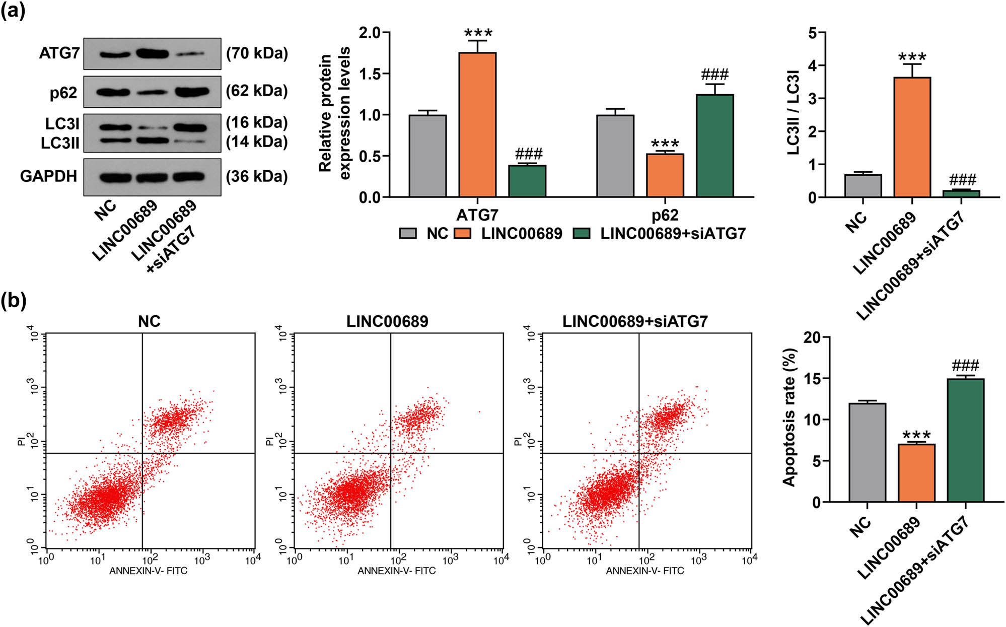
SiATG7 abolished the effect of overexpressed LINC00689 on promoting the autophagy and on inhibiting the apoptosis of NPCs. (a) The levels of autophagy-related proteins (ATG7, p62, and LC3Ⅱ/LC3I) were quantified by the Western blot. (b) Following the transfection of NPCs with LINC00689 overexpression plasmid and siATG7, the apoptosis of NPCs was detected by using flow cytometry. *** P < 0.001 vs NC; ### P < 0.001 vs LINC00689 (NPCs: human nucleus pulposus cells, ATG7: autophagy related 7).
3.3 LINC00689 could competitively bind with miR-3127-5p, and ATG7 was targeted by miR-3127-5p in NPCs
As shown in Figure 4a, five potential upstream miRNAs that could target ATG7 were obtained, namely, miR-3127-5p, miR-769-5p, miR-3179, miR-129-5p, and miR-766-5p. A previous study has reported the high expression of miR-3127-5p in lumbar IDD [9]. Here, the complementary binding sites in between LINC00689/ATG7 and miR-3127-5p are shown in Figure 4b and c. Moreover, we found that the luciferase activity was decreased when NPCs were co-transfected with ATG7-WT or LINC00689-WT and miR-3127-5p mimic (Figure 4d and e, P < 0.001). Besides, there was no difference in the luciferase activity when NPCs were co-transfected with ATG7-MUT or LINC00689-MUT and miR-3127-5p mimic or mimic control. These results thus signified that ATG7 was indeed targeted by miR-3127-5p, and miR-3127-5p was further targeted by LINC00689 in NPCs. Additionally, miR-3127-5p expression was proved to be up-regulated in NP tissues of the IDD group when compared with that in LVF group (Figure 4f, P < 0.001). It was clearly mirrored in correlation analysis that LINC00689 expression was negatively correlated with miR-3127-5p expression in IDD tissues (Figure 4g, r = −0.684, P = 0.029). These results confirmed that LINC00689 could competitively bind with miR-3127-5p, the miRNA which could target ATG7 in IDD.

LINC00689 could competitively bind with miR-3127-5p, and ATG7 was targeted by miR-3127-5p in NPCs. (a) The upstream miRNAs of ATG7 were predicted by Starbase, TargetScan, and LncBase Predicted v.2 websites. By analyzing the data, five potential upstream miRNAs of ATG7 (miR-3127-5p, miR-769-5p, miR-3179, miR-129-5p, and miR-766-5p) were obtained. (b) TargetScan was used to predict the binding site of miR-3127-5p and ATG7. (c) The binding site of LINC00689 and miR-3127-5p was predicted via Starbase. (d and e) The interrelations of LINC00689, miR-3127-5p, and ATG7 were analyzed using dual-luciferase reporter assay. (f) The level of miR-3127-5p in NP tissue samples was measured by qRT-PCR. (g) The correlation between miR-3127-5p and LINC00689 levels was analyzed. ^^^ P < 0.001 vs MC; *** P < 0.001 vs LVF (NPCs: human nucleus pulposus cells, qRT-PCR: quantitative RT-PCR, M: miR-3127-5p mimic, MC: mimic control, LVF: lumbar vertebrae fractures, IDD: intervertebral disc degeneration, ATG7: autophagy related 7, NP: nucleus pulposus).
3.4 Up-regulation of miR-3127-5p reversed the effects of overexpressed LINC00689 on promoting the proliferation and autophagy and on inhibiting the apoptosis of NPCs
The results of qRT-PCR manifested that miR-3127-5p mimic notably enhanced the level of miR-3127-5p in NPCs (Figure 5a, P < 0.001). As depicted in Figure 5b, overexpressed LINC00689 inhibited the expression of miR-3127-5p, while miR-3127-5p mimic enhanced miR-3127-5p level and reversed the inhibitory effect of LINC00689 overexpression on miR-3127-5p expression in NPCs (Figure 5b, P < 0.001). Meanwhile, LINC00689 overexpression obviously facilitated the proliferation of NPCs, while overexpressed miR-3127-5p inhibited the proliferation of NPCs (Figure 5c). Additionally, the up-regulation of miR-3127-5p reversed the effect of LINC00689 overexpression on promoting the proliferation of NPCs (Figure 5c). Moreover, the apoptosis of NPCs was reduced following the overexpression of LINC00689 yet raised by the up-regulation of miR-3127-5p, and more importantly, the up-regulation of miR-3127-5p reversed the effect of overexpressed LINC00689 on inhibiting the apoptosis of NPCs (Figure 5d and e, P < 0.001).

Up-regulation of miR-3127-5p reversed the effect of overexpressed LINC00689 on promoting the proliferation and autophagy, and on inhibiting the apoptosis of NPCs. (a and b) The expression of miR-3127-5p in transfected NPCs was analyzed using the qRT-PCR. (c) The proliferation of NPCs was measured by EdU staining (under 200× magnification, scale bar = 100 μm). (d and e) The apoptosis of NPCs was detected by flow cytometer. (f) The expressions of apoptosis-related factors (Bax, Bcl-2, and Cleaved caspase-3) in transfected NPCs were examined using the Western blot. (g) The levels of autophagy-related proteins (ATG7, p62, and LC3Ⅱ/LC3I) in transfected NPCs were analyzed by the Western blot. ** P < 0.01, *** P < 0.001 vs MC (Figure 5a) or NC + MC (Figure 5b–g); ^^ P < 0.01, ^^^ P < 0.001 vs LINC00689 + MC; ### P < 0.001 vs NC + M. (LINC00689: long non-coding RNAs LINC00689, NPCs: human nucleus pulposus cells, M: miR-3127-5p mimic, MC: mimic control, NC: negative control, ATG7: autophagy related 7).
The results of Western blot displayed that overexpressed LINC00689 suppressed the levels of Bax and Cleaved caspase-3 and promoted that of Bcl-2 in NPCs, while the overexpression of miR-3127-5p up-regulated the levels of Bax and Cleaved caspase-3 yet down-regulated the levels of Bcl-2 in NPCs (Figure 5f, P < 0.001). Also, the up-regulation of miR-3127-5p counteracted the effects of overexpressed LINC00689 on inhibiting the levels of Bax and Cleaved caspase-3 yet promoting the Bcl-2 level in NPCs (Figure 5f, P < 0.01). In addition, the up-regulation of LINC00689 raised the ATG7 level and LC3Ⅱ/LC3I value, while reducing p62 level (Figure 5g, P < 0.001). However, the overexpression of miR-3127-5p not only led to the inhibited ATG7 level and LC3Ⅱ/LC3I ratio and the promoted p62 level, but also reversed the effects of LINC00689 overexpression on increasing the ATG7 level and LC3Ⅱ/LC3I ratio yet decreasing the p62 expression in NPCs (Figure 5g, P < 0.001). These results suggested that up-regulation of miR-3127-5p has the ability to reverse the effects of LINC00689 overexpression on promoting the proliferation and autophagy and on inhibiting the apoptosis of NPCs.
4 Discussion
IDD is one of the main causes of back pain [27]. The IVD is comprised of the inner NP, which is encircled by the cartilaginous endplates and annulus fibrosis lying between the adjacent vertebral bodies and IVD [28]. It has already been suggested that the functional changes of NPCs are considered to be the initiating factors of IDD [28]. Additionally, current research has already shown that the molecular biological process of IDD is abnormally complex and that numerous cytokines and proteins, such as inflammatory factors, growth factors, and matrix-degrading enzymes, are abnormally expressed at the molecular level [27,29,30].
A great deal of researchers have reported that lncRNAs are involved in numerous processes, with the regulatory effects on gene expression [31]. In addition, dysregulated expression of lncRNAs is closely linked to many human diseases, such as neurological diseases, cancer, osteoarthritis, and IDD [32]. Similarly, aberrantly expressed lncRNAs are involved in the initiation and development of IDD by regulating the abnormal phenotypes of NPCs, the proliferation and apoptosis of cells, for instance [33]. In this study, some significant changes concerning the morphology of NPCs have been evidenced in IDD patients. It has been reported that lncRNA ANPODRT expression was reduced in degenerative NP tissues and that lncRNA ANPODRT inhibited the apoptosis of NPCs via activating Nrf2 signaling [34]. Chen et al. have demonstrated that LINC00324 level was increased in IDD patients, and LINC00324 may accelerate the IDD progression via up-regulating the expression of Fas ligand [35]. Also, recent evidence has additionally suggested that lncRNA LINC00689 expression was down-regulated in IDD, despite its vague effect in IDD, which awaited to be further elucidated [9]. In this research, we also found that LINC00689 expression was down-regulated in IDD tissue. Apart from this, for the first time, we found that overexpressed LINC00689 promoted the proliferation yet inhibited the apoptosis of NPCs, whereas LINC00689 silencing did the opposite. These results suggested that LINC00689 indeed regulated the biological behaviors of NPCs.
To further validate the experimental results above, we examined the expressions of apoptosis-related factors (Bax, Bcl-2, and Cleaved caspase-3) in treated NPCs as needed [36]. Bcl-2 family plays a vital role in the intrinsic apoptosis of cells. Bax and Bcl-2 belong to the Bcl-2-related family, in which Bcl-2 is an apoptosis inhibitor, while Bax is an apoptosis promoter [37,38]. Moreover, the cell apoptosis has been proposed to be orchestrated by caspases family, among which caspase-3 is responsible for the majority of proteolysis during apoptosis, making Cleaved caspase-3 level thereby considered as a marker for evaluating the apoptosis of cells [39]. In this study, we discovered that overexpressed LINC00689 inhibited the levels of Bax and Cleaved caspase-3, while promoting Bcl-2 level. On the contrary, the silence of LINC00689 increased the levels of Bax and Cleaved caspase-3 yet decreased the level of Bcl-2 in NPCs. In the previous studies it has been suggested that activating the autophagy of NPCs reduced cell senescence and apoptosis [20,21]. ATG5 and ATG7 are thought to be essential for the induction of autophagy [40]. A recent evidence has profiled that miR-210 promoted extracellular matrix degradation via suppressing ATG7-mediated autophagy in human degenerated NPCs [41]. Additionally, p62 and LC3Ⅱ/LC3I belong to the autophagy-related proteins, of which p62 level is accumulated yet LC3Ⅱ/LC3I ratio is reduced upon the inhibition of autophagy [42,43]. In this study, we also found that the overexpression of LINC00689 raised the ATG7 level and LC3Ⅱ/LC3I value yet reduced that of p62, whereas the knockdown of LINC00689 weakened the ATG7 level and LC3Ⅱ/LC3I ratio, but elevated the p62 level in NPCs. However, ATG7 silencing abolished the effect of overexpressed LINC00689 in NPCs. These data, collectively, indicated that overexpressed LINC00689 inhibited the apoptosis of NPCs via activating ATG7-dependent canonical autophagy in NPCs, whereas LINC00689 silencing exerted the opposite effect.
Furthermore, it has already been reported that lncRNA–miRNA–mRNA network plays a critical role in IDD [9]. Zhang et al. have demonstrated that the up-regulation of lncRNA MALAT1 promoted the proliferation of NPCs and attenuated the severity of disc degeneration in IDD-modeled rats via sponging miR-503 [44]. Yang et al. have indicated that lncRNA-SLC20A1 elevated the extracellular matrix degradation in IDD NP cells via regulating the miR-31-5p/MMP3 axis [45]. In addition to that, the expression of miR-3127-5p was previously reported to be up-regulated in lumbar IDD [9]. Likewise, in our current research, we discovered that miR-3127-5p expression was up-regulated in IDD, and that LINC00689 could competitively bind with miR-3127-5p, an miRNA which could target ATG7 in NPCs. Besides, the up-regulation of miR-3127-5p reversed the effects of overexpressed LINC00689 in NPCs. These results uncovered that LINC00689 regulated the apoptosis of NPCs via miR-3127-5p/ATG7 axis-mediated autophagy.
In conclusion, in this research, we unveiled that the up-regulation of LINC00689 inhibited the apoptosis of NPCs via miR-3127-5p/ATG7 axis-mediated autophagy. These results may offer some important insights for the gene therapy of IDD.
Acknowledgment
Not applicable.
-
Funding information: None.
-
Author contributions: C.W. and X.Z. performed the immunohistochemistry, X.Z. carried out the molecular genetics studies, C.W. and R.C. designed the study and drafted the manuscript. All authors have read and approved the final manuscript.
-
Conflict of interest: The authors declare no conflicts of interest.
-
Data availability statement: The analyzed datasets generated during the study are available from the corresponding author on reasonable request.
References
[1] Yang J, Liu J, Zhao S, Tian F. N(6)-methyladenosine METTL3 modulates the proliferation and apoptosis of lens epithelial cells in diabetic cataract. Mol Ther Nucleic Acids. 2020;20:111–6. 10.1016/j.omtn.2020.02.002.Suche in Google Scholar PubMed PubMed Central
[2] Kos N, Gradisnik L, Velnar T. A brief review of the degenerative intervertebral disc disease. Med Arch. 2019;73(6):421–4. 10.5455/medarh.2019.73.421-424.Suche in Google Scholar PubMed PubMed Central
[3] Wang Y, Che M, Xin J, Zheng Z, Li J, Zhang S. The role of IL-1beta and TNF-alpha in intervertebral disc degeneration. Biomed Pharmacother. 2020;131:110660. 10.1016/j.biopha.2020.110660.Suche in Google Scholar PubMed
[4] Tang N, Dong Y, Xiao T, Zhao H. LncRNA TUG1 promotes the intervertebral disc degeneration and nucleus pulposus cell apoptosis though modulating miR-26a/HMGB1 axis and regulating NF-kappaB activation. Am J Transl Res. 2020;12(9):5449–64.10.2139/ssrn.3493212Suche in Google Scholar
[5] Zhang GZ, Deng YJ, Xie QQ, Ren EH, Ma ZJ, He XG, et al. Sirtuins and intervertebral disc degeneration: roles in inflammation, oxidative stress, and mitochondrial function. Clin Chim Acta. 2020;508:33–42. 10.1016/j.cca.2020.04.016.Suche in Google Scholar PubMed
[6] Dowdell J, Erwin M, Choma T, Vaccaro A, Iatridis J, Cho SK. Intervertebral disk degeneration and repair. Neurosurgery. 2017;80(3S):S46–54. 10.1093/neuros/nyw078.Suche in Google Scholar PubMed PubMed Central
[7] Oichi T, Taniguchi Y, Oshima Y, Tanaka S, Saito T. Pathomechanism of intervertebral disc degeneration. JOR Spine. 2020;3(1):e1076. 10.1002/jsp2.1076.Suche in Google Scholar PubMed PubMed Central
[8] Chen S, Luo M, Kou H, Shang G, Ji Y, Liu H. A review of gene therapy delivery systems for intervertebral disc degeneration. Curr Pharm Biotechnol. 2020;21(3):194–205. 10.2174/1389201020666191024171618.Suche in Google Scholar PubMed
[9] Zhu J, Zhang X, Gao W, Hu H, Wang X, Hao D. lncRNA/circRNAmiRNAmRNA ceRNA network in lumbar intervertebral disc degeneration. Mol Med Rep. 2019;20(4):3160–74. 10.3892/mmr.2019.10569.Suche in Google Scholar PubMed PubMed Central
[10] Hombach S, Kretz M. Non-coding RNAs: classification, biology and functioning. Adv Exp Med Biol. 2016;937:3–17. 10.1007/978-3-319-42059-2_1.Suche in Google Scholar PubMed
[11] Matsui M, Corey DR. Non-coding RNAs as drug targets. Nat Rev Drug Discov. 2017;16(3):167–79. 10.1038/nrd.2016.117.Suche in Google Scholar PubMed PubMed Central
[12] Dastmalchi N, Safaralizadeh R, Nargesi MM. LncRNAs: potential novel prognostic and diagnostic biomarkers in colorectal cancer. Curr Med Chem. 2020;27(30):5067–77. 10.2174/0929867326666190227230024.Suche in Google Scholar PubMed
[13] Chen WK, Yu XH, Yang W, Wang C, He WS, Yan YG, et al. lncRNAs: novel players in intervertebral disc degeneration and osteoarthritis. Cell Prolif. 2017;50(1):e12313. 10.1111/cpr.12313.Suche in Google Scholar PubMed PubMed Central
[14] Zhan S, Wang K, Song Y, Li S, Yin H, Luo R, et al. Long non-coding RNA HOTAIR modulates intervertebral disc degenerative changes via Wnt/beta-catenin pathway. Arthritis Res Ther. 2019;21(1):201. 10.1186/s13075-019-1986-8.Suche in Google Scholar PubMed PubMed Central
[15] Cui S, Liu Z, Tang B, Wang Z, Li B. LncRNA MAGI2-AS3 is down-regulated in intervertebral disc degeneration and participates in the regulation of FasL expression in nucleus pulposus cells. BMC Musculoskelet Disord. 2020;21(1):149. 10.1186/s12891-020-3086-y.Suche in Google Scholar PubMed PubMed Central
[16] Zhang Z, Huo Y, Zhou Z, Zhang P, Hu J. Role of lncRNA PART1 in intervertebral disc degeneration and associated underlying mechanism. Exp Ther Med. 2021;21(2):131. 10.3892/etm.2020.9563.Suche in Google Scholar PubMed PubMed Central
[17] Yu Z, Wang Y, Wang B, Zhai J. Metformin affects paclitaxel sensitivity of ovarian cancer cells through autophagy mediated by long noncoding RNASNHG7/miR-3127-5p axis. Cancer Biother Radiopharm. 2020. 10.1089/cbr.2019.3390.Suche in Google Scholar PubMed
[18] Li F, Sun X, Zheng B, Sun K, Zhu J, Ji C, et al. Arginase II promotes intervertebral disc degeneration through exacerbating senescence and apoptosis caused by oxidative stress and inflammation via the NF-κB pathway. Front Cell Dev Biol. 2021;9:737809. 10.3389/fcell.2021.737809.Suche in Google Scholar PubMed PubMed Central
[19] Novais EJ, Tran VA, Johnston SN, Darris KR, Roupas AJ, Sessions GA, et al. Long-term treatment with senolytic drugs Dasatinib and Quercetin ameliorates age-dependent intervertebral disc degeneration in mice. Nat Commun. 2021;12(1):5213. 10.1038/s41467-021-25453-2.Suche in Google Scholar PubMed PubMed Central
[20] Yi W, Wen Y, Tan F, Liu X, Lan H, Ye H, et al. Impact of NF-κB pathway on the apoptosis-inflammation-autophagy crosstalk in human degenerative nucleus pulposus cells. Aging (Albany NY). 2019;11(17):7294–306. 10.18632/aging.102266.Suche in Google Scholar PubMed PubMed Central
[21] Wang XY, Jiao LY, He JL, Fu ZA, Guo RJ. Parathyroid hormone 1‑34 inhibits senescence in rat nucleus pulposus cells by activating autophagy via the m‑TOR pathway. Mol Med Rep. 2018;18(3):2681–8. 10.3892/mmr.2018.9229.Suche in Google Scholar PubMed PubMed Central
[22] He R, Cui M, Lin H, Zhao L, Wang J, Chen S, et al. Melatonin resists oxidative stress-induced apoptosis in nucleus pulposus cells. Life Sci. 2018;199:122–30. 10.1016/j.lfs.2018.03.020.Suche in Google Scholar PubMed
[23] Gong C, Pan W, Hu W, Chen L. Bone morphogenetic protein-7 retards cell subculture-induced senescence of human nucleus pulposus cells through activating the PI3K/Akt pathway. Biosci Rep. 2019;39(3):BSR20182312. 10.1042/BSR20182312.Suche in Google Scholar PubMed PubMed Central
[24] Chen D, Xia D, Pan Z, Xu D, Zhou Y, Wu Y, et al. Metformin protects against apoptosis and senescence in nucleus pulposus cells and ameliorates disc degeneration in vivo. Cell Death Dis. 2016;7(10):e2441. 10.1038/cddis.2016.334.Suche in Google Scholar PubMed PubMed Central
[25] Livak KJ, Schmittgen TD. Analysis of relative gene expression data using real-time quantitative PCR and the 2(-Delta Delta C(T)) method. Methods (San Diego, CA). 2001;25(4):402–8. 10.1006/meth.2001.1262.Suche in Google Scholar PubMed
[26] Chen J, Xie JJ, Jin MY, Gu YT, Wu CC, Guo WJ, et al. Sirt6 overexpression suppresses senescence and apoptosis of nucleus pulposus cells by inducing autophagy in a model of intervertebral disc degeneration. Cell Death Dis. 2018;9(2):56. 10.1038/s41419-017-0085-5.Suche in Google Scholar PubMed PubMed Central
[27] Navone SE, Marfia G, Giannoni A, Beretta M, Guarnaccia L, Gualtierotti R, et al. Inflammatory mediators and signalling pathways controlling intervertebral disc degeneration. Histol Histopathol. 2017;32(6):523–42. 10.14670/HH-11-846.Suche in Google Scholar PubMed
[28] Cui S, Zhang L. circ_001653 silencing promotes the proliferation and ECM synthesis of NPCs in IDD by downregulating miR-486-3p-mediated CEMIP. Mol Ther Nucleic Acids. 2020;20:385–99. 10.1016/j.omtn.2020.01.026.Suche in Google Scholar PubMed PubMed Central
[29] Zhang Y, He F, Chen Z, Su Q, Yan M, Zhang Q, et al. Melatonin modulates IL-1beta-induced extracellular matrix remodeling in human nucleus pulposus cells and attenuates rat intervertebral disc degeneration and inflammation. Aging (Albany NY). 2019;11(22):10499–512. 10.18632/aging.102472.Suche in Google Scholar PubMed PubMed Central
[30] McCann MR, Veras MA, Yeung C, Lalli G, Patel P, Leitch KM, et al. Whole-body vibration of mice induces progressive degeneration of intervertebral discs associated with increased expression of Il-1beta and multiple matrix degrading enzymes. Osteoarthr Cartil. 2017;25(5):779–89. 10.1016/j.joca.2017.01.004.Suche in Google Scholar PubMed
[31] Gil N, Ulitsky I. Regulation of gene expression by cis-acting long non-coding RNAs. Nat Rev Genet. 2020;21(2):102–17. 10.1038/s41576-019-0184-5.Suche in Google Scholar PubMed
[32] Chen Y, Zhou J. LncRNAs: macromolecules with big roles in neurobiology and neurological diseases. Metab Brain Dis. 2017;32(2):281–91. 10.1007/s11011-017-9965-8.Suche in Google Scholar PubMed
[33] Li Z, Li X, Chen C, Li S, Shen J, Tse G, et al. Long non-coding RNAs in nucleus pulposus cell function and intervertebral disc degeneration. Cell Prolif. 2018;51(5):e12483. 10.1111/cpr.12483.Suche in Google Scholar PubMed PubMed Central
[34] Kang L, Tian Y, Guo X, Chu X, Xue Y. Long noncoding RNA ANPODRT overexpression protects nucleus pulposus cells from oxidative stress and apoptosis by activating Keap1-Nrf2 signaling. Oxid Med Cell Longev. 2021;2021:6645005. 10.1155/2021/6645005.Suche in Google Scholar PubMed PubMed Central
[35] Chen Y, Wu Y, Chen R, Xu C, Chen Q. LncRNA LINC00324 is upregulated in intervertebral disk degeneration and upregulates FasL in nucleus pulposus cells. Mol Cell Biochem. 2021;476(5):1995–2000. 10.1007/s11010-021-04058-9.Suche in Google Scholar PubMed
[36] Dai S, Liang T, Shi X, Luo Z, Yang H. Salvianolic acid B protects intervertebral discs from oxidative stress-induced degeneration via activation of the JAK2/STAT3 signaling pathway. Oxid Med Cell Longev. 2021;2021:6672978. 10.1155/2021/6672978.Suche in Google Scholar PubMed PubMed Central
[37] Banjara S, Suraweera CD, Hinds MG, Kvansakul M. The Bcl-2 family: ancient origins, conserved structures, and divergent mechanisms. Biomolecules. 2020;10(1):128. 10.3390/biom10010128.Suche in Google Scholar PubMed PubMed Central
[38] Luo X, O’Neill KL, Huang K. The third model of Bax/Bak activation: a Bcl-2 family feud finally resolved? F1000Res. 2020;9:F1000 Faculty Rev-935. 10.12688/f1000research.25607.1.Suche in Google Scholar PubMed PubMed Central
[39] Crowley LC, Waterhouse NJ. Detecting Cleaved Caspase-3 in apoptotic cells by flow cytometry. Cold Spring Harb Protoc. 2016;2016(11). 10.1101/pdb.prot087312.Suche in Google Scholar PubMed
[40] Arakawa S, Honda S, Yamaguchi H, Shimizu S. Molecular mechanisms and physiological roles of Atg5/Atg7-independent alternative autophagy. Proc Jpn Acad Ser B Phys Biol Sci. 2017;93(6):378–85. 10.2183/pjab.93.023.Suche in Google Scholar PubMed PubMed Central
[41] Wang C, Zhang ZZ, Yang W, Ouyang ZH, Xue JB, Li XL, et al. MiR-210 facilitates ECM degradation by suppressing autophagy via silencing of ATG7 in human degenerated NP cells. Biomed Pharmacother. 2017;93:470–9. 10.1016/j.biopha.2017.06.048.Suche in Google Scholar PubMed
[42] Bjorkoy G, Lamark T, Pankiv S, Overvatn A, Brech A, Johansen T. Monitoring autophagic degradation of p62/SQSTM1. Methods Enzymol. 2009;452:181–97. 10.1016/S0076-6879(08)03612-4.Suche in Google Scholar PubMed
[43] Kang Y, Jia Y, Wang Q, Zhao Q, Song M, Ni R, et al. Long noncoding RNA KCNQ1OT1 promotes the progression of non-small cell lung cancer via regulating miR-204-5p/ATG3 axis. Onco Targets Ther. 2019;12:10787–97. 10.2147/OTT.S226044.Suche in Google Scholar PubMed PubMed Central
[44] Zheng H, Wang T, Li X, He W, Gong Z, Lou Z, et al. LncRNA MALAT1 exhibits positive effects on nucleus pulposus cell biology in vivo and in vitro by sponging miR-503. BMC Mol Cell Biol. 2020;21(1):23. 10.1186/s12860-020-00265-2.Suche in Google Scholar PubMed PubMed Central
[45] Yang Y, Zhong Z, Zhao Y, Ren K, Li N. LincRNA-SLC20A1 (SLC20A1) promotes extracellular matrix degradation in nucleus pulposus cells in human intervertebral disc degeneration by targeting the miR-31-5p/MMP3 axis. Int J Clin Exp Pathol. 2019;12(9):3632–43.Suche in Google Scholar
© 2022 the author(s), published by De Gruyter
This work is licensed under the Creative Commons Attribution 4.0 International License.
Artikel in diesem Heft
- Research Articles
- AMBRA1 attenuates the proliferation of uveal melanoma cells
- A ceRNA network mediated by LINC00475 in papillary thyroid carcinoma
- Differences in complications between hepatitis B-related cirrhosis and alcohol-related cirrhosis
- Effect of gestational diabetes mellitus on lipid profile: A systematic review and meta-analysis
- Long noncoding RNA NR2F1-AS1 stimulates the tumorigenic behavior of non-small cell lung cancer cells by sponging miR-363-3p to increase SOX4
- Promising novel biomarkers and candidate small-molecule drugs for lung adenocarcinoma: Evidence from bioinformatics analysis of high-throughput data
- Plasmapheresis: Is it a potential alternative treatment for chronic urticaria?
- The biomarkers of key miRNAs and gene targets associated with extranodal NK/T-cell lymphoma
- Gene signature to predict prognostic survival of hepatocellular carcinoma
- Effects of miRNA-199a-5p on cell proliferation and apoptosis of uterine leiomyoma by targeting MED12
- Does diabetes affect paraneoplastic thrombocytosis in colorectal cancer?
- Is there any effect on imprinted genes H19, PEG3, and SNRPN during AOA?
- Leptin and PCSK9 concentrations are associated with vascular endothelial cytokines in patients with stable coronary heart disease
- Pericentric inversion of chromosome 6 and male fertility problems
- Staple line reinforcement with nebulized cyanoacrylate glue in laparoscopic sleeve gastrectomy: A propensity score-matched study
- Retrospective analysis of crescent score in clinical prognosis of IgA nephropathy
- Expression of DNM3 is associated with good outcome in colorectal cancer
- Activation of SphK2 contributes to adipocyte-induced EOC cell proliferation
- CRRT influences PICCO measurements in febrile critically ill patients
- SLCO4A1-AS1 mediates pancreatic cancer development via miR-4673/KIF21B axis
- lncRNA ACTA2-AS1 inhibits malignant phenotypes of gastric cancer cells
- circ_AKT3 knockdown suppresses cisplatin resistance in gastric cancer
- Prognostic value of nicotinamide N-methyltransferase in human cancers: Evidence from a meta-analysis and database validation
- GPC2 deficiency inhibits cell growth and metastasis in colon adenocarcinoma
- A pan-cancer analysis of the oncogenic role of Holliday junction recognition protein in human tumors
- Radiation increases COL1A1, COL3A1, and COL1A2 expression in breast cancer
- Association between preventable risk factors and metabolic syndrome
- miR-29c-5p knockdown reduces inflammation and blood–brain barrier disruption by upregulating LRP6
- Cardiac contractility modulation ameliorates myocardial metabolic remodeling in a rabbit model of chronic heart failure through activation of AMPK and PPAR-α pathway
- Quercitrin protects human bronchial epithelial cells from oxidative damage
- Smurf2 suppresses the metastasis of hepatocellular carcinoma via ubiquitin degradation of Smad2
- circRNA_0001679/miR-338-3p/DUSP16 axis aggravates acute lung injury
- Sonoclot’s usefulness in prediction of cardiopulmonary arrest prognosis: A proof of concept study
- Four drug metabolism-related subgroups of pancreatic adenocarcinoma in prognosis, immune infiltration, and gene mutation
- Decreased expression of miR-195 mediated by hypermethylation promotes osteosarcoma
- LMO3 promotes proliferation and metastasis of papillary thyroid carcinoma cells by regulating LIMK1-mediated cofilin and the β-catenin pathway
- Cx43 upregulation in HUVECs under stretch via TGF-β1 and cytoskeletal network
- Evaluation of menstrual irregularities after COVID-19 vaccination: Results of the MECOVAC survey
- Histopathologic findings on removed stomach after sleeve gastrectomy. Do they influence the outcome?
- Analysis of the expression and prognostic value of MT1-MMP, β1-integrin and YAP1 in glioma
- Optimal diagnosis of the skin cancer using a hybrid deep neural network and grasshopper optimization algorithm
- miR-223-3p alleviates TGF-β-induced epithelial-mesenchymal transition and extracellular matrix deposition by targeting SP3 in endometrial epithelial cells
- Clinical value of SIRT1 as a prognostic biomarker in esophageal squamous cell carcinoma, a systematic meta-analysis
- circ_0020123 promotes cell proliferation and migration in lung adenocarcinoma via PDZD8
- miR-22-5p regulates the self-renewal of spermatogonial stem cells by targeting EZH2
- hsa-miR-340-5p inhibits epithelial–mesenchymal transition in endometriosis by targeting MAP3K2 and inactivating MAPK/ERK signaling
- circ_0085296 inhibits the biological functions of trophoblast cells to promote the progression of preeclampsia via the miR-942-5p/THBS2 network
- TCD hemodynamics findings in the subacute phase of anterior circulation stroke patients treated with mechanical thrombectomy
- Development of a risk-stratification scoring system for predicting risk of breast cancer based on non-alcoholic fatty liver disease, non-alcoholic fatty pancreas disease, and uric acid
- Tollip promotes hepatocellular carcinoma progression via PI3K/AKT pathway
- circ_0062491 alleviates periodontitis via the miR-142-5p/IGF1 axis
- Human amniotic fluid as a source of stem cells
- lncRNA NONRATT013819.2 promotes transforming growth factor-β1-induced myofibroblastic transition of hepatic stellate cells by miR24-3p/lox
- NORAD modulates miR-30c-5p-LDHA to protect lung endothelial cells damage
- Idiopathic pulmonary fibrosis telemedicine management during COVID-19 outbreak
- Risk factors for adverse drug reactions associated with clopidogrel therapy
- Serum zinc associated with immunity and inflammatory markers in Covid-19
- The relationship between night shift work and breast cancer incidence: A systematic review and meta-analysis of observational studies
- LncRNA expression in idiopathic achalasia: New insight and preliminary exploration into pathogenesis
- Notoginsenoside R1 alleviates spinal cord injury through the miR-301a/KLF7 axis to activate Wnt/β-catenin pathway
- Moscatilin suppresses the inflammation from macrophages and T cells
- Zoledronate promotes ECM degradation and apoptosis via Wnt/β-catenin
- Epithelial-mesenchymal transition-related genes in coronary artery disease
- The effect evaluation of traditional vaginal surgery and transvaginal mesh surgery for severe pelvic organ prolapse: 5 years follow-up
- Repeated partial splenic artery embolization for hypersplenism improves platelet count
- Low expression of miR-27b in serum exosomes of non-small cell lung cancer facilitates its progression by affecting EGFR
- Exosomal hsa_circ_0000519 modulates the NSCLC cell growth and metastasis via miR-1258/RHOV axis
- miR-455-5p enhances 5-fluorouracil sensitivity in colorectal cancer cells by targeting PIK3R1 and DEPDC1
- The effect of tranexamic acid on the reduction of intraoperative and postoperative blood loss and thromboembolic risk in patients with hip fracture
- Isocitrate dehydrogenase 1 mutation in cholangiocarcinoma impairs tumor progression by sensitizing cells to ferroptosis
- Artemisinin protects against cerebral ischemia and reperfusion injury via inhibiting the NF-κB pathway
- A 16-gene signature associated with homologous recombination deficiency for prognosis prediction in patients with triple-negative breast cancer
- Lidocaine ameliorates chronic constriction injury-induced neuropathic pain through regulating M1/M2 microglia polarization
- MicroRNA 322-5p reduced neuronal inflammation via the TLR4/TRAF6/NF-κB axis in a rat epilepsy model
- miR-1273h-5p suppresses CXCL12 expression and inhibits gastric cancer cell invasion and metastasis
- Clinical characteristics of pneumonia patients of long course of illness infected with SARS-CoV-2
- circRNF20 aggravates the malignancy of retinoblastoma depending on the regulation of miR-132-3p/PAX6 axis
- Linezolid for resistant Gram-positive bacterial infections in children under 12 years: A meta-analysis
- Rack1 regulates pro-inflammatory cytokines by NF-κB in diabetic nephropathy
- Comprehensive analysis of molecular mechanism and a novel prognostic signature based on small nuclear RNA biomarkers in gastric cancer patients
- Smog and risk of maternal and fetal birth outcomes: A retrospective study in Baoding, China
- Let-7i-3p inhibits the cell cycle, proliferation, invasion, and migration of colorectal cancer cells via downregulating CCND1
- β2-Adrenergic receptor expression in subchondral bone of patients with varus knee osteoarthritis
- Possible impact of COVID-19 pandemic and lockdown on suicide behavior among patients in Southeast Serbia
- In vitro antimicrobial activity of ozonated oil in liposome eyedrop against multidrug-resistant bacteria
- Potential biomarkers for inflammatory response in acute lung injury
- A low serum uric acid concentration predicts a poor prognosis in adult patients with candidemia
- Antitumor activity of recombinant oncolytic vaccinia virus with human IL2
- ALKBH5 inhibits TNF-α-induced apoptosis of HUVECs through Bcl-2 pathway
- Risk prediction of cardiovascular disease using machine learning classifiers
- Value of ultrasonography parameters in diagnosing polycystic ovary syndrome
- Bioinformatics analysis reveals three key genes and four survival genes associated with youth-onset NSCLC
- Identification of autophagy-related biomarkers in patients with pulmonary arterial hypertension based on bioinformatics analysis
- Protective effects of glaucocalyxin A on the airway of asthmatic mice
- Overexpression of miR-100-5p inhibits papillary thyroid cancer progression via targeting FZD8
- Bioinformatics-based analysis of SUMOylation-related genes in hepatocellular carcinoma reveals a role of upregulated SAE1 in promoting cell proliferation
- Effectiveness and clinical benefits of new anti-diabetic drugs: A real life experience
- Identification of osteoporosis based on gene biomarkers using support vector machine
- Tanshinone IIA reverses oxaliplatin resistance in colorectal cancer through microRNA-30b-5p/AVEN axis
- miR-212-5p inhibits nasopharyngeal carcinoma progression by targeting METTL3
- Association of ST-T changes with all-cause mortality among patients with peripheral T-cell lymphomas
- LINC00665/miRNAs axis-mediated collagen type XI alpha 1 correlates with immune infiltration and malignant phenotypes in lung adenocarcinoma
- The perinatal factors that influence the excretion of fecal calprotectin in premature-born children
- Effect of femoral head necrosis cystic area on femoral head collapse and stress distribution in femoral head: A clinical and finite element study
- Does the use of 3D-printed cones give a chance to postpone the use of megaprostheses in patients with large bone defects in the knee joint?
- lncRNA HAGLR modulates myocardial ischemia–reperfusion injury in mice through regulating miR-133a-3p/MAPK1 axis
- Protective effect of ghrelin on intestinal I/R injury in rats
- In vivo knee kinematics of an innovative prosthesis design
- Relationship between the height of fibular head and the incidence and severity of knee osteoarthritis
- lncRNA WT1-AS attenuates hypoxia/ischemia-induced neuronal injury during cerebral ischemic stroke via miR-186-5p/XIAP axis
- Correlation of cardiac troponin T and APACHE III score with all-cause in-hospital mortality in critically ill patients with acute pulmonary embolism
- LncRNA LINC01857 reduces metastasis and angiogenesis in breast cancer cells via regulating miR-2052/CENPQ axis
- Endothelial cell-specific molecule 1 (ESM1) promoted by transcription factor SPI1 acts as an oncogene to modulate the malignant phenotype of endometrial cancer
- SELENBP1 inhibits progression of colorectal cancer by suppressing epithelial–mesenchymal transition
- Visfatin is negatively associated with coronary artery lesions in subjects with impaired fasting glucose
- Treatment and outcomes of mechanical complications of acute myocardial infarction during the Covid-19 era: A comparison with the pre-Covid-19 period. A systematic review and meta-analysis
- Neonatal stroke surveillance study protocol in the United Kingdom and Republic of Ireland
- Oncogenic role of TWF2 in human tumors: A pan-cancer analysis
- Mean corpuscular hemoglobin predicts the length of hospital stay independent of severity classification in patients with acute pancreatitis
- Association of gallstone and polymorphisms of UGT1A1*27 and UGT1A1*28 in patients with hepatitis B virus-related liver failure
- TGF-β1 upregulates Sar1a expression and induces procollagen-I secretion in hypertrophic scarring fibroblasts
- Antisense lncRNA PCNA-AS1 promotes esophageal squamous cell carcinoma progression through the miR-2467-3p/PCNA axis
- NK-cell dysfunction of acute myeloid leukemia in relation to the renin–angiotensin system and neurotransmitter genes
- The effect of dilution with glucose and prolonged injection time on dexamethasone-induced perineal irritation – A randomized controlled trial
- miR-146-5p restrains calcification of vascular smooth muscle cells by suppressing TRAF6
- Role of lncRNA MIAT/miR-361-3p/CCAR2 in prostate cancer cells
- lncRNA NORAD promotes lung cancer progression by competitively binding to miR-28-3p with E2F2
- Noninvasive diagnosis of AIH/PBC overlap syndrome based on prediction models
- lncRNA FAM230B is highly expressed in colorectal cancer and suppresses the maturation of miR-1182 to increase cell proliferation
- circ-LIMK1 regulates cisplatin resistance in lung adenocarcinoma by targeting miR-512-5p/HMGA1 axis
- LncRNA SNHG3 promoted cell proliferation, migration, and metastasis of esophageal squamous cell carcinoma via regulating miR-151a-3p/PFN2 axis
- Risk perception and affective state on work exhaustion in obstetrics during the COVID-19 pandemic
- lncRNA-AC130710/miR-129-5p/mGluR1 axis promote migration and invasion by activating PKCα-MAPK signal pathway in melanoma
- SNRPB promotes cell cycle progression in thyroid carcinoma via inhibiting p53
- Xylooligosaccharides and aerobic training regulate metabolism and behavior in rats with streptozotocin-induced type 1 diabetes
- Serpin family A member 1 is an oncogene in glioma and its translation is enhanced by NAD(P)H quinone dehydrogenase 1 through RNA-binding activity
- Silencing of CPSF7 inhibits the proliferation, migration, and invasion of lung adenocarcinoma cells by blocking the AKT/mTOR signaling pathway
- Ultrasound-guided lumbar plexus block versus transversus abdominis plane block for analgesia in children with hip dislocation: A double-blind, randomized trial
- Relationship of plasma MBP and 8-oxo-dG with brain damage in preterm
- Identification of a novel necroptosis-associated miRNA signature for predicting the prognosis in head and neck squamous cell carcinoma
- Delayed femoral vein ligation reduces operative time and blood loss during hip disarticulation in patients with extremity tumors
- The expression of ASAP3 and NOTCH3 and the clinicopathological characteristics of adult glioma patients
- Longitudinal analysis of factors related to Helicobacter pylori infection in Chinese adults
- HOXA10 enhances cell proliferation and suppresses apoptosis in esophageal cancer via activating p38/ERK signaling pathway
- Meta-analysis of early-life antibiotic use and allergic rhinitis
- Marital status and its correlation with age, race, and gender in prognosis of tonsil squamous cell carcinomas
- HPV16 E6E7 up-regulates KIF2A expression by activating JNK/c-Jun signal, is beneficial to migration and invasion of cervical cancer cells
- Amino acid profiles in the tissue and serum of patients with liver cancer
- Pain in critically ill COVID-19 patients: An Italian retrospective study
- Immunohistochemical distribution of Bcl-2 and p53 apoptotic markers in acetamiprid-induced nephrotoxicity
- Estradiol pretreatment in GnRH antagonist protocol for IVF/ICSI treatment
- Long non-coding RNAs LINC00689 inhibits the apoptosis of human nucleus pulposus cells via miR-3127-5p/ATG7 axis-mediated autophagy
- The relationship between oxygen therapy, drug therapy, and COVID-19 mortality
- Monitoring hypertensive disorders in pregnancy to prevent preeclampsia in pregnant women of advanced maternal age: Trial mimicking with retrospective data
- SETD1A promotes the proliferation and glycolysis of nasopharyngeal carcinoma cells by activating the PI3K/Akt pathway
- The role of Shunaoxin pills in the treatment of chronic cerebral hypoperfusion and its main pharmacodynamic components
- TET3 governs malignant behaviors and unfavorable prognosis of esophageal squamous cell carcinoma by activating the PI3K/AKT/GSK3β/β-catenin pathway
- Associations between morphokinetic parameters of temporary-arrest embryos and the clinical prognosis in FET cycles
- Long noncoding RNA WT1-AS regulates trophoblast proliferation, migration, and invasion via the microRNA-186-5p/CADM2 axis
- The incidence of bronchiectasis in chronic obstructive pulmonary disease
- Integrated bioinformatics analysis shows integrin alpha 3 is a prognostic biomarker for pancreatic cancer
- Inhibition of miR-21 improves pulmonary vascular responses in bronchopulmonary dysplasia by targeting the DDAH1/ADMA/NO pathway
- Comparison of hospitalized patients with severe pneumonia caused by COVID-19 and influenza A (H7N9 and H1N1): A retrospective study from a designated hospital
- lncRNA ZFAS1 promotes intervertebral disc degeneration by upregulating AAK1
- Pathological characteristics of liver injury induced by N,N-dimethylformamide: From humans to animal models
- lncRNA ELFN1-AS1 enhances the progression of colon cancer by targeting miR-4270 to upregulate AURKB
- DARS-AS1 modulates cell proliferation and migration of gastric cancer cells by regulating miR-330-3p/NAT10 axis
- Dezocine inhibits cell proliferation, migration, and invasion by targeting CRABP2 in ovarian cancer
- MGST1 alleviates the oxidative stress of trophoblast cells induced by hypoxia/reoxygenation and promotes cell proliferation, migration, and invasion by activating the PI3K/AKT/mTOR pathway
- Bifidobacterium lactis Probio-M8 ameliorated the symptoms of type 2 diabetes mellitus mice by changing ileum FXR-CYP7A1
- circRNA DENND1B inhibits tumorigenicity of clear cell renal cell carcinoma via miR-122-5p/TIMP2 axis
- EphA3 targeted by miR-3666 contributes to melanoma malignancy via activating ERK1/2 and p38 MAPK pathways
- Pacemakers and methylprednisolone pulse therapy in immune-related myocarditis concomitant with complete heart block
- miRNA-130a-3p targets sphingosine-1-phosphate receptor 1 to activate the microglial and astrocytes and to promote neural injury under the high glucose condition
- Review Articles
- Current management of cancer pain in Italy: Expert opinion paper
- Hearing loss and brain disorders: A review of multiple pathologies
- The rationale for using low-molecular weight heparin in the therapy of symptomatic COVID-19 patients
- Amyotrophic lateral sclerosis and delayed onset muscle soreness in light of the impaired blink and stretch reflexes – watch out for Piezo2
- Interleukin-35 in autoimmune dermatoses: Current concepts
- Recent discoveries in microbiota dysbiosis, cholangiocytic factors, and models for studying the pathogenesis of primary sclerosing cholangitis
- Advantages of ketamine in pediatric anesthesia
- Congenital adrenal hyperplasia. Role of dentist in early diagnosis
- Migraine management: Non-pharmacological points for patients and health care professionals
- Atherogenic index of plasma and coronary artery disease: A systematic review
- Physiological and modulatory role of thioredoxins in the cellular function
- Case Reports
- Intrauterine Bakri balloon tamponade plus cervical cerclage for the prevention and treatment of postpartum haemorrhage in late pregnancy complicated with acute aortic dissection: Case series
- A case of successful pembrolizumab monotherapy in a patient with advanced lung adenocarcinoma: Use of multiple biomarkers in combination for clinical practice
- Unusual neurological manifestations of bilateral medial medullary infarction: A case report
- Atypical symptoms of malignant hyperthermia: A rare causative mutation in the RYR1 gene
- A case report of dermatomyositis with the missed diagnosis of non-small cell lung cancer and concurrence of pulmonary tuberculosis
- A rare case of endometrial polyp complicated with uterine inversion: A case report and clinical management
- Spontaneous rupturing of splenic artery aneurysm: Another reason for fatal syncope and shock (Case report and literature review)
- Fungal infection mimicking COVID-19 infection – A case report
- Concurrent aspergillosis and cystic pulmonary metastases in a patient with tongue squamous cell carcinoma
- Paraganglioma-induced inverted takotsubo-like cardiomyopathy leading to cardiogenic shock successfully treated with extracorporeal membrane oxygenation
- Lineage switch from lymphoma to myeloid neoplasms: First case series from a single institution
- Trismus during tracheal extubation as a complication of general anaesthesia – A case report
- Simultaneous treatment of a pubovesical fistula and lymph node metastasis secondary to multimodal treatment for prostate cancer: Case report and review of the literature
- Two case reports of skin vasculitis following the COVID-19 immunization
- Ureteroiliac fistula after oncological surgery: Case report and review of the literature
- Synchronous triple primary malignant tumours in the bladder, prostate, and lung harbouring TP53 and MEK1 mutations accompanied with severe cardiovascular diseases: A case report
- Huge mucinous cystic neoplasms with adhesion to the left colon: A case report and literature review
- Commentary
- Commentary on “Clinicopathological features of programmed cell death-ligand 1 expression in patients with oral squamous cell carcinoma”
- Rapid Communication
- COVID-19 fear, post-traumatic stress, growth, and the role of resilience
- Erratum
- Erratum to “Tollip promotes hepatocellular carcinoma progression via PI3K/AKT pathway”
- Erratum to “Effect of femoral head necrosis cystic area on femoral head collapse and stress distribution in femoral head: A clinical and finite element study”
- Erratum to “lncRNA NORAD promotes lung cancer progression by competitively binding to miR-28-3p with E2F2”
- Retraction
- Expression and role of ABIN1 in sepsis: In vitro and in vivo studies
- Retraction to “miR-519d downregulates LEP expression to inhibit preeclampsia development”
- Special Issue Computational Intelligence Methodologies Meets Recurrent Cancers - Part II
- Usefulness of close surveillance for rectal cancer patients after neoadjuvant chemoradiotherapy