Activation of SphK2 contributes to adipocyte-induced EOC cell proliferation
-
Lan Dai
 ,
Chen Wang
,
Chen Wang

Abstract
Epithelial ovarian cancer (EOC) is the leading cause of deaths due to cancer in women. Adipocytes have been suggested to play a key role in the stimulation of EOC growth. However, the mechanisms underlying the adipocyte-induced EOC proliferation remain undefined. Here, we provide the first evidence that adipocytes induce the activation of sphingosine kinase (SphK) 2 in EOC, which represents a novel pathway that mediates the adipocyte-induced EOC growth. SphK2 inhibition in EOC cells led to a remarkable inhibition of the adipocyte-induced cell proliferation. Moreover, the adipocyte-induced SphK2 activation in EOC cells was extracellular signal-regulated protein kinases (ERK) dependent. Furthermore, silencing SphK2 in EOC significantly inhibited the adipocyte-induced expression of phospho-ERK and c-Myc, two crucial players in EOC growth. Collectively, the current study unraveled a previously unrecognized role of SphK2 in the adipocyte-induced growth-promoting action in EOC, suggesting a novel target for EOC treatment.
1 Introduction
Epithelial ovarian cancer (EOC) is the deadliest gynecological malignancy [1]. Most EOC patients are found to have tumors in an advanced stage at initial diagnosis, which may lead to high disease mortality [2]. Therefore, understanding the mechanisms that regulate EOC growth may have an important impact on the outcome of this fatal cancer. EOC growth is particularly affected by adipocytes [3,4,5,6,7]. EOC cells have a predilection to proliferate in the omentum, an organ primarily composed of adipocytes [3,6,7]. In vitro, coculture of EOC cells with adipocytes can promote the proliferation of EOC. In vivo, subcutaneous injection of EOC cells with adipocytes into nude mice can produce tumors larger than those produced using EOC cells alone [3]. Moreover, as a source of various adipokines, adipocytes can provide high-energy metabolites and a series of factors for EOC growth [5,8]. However, the molecular mechanisms responsible for the growth-promoting effect of adipocytes in EOC remain unclear.
Sphingolipid metabolism dysregulation is often associated with cancer initiation and progression [9,10]. Sphingosine kinases (SphKs), the key enzymes controlling sphingolipid metabolism, are emerging as exploitable targets for cancer therapy [10,11]. SphKs contain two isoforms, SphK1 and SphK2. SphK1 has been reported to be involved in many aspects of EOC progression [12,13]. SphK1, which was overexpressed in EOC tissue [14], was inversely correlated with overall survival (OS) in EOC patients [15]. Moreover, elevated SphK1 levels were associated with EOC growth [16], metastasis [14], angiogenesis [17], and chemotherapeutic resistance [18]. Furthermore, knockout of SphK1 significantly blocked the EOC progression [19]. Unlike extensively studied SphK1, the roles of SphK2 are controversial and still poorly characterized. Initially, SphK2 was considered to be a proapoptotic protein because overexpression of SphK2 promoted tumor apoptosis [20]. However, it was subsequently found to be a prosurvival factor, as inhibition of SphK2 suppressed the tumor growth [21]. Our earlier studies showed that SphK2 is mainly located in the nucleus of ovarian cancer cells [15,17] and is potentially involved in the regulation of gene activation [22,23]. SphK2 activation was a prognostic indicator of OS in EOC patients [15]. In addition, knockdown of SphK2 arrested the cell cycle progression and inhibited the EOC cell proliferation both in vitro and in vivo [23]. Moreover, inhibition of SphK2 was shown to sensitize EOC to paclitaxel [24]. Although SphK2 is an important signaling enzyme in EOC progression, its regulatory mechanisms are far from clarified. In this study, we provide the first evidence that adipocytes are capable of activating SphK2 and unravel a previously unrecognized role of SphK2 in the adipocyte-induced growth-promoting action in ovarian cancer.
2 Materials and methods
2.1 Reagents and antibodies
Antibodies against SphK2 (ab264042, rabbit), c-Myc (ab32072, rabbit), and glyceraldehyde-3-phosphate dehydrogenase (GAPDH) (ab8245, mouse) were purchased from Abcam (Cambridge, MA, USA). Antibodies against ERK1/2 (4696, rabbit) and phospho-ERK1 (Thr202/Tyr204)/ERK2 (Thr185/Tyr187) (4370, rabbit) were ordered from Cell Signaling Technology (Danvers, MA, USA). Antibodies against phosphor-SphK2 (Thr578) (SP4631, rabbit) were purchased from ECM Biosciences (Versailles, KY, USA). U0126, ABC294640, insulin, dexamethasone, and 3-isobutyl-1-methylxanthine were ordered from Sigma-Aldrich (St. Louis, MO, USA).
2.2 Cell lines and culture conditions
The human EOC cell line A2780 was obtained from the China Center for Type Culture Collection. SKOV3 cells were purchased from American Type Culture Collection. These two EOC cell lines were cultured in Dulbecco's Modified Eagle Medium (DMEM) (Invitrogen, Carlsbad, CA, USA) supplemented with 10% fetal bovine serum (Invitrogen) and 1% antibiotics. The murine 3T3-L1 preadipocyte cell line was ordered from the Cell Bank of the Chinese Academy of Sciences (Shanghai, China). 3T3-L1 cells were cultured in DMEM supplemented with 10% calf serum and 1% antibiotics. 3T3-L1 preadipocytes were induced into mature adipocytes by treatment with insulin, dexamethasone, and 3-isobutyl-1-methylxanthine as described earlier [25]. To make adipocyte-conditioned medium (Adi-CM), mature adipocytes were cultured with serum-free medium (SFM) for 24 h after being washed twice with phosphate buffered saline. Adi-CM was then collected and filtered.
2.3 Small interfering RNA (siRNA) and transient transfection
The chemically synthesized siRNAs targeting human SphK2 (5′-AACCUCAUCCAGACAGAACGA-3′) and the control siRNA (5′-AAUUCUCCGAACGUGUCACGU-3′) were ordered from GenePharma (Shanghai, China) [17]. SiRNA transfection was performed by using Lipofectamine (Invitrogen). The levels of SphK2 were detected by reverse transcription-polymerase chain reaction (RT-PCR) and Western blot 24–48 h after transfection.
2.4 Real-time RT-PCR
RNA was extracted by using TRIzol Reagent (Invitrogen). The mRNA levels were detected by using Synergy Brands Green RT-PCR and calculated by the 2−ΔΔCt method. Primers were as follows: SphK2, 5′-GGTTGCTTCTATTGGTCAATCC-3′ (forward) and 5′-GTTCTGTCGTTCTGTCTGGATG-3′ (reverse); and GAPDH, 5′-TGCACCACCAACTGCTTAGC-3′ (forward) and 5′-GGCATGGACTGTGGTCATGAG-3′ (reverse).
2.5 Western blot analysis
Western blot analysis was performed as described earlier [26]. Briefly, cells were harvested and lysed with RIPA buffer plus protease inhibitors. The proteins were resolved by sodium dodecyl sulfate polyacrylamide gel electrophoresis and transferred to polyvinylidene fluoride membranes. The membranes were then incubated with appropriate antibodies. Finally, the proteins were visualized using an enhanced chemiluminescence detection kit (Pierce, Rockford, IL, USA).
2.6 Cellular proliferation assay
Cell proliferation was assessed using a CCK-8 (Dojindo) assay as described earlier [27]. Briefly, cells were seeded into 96-well plates. CCK-8 assay reagent was added to each well and cultured at 37°C for 2 h. Optical density values of the supernatant from each well were then measured in a microplate reader.
2.7 Statistical analysis
Statistical analyses were performed using the SPSS software (IBM, Armonk, NY, USA). The values are presented as the mean ± SD and were analyzed by t test. A P value less than 0.05 was considered statistically significant.
3 Results
3.1 SphK2 contributes to the adipocyte-induced EOC cell proliferation
To investigate whether the SphK2 pathway participates in the adipocyte-induced EOC proliferation, we used ABC294640 [28], an inhibitor of SphK2. Consistent with earlier reports [3], Adi-CM significantly increased the proliferation rate of EOC cells (Figure 1a and b). Remarkably, ABC294640 significantly inhibited the Adi-CM-induced EOC cell proliferation. As a control, ABC294640 alone did not significantly affect EOC growth (Figure 1a and b). Moreover, SphK2 siRNA significantly inhibited both the mRNA and protein expression levels of SphK2, as shown in Figure 1c and d. SphK2 silencing significantly inhibited the Adi-CM-induced EOC cell proliferation (Figure 1e and f). Collectively, these results suggested that SphK2 contributed to the Adi-CM-induced EOC proliferation.
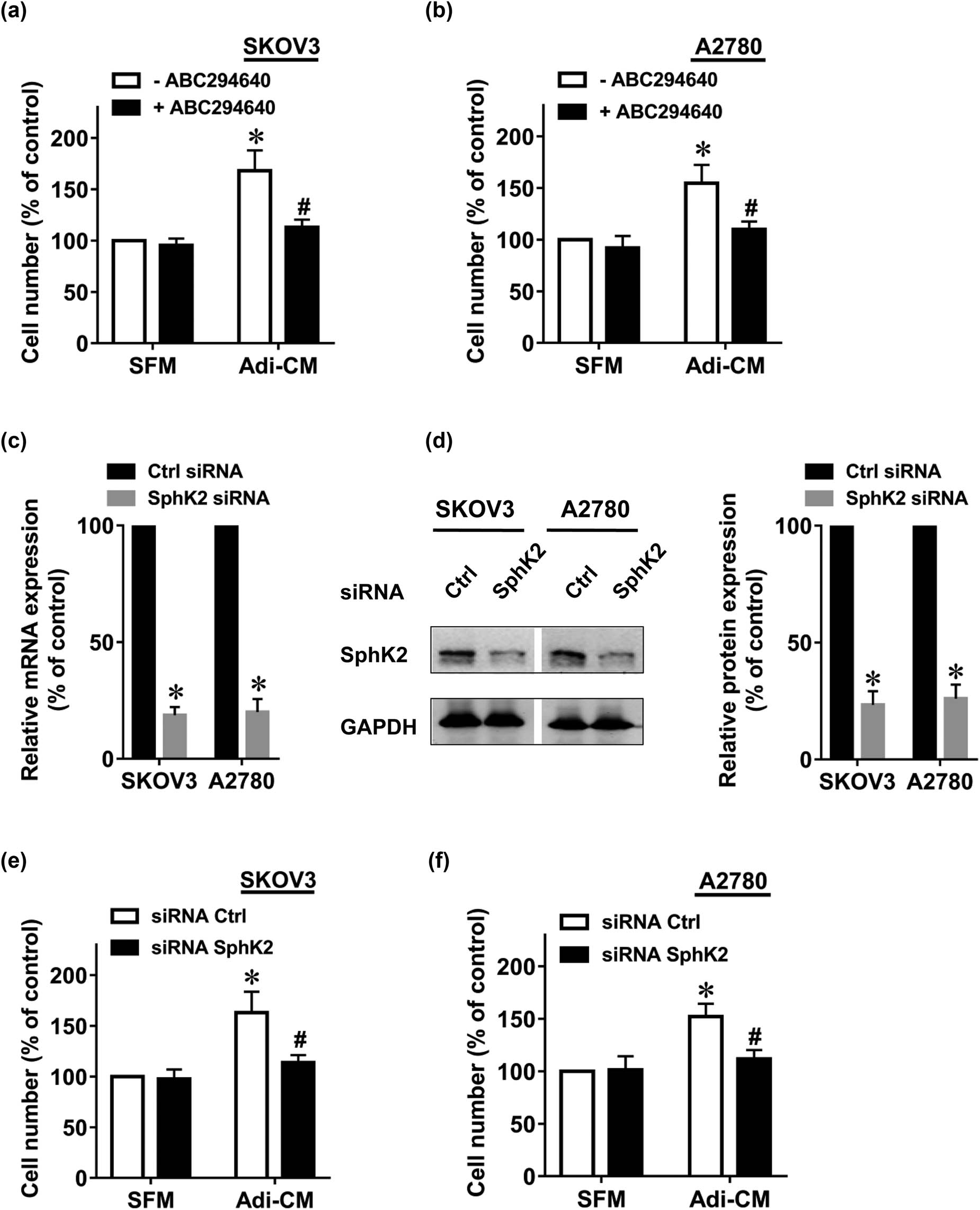
SphK2 inhibition suppresses the adipocyte-induced EOC cell growth. The human EOC cell lines (a) SKOV3 and (b) A2780 were serum-starved overnight and then cultured with SFM or Adi-CM in the presence or absence of ABC294640 (10 μM) for 48 h. Cell proliferation was measured by CCK-8 assay. (c) Twenty-four hours after siRNA transfection, SphK2 mRNA levels were determined by RT-PCR. (d) Forty-eight hours after siRNA transfection, SphK2 protein levels were determined by using Western blot. Densitometric analysis of SphK2 (normalized to GAPDH) is shown on the right. (e) SKOV3 and (f) A2780 cells were transfected with the indicated siRNAs, followed by culture with SFM or Adi-CM for 48 h. Cell proliferation was measured by CCK-8 assays. Molecular weight of SphK2 is 69 kDa, and molecular weight of GAPDH is 36 kDa. Data are mean ± SD. *, P < 0.05 vs control; #, P < 0.05 vs Adi-CM alone.
3.2 Adipocytes mediate the activation of SphK2 in EOC cells
As SphK2 is an important enzyme in EOC proliferation, we explored the role of adipocytes in SphK2 activation. It has been reported that SphK2 can be activated by phosphorylation [29]. Therefore, we measured SphK2 phosphorylation in EOC after Adi-CM treatment. The results showed that Adi-CM treatment induced an increase in SphK2 phosphorylation in EOC (Figure 2a). Adi-CM treatment also resulted in increased phosphorylation of ERK (Figure 2b), a key enzyme controlling EOC proliferation [30]. Our earlier study suggested that ERK could be activated through SphK2 [12]. In agreement with this finding, SphK2 blockade significantly inhibited the Adi-CM-induced ERK phosphorylation, which indicated that SphK2 contributed to the Adi-CM-induced ERK activation in EOC. ERK is a key enzyme causing SphK2 activation [29]. Indeed, U0126, an inhibitor of ERK, significantly blocked the adipocyte-induced SphK2 activation (Figure 3a and b).

Adipocytes activate the SphK2/ERK pathway in EOC cells. (a) SKOV3 and A2780 cells were cultured with SFM or Adi-CM for 24 h. SphK2 phosphorylation was determined by using Western blot. Densitometric analysis of pSphK2 (normalized to total SphK2) is shown on the right. (b) SKOV3 and (c) A2780 cells were transfected with the indicated siRNAs and cultured with SFM or Adi-CM for 24 h. Total and phosphorylated ERK (pERK) levels were then determined by using Western blot. The right panel shows densitometric analysis of pERK (normalized to total ERK). Molecular weight of pSphK2 is 70 kDa, molecular weight of SphK2 is 69 kDa, molecular weight of GAPDH is 36 kDa, molecular weight of pERK is 42, 44 kDa, and molecular weight of ERK is 42, 44 kDa. Data are the mean ± SD. *, P < 0.05.
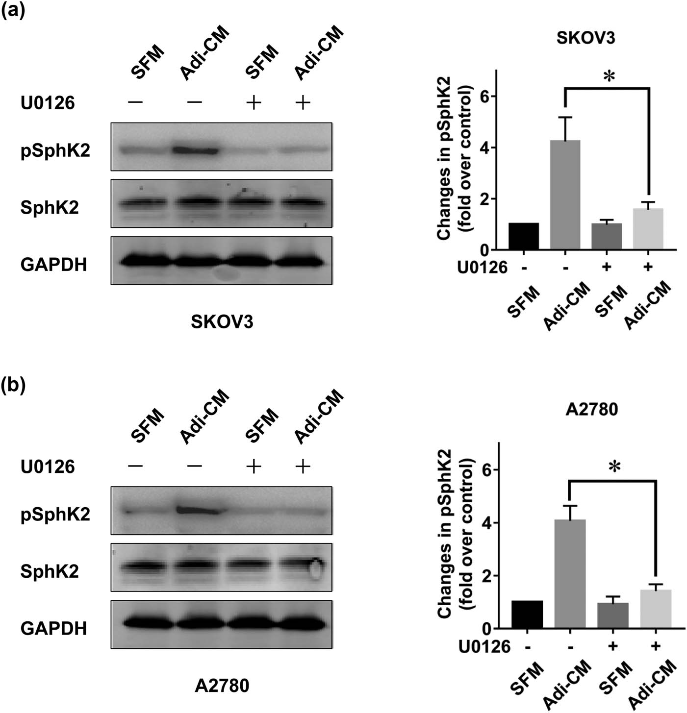
Adipocyte-induced SphK2 activation is ERK dependent. (a) SKOV3 and (b) A2780 cells were serum starved overnight and pretreated with U0126 (5 μM) for 2 h. Cells were then cultured with SFM or Adi-CM for 24 h. Total and phosphorylated SphK2 (pSphK2) levels were then determined by using Western blot. Right panels show the densitometric analysis of pSphK2 (normalized to total SphK2) corresponding to the bands shown in the Western blots. Molecular weight of pSphK2 is 70 kDa, molecular weight of SphK2 is 69 kDa, and molecular weight of GAPDH is 36 kDa. Data are the mean ± SD. *, P < 0.05.
3.3 Adipocyte-induced c-Myc expression in EOC cells occurs partly through the SphK2 pathway
It is well established that c-Myc is a key mediator of EOC proliferation [31]. Therefore, we detected the expression level of c-Myc and confirmed that Adi-CM treatment significantly increased the c-Myc protein level in EOC cells (Figure 4a). Our earlier studies indicated that c-Myc could be regulated by SphK2 in EOC cells [27]. To determine whether adipocytes induced c-Myc expression in EOC through the SphK2 pathway, we tested the expression level of c-Myc after SphK2 blockade. As expected, SphK2 silencing by siRNA significantly inhibited the Adi-CM-induced c-Myc expression in the two EOC cell lines (Figure 4b and c). Together, these results suggested that adipocytes could activate the c-Myc pathway in EOC cells. The adipocyte-induced c-Myc expression was partly SphK2 dependent.

Adipocyte-induced c-Myc expression is partly SphK2 dependent. (a) SKOV3 and A2780 cells were cultured with SFM or Adi-CM for 24 h. c-Myc expression level was determined by using Western blot. Densitometric analysis of c-Myc is shown on the right. (b) SKOV3 and (c) A2780 cells were transfected with the indicated siRNAs and cultured with SFM or Adi-CM for 24 h. c-Myc levels were then determined by using Western blot. Right panel shows the densitometric analysis of c-Myc. Molecular weight of c-Myc is 57 kDa and molecular weight of GAPDH is 36 kDa. Data are the mean ± SD. *, P < 0.05.
4 Discussion
In this study, we unraveled a previously unrecognized role of SphK2 in mediating the growth-promoting functions of adipocytes in EOC. SphK2, an enzyme that exhibits controversial roles in the regulation of cell growth, is responsible for the adipocyte-induced EOC proliferation. Moreover, SphK2 contributes to the adipocyte-induced ERK and c-Myc pathway activation, both of which are well recognized as key signals that facilitate EOC growth. These results suggested that SphK2 could be a new target for the management of EOC.
Ovarian cancer cells preferentially proliferate in the omentum, an organ primarily composed of adipocytes [3]. Indeed, omental metastases typically represent the largest tumor in the abdominal cavities of women with ovarian cancer. Moreover, adipocytes were reported to promote EOC growth both in vitro and in vivo [3]. Therefore, understanding the mechanisms involved in the adipocyte-promoted EOC growth is an important research topic. Our results demonstrated that SphK2 was not only activated by adipocytes but also responsible for the adipocyte-induced EOC growth. Adipocyte culture medium was able to stimulate the phosphorylation of SphK2 in EOC cells. Moreover, drug inhibition of SphK2 significantly suppressed the adipocyte-dependent EOC growth. Furthermore, the siRNA-mediated knockdown of SphK2 resulted in a significant suppression of the adipocyte-promoted cell growth, suggesting an important role of SphK2 in the growth-promoting action of adipocytes in EOC cells. This is consistent with our earlier study showing that SphK2 is important for the follicle-stimulating hormone-induced EOC growth [12]. However, studies have indicated the proapoptotic effect of SphK2 on certain cell types [20,32]. Therefore, SphK2 regulates cell growth in a highly cell type-specific fashion. The underlying mechanisms of this cell type-dependent effect of SphK2 remain to be addressed. The SphK/S1P pathway was also reported to play potential roles in the development of drug resistance. For example, targeting SphK2 reversed the acquired resistance to regorafenib in hepatocellular carcinoma [33]. Tamoxifen-resistant breast cancer cells showed increased levels of SphK expression and activity [34]. S1P receptor expression levels were influenced by tamoxifen treatment in breast cancer cells [35]. Our results showed that adipocytes could induce the activation of SphK in EOC cells. Therefore, further studies are warranted to explore the roles of adipocytes in the development of EOC drug resistance. In addition to EOC cells, SphK/S1P signaling could also regulate endothelial cell functions [36,37], such as cell proliferation, migration, and survival. These processes in endothelial cells are essential components of angiogenesis [37]. Therefore, adipocytes may also provide nutrition and oxygen for sustained EOC growth by activating SphK in endothelial cells and inducing angiogenesis.
The ERK pathway is well recognized as a critical signaling pathway that facilitates EOC growth [38,39]. For instance, ERK signaling is constitutively active in EOC cells [30]. Downregulation of the ERK pathway could lead to the complete suppression of EOC proliferation [30]. Moreover, a variety of cytokines and growth factors have been shown to promote EOC growth by activating ERK [40,41]. As important secretory cells, adipocytes release a variety of adipokines, including leptin, IL-8, and IL-6 [42]. Many of these adipokines could promote EOC growth by activating the ERK pathway [43,44]. Indeed, exposure of EOC cells to Adi-CM resulted in increased ERK phosphorylation. We previously found that SphK2 is an important regulator of ERK [12]. Having demonstrated the ability of adipocytes to cause SphK2 activation in EOC, a potential role of SphK2 in the adipocyte-induced ERK activation was suggested. As expected, SphK2 blockade by siRNA significantly inhibited ERK phosphorylation induced by adipocytes. This result indicated that SphK2 plays an important role in mediating the adipocyte-induced ERK activation in EOC. Earlier studies indicated that ERK is a key enzyme that mediates SphK2 phosphorylation [29]. Consistent with this finding, the treatment of EOC cells with U0126, an inhibitor of ERK signaling, significantly blocked the adipocyte-induced ERK phosphorylation. Collectively, these data indicated that ERK resided both upstream and downstream of SphK2, propagating a positive feedback loop.
Another new finding of this study is that the adipocyte-induced c-Myc expression is partly SphK2 dependent. As an important oncogene, c-Myc has been reported to be a crucial mediator of EOC progression. The disease-free survival and OS of ovarian cancer patients were decreased with high c-Myc mRNA levels [45]. Moreover, c-Myc silencing significantly inhibited the growth of EOC cells [31,45]. Given the importance of c-Myc in EOC growth, we examined the effect of adipocytes on the expression of c-Myc. We found that adipocyte CM significantly increased the c-Myc protein expression level in EOC cells, which may participate in the adipocyte-induced EOC growth. Earlier studies have shown that SphK2 regulates c-Myc in a number of cancer cells [46,47]. Our recent studies also found that SphK2 inhibition downregulates c-Myc expression in EOC [27]. Consistent with these findings, SphK2 blockade by siRNA significantly inhibited the adipocyte-induced c-Myc expression. These results suggested that adipocyte-mediated c-Myc expression was partly mediated through SphK2. However, the exact mechanism of the adipocyte-induced c-Myc expression is not yet clear. c-Myc could be targeted by other adipocyte-regulated pathways, such as the SphK1 [16,48], AKT [25,49], and ERK [23,50] pathways. In addition, c-Myc could also be activated by adipocyte-secreted factors, such as estrogen [51,52], interleukin-6 [3,53], and interleukin-8 [3,54]. Therefore, we speculate that adipocytes can also induce c-Myc expression through SphK2-independent pathways and some unknown signaling pathways. Moreover, SphK1 and SphK2 may play similar roles in mediating the adipocyte-induced c-Myc expression in EOC cells. These points need further investigation.
The present study has several limitations. First, the experiment was only performed in EOC cell lines. This would be improved by verification of the key results in mouse ovarian cancer models. Second, the mechanisms by which adipocytes activate the SphK2 pathway in EOC cells need to be further explored. Third, adipocytes may also affect tumor growth by acting on endothelial cells, and their mechanism needs to be studied in the future. Finally, SphK was also reported to play other roles in EOC progression, such as invasion and angiogenesis. Whether the adipocyte-induced SphK activation affects EOC metastasis and vascularization needs further study.
Acknowledgments
Not applicable.
-
Funding information: This study was supported by the grants from National Natural Science Foundation of China (NSFC) (No. 81974401 to Lan Dai and No. 81974454 to Wen Di), the grant from science and technology commission of Shanghai municipality (No. 18ZR1423100 to Lan Dai) and the grants from Shanghai Municipal Commission of Health and Family Planning (No. 2017YQ035 to Lan Dai).
-
Author contributions: WD, LD and JZ designed and supervised the whole project. CW, WW, KS and TY performed the experiments. All the authors read and approved the manuscript.
-
Conflict of interest: The authors stated that there was no conflict of interest in this study.
-
Data availability statement: The data that support the findings of this study are available on request from the corresponding author.
References
[1] Siegel RL, Miller KD, Jemal A. Cancer statistics, 2020. CA: A Cancer J Clinic. 2020;70(1):7–30.10.3322/caac.21590Search in Google Scholar PubMed
[2] Bowtell DD, Bh S, Ahmed AA, Aspuria PJ, Bast RC, Jr, Beral V, et al. Rethinking ovarian cancer II: reducing mortality from high-grade serous ovarian cancer. Nat Rev Cancer. 2015;15(11):668–79.10.1038/nrc4019Search in Google Scholar PubMed PubMed Central
[3] Nieman KM, Kenny HA, Penicka CV, Ladanyi A, Buell-Gutbrod R, Zillhardt MR, et al. Adipocytes promote ovarian cancer metastasis and provide energy for rapid tumor growth. Nat Med. 2011;17(11):1498–503.10.1038/nm.2492Search in Google Scholar PubMed PubMed Central
[4] Yang J, Zaman MM, Vlasakov I, Roy R, Huang L, Martin CR, et al. Adipocytes promote ovarian cancer chemoresistance. Sci Rep. 2019;9(1):13316.10.1038/s41598-019-49649-1Search in Google Scholar PubMed PubMed Central
[5] Wroblewski M, Szewczyk-Golec K, Holynska-Iwan I, Wroblewska J, Wozniak A. Characteristics of selected adipokines in ascites and blood of ovarian cancer patients. Cancers (Basel). 2021;13(18):4072.10.3390/cancers13184702Search in Google Scholar PubMed PubMed Central
[6] Mukherjee A, Chiang CY, Daifotis HA, Nieman KM, Fahrmann JF, Lastra RR, et al. Adipocyte-induced FABP4 expression in ovarian cancer cells promotes metastasis and mediates carboplatin resistance. Cancer Res. 2020;80(8):1748–61.10.1158/0008-5472.CAN-19-1999Search in Google Scholar PubMed
[7] John B, Naczki C, Patel C, Ghoneum A, Qasem S, Salih Z, et al. Regulation of the bi-directional cross-talk between ovarian cancer cells and adipocytes by SPARC. Oncogene. 2019;38(22):4366–83.10.1038/s41388-019-0728-3Search in Google Scholar PubMed PubMed Central
[8] Dai L, Song K, Di W. Adipocytes: active facilitators in epithelial ovarian cancer progression. J Ovarian Res. 2020;13(1):115.10.1186/s13048-020-00718-4Search in Google Scholar PubMed PubMed Central
[9] Ryland LK, Fox TE, Liu X, Loughran TP, Kester M. Dysregulation of sphingolipid metabolism in cancer. Cancer Biol Ther. 2011;11(2):138–49.10.4161/cbt.11.2.14624Search in Google Scholar PubMed
[10] Sukocheva OA. Expansion of sphingosine kinase and sphingosine-1-phosphate receptor function in normal and cancer cells: from membrane restructuring to mediation of estrogen signaling and stem cell programming. Int J Mol Sci. 2018;19(2):420.10.3390/ijms19020420Search in Google Scholar PubMed PubMed Central
[11] Ogretmen B. Sphingolipid metabolism in cancer signalling and therapy. Nat Rev Cancer. 2018;18(1):33–50.10.1038/nrc.2017.96Search in Google Scholar PubMed PubMed Central
[12] Song K, Dai L, Long X, Wang W, Di W. Follicle-stimulating hormone? promotes the proliferation of? epithelial ovarian cancer? cells by activating sphingosine kinase. Sci Rep. 2020;10(1):13834.10.1038/s41598-020-70896-0Search in Google Scholar PubMed PubMed Central
[13] Pitman M, Oehler MK, Pitson SM. Sphingolipids as multifaceted mediators in ovarian cancer. Cell Signal. 2021;81:109949.10.1016/j.cellsig.2021.109949Search in Google Scholar PubMed
[14] Zhang H, Wang Q, Zhao Q, Di W. MiR-124 inhibits the migration and invasion of ovarian cancer cells by targeting SphK1. J Ovarian Res. 2013;6(1):84.10.1186/1757-2215-6-84Search in Google Scholar PubMed PubMed Central
[15] Song K, Dai L, Long X, Wang W, Di W. Follicle-stimulating hormone promotes the proliferation of epithelial ovarian cancer cells by activating sphingosine kinase. Sci Rep. 2020;10(1):13834.10.1038/s41598-020-70896-0Search in Google Scholar
[16] Dai L, Wang C, Song K, Wang W, Di W. Activation of SphK1 by adipocytes mediates epithelial ovarian cancer cell proliferation. J Ovarian Res. 2021;14(1):62.10.1186/s13048-021-00815-ySearch in Google Scholar PubMed PubMed Central
[17] Dai L, Liu Y, Xie L, Wu X, Qiu L, Di W. Sphingosine kinase 1/sphingosine-1-phosphate (S1P)/S1P receptor axis is involved in ovarian cancer angiogenesis. Oncotarget. 2017;8(43):74947–61.10.18632/oncotarget.20471Search in Google Scholar PubMed PubMed Central
[18] Yang YL, Ji C, Cheng L, He L, Lu CC, Wang R, et al. Sphingosine kinase-1 inhibition sensitizes curcumin-induced growth inhibition and apoptosis in ovarian cancer cells. Cancer Sci. 2012;103(8):1538–45.10.1111/j.1349-7006.2012.02335.xSearch in Google Scholar PubMed PubMed Central
[19] Beach JA, Aspuria PJ, Cheon DJ, Lawrenson K, Agadjanian H, Walsh CS, et al. Sphingosine kinase 1 is required for TGF-beta mediated fibroblastto- myofibroblast differentiation in ovarian cancer. Oncotarget. 2016;7(4):4167–82.10.18632/oncotarget.6703Search in Google Scholar PubMed PubMed Central
[20] Liu H, Toman RE, Goparaju SK, Maceyka M, Nava VE, Sankala H, et al. Sphingosine kinase type 2 is a putative BH3-only protein that induces apoptosis. J Biol Chem. 2003;278(41):40330–6.10.1074/jbc.M304455200Search in Google Scholar PubMed
[21] Liang J, Zhang X, He S, Miao Y, Wu N, Li J, et al. Sphk2 RNAi nanoparticles suppress tumor growth via downregulating cancer cell derived exosomal microRNA. J Control Rel: Off J Control Release Soc. 2018;286:348–57.10.1016/j.jconrel.2018.07.039Search in Google Scholar PubMed
[22] Diaz Escarcega R, McCullough LD, Tsvetkov AS. The functional role of sphingosine kinase 2. Front Mol Biosci. 2021;8:683767.10.3389/fmolb.2021.683767Search in Google Scholar PubMed PubMed Central
[23] Dai L, Wang W, Liu Y, Song K, Di W. Inhibition of sphingosine kinase 2 down-regulates ERK/c-Myc pathway and reduces cell proliferation in human epithelial ovarian cancer. Ann Transl Med. 2021;9(8):645.10.21037/atm-20-6742Search in Google Scholar PubMed PubMed Central
[24] White MD, Chan L, Antoon JW, Beckman BS. Targeting ovarian cancer and chemoresistance through selective inhibition of sphingosine kinase-2 with ABC294640. Anticancer Res. 2013;33(9):3573–9.Search in Google Scholar
[25] Park JY, Kang SE, Ahn KS, Um JY, Yang WM, Yun M, et al. Inhibition of the PI3K-AKT-mTOR pathway suppresses the adipocyte-mediated proliferation and migration of breast cancer cells. J Cancer. 2020;11(9):2552–9.10.7150/jca.37975Search in Google Scholar PubMed PubMed Central
[26] Dai L, Qi Y, Chen J, Kaczorowski D, Di W, Wang W, et al. Sphingosine kinase (SphK) 1 and SphK2 play equivalent roles in mediating insulin’s mitogenic action. Mol Endocrinol (Baltimore, Md). 2014;28(2):197–207.10.1210/me.2013-1237Search in Google Scholar PubMed PubMed Central
[27] Song K, Dai L, Long X, Cui X, Liu Y, Di W. Sphingosine kinase 2 inhibitor ABC294640 displays anti-epithelial ovarian cancer activities in vitro and in vivo. Onco Targets Ther. 2019;12:4437–49.10.2147/OTT.S208519Search in Google Scholar PubMed PubMed Central
[28] French KJ, Zhuang Y, Maines LW, Gao P, Wang W, Beljanski V, et al. Pharmacology and antitumor activity of ABC294640, a selective inhibitor of sphingosine kinase-2. J Pharmacol Exp Therap. 2010;333(1):129–39.10.1124/jpet.109.163444Search in Google Scholar PubMed PubMed Central
[29] Hait NC, Bellamy A, Milstien S, Kordula T, Spiegel S. Sphingosine kinase type 2 activation by ERK-mediated phosphorylation. J Biol Chem. 2007;282(16):12058–65.10.1074/jbc.M609559200Search in Google Scholar PubMed
[30] Steinmetz R, Wagoner HA, Zeng P, Hammond JR, Hannon TS, Meyers JL, et al. Mechanisms regulating the constitutive activation of the extracellular signal-regulated kinase (ERK) signaling pathway in ovarian cancer and the effect of ribonucleic acid interference for ERK1/2 on cancer cell proliferation. Mol Endocrinol (Baltimore. Md). 2004;18(10):2570–82.10.1210/me.2004-0082Search in Google Scholar PubMed
[31] Zeng M, Kwiatkowski NP, Zhang T, Nabet B, Xu M, Liang Y, et al. Targeting MYC dependency in ovarian cancer through inhibition of CDK7 and CDK12/13. eLife. 2018;7:e39030.10.7554/eLife.39030Search in Google Scholar PubMed PubMed Central
[32] Maceyka M, Sankala H, Hait NC, Le Stunff H, Liu H, Toman R, et al. SphK1 and SphK2, sphingosine kinase isoenzymes with opposing functions in sphingolipid metabolism. J Biol Chem. 2005;280(44):37118–29.10.1074/jbc.M502207200Search in Google Scholar PubMed
[33] Shi W, Zhang S, Ma D, Yan D, Zhang G, Cao Y, et al. Targeting SphK2 reverses acquired resistance of regorafenib in hepatocellular carcinoma. Front Oncol. 2020;10:694.10.3389/fonc.2020.00694Search in Google Scholar PubMed PubMed Central
[34] Sukocheva O, Wang L, Verrier E, Vadas MA, Xia P. Restoring endocrine response in breast cancer cells by inhibition of the sphingosine kinase-1 signaling pathway. Endocrinology. 2009;150(10):4484–92.10.1210/en.2009-0391Search in Google Scholar PubMed
[35] Ghosal P, Sukocheva OA, Wang T, Mayne GC, Watson DI, Hussey DJ. Effects of chemotherapy agents on Sphingosine-1-Phosphate receptors expression in MCF-7 mammary cancer cells. Biomed Pharma Biomed Pharma. 2016;81:218–4.10.1016/j.biopha.2016.04.016Search in Google Scholar PubMed
[36] Sukocheva O, Wadham C, Gamble J, Xia P. Sphingosine-1-phosphate receptor 1 transmits estrogens’ effects in endothelial cells. Steroids. 2015;104:237–45.10.1016/j.steroids.2015.10.009Search in Google Scholar PubMed
[37] Limaye V. The role of sphingosine kinase and sphingosine-1-phosphate in the regulation of endothelial cell biology. Endothelium: J Endothelial Cell Res. 2008;15(3):101–12.10.1080/10623320802125342Search in Google Scholar PubMed
[38] Jiang XL, Gao JC, Jiang L, Zhang PX, Kang TJ, Sun Q, et al. [Expression and significance of MAPK/ERK in the specimens and cells of epithelial ovarian cancer]. Zhonghua Fu Chan Ke Za Zhi. 2019;54(8):541–7.Search in Google Scholar
[39] Liu SB, Lin XP, Xu Y, Shen ZF, Pan WW. DAXX promotes ovarian cancer ascites cell proliferation and migration by activating the ERK signaling pathway. J Ovarian Res. 2018;11(1):90.10.1186/s13048-018-0462-4Search in Google Scholar PubMed PubMed Central
[40] Zeng XY, Xie H, Yuan J, Jiang XY, Yong JH, Zeng D, et al. M2-like tumor-associated macrophages-secreted EGF promotes epithelial ovarian cancer metastasis via activating EGFR-ERK signaling and suppressing lncRNA LIMT expression. Cancer Biol Ther. 2019;20(7):956–66.10.1080/15384047.2018.1564567Search in Google Scholar PubMed PubMed Central
[41] Mao W, Peters HL, Sutton MN, Orozco AF, Pang L, Yang H, et al. The role of vascular endothelial growth factor, interleukin 8, and insulinlike growth factor in sustaining autophagic DIRAS3-induced dormant ovarian cancer xenografts. Cancer. 2019;125(8):1267–80.10.1002/cncr.31935Search in Google Scholar PubMed PubMed Central
[42] Zhong J, Krawczyk SA, Chaerkady R, Huang H, Goel R, Bader JS, et al. Temporal profiling of the secretome during adipogenesis in humans. J Proteome Res. 2010;9(10):5228–38.10.1021/pr100521cSearch in Google Scholar PubMed PubMed Central
[43] Chin YT, Wang LM, Hsieh MT, Shih YJ, Nana AW, Changou CA, et al. Leptin OB3 peptide suppresses leptin-induced signaling and progression in ovarian cancer cells. J Biomed Sci. 2017;24(1):51.10.1186/s12929-017-0356-6Search in Google Scholar PubMed PubMed Central
[44] Wang Y, Xu RC, Zhang XL, Niu XL, Qu Y, Li LZ, et al. Interleukin-8 secretion by ovarian cancer cells increases anchorage-independent growth, proliferation, angiogenic potential, adhesion and invasion. Cytokine. 2012;59(1):145–55.10.1016/j.cyto.2012.04.013Search in Google Scholar PubMed
[45] Reyes-González JM, Armaiz-Peña GN, Mangala LS, Valiyeva F, Ivan C, Pradeep S, et al. Targeting c-MYC in platinum-resistant ovarian cancer. Mol cancer therapeutics. 2015;14(10):2260–9.10.1158/1535-7163.MCT-14-0801Search in Google Scholar PubMed PubMed Central
[46] Venkata JK, An N, Stuart R, Costa LJ, Cai H, Coker W, et al. Inhibition of sphingosine kinase 2 downregulates the expression of c-Myc and Mcl-1 and induces apoptosis in multiple myeloma. Blood. 2014;124(12):1915–25.10.1182/blood-2014-03-559385Search in Google Scholar PubMed PubMed Central
[47] Wallington-Beddoe CT, Powell JA, Tong D, Pitson SM, Bradstock KF, Bendall LJ. Sphingosine kinase 2 promotes acute lymphoblastic leukemia by enhancing MYC expression. Cancer Res. 2014;74(10):2803–15.10.1158/0008-5472.CAN-13-2732Search in Google Scholar PubMed
[48] Chen J, Qi Y, Zhao Y, Kaczorowski D, Couttas TA, Coleman PR, et al. Deletion of sphingosine kinase 1 inhibits liver tumorigenesis in diethylnitrosamine-treated mice. Oncotarget. 2018;9(21):15635–49.10.18632/oncotarget.24583Search in Google Scholar PubMed PubMed Central
[49] Hongwiangchan N, Sriratanasak N, Wichadakul D, Aksorn N, Chamni S, Chanvorachote P. Hydroquinone 5-O-cinnamoyl ester of renieramycin m suppresses lung cancer stem cells by targeting akt and destabilizes c-Myc. Pharmaceuticals. 2021;14(11):1112.10.3390/ph14111112Search in Google Scholar PubMed PubMed Central
[50] Cheng SP, Yin PH, Hsu YC, Chang YC, Huang SY, Lee JJ, et al. Leptin enhances migration of human papillary thyroid cancer cells through the PI3K/AKT and MEK/ERK signaling pathways. Oncol Rep. 2011;26(5):1265–71.Search in Google Scholar
[51] Pu X, Chen D. Targeting adipokines in obesity-related tumors. Front Oncol. 2021;11:685923.10.3389/fonc.2021.685923Search in Google Scholar PubMed PubMed Central
[52] Fallah Y, Brundage J, Allegakoen P, Shajahan-Haq AN. MYC-driven pathways in breast cancer subtypes. Biomolecules. 2017;7(3):53.10.3390/biom7030053Search in Google Scholar PubMed PubMed Central
[53] Zhang W, Liu Y, Yan Z, Yang H, Sun W, Yao Y, et al. IL-6 promotes PD-L1 expression in monocytes and macrophages by decreasing protein tyrosine phosphatase receptor type O expression in human hepatocellular carcinoma. J Immuno Cancer. 2020;8(1):e000285.10.1136/jitc-2019-000285Search in Google Scholar PubMed PubMed Central
[54] Sun L, Wang Q, Chen B, Zhao Y, Shen B, Wang H, et al. Gastric cancer mesenchymal stem cells derived IL-8 induces PD-L1 expression in gastric cancer cells via STAT3/mTOR-c-Myc signal axis. Cell Death Dis. 2018;9(9):928.10.1038/s41419-018-0988-9Search in Google Scholar PubMed PubMed Central
© 2022 Lan Dai et al., published by De Gruyter
This work is licensed under the Creative Commons Attribution 4.0 International License.
Articles in the same Issue
- Research Articles
- AMBRA1 attenuates the proliferation of uveal melanoma cells
- A ceRNA network mediated by LINC00475 in papillary thyroid carcinoma
- Differences in complications between hepatitis B-related cirrhosis and alcohol-related cirrhosis
- Effect of gestational diabetes mellitus on lipid profile: A systematic review and meta-analysis
- Long noncoding RNA NR2F1-AS1 stimulates the tumorigenic behavior of non-small cell lung cancer cells by sponging miR-363-3p to increase SOX4
- Promising novel biomarkers and candidate small-molecule drugs for lung adenocarcinoma: Evidence from bioinformatics analysis of high-throughput data
- Plasmapheresis: Is it a potential alternative treatment for chronic urticaria?
- The biomarkers of key miRNAs and gene targets associated with extranodal NK/T-cell lymphoma
- Gene signature to predict prognostic survival of hepatocellular carcinoma
- Effects of miRNA-199a-5p on cell proliferation and apoptosis of uterine leiomyoma by targeting MED12
- Does diabetes affect paraneoplastic thrombocytosis in colorectal cancer?
- Is there any effect on imprinted genes H19, PEG3, and SNRPN during AOA?
- Leptin and PCSK9 concentrations are associated with vascular endothelial cytokines in patients with stable coronary heart disease
- Pericentric inversion of chromosome 6 and male fertility problems
- Staple line reinforcement with nebulized cyanoacrylate glue in laparoscopic sleeve gastrectomy: A propensity score-matched study
- Retrospective analysis of crescent score in clinical prognosis of IgA nephropathy
- Expression of DNM3 is associated with good outcome in colorectal cancer
- Activation of SphK2 contributes to adipocyte-induced EOC cell proliferation
- CRRT influences PICCO measurements in febrile critically ill patients
- SLCO4A1-AS1 mediates pancreatic cancer development via miR-4673/KIF21B axis
- lncRNA ACTA2-AS1 inhibits malignant phenotypes of gastric cancer cells
- circ_AKT3 knockdown suppresses cisplatin resistance in gastric cancer
- Prognostic value of nicotinamide N-methyltransferase in human cancers: Evidence from a meta-analysis and database validation
- GPC2 deficiency inhibits cell growth and metastasis in colon adenocarcinoma
- A pan-cancer analysis of the oncogenic role of Holliday junction recognition protein in human tumors
- Radiation increases COL1A1, COL3A1, and COL1A2 expression in breast cancer
- Association between preventable risk factors and metabolic syndrome
- miR-29c-5p knockdown reduces inflammation and blood–brain barrier disruption by upregulating LRP6
- Cardiac contractility modulation ameliorates myocardial metabolic remodeling in a rabbit model of chronic heart failure through activation of AMPK and PPAR-α pathway
- Quercitrin protects human bronchial epithelial cells from oxidative damage
- Smurf2 suppresses the metastasis of hepatocellular carcinoma via ubiquitin degradation of Smad2
- circRNA_0001679/miR-338-3p/DUSP16 axis aggravates acute lung injury
- Sonoclot’s usefulness in prediction of cardiopulmonary arrest prognosis: A proof of concept study
- Four drug metabolism-related subgroups of pancreatic adenocarcinoma in prognosis, immune infiltration, and gene mutation
- Decreased expression of miR-195 mediated by hypermethylation promotes osteosarcoma
- LMO3 promotes proliferation and metastasis of papillary thyroid carcinoma cells by regulating LIMK1-mediated cofilin and the β-catenin pathway
- Cx43 upregulation in HUVECs under stretch via TGF-β1 and cytoskeletal network
- Evaluation of menstrual irregularities after COVID-19 vaccination: Results of the MECOVAC survey
- Histopathologic findings on removed stomach after sleeve gastrectomy. Do they influence the outcome?
- Analysis of the expression and prognostic value of MT1-MMP, β1-integrin and YAP1 in glioma
- Optimal diagnosis of the skin cancer using a hybrid deep neural network and grasshopper optimization algorithm
- miR-223-3p alleviates TGF-β-induced epithelial-mesenchymal transition and extracellular matrix deposition by targeting SP3 in endometrial epithelial cells
- Clinical value of SIRT1 as a prognostic biomarker in esophageal squamous cell carcinoma, a systematic meta-analysis
- circ_0020123 promotes cell proliferation and migration in lung adenocarcinoma via PDZD8
- miR-22-5p regulates the self-renewal of spermatogonial stem cells by targeting EZH2
- hsa-miR-340-5p inhibits epithelial–mesenchymal transition in endometriosis by targeting MAP3K2 and inactivating MAPK/ERK signaling
- circ_0085296 inhibits the biological functions of trophoblast cells to promote the progression of preeclampsia via the miR-942-5p/THBS2 network
- TCD hemodynamics findings in the subacute phase of anterior circulation stroke patients treated with mechanical thrombectomy
- Development of a risk-stratification scoring system for predicting risk of breast cancer based on non-alcoholic fatty liver disease, non-alcoholic fatty pancreas disease, and uric acid
- Tollip promotes hepatocellular carcinoma progression via PI3K/AKT pathway
- circ_0062491 alleviates periodontitis via the miR-142-5p/IGF1 axis
- Human amniotic fluid as a source of stem cells
- lncRNA NONRATT013819.2 promotes transforming growth factor-β1-induced myofibroblastic transition of hepatic stellate cells by miR24-3p/lox
- NORAD modulates miR-30c-5p-LDHA to protect lung endothelial cells damage
- Idiopathic pulmonary fibrosis telemedicine management during COVID-19 outbreak
- Risk factors for adverse drug reactions associated with clopidogrel therapy
- Serum zinc associated with immunity and inflammatory markers in Covid-19
- The relationship between night shift work and breast cancer incidence: A systematic review and meta-analysis of observational studies
- LncRNA expression in idiopathic achalasia: New insight and preliminary exploration into pathogenesis
- Notoginsenoside R1 alleviates spinal cord injury through the miR-301a/KLF7 axis to activate Wnt/β-catenin pathway
- Moscatilin suppresses the inflammation from macrophages and T cells
- Zoledronate promotes ECM degradation and apoptosis via Wnt/β-catenin
- Epithelial-mesenchymal transition-related genes in coronary artery disease
- The effect evaluation of traditional vaginal surgery and transvaginal mesh surgery for severe pelvic organ prolapse: 5 years follow-up
- Repeated partial splenic artery embolization for hypersplenism improves platelet count
- Low expression of miR-27b in serum exosomes of non-small cell lung cancer facilitates its progression by affecting EGFR
- Exosomal hsa_circ_0000519 modulates the NSCLC cell growth and metastasis via miR-1258/RHOV axis
- miR-455-5p enhances 5-fluorouracil sensitivity in colorectal cancer cells by targeting PIK3R1 and DEPDC1
- The effect of tranexamic acid on the reduction of intraoperative and postoperative blood loss and thromboembolic risk in patients with hip fracture
- Isocitrate dehydrogenase 1 mutation in cholangiocarcinoma impairs tumor progression by sensitizing cells to ferroptosis
- Artemisinin protects against cerebral ischemia and reperfusion injury via inhibiting the NF-κB pathway
- A 16-gene signature associated with homologous recombination deficiency for prognosis prediction in patients with triple-negative breast cancer
- Lidocaine ameliorates chronic constriction injury-induced neuropathic pain through regulating M1/M2 microglia polarization
- MicroRNA 322-5p reduced neuronal inflammation via the TLR4/TRAF6/NF-κB axis in a rat epilepsy model
- miR-1273h-5p suppresses CXCL12 expression and inhibits gastric cancer cell invasion and metastasis
- Clinical characteristics of pneumonia patients of long course of illness infected with SARS-CoV-2
- circRNF20 aggravates the malignancy of retinoblastoma depending on the regulation of miR-132-3p/PAX6 axis
- Linezolid for resistant Gram-positive bacterial infections in children under 12 years: A meta-analysis
- Rack1 regulates pro-inflammatory cytokines by NF-κB in diabetic nephropathy
- Comprehensive analysis of molecular mechanism and a novel prognostic signature based on small nuclear RNA biomarkers in gastric cancer patients
- Smog and risk of maternal and fetal birth outcomes: A retrospective study in Baoding, China
- Let-7i-3p inhibits the cell cycle, proliferation, invasion, and migration of colorectal cancer cells via downregulating CCND1
- β2-Adrenergic receptor expression in subchondral bone of patients with varus knee osteoarthritis
- Possible impact of COVID-19 pandemic and lockdown on suicide behavior among patients in Southeast Serbia
- In vitro antimicrobial activity of ozonated oil in liposome eyedrop against multidrug-resistant bacteria
- Potential biomarkers for inflammatory response in acute lung injury
- A low serum uric acid concentration predicts a poor prognosis in adult patients with candidemia
- Antitumor activity of recombinant oncolytic vaccinia virus with human IL2
- ALKBH5 inhibits TNF-α-induced apoptosis of HUVECs through Bcl-2 pathway
- Risk prediction of cardiovascular disease using machine learning classifiers
- Value of ultrasonography parameters in diagnosing polycystic ovary syndrome
- Bioinformatics analysis reveals three key genes and four survival genes associated with youth-onset NSCLC
- Identification of autophagy-related biomarkers in patients with pulmonary arterial hypertension based on bioinformatics analysis
- Protective effects of glaucocalyxin A on the airway of asthmatic mice
- Overexpression of miR-100-5p inhibits papillary thyroid cancer progression via targeting FZD8
- Bioinformatics-based analysis of SUMOylation-related genes in hepatocellular carcinoma reveals a role of upregulated SAE1 in promoting cell proliferation
- Effectiveness and clinical benefits of new anti-diabetic drugs: A real life experience
- Identification of osteoporosis based on gene biomarkers using support vector machine
- Tanshinone IIA reverses oxaliplatin resistance in colorectal cancer through microRNA-30b-5p/AVEN axis
- miR-212-5p inhibits nasopharyngeal carcinoma progression by targeting METTL3
- Association of ST-T changes with all-cause mortality among patients with peripheral T-cell lymphomas
- LINC00665/miRNAs axis-mediated collagen type XI alpha 1 correlates with immune infiltration and malignant phenotypes in lung adenocarcinoma
- The perinatal factors that influence the excretion of fecal calprotectin in premature-born children
- Effect of femoral head necrosis cystic area on femoral head collapse and stress distribution in femoral head: A clinical and finite element study
- Does the use of 3D-printed cones give a chance to postpone the use of megaprostheses in patients with large bone defects in the knee joint?
- lncRNA HAGLR modulates myocardial ischemia–reperfusion injury in mice through regulating miR-133a-3p/MAPK1 axis
- Protective effect of ghrelin on intestinal I/R injury in rats
- In vivo knee kinematics of an innovative prosthesis design
- Relationship between the height of fibular head and the incidence and severity of knee osteoarthritis
- lncRNA WT1-AS attenuates hypoxia/ischemia-induced neuronal injury during cerebral ischemic stroke via miR-186-5p/XIAP axis
- Correlation of cardiac troponin T and APACHE III score with all-cause in-hospital mortality in critically ill patients with acute pulmonary embolism
- LncRNA LINC01857 reduces metastasis and angiogenesis in breast cancer cells via regulating miR-2052/CENPQ axis
- Endothelial cell-specific molecule 1 (ESM1) promoted by transcription factor SPI1 acts as an oncogene to modulate the malignant phenotype of endometrial cancer
- SELENBP1 inhibits progression of colorectal cancer by suppressing epithelial–mesenchymal transition
- Visfatin is negatively associated with coronary artery lesions in subjects with impaired fasting glucose
- Treatment and outcomes of mechanical complications of acute myocardial infarction during the Covid-19 era: A comparison with the pre-Covid-19 period. A systematic review and meta-analysis
- Neonatal stroke surveillance study protocol in the United Kingdom and Republic of Ireland
- Oncogenic role of TWF2 in human tumors: A pan-cancer analysis
- Mean corpuscular hemoglobin predicts the length of hospital stay independent of severity classification in patients with acute pancreatitis
- Association of gallstone and polymorphisms of UGT1A1*27 and UGT1A1*28 in patients with hepatitis B virus-related liver failure
- TGF-β1 upregulates Sar1a expression and induces procollagen-I secretion in hypertrophic scarring fibroblasts
- Antisense lncRNA PCNA-AS1 promotes esophageal squamous cell carcinoma progression through the miR-2467-3p/PCNA axis
- NK-cell dysfunction of acute myeloid leukemia in relation to the renin–angiotensin system and neurotransmitter genes
- The effect of dilution with glucose and prolonged injection time on dexamethasone-induced perineal irritation – A randomized controlled trial
- miR-146-5p restrains calcification of vascular smooth muscle cells by suppressing TRAF6
- Role of lncRNA MIAT/miR-361-3p/CCAR2 in prostate cancer cells
- lncRNA NORAD promotes lung cancer progression by competitively binding to miR-28-3p with E2F2
- Noninvasive diagnosis of AIH/PBC overlap syndrome based on prediction models
- lncRNA FAM230B is highly expressed in colorectal cancer and suppresses the maturation of miR-1182 to increase cell proliferation
- circ-LIMK1 regulates cisplatin resistance in lung adenocarcinoma by targeting miR-512-5p/HMGA1 axis
- LncRNA SNHG3 promoted cell proliferation, migration, and metastasis of esophageal squamous cell carcinoma via regulating miR-151a-3p/PFN2 axis
- Risk perception and affective state on work exhaustion in obstetrics during the COVID-19 pandemic
- lncRNA-AC130710/miR-129-5p/mGluR1 axis promote migration and invasion by activating PKCα-MAPK signal pathway in melanoma
- SNRPB promotes cell cycle progression in thyroid carcinoma via inhibiting p53
- Xylooligosaccharides and aerobic training regulate metabolism and behavior in rats with streptozotocin-induced type 1 diabetes
- Serpin family A member 1 is an oncogene in glioma and its translation is enhanced by NAD(P)H quinone dehydrogenase 1 through RNA-binding activity
- Silencing of CPSF7 inhibits the proliferation, migration, and invasion of lung adenocarcinoma cells by blocking the AKT/mTOR signaling pathway
- Ultrasound-guided lumbar plexus block versus transversus abdominis plane block for analgesia in children with hip dislocation: A double-blind, randomized trial
- Relationship of plasma MBP and 8-oxo-dG with brain damage in preterm
- Identification of a novel necroptosis-associated miRNA signature for predicting the prognosis in head and neck squamous cell carcinoma
- Delayed femoral vein ligation reduces operative time and blood loss during hip disarticulation in patients with extremity tumors
- The expression of ASAP3 and NOTCH3 and the clinicopathological characteristics of adult glioma patients
- Longitudinal analysis of factors related to Helicobacter pylori infection in Chinese adults
- HOXA10 enhances cell proliferation and suppresses apoptosis in esophageal cancer via activating p38/ERK signaling pathway
- Meta-analysis of early-life antibiotic use and allergic rhinitis
- Marital status and its correlation with age, race, and gender in prognosis of tonsil squamous cell carcinomas
- HPV16 E6E7 up-regulates KIF2A expression by activating JNK/c-Jun signal, is beneficial to migration and invasion of cervical cancer cells
- Amino acid profiles in the tissue and serum of patients with liver cancer
- Pain in critically ill COVID-19 patients: An Italian retrospective study
- Immunohistochemical distribution of Bcl-2 and p53 apoptotic markers in acetamiprid-induced nephrotoxicity
- Estradiol pretreatment in GnRH antagonist protocol for IVF/ICSI treatment
- Long non-coding RNAs LINC00689 inhibits the apoptosis of human nucleus pulposus cells via miR-3127-5p/ATG7 axis-mediated autophagy
- The relationship between oxygen therapy, drug therapy, and COVID-19 mortality
- Monitoring hypertensive disorders in pregnancy to prevent preeclampsia in pregnant women of advanced maternal age: Trial mimicking with retrospective data
- SETD1A promotes the proliferation and glycolysis of nasopharyngeal carcinoma cells by activating the PI3K/Akt pathway
- The role of Shunaoxin pills in the treatment of chronic cerebral hypoperfusion and its main pharmacodynamic components
- TET3 governs malignant behaviors and unfavorable prognosis of esophageal squamous cell carcinoma by activating the PI3K/AKT/GSK3β/β-catenin pathway
- Associations between morphokinetic parameters of temporary-arrest embryos and the clinical prognosis in FET cycles
- Long noncoding RNA WT1-AS regulates trophoblast proliferation, migration, and invasion via the microRNA-186-5p/CADM2 axis
- The incidence of bronchiectasis in chronic obstructive pulmonary disease
- Integrated bioinformatics analysis shows integrin alpha 3 is a prognostic biomarker for pancreatic cancer
- Inhibition of miR-21 improves pulmonary vascular responses in bronchopulmonary dysplasia by targeting the DDAH1/ADMA/NO pathway
- Comparison of hospitalized patients with severe pneumonia caused by COVID-19 and influenza A (H7N9 and H1N1): A retrospective study from a designated hospital
- lncRNA ZFAS1 promotes intervertebral disc degeneration by upregulating AAK1
- Pathological characteristics of liver injury induced by N,N-dimethylformamide: From humans to animal models
- lncRNA ELFN1-AS1 enhances the progression of colon cancer by targeting miR-4270 to upregulate AURKB
- DARS-AS1 modulates cell proliferation and migration of gastric cancer cells by regulating miR-330-3p/NAT10 axis
- Dezocine inhibits cell proliferation, migration, and invasion by targeting CRABP2 in ovarian cancer
- MGST1 alleviates the oxidative stress of trophoblast cells induced by hypoxia/reoxygenation and promotes cell proliferation, migration, and invasion by activating the PI3K/AKT/mTOR pathway
- Bifidobacterium lactis Probio-M8 ameliorated the symptoms of type 2 diabetes mellitus mice by changing ileum FXR-CYP7A1
- circRNA DENND1B inhibits tumorigenicity of clear cell renal cell carcinoma via miR-122-5p/TIMP2 axis
- EphA3 targeted by miR-3666 contributes to melanoma malignancy via activating ERK1/2 and p38 MAPK pathways
- Pacemakers and methylprednisolone pulse therapy in immune-related myocarditis concomitant with complete heart block
- miRNA-130a-3p targets sphingosine-1-phosphate receptor 1 to activate the microglial and astrocytes and to promote neural injury under the high glucose condition
- Review Articles
- Current management of cancer pain in Italy: Expert opinion paper
- Hearing loss and brain disorders: A review of multiple pathologies
- The rationale for using low-molecular weight heparin in the therapy of symptomatic COVID-19 patients
- Amyotrophic lateral sclerosis and delayed onset muscle soreness in light of the impaired blink and stretch reflexes – watch out for Piezo2
- Interleukin-35 in autoimmune dermatoses: Current concepts
- Recent discoveries in microbiota dysbiosis, cholangiocytic factors, and models for studying the pathogenesis of primary sclerosing cholangitis
- Advantages of ketamine in pediatric anesthesia
- Congenital adrenal hyperplasia. Role of dentist in early diagnosis
- Migraine management: Non-pharmacological points for patients and health care professionals
- Atherogenic index of plasma and coronary artery disease: A systematic review
- Physiological and modulatory role of thioredoxins in the cellular function
- Case Reports
- Intrauterine Bakri balloon tamponade plus cervical cerclage for the prevention and treatment of postpartum haemorrhage in late pregnancy complicated with acute aortic dissection: Case series
- A case of successful pembrolizumab monotherapy in a patient with advanced lung adenocarcinoma: Use of multiple biomarkers in combination for clinical practice
- Unusual neurological manifestations of bilateral medial medullary infarction: A case report
- Atypical symptoms of malignant hyperthermia: A rare causative mutation in the RYR1 gene
- A case report of dermatomyositis with the missed diagnosis of non-small cell lung cancer and concurrence of pulmonary tuberculosis
- A rare case of endometrial polyp complicated with uterine inversion: A case report and clinical management
- Spontaneous rupturing of splenic artery aneurysm: Another reason for fatal syncope and shock (Case report and literature review)
- Fungal infection mimicking COVID-19 infection – A case report
- Concurrent aspergillosis and cystic pulmonary metastases in a patient with tongue squamous cell carcinoma
- Paraganglioma-induced inverted takotsubo-like cardiomyopathy leading to cardiogenic shock successfully treated with extracorporeal membrane oxygenation
- Lineage switch from lymphoma to myeloid neoplasms: First case series from a single institution
- Trismus during tracheal extubation as a complication of general anaesthesia – A case report
- Simultaneous treatment of a pubovesical fistula and lymph node metastasis secondary to multimodal treatment for prostate cancer: Case report and review of the literature
- Two case reports of skin vasculitis following the COVID-19 immunization
- Ureteroiliac fistula after oncological surgery: Case report and review of the literature
- Synchronous triple primary malignant tumours in the bladder, prostate, and lung harbouring TP53 and MEK1 mutations accompanied with severe cardiovascular diseases: A case report
- Huge mucinous cystic neoplasms with adhesion to the left colon: A case report and literature review
- Commentary
- Commentary on “Clinicopathological features of programmed cell death-ligand 1 expression in patients with oral squamous cell carcinoma”
- Rapid Communication
- COVID-19 fear, post-traumatic stress, growth, and the role of resilience
- Erratum
- Erratum to “Tollip promotes hepatocellular carcinoma progression via PI3K/AKT pathway”
- Erratum to “Effect of femoral head necrosis cystic area on femoral head collapse and stress distribution in femoral head: A clinical and finite element study”
- Erratum to “lncRNA NORAD promotes lung cancer progression by competitively binding to miR-28-3p with E2F2”
- Retraction
- Expression and role of ABIN1 in sepsis: In vitro and in vivo studies
- Retraction to “miR-519d downregulates LEP expression to inhibit preeclampsia development”
- Special Issue Computational Intelligence Methodologies Meets Recurrent Cancers - Part II
- Usefulness of close surveillance for rectal cancer patients after neoadjuvant chemoradiotherapy