Protective effect of ghrelin on intestinal I/R injury in rats
-
Meng Jiang


Abstract
This study aimed to investigate whether ghrelin affected the autophagy and inflammatory response of intestinal intraepithelial lymphocytes (IELs) by regulating the NOD2/Beclin-1 pathway in an intestinal ischemia–reperfusion (I/R) injury model. Twenty hours after implementing the intestinal I/R injury rat model, the small intestine and both lungs were collected for histological analysis. The morphological changes in the intestinal mucosa epithelium and lung tissues were evaluated using hematoxylin-eosin staining. The activity of autophagic vacuoles and organ injury were evaluated using electron microscopy. The cytokine levels (IL-10 and TNF-α) in IEL cells and lung tissue were determined using enzyme-linked immunosorbent assay. RT-qPCR and western blot assays were conducted to check the NOD2, Beclin-1, and ATG16 levels. Ghrelin relieved the I/R-induced destruction of the intestinal mucosa epithelium and lung tissues. Moreover, ghrelin enhanced autophagy in the intestinal epithelium and lungs of I/R rats. In addition, the levels of autophagy-associated proteins (Beclin-1, ATG16, and NOD2) were higher in the ghrelin treatment group than in rats with I/R. Ghrelin reduced significantly the IL-10 and TNF-α levels. However, these changes were reversed by the NOD2 antagonist. In conclusion, ghrelin may relieve I/R-induced acute intestinal mucosal damage, autophagy disorder, and inflammatory response in IELs by regulating the NOD2/Beclin-1 pathway.
1 Introduction
Intestinal ischemia–reperfusion (I/R) injury, a serious abdominal emergency with high mortality rate, is commonly found in patients affected by trauma, burns, and hemorrhagic shock as well as organ transplantation [1,2]. Reperfusion may induce remote organ damages and dysfunction by producing various pro-inflammatory cytokines and activating intestinal immune cells, thereby not only resulting in intestinal motility injury but also resulting in immune imbalance [3,4]. However, the latent mechanism of immune response following I/R remains poorly understood [5].
Autophagy is a self-protection process of cells, which degrades and recycles organelles and proteins through the autophagy lysosomal process; this process is vital for maintaining the energy balance and cell homeostasis during starvation [6,7]. Several studies have suggested that I/R injury may result in autophagic dysfunction [8], the abnormal activation of intestinal intraepithelial lymphocyte (IEL) [9], and intestinal, immune, and distant organ damage [10]. Many genes have been reported to mediate autophagy in mammals. For example, in intestinal epithelial cells, impaired autophagy causes intestinal inflammation-related damage and adhesion dysfunction [11]. Therefore, inhibiting autophagy-related injury may provide a new therapeutic approach for the protection of intestinal barrier function in I/R injury.
NOD2, a family member of NOD-like receptors, is identified as a vital intracellular sensor in autophagy and intestinal cell immune balance. Our previous studies have confirmed that NOD2-RIP2 is involved in the small intestine damage regulated by MDP [12]. Moreover, Wu et al. have confirmed that muramyl dipeptide enhances thermal injury-induced autophagy and the inflammatory cytokine response of lungs by the activation of the NOD2/RICK signaling pathway in rats [13]. NOD2 is highly expressed in intestinal epithelial cells and immune cells, including dendritic [14], Paneth [15], and lymphoid cells [16], which play important roles in autophagy, the regulation of the intestinal immune function, and the balance of the intestinal inflammatory response. Previous studies demonstrated that the NOD2/Beclin-1 pathway is involved in autophagy under intestinal stress, which closely related to the amplification of the inflammatory response cascade [17,18]. However, the relationship between IEL autophagy and the NOD2/Beclin-1 pathway in intestinal I/R injury remains elusive.
Ghrelin, an orexigenic hormone, was first identified as an endogenous ligand for GHSR-1a. Ghrelin can regulate autophagy, anti-inflammation, and anti-oxidation as well as downregulate stress [19,20]. In addition, ghrelin has a natural regulatory effect on many diseases, including sepsis [21], chronic inflammatory bowel disease, and intestinal injury [22]. Our previous study showed that ghrelin not only promotes autophagy-related genes in septic intestinal epithelial cells [23] but also regulates the level of NOD2 mRNA and improves lung injury in septic rats [24]. Studies have shown that ghrelin regulates autophagy in IEL cells after intestinal I/R injury and inflammation after IEL activation. However, whether ghrelin can affect IEL autophagy and immune activity through the NOD2/Beclin-1 pathway, especially in the small intestine, needs to be further researched and confirmed.
The aforementioned studies led us to hypothesize that intestinal I/R injury would have a significant effect on IEL autophagy, which would lead to epithelial dysfunction after intestinal I/R. Our research was designed to: (1) investigate the relationship among IEL autophagy, NOD2/Beclin-1, and intestinal and lung injury after I/R injury; (2) evaluate the regulatory effect of ghrelin on IEL autophagy, abnormal intestinal immune activation, and intestinal inflammation after intestinal I/R; and (3) examine the roles of NOD2/Beclin-1 in ghrelin regulatory functions in intestinal I/R. Our findings revealed the protective role of ghrelin in the intestinal I/R injury model by regulating organ damage, autophagy disorder, and the inflammatory response via the NOD2/Beclin-1 pathway. These findings provide a theoretical and therapeutic basis for intestinal I/R injury.
2 Methods
2.1 Animal model of gut I/R
Male Sprague‒Dawley rats (275–325 g) were provided by the animal experimental center of Tongji University Medical College. The rats were divided into four groups (n = 10): sham, intestinal I/R, I/R + ghrelin, and I/R + ghrelin + NOD2 antagonist groups. The rats were kept in a single ventilated cage and allowed to eat and drink enough water in a sterile standard experimental environment. Before ischemia induction, the rats were fasted overnight but allowed to drink freely. Ketamine (75 mg/kg) and midazolam (5 mg/kg) were injected intraperitoneally to anesthetize the animals. After anesthetizing the animals, a middle line incision was made in the lower xiphoid abdomen to separate the superior mesenteric artery. The root of the superior mesenteric artery was clamped with a noninvasive microvascular clamp to restore the blood supply after 75 min of intestinal ischemia. Then I/R models were implemented after reperfusion. All experiments in the present study were approved by the Animal Ethics Committee of Yangpu Hospital, Tongji University, and were conducted in line with the guidelines for the Care and Use of Laboratory Animals of the National Institutes of Health.
2.2 Intravenous administration of ghrelin and NOD2 antagonist
Thirty minutes after the administration of an intravenous injection of 2.5 μm camotaxel, I/R was performed after laparotomy. Immediately after removing the microvascular clip, ghrelin (2 nmol; Phoenix Pharmaceuticals, CA, USA) or a vehicle (100 μM normal saline) was administered intravenously for 20 h (8 μL/h) through a 200-μL micropump until the sampling site was killed. Peripheral blood and double lobes were collected from small intestine blood and tissue samples for subsequent studies.
2.3 IEL isolation
The small intestine of rats was removed, rinsed, and placed in a culture medium (RPMI-1640, with 10% fetal bovine serum). Then, the intestine was cut into 3–5 cm fragments, washed with extraction buffer, and hatched in buffer with continuous stirring for 30 min. Next, the supernatant was filtered fleetly via a glass wool column and centrifuged at 400×g for 5 min. Then, the pellets were purified in 40% Percoll (GE Healthcare Biosciences) and re-suspended in RPMI-1640 culture medium. The cells were cultured in six-well plates and frozen stored for subsequent experiments.
2.4 Enzyme-linked immunosorbent assay (ELISA)
After the treatment, the IEL cells or pulmonary tissue were collected and centrifuged for 10 min at 400×g. Then, the inflammatory cytokines (IL-10 and TNF-α) in IEL cells and pulmonary tissue were quantified using ELISA kits (BioLegend, Inc., CA, USA) according to the manufacturer’s instructions. The optical density value at 450 nm was determined on a Multiscan Spectrum (Bio-Tek, China) following the manufacturer’s instructions.
2.5 Electron microscopy
Intestinal tissues were fixed using 2% glutaraldehyde, washed using 0.1 M phosphate buffer, and cut into ultrathin slices. Then, the tissue slices were dehydrated and stained. We used electron microscopy (Nikon Corporation, China) to evaluate the activity of autophagic vacuoles and organ injury. The autophagosome density was measured using an image software (Experimental Centre of Fudan University, China).
2.6 Histological analysis
Small intestine tissue and lungs were harvested, fixed with paraformaldehyde, dehydrated, and embedded in paraffin. The sections were stained with hematoxylin and eosin, and the morphological changes in the stained sections were observed under an optical microscope (CX41, Tokyo, Japan). The histological score was evaluated following the previous study.
2.7 qPCR analysis
Total RNA from IEL cells was extracted using a TRIzol reagent (Invitrogen, USA) according to the manufacturer’s protocol. Then, the RNA was reverse-transcribed to cDNA using a cDNA Synthesis Kit (TaKaRa, Beijing, China), and qRT-PCR was performed using an ABI 7500 Real-Time PCR System (Applied Biosystems) with SYBR Premix Ex TaqTM II (TaKaRa). The following primers were used:
β-actin-forward, 5ʹ-CTTCTTTGCAGCTCCTTCGTT-3ʹ;
reverse, 5ʹ-AGGAGTCCTTCTGACCCATTC-3ʹ;
Atg16-forward, 5ʹ-ATGCGCGGATTGTCTCAGGG-3ʹ;
reverse, 5ʹ-GTCCACTCATTACACATTGCTCT-3ʹ;
Beclin-1-forward, 5ʹ-GGCTGAGAGACTGGATCAGG-3ʹ;
reverse, 5ʹ-CTGCGTCTGGGCATAACG-3ʹ;
NOD2-forward, 5ʹ-ATGAGATCCAGCTGTTGTGACATGTG-3ʹ;
reverse, 5ʹ-CTACAGTCCACTCACAAACGGAGAC-3ʹ;
The expression of target genes was calculated using the 2−ΔΔCt method.
2.8 Western blot assay
Total proteins from IEL cells were lysed using a protein buffer (Beyotime, China). Protein concentration was determined using the BCA protein quantitative kits (Sigma, USA). The extracted protein samples were separated by 7.5% sodium dodecyl sulfate-polyacrylamide gel electrophoresis and transferred to polyvinylidene fluoride membranes (Millipore, USA). The membranes were then blocked with 7.5% fat-free milk overnight and incubated with the primary antibodies against NOD2, Beclin 1, Atg16, and GAPDH (at a 1:1,500 dilution; Abcam, USA) at room temperature for 1 h. After washing with tris-buffered saline with Tween-20, the membranes were cultured along with secondary antibodies at room temperature for 1 h (1:5,000; Abcam). Finally, the protein bands were analyzed using the enhanced chemiluminescence detection kits (BestBio, China) according to the manufacturer’s instructions.
2.9 Statistical analysis
IBM SPSS Statistics for Windows, version 21.0 (IBM Corp., Armonk, NY, USA) was used for statistical analysis. The statistical values were expressed as mean ± standard deviation from at least three independent experiments. Measurement differences among groups were analyzed using one-way analysis of variance and Student’s t-test. Data from the analysis of histopathology were compared using the Kruskal–Wallis analysis of variance on ranks test followed by the Dunnett T3 post hoc test for multiple comparisons. *P < 0.05 and **P < 0.01 denoted significant differences.
3 Results
3.1 Ghrelin improved microscopic intestinal damage following intestinal I/R injury
In this study, we established intestinal I/R injury models and treated Sprague–Dawley rats with ghrelin or a NOD2 antagonist. Then, the animals were euthanized 20 h after treatment to collect their small intestines. Intestinal sections from rats of different groups were stained with hematoxylin and eosin. As illustrated in Figure 1a, the epithelial structure was clearly demarcated in the sham group. Moreover, we observed more serious mucosal injury in the I/R group than in the sham group. However, compared to the I/R group, the mucosal injury was ameliorated by ghrelin, and this effect was abolished by the NOD2 antagonist (Figure 1a). Figure 1b shows that the reduced histological score in IR + ghrelin group was significantly reversed by NOD2 antagonist. These findings revealed that ghrelin attenuated microscopic intestinal damage following intestinal I/R injury by regulating NOD2.
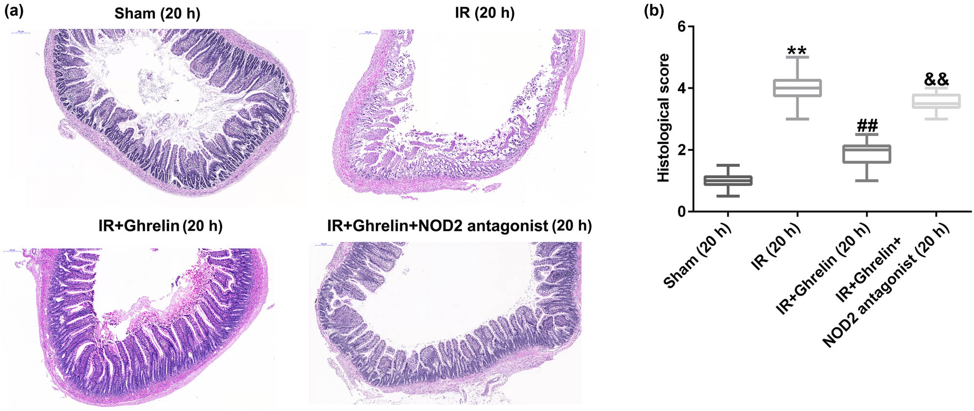
NOD2 antagonist reversed the effects of ghrelin on small bowel tissue injury. Rats were euthanized at either 4 or 20 h after treatment, and their small intestine tissue samples were obtained. (a) Representative intestinal sections from rats of different groups were stained with hematoxylin and eosin. The epithelial structure was clearly demarcated in the sham group. More serious mucosal damage was found in the I/R group than in the sham group. Ghrelin relieved mucosal injury compared to the I/R group. The NOD2 antagonist aggravated mucosal injury in the I/R group. (b) The histological score was determined. Data were expressed as mean ± SD, n = 6; **P < 0.01 vs sham; ## P < 0.01 vs IR; and P < 0.01 vs IR + ghrelin.
3.2 Ghrelin promoted autophagy in the small intestinal epithelial cells of rats with intestinal I/R injury
Next, we investigated whether ghrelin influenced autophagy. Either ghrelin or the NOD2 antagonist was continuously administered after intestinal I/R injury. Intestinal epithelium tissues were obtained 0, 4, or 20 h after the treatment and analyzed using electron microscopy. As shown in Figure 2, the numbers of autophagosomes and autolysosomes increased 4 h and decreased 20 h after I/R. Moreover, ghrelin treatment obviously enhanced the numbers of autophagosomes and autolysosomes compared to those in the sham group (Figures 2 and 3). In addition, the numbers of autophagosomes and autolysosomes were remarkably lower in ghrelin + NOD2 antagonist rats than in ghrelin-treated rats (Figures 2 and 3). Our results revealed that the NOD2 antagonist suppressed the phagocytic functions of ghrelin in the intestinal epithelium of rats with I/R injury.

NOD2 antagonist inhibited the phagocytic functions of ghrelin in the intestinal epithelium of rats. Representative electron microscopy images of the intestinal epithelium. Double arrowheads indicated autophagosomes and single arrowheads indicated autolysosomes containing organelles. n = 6.
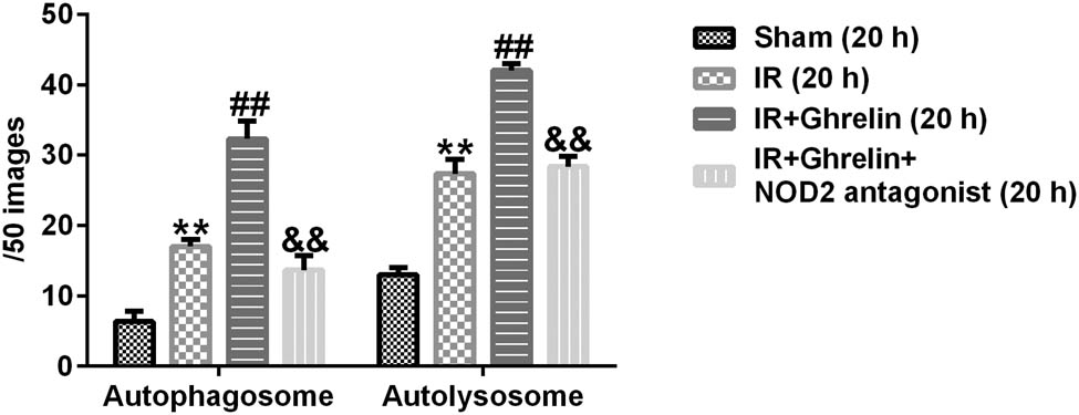
The number of autophagosomes and autolysosomes in the intestinal epithelium of rats. The number of autophagosomes and autolysosomes in the IR group, the ghrelin-treated group, the ghrelin and NOD2 antagonist treatment group, and the sham group at 20 h after I/R. Data were expressed as mean ± SD, n = 6; **P < 0.01 vs. sham; ## P < 0.01 vs. IR; and P < 0.01 vs. IR + ghrelin.
3.3 Ghrelin inhibited inflammatory responses after intestinal I/R injury
Inflammatory responses were involved in the damage of organs after intestinal I/R. To explore whether the cytokines TNF-α and IL-10 were influenced by ghrelin treatment, we determined their levels using ELISA. We observed that the administration of ghrelin obviously inhibited the release of inflammatory factors in IEL cells, which increased in intestinal I/R injury models. However, the opposite results were observed in the ghrelin + NOD2 antagonist group (Figure 4a and b). In addition, we determined TNF-α and IL-10 production in the lungs using ELISA. We observed similar inhibition effects of ghrelin on the expression of the aforementioned cytokines in the pulmonary tissues (Figure 4c and d). Our findings suggested that the NOD2 antagonist reversed the effects of ghrelin on the production of inflammatory factors in IEL cells and lung tissues.

NOD2 antagonist reversed the effects of ghrelin on the release of inflammatory factors in IEL cells and lung tissue. Pro-inflammatory cytokine levels in IEL cells and lung tissue were evaluated using an ELISA. Changes were observed in the levels of IL-10 (a and c) and TNF-α (b and d) in IEL cells and lung tissue in the sham, I/R injury, ghrelin, and ghrelin and NOD2 antagonist groups after reperfusion. Data were expressed as mean ± SD, n = 6; **P < 0.01 vs sham; ## P < 0.01 vs IR; and P < 0.01 vs IR + ghrelin.
3.4 Ghrelin regulated IEL cell autophagy after I/R via the NOD2/Beclin-1 pathway
To further investigate whether ghrelin regulated the autophagy of IEL cells after I/R via the NOD2/Beclin-1 pathway, we determined the expression of autophagy-associated proteins using qRT-PCR and western blot assays. Our results revealed that the levels of NOD2, ATG16, and Beclin-1 were upregulated after the ghrelin treatment, while the levels of NOD2, ATG16, and Beclin-1 were downregulated in the I/R + ghrelin + NOD2 antagonist group due to the action of NOD2 antagonist (Figure 5a‒g). Our findings indicated that ghrelin enhanced the autophagy of IEL cells by activating the NOD2/Beclin-1 pathway.

NOD2 antagonist reversed the effect of ghrelin on autophagy-associated proteins. (a–d) The protein levels of NOD2, Beclin-1, and ATG16 were determined using western blotting. (e–g) Detection of the NOD2, Beclin-1, and ATG16 mRNA expression levels in different groups (sham, I/R injury, ghrelin, or a combination of ghrelin and the NOD2 antagonist) after reperfusion. Data were expressed as mean ± SD, n = 6; **P < 0.01 vs sham; ## P < 0.01 vs IR; and P < 0.01 vs IR + ghrelin.
3.5 NOD2 antagonist reversed the protective effects of ghrelin on lung tissue damage
To further explore the distant organ damage in rats of intestinal I/R injury, we determined the damage to the lung tissues in different groups. The hematoxylin and eosin staining results revealed that ghrelin relieved the degree of lung tissue damage, which was induced by I/R injury, while these protective effects were abolished by the NOD2 antagonist (Figure 6a). Meanwhile, compared with the sham group, the histological score in the IR group significantly enhanced. Compared with the IR group, the histological score in the IR + ghrelin group significantly reduced, while this reduction was significantly increased by NOD2 antagonist (Figure 6b). In summary, our findings verified the protective role of ghrelin in the intestinal I/R injury model by regulating autophagy disorder and inflammatory response via the NOD2/Beclin-1 pathway.

NOD2 antagonist reversed the effects of ghrelin on lung tissue damage. (a) Lung tissue samples from rats of different groups were stained with hematoxylin and eosin. (b) The histological score was determined. Data were expressed as mean ± SD, n = 6; **P < 0.01 vs sham; ## P < 0.01 vs IR; and P < 0.01 vs IR + ghrelin.
4 Discussion
Intestinal I/R injury is a serious, multifactorial complication in various pathophysiological and clinical conditions, such as sepsis, vascular surgery, and absorption dysfunctions [25,26]. Moreover, intestinal I/R injury may lead to autophagy dysfunction and the abnormal activation of IEL. The activation of immune cells, which lie adjacent to the surface area of endothelial cells, may lead to systemic inflammatory responses and severe tissue injury [27]. Previous studies confirmed that intracellular pattern recognition receptor NOD2 is involved in the regulation of autophagy and activation of immune cells, while the role of NOD2/Beclin-1 in intestinal I/R injury remains unknown. This study for the first time investigated the role of NOD2/Beclin-1 in intestinal I/R injury.
Zhang et al. confirmed that the plasma levels of ghrelin are obviously suppressed after intestinal I/R injury [28]. Ghrelin, a gastrointestinal hormone, plays pivotal roles both in the central and peripheral nervous systems [29]. Raghay et al. showed that ghrelin acts as an anti-inflammatory and protective factor in I/R injury [30]. Moreover, Wang et al. revealed that ghrelin protects the heart against I/R injury by inhibiting the TLR4/NLRP3 pathway [31]. However, the regulation of ghrelin in the NOD2/Beclin-1 pathway remains to be explored.
According to previous reports, we established an animal model of intestinal I/R injury in Sprague–Dawley rats using ghrelin or a NOD2 antagonist [32]. The animals were euthanized 4 or 20 h after the treatment, at which point we collected their small intestines, peripheral blood, and lungs. In this study, ghrelin relieved the I/R-induced destruction of the intestinal mucosa epithelium and lung tissues. Electron microscopy revealed that ghrelin enhanced autophagy in the intestinal epithelium of rats with I/R injury. Studies have reported that autophagy is involved in CLP-induced sepsis, and autophagy is upregulated in the early phase but downregulated later [23,33,34]. In this study, the findings for the first time indicated that the autophagy levels in IEL significantly increased at 4 h after intestinal I/R but decreased again at 20 h compared to 4 h after intestinal I/R, suggesting that autophagy might be upregulated in the early phase of intestinal I/R injury but downregulated later.
Our previous study demonstrated that ghrelin not only promotes autophagy-related genes, including Beclin-1 and Atg16, in septic intestinal epithelial cells, but also protects them by promoting autophagy [23]. In addition, ghrelin regulates the NOD2 mRNA levels and improves lung injury in septic rats, suggesting that ghrelin has a potential role in the regulation of NOD2-mediated autophagy [24]. Then, we investigated whether ghrelin can affect autophagy via the NOD2/Beclin-1 pathway and further affect the small intestine. In our study, we checked the levels of autophagy-associated proteins and genes using western blot and qRT-PCR analyses. Our findings suggested that ghrelin upregulated NOD2, Atg16, and Beclin-1 mRNA as well as protein expression levels, while the NOD2 antagonist produced the opposite results. Lucchi et al. revealed that the involvement of the ghrelin receptor antagonist enhanced pro-inflammatory factor release in healthy animals, promoted intestinal barrier dysfunction, and aggravated organ injury [35]. We isolated IEL cells from the small intestine of rats with I/R injury and determined the release of pro-inflammatory cytokines and autophagy-related protein expression in IEL cells. Our results indicated that the TNF-α and IL-10 levels in IELs were obviously enhanced after I/R; this increase may lead to IEL detachment from the epithelium. Meanwhile, the TNF-α and IL-10 levels decreased in the ghrelin-stimulated group, while the TNF-α and IL-10 levels increased in the I/R + ghrelin + NOD2 antagonist group due to the action of NOD2 antagonist. We obtained similar findings in the lung tissues, suggesting that ghrelin reduced excessive inflammation and organ injury through the NOD2/Beclin-1 pathway. Taken together, these findings indicate that ghrelin enhanced IEL autophagy and reduced excessive inflammation and organ injury by activating the NOD2/Beclin-1 pathway. However, this study still has some limitations. For example, how ghrelin acts on NOD2 remains to be further explored. In addition, the significance of the significant decrease in IEL autophagy levels at 20 h after intestinal I/R compared to 4 h after intestinal I/R remains to be explored. We will explore this more deeply in future research.
5 Conclusion
Our findings verified the protective role of ghrelin in an intestinal I/R injury model by regulating organ damage, autophagy disorders, and the inflammatory response via the NOD2/Beclin-1 pathway. These results provide a therapeutic basis for ghrelin in the treatment of intestinal I/R injury.
Abbreviations
- IEL
-
intestinal intraepithelial lymphocytes
- I/R
-
ischemia–reperfusion
-
Funding information: The present study was supported by the Research Project Fund of Shanghai Municipal Health Commission (grant no. 201840134).
-
Conflict of interest: Authors state no conflict of interest.
-
Data availability statement: The datasets used and/or analyzed during the present study are available from the corresponding author on reasonable request.
References
[1] Ito H, Sadatomo A, Inoue Y, Yamada N, Aizawa E, Hishida E, et al. Role of TLR5 in inflammation and tissue damage after intestinal ischemia-reperfusion injury. Biochem Biophys Res Commun. 2019;519(1):15–22.10.1016/j.bbrc.2019.08.083Suche in Google Scholar PubMed
[2] Wu MB, Ma B, Zhang TX, Zhao K, Cui SM, He SC. Propofol improves intestinal ischemia-reperfusion injury in rats through NF-kappaB pathway. Eur Rev Med Pharmacol Sci. 2020;24(11):6463–9.Suche in Google Scholar
[3] Zhang XY, Guan S, Zhang HF, Li RY, Liu ZM. Activation of PD-1 protects intestinal immune defense through IL-10/miR-155 pathway after intestinal ischemia reperfusion. Dig Dis Sci. 2018;63(12):3307–16.10.1007/s10620-018-5282-2Suche in Google Scholar PubMed
[4] Gregová K, Číkoš Š, Bilecová-Rabajdová M, Urban P, Varga J, Feterik Š, et al. Intestinal ischemia-reperfusion injury mediates expression of inflammatory cytokines in rats. Gen Physiol Biophys. 2015;34(1):95–9.10.4149/gpb_2014030Suche in Google Scholar PubMed
[5] Amani H, Habibey R, Hajmiresmail SJ, Latifi S, Pazoki-Toroudi H, Akhavan O. Antioxidant nanomaterials in advanced diagnoses and treatments of ischemia reperfusion injuries. J Mater Chem B. 2017;5(48):9452–76.10.1039/C7TB01689ASuche in Google Scholar
[6] Chichger H, Rounds S, Harrington EO. Endosomes and autophagy: regulators of pulmonary endothelial cell homeostasis in health and disease. Antioxid Redox Signal. 2019;31(13):994–1008.10.1089/ars.2019.7817Suche in Google Scholar PubMed PubMed Central
[7] Arnold J, Murera D, Arbogast F, Muller S, Gros F. [Autophagy in T and B cell homeostasis: recycling for sustainable growth]. Med Sci (Paris). 2016;32(3):281–9.10.1051/medsci/20163203013Suche in Google Scholar PubMed
[8] Shi B, Ma M, Zheng Y, Pan Y, Lin X. mTOR and Beclin1: Two key autophagy-related molecules and their roles in myocardial ischemia/reperfusion injury. J Cell Physiol. 2019;234(8):12562–8.10.1002/jcp.28125Suche in Google Scholar PubMed
[9] Qiu Y, Yu M, Yang Y, Sheng H, Wang W, Sun L, et al. Disturbance of intraepithelial lymphocytes in a murine model of acute intestinal ischemia/reperfusion. J Mol Histol. 2014;45(2):217–27.10.1007/s10735-013-9544-1Suche in Google Scholar PubMed
[10] Minutoli L, Puzzolo D, Rinaldi M, Irrera N, Marini H, Arcoraci V, et al. ROS-mediated NLRP3 inflammasome activation in brain, heart, kidney, and testis ischemia/reperfusion injury. Oxid Med Cell Longev. 2016;2016:2183026.10.1155/2016/2183026Suche in Google Scholar PubMed PubMed Central
[11] Yang Y, Huang J, Li J, Yang H, Yin Y. Effects of stearic acid on proliferation, differentiation, apoptosis, and autophagy in porcine intestinal epithelial cells. Curr Mol Med. 2020;20(2):157–66.10.2174/1566524019666190917144127Suche in Google Scholar PubMed
[12] Ma G, Shi B, Liu J, Zhang H, YinTao Z, Lou X, et al. NOD2-RIP2 signaling contributes to intestinal injury induced by muramyl dipeptide via oligopeptide transporter in rats. Dig Dis Sci. 2015;60(11):3264–70.10.1007/s10620-015-3762-1Suche in Google Scholar PubMed
[13] Wu XJ, Liang H, Zhang Y, Yang XM, Wang HY, Li H, et al. Muramyl dipeptide enhances thermal injury-induced autophagy and inflammatory cytokine response of lungs via activation of NOD2/RICK signaling pathway in rats. Shock. 2018;50(5):606–12.10.1097/SHK.0000000000001077Suche in Google Scholar PubMed
[14] Hu J, Peter I. Evidence of expression variation and allelic imbalance in Crohn’s disease susceptibility genes NOD2 and ATG16L1 in human dendritic cells. Gene. 2013;527(2):496–502.10.1016/j.gene.2013.06.066Suche in Google Scholar PubMed PubMed Central
[15] Wang H, Zhang X, Zuo Z, Zhang Q, Pan Y, Zeng B, et al. RIP2 is required for NOD2-mediated lysozyme sorting in paneth cells. J Immunol. 2017;198(9):3729–36.10.4049/jimmunol.1601583Suche in Google Scholar PubMed
[16] De Salvo C, Buela KA, Creyns B, Corridoni D, Rana N, Wargo HL, et al. NOD2 drives early IL-33-dependent expansion of group 2 innate lymphoid cells during Crohn’s disease-like ileitis. J Clin Invest. 2021;131(5):e140624.10.1172/JCI140624Suche in Google Scholar PubMed PubMed Central
[17] Negroni A, Pierdomenico M, Cucchiara S, Stronati L. NOD2 and inflammation: current insights. J Inflamm Res. 2018;11:49–60.10.2147/JIR.S137606Suche in Google Scholar PubMed PubMed Central
[18] Negroni A, Colantoni E, Vitali R, Palone F, Pierdomenico M, Costanzo M, et al. NOD2 induces autophagy to control AIEC bacteria infectiveness in intestinal epithelial cells. Inflamm Res. 2016;65(10):803–13.10.1007/s00011-016-0964-8Suche in Google Scholar PubMed
[19] Wang H, Dou S, Zhu J, Shao Z, Wang C, Cheng B. Regulatory effects of ghrelin on endoplasmic reticulum stress, oxidative stress, and autophagy: Therapeutic potential. Neuropeptides. 2021;85:102112.10.1016/j.npep.2020.102112Suche in Google Scholar PubMed
[20] Huang CX, Yuan MJ, Huang H, Wu G, Liu Y, Yu SB, et al. Ghrelin inhibits post-infarct myocardial remodeling and improves cardiac function through anti-inflammation effect. Peptides. 2009;30(12):2286–91.10.1016/j.peptides.2009.09.004Suche in Google Scholar PubMed
[21] Nikitopoulou I, Kampisiouli E, Jahaj E, Vassiliou AG, Dimopoulou I, Mastora Z, et al. Ghrelin alterations during experimental and human sepsis. Cytokine. 2020;127:154937.10.1016/j.cyto.2019.154937Suche in Google Scholar PubMed
[22] Kiang JG, Smith JT, Cannon G, Anderson MN, Ho C, Zhai M, et al. Ghrelin, a novel therapy, corrects cytokine and NF-kappaB-AKT-MAPK network and mitigates intestinal injury induced by combined radiation and skin-wound trauma. Cell Biosci. 2020;10:63.10.1186/s13578-020-00425-zSuche in Google Scholar PubMed PubMed Central
[23] Wan SX, Shi B, Lou XL, Liu JQ, Ma GG, Liang DY, et al. Ghrelin protects small intestinal epithelium against sepsis-induced injury by enhancing the autophagy of intestinal epithelial cells. Biomed Pharmacother. 2016;83:1315–20.10.1016/j.biopha.2016.08.048Suche in Google Scholar PubMed
[24] Peng Z, Zhu Y, Zhang Y, Wilhelmsen K, Jia C, Jin J, et al. Effects of ghrelin on pulmonary NOD2 mRNA expression and NF-kappaB activation when protects against acute lung injury in rats challenged with cecal ligation and puncture. Int Immunopharmacol. 2012;13(4):440–5.10.1016/j.intimp.2012.04.006Suche in Google Scholar PubMed
[25] Jia Y, Cui R, Wang C, Feng Y, Li Z, Tong Y, et al. Metformin protects against intestinal ischemia-reperfusion injury and cell pyroptosis via TXNIP-NLRP3-GSDMD pathway. Redox Biol. 2020;32:101534.10.1016/j.redox.2020.101534Suche in Google Scholar PubMed PubMed Central
[26] Chen F, Wang D, Li X, Wang H. Molecular mechanisms underlying intestinal ischemia/reperfusion injury: bioinformatics analysis and in vivo validation. Med Sci Monit. 2020;26:e927476.10.12659/MSM.927476Suche in Google Scholar PubMed PubMed Central
[27] Chen L, Deng H, Cui H, Fang J, Zuo Z, Deng J, et al. Inflammatory responses and inflammation-associated diseases in organs. Oncotarget. 2018;9(6):7204–18.10.18632/oncotarget.23208Suche in Google Scholar PubMed PubMed Central
[28] Zhang H, Cui Z, Luo G, Zhang J, Ma T, Hu N, et al. Ghrelin attenuates intestinal ischemia/reperfusion injury in mice by activating the mTOR signaling pathway. Int j Mol Med. 2013;32(4):851–9.10.3892/ijmm.2013.1452Suche in Google Scholar PubMed
[29] Muta H, Sugita Y, Furuta T, Shiimura Y, Ohshima K, Nakashima K, et al. Expression of the ghrelin/growth hormone secretagogue receptor axis and its functional role in promoting tumor growth in primary central nervous system lymphomas. Neuropathology. 2020;40(3):232–9.10.1111/neup.12634Suche in Google Scholar PubMed
[30] Raghay K, Akki R, Bensaid D, Errami M. Ghrelin as an anti-inflammatory and protective agent in ischemia/reperfusion injury. Peptides. 2020;124:170226.10.1016/j.peptides.2019.170226Suche in Google Scholar PubMed
[31] Wang Q, Lin P, Li P, Feng L, Ren Q, Xie X, et al. Ghrelin protects the heart against ischemia/reperfusion injury via inhibition of TLR4/NLRP3 inflammasome pathway. Life Sci. 2017;186:50–8.10.1016/j.lfs.2017.08.004Suche in Google Scholar PubMed
[32] Chen GL, Xu S, Wu ZJ, Wu Y. [Protective effect of salvianolic acid b on intestinal ischemia-reperfusion injury in rats]. Zhongguo Yi Xue Ke Xue Yuan Xue Bao. 2020;42(1):30–6.Suche in Google Scholar
[33] Hsieh CH, Pai PY, Hsueh HW, Yuan SS, Hsieh YC. Complete induction of autophagy is essential for cardioprotection in sepsis. Ann Surg. 2011;253(6):1190–200.10.1097/SLA.0b013e318214b67eSuche in Google Scholar PubMed
[34] Hsiao HW, Tsai KL, Wang LF, Chen YH, Chiang PC, Chuang SM, et al. The decline of autophagy contributes to proximal tubular dysfunction during sepsis. Shock. 2012;37(3):289–96.10.1097/SHK.0b013e318240b52aSuche in Google Scholar PubMed
[35] Lucchi C, Costa AM, Giordano C, Curia G, Piat M, Leo G, et al. Involvement of PPARgamma in the anticonvulsant activity of EP-80317, a Ghrelin Receptor Antagonist. Front Pharmacol. 2017;8:676.10.3389/fphar.2017.00676Suche in Google Scholar PubMed PubMed Central
© 2022 Meng Jiang et al., published by De Gruyter
This work is licensed under the Creative Commons Attribution 4.0 International License.
Artikel in diesem Heft
- Research Articles
- AMBRA1 attenuates the proliferation of uveal melanoma cells
- A ceRNA network mediated by LINC00475 in papillary thyroid carcinoma
- Differences in complications between hepatitis B-related cirrhosis and alcohol-related cirrhosis
- Effect of gestational diabetes mellitus on lipid profile: A systematic review and meta-analysis
- Long noncoding RNA NR2F1-AS1 stimulates the tumorigenic behavior of non-small cell lung cancer cells by sponging miR-363-3p to increase SOX4
- Promising novel biomarkers and candidate small-molecule drugs for lung adenocarcinoma: Evidence from bioinformatics analysis of high-throughput data
- Plasmapheresis: Is it a potential alternative treatment for chronic urticaria?
- The biomarkers of key miRNAs and gene targets associated with extranodal NK/T-cell lymphoma
- Gene signature to predict prognostic survival of hepatocellular carcinoma
- Effects of miRNA-199a-5p on cell proliferation and apoptosis of uterine leiomyoma by targeting MED12
- Does diabetes affect paraneoplastic thrombocytosis in colorectal cancer?
- Is there any effect on imprinted genes H19, PEG3, and SNRPN during AOA?
- Leptin and PCSK9 concentrations are associated with vascular endothelial cytokines in patients with stable coronary heart disease
- Pericentric inversion of chromosome 6 and male fertility problems
- Staple line reinforcement with nebulized cyanoacrylate glue in laparoscopic sleeve gastrectomy: A propensity score-matched study
- Retrospective analysis of crescent score in clinical prognosis of IgA nephropathy
- Expression of DNM3 is associated with good outcome in colorectal cancer
- Activation of SphK2 contributes to adipocyte-induced EOC cell proliferation
- CRRT influences PICCO measurements in febrile critically ill patients
- SLCO4A1-AS1 mediates pancreatic cancer development via miR-4673/KIF21B axis
- lncRNA ACTA2-AS1 inhibits malignant phenotypes of gastric cancer cells
- circ_AKT3 knockdown suppresses cisplatin resistance in gastric cancer
- Prognostic value of nicotinamide N-methyltransferase in human cancers: Evidence from a meta-analysis and database validation
- GPC2 deficiency inhibits cell growth and metastasis in colon adenocarcinoma
- A pan-cancer analysis of the oncogenic role of Holliday junction recognition protein in human tumors
- Radiation increases COL1A1, COL3A1, and COL1A2 expression in breast cancer
- Association between preventable risk factors and metabolic syndrome
- miR-29c-5p knockdown reduces inflammation and blood–brain barrier disruption by upregulating LRP6
- Cardiac contractility modulation ameliorates myocardial metabolic remodeling in a rabbit model of chronic heart failure through activation of AMPK and PPAR-α pathway
- Quercitrin protects human bronchial epithelial cells from oxidative damage
- Smurf2 suppresses the metastasis of hepatocellular carcinoma via ubiquitin degradation of Smad2
- circRNA_0001679/miR-338-3p/DUSP16 axis aggravates acute lung injury
- Sonoclot’s usefulness in prediction of cardiopulmonary arrest prognosis: A proof of concept study
- Four drug metabolism-related subgroups of pancreatic adenocarcinoma in prognosis, immune infiltration, and gene mutation
- Decreased expression of miR-195 mediated by hypermethylation promotes osteosarcoma
- LMO3 promotes proliferation and metastasis of papillary thyroid carcinoma cells by regulating LIMK1-mediated cofilin and the β-catenin pathway
- Cx43 upregulation in HUVECs under stretch via TGF-β1 and cytoskeletal network
- Evaluation of menstrual irregularities after COVID-19 vaccination: Results of the MECOVAC survey
- Histopathologic findings on removed stomach after sleeve gastrectomy. Do they influence the outcome?
- Analysis of the expression and prognostic value of MT1-MMP, β1-integrin and YAP1 in glioma
- Optimal diagnosis of the skin cancer using a hybrid deep neural network and grasshopper optimization algorithm
- miR-223-3p alleviates TGF-β-induced epithelial-mesenchymal transition and extracellular matrix deposition by targeting SP3 in endometrial epithelial cells
- Clinical value of SIRT1 as a prognostic biomarker in esophageal squamous cell carcinoma, a systematic meta-analysis
- circ_0020123 promotes cell proliferation and migration in lung adenocarcinoma via PDZD8
- miR-22-5p regulates the self-renewal of spermatogonial stem cells by targeting EZH2
- hsa-miR-340-5p inhibits epithelial–mesenchymal transition in endometriosis by targeting MAP3K2 and inactivating MAPK/ERK signaling
- circ_0085296 inhibits the biological functions of trophoblast cells to promote the progression of preeclampsia via the miR-942-5p/THBS2 network
- TCD hemodynamics findings in the subacute phase of anterior circulation stroke patients treated with mechanical thrombectomy
- Development of a risk-stratification scoring system for predicting risk of breast cancer based on non-alcoholic fatty liver disease, non-alcoholic fatty pancreas disease, and uric acid
- Tollip promotes hepatocellular carcinoma progression via PI3K/AKT pathway
- circ_0062491 alleviates periodontitis via the miR-142-5p/IGF1 axis
- Human amniotic fluid as a source of stem cells
- lncRNA NONRATT013819.2 promotes transforming growth factor-β1-induced myofibroblastic transition of hepatic stellate cells by miR24-3p/lox
- NORAD modulates miR-30c-5p-LDHA to protect lung endothelial cells damage
- Idiopathic pulmonary fibrosis telemedicine management during COVID-19 outbreak
- Risk factors for adverse drug reactions associated with clopidogrel therapy
- Serum zinc associated with immunity and inflammatory markers in Covid-19
- The relationship between night shift work and breast cancer incidence: A systematic review and meta-analysis of observational studies
- LncRNA expression in idiopathic achalasia: New insight and preliminary exploration into pathogenesis
- Notoginsenoside R1 alleviates spinal cord injury through the miR-301a/KLF7 axis to activate Wnt/β-catenin pathway
- Moscatilin suppresses the inflammation from macrophages and T cells
- Zoledronate promotes ECM degradation and apoptosis via Wnt/β-catenin
- Epithelial-mesenchymal transition-related genes in coronary artery disease
- The effect evaluation of traditional vaginal surgery and transvaginal mesh surgery for severe pelvic organ prolapse: 5 years follow-up
- Repeated partial splenic artery embolization for hypersplenism improves platelet count
- Low expression of miR-27b in serum exosomes of non-small cell lung cancer facilitates its progression by affecting EGFR
- Exosomal hsa_circ_0000519 modulates the NSCLC cell growth and metastasis via miR-1258/RHOV axis
- miR-455-5p enhances 5-fluorouracil sensitivity in colorectal cancer cells by targeting PIK3R1 and DEPDC1
- The effect of tranexamic acid on the reduction of intraoperative and postoperative blood loss and thromboembolic risk in patients with hip fracture
- Isocitrate dehydrogenase 1 mutation in cholangiocarcinoma impairs tumor progression by sensitizing cells to ferroptosis
- Artemisinin protects against cerebral ischemia and reperfusion injury via inhibiting the NF-κB pathway
- A 16-gene signature associated with homologous recombination deficiency for prognosis prediction in patients with triple-negative breast cancer
- Lidocaine ameliorates chronic constriction injury-induced neuropathic pain through regulating M1/M2 microglia polarization
- MicroRNA 322-5p reduced neuronal inflammation via the TLR4/TRAF6/NF-κB axis in a rat epilepsy model
- miR-1273h-5p suppresses CXCL12 expression and inhibits gastric cancer cell invasion and metastasis
- Clinical characteristics of pneumonia patients of long course of illness infected with SARS-CoV-2
- circRNF20 aggravates the malignancy of retinoblastoma depending on the regulation of miR-132-3p/PAX6 axis
- Linezolid for resistant Gram-positive bacterial infections in children under 12 years: A meta-analysis
- Rack1 regulates pro-inflammatory cytokines by NF-κB in diabetic nephropathy
- Comprehensive analysis of molecular mechanism and a novel prognostic signature based on small nuclear RNA biomarkers in gastric cancer patients
- Smog and risk of maternal and fetal birth outcomes: A retrospective study in Baoding, China
- Let-7i-3p inhibits the cell cycle, proliferation, invasion, and migration of colorectal cancer cells via downregulating CCND1
- β2-Adrenergic receptor expression in subchondral bone of patients with varus knee osteoarthritis
- Possible impact of COVID-19 pandemic and lockdown on suicide behavior among patients in Southeast Serbia
- In vitro antimicrobial activity of ozonated oil in liposome eyedrop against multidrug-resistant bacteria
- Potential biomarkers for inflammatory response in acute lung injury
- A low serum uric acid concentration predicts a poor prognosis in adult patients with candidemia
- Antitumor activity of recombinant oncolytic vaccinia virus with human IL2
- ALKBH5 inhibits TNF-α-induced apoptosis of HUVECs through Bcl-2 pathway
- Risk prediction of cardiovascular disease using machine learning classifiers
- Value of ultrasonography parameters in diagnosing polycystic ovary syndrome
- Bioinformatics analysis reveals three key genes and four survival genes associated with youth-onset NSCLC
- Identification of autophagy-related biomarkers in patients with pulmonary arterial hypertension based on bioinformatics analysis
- Protective effects of glaucocalyxin A on the airway of asthmatic mice
- Overexpression of miR-100-5p inhibits papillary thyroid cancer progression via targeting FZD8
- Bioinformatics-based analysis of SUMOylation-related genes in hepatocellular carcinoma reveals a role of upregulated SAE1 in promoting cell proliferation
- Effectiveness and clinical benefits of new anti-diabetic drugs: A real life experience
- Identification of osteoporosis based on gene biomarkers using support vector machine
- Tanshinone IIA reverses oxaliplatin resistance in colorectal cancer through microRNA-30b-5p/AVEN axis
- miR-212-5p inhibits nasopharyngeal carcinoma progression by targeting METTL3
- Association of ST-T changes with all-cause mortality among patients with peripheral T-cell lymphomas
- LINC00665/miRNAs axis-mediated collagen type XI alpha 1 correlates with immune infiltration and malignant phenotypes in lung adenocarcinoma
- The perinatal factors that influence the excretion of fecal calprotectin in premature-born children
- Effect of femoral head necrosis cystic area on femoral head collapse and stress distribution in femoral head: A clinical and finite element study
- Does the use of 3D-printed cones give a chance to postpone the use of megaprostheses in patients with large bone defects in the knee joint?
- lncRNA HAGLR modulates myocardial ischemia–reperfusion injury in mice through regulating miR-133a-3p/MAPK1 axis
- Protective effect of ghrelin on intestinal I/R injury in rats
- In vivo knee kinematics of an innovative prosthesis design
- Relationship between the height of fibular head and the incidence and severity of knee osteoarthritis
- lncRNA WT1-AS attenuates hypoxia/ischemia-induced neuronal injury during cerebral ischemic stroke via miR-186-5p/XIAP axis
- Correlation of cardiac troponin T and APACHE III score with all-cause in-hospital mortality in critically ill patients with acute pulmonary embolism
- LncRNA LINC01857 reduces metastasis and angiogenesis in breast cancer cells via regulating miR-2052/CENPQ axis
- Endothelial cell-specific molecule 1 (ESM1) promoted by transcription factor SPI1 acts as an oncogene to modulate the malignant phenotype of endometrial cancer
- SELENBP1 inhibits progression of colorectal cancer by suppressing epithelial–mesenchymal transition
- Visfatin is negatively associated with coronary artery lesions in subjects with impaired fasting glucose
- Treatment and outcomes of mechanical complications of acute myocardial infarction during the Covid-19 era: A comparison with the pre-Covid-19 period. A systematic review and meta-analysis
- Neonatal stroke surveillance study protocol in the United Kingdom and Republic of Ireland
- Oncogenic role of TWF2 in human tumors: A pan-cancer analysis
- Mean corpuscular hemoglobin predicts the length of hospital stay independent of severity classification in patients with acute pancreatitis
- Association of gallstone and polymorphisms of UGT1A1*27 and UGT1A1*28 in patients with hepatitis B virus-related liver failure
- TGF-β1 upregulates Sar1a expression and induces procollagen-I secretion in hypertrophic scarring fibroblasts
- Antisense lncRNA PCNA-AS1 promotes esophageal squamous cell carcinoma progression through the miR-2467-3p/PCNA axis
- NK-cell dysfunction of acute myeloid leukemia in relation to the renin–angiotensin system and neurotransmitter genes
- The effect of dilution with glucose and prolonged injection time on dexamethasone-induced perineal irritation – A randomized controlled trial
- miR-146-5p restrains calcification of vascular smooth muscle cells by suppressing TRAF6
- Role of lncRNA MIAT/miR-361-3p/CCAR2 in prostate cancer cells
- lncRNA NORAD promotes lung cancer progression by competitively binding to miR-28-3p with E2F2
- Noninvasive diagnosis of AIH/PBC overlap syndrome based on prediction models
- lncRNA FAM230B is highly expressed in colorectal cancer and suppresses the maturation of miR-1182 to increase cell proliferation
- circ-LIMK1 regulates cisplatin resistance in lung adenocarcinoma by targeting miR-512-5p/HMGA1 axis
- LncRNA SNHG3 promoted cell proliferation, migration, and metastasis of esophageal squamous cell carcinoma via regulating miR-151a-3p/PFN2 axis
- Risk perception and affective state on work exhaustion in obstetrics during the COVID-19 pandemic
- lncRNA-AC130710/miR-129-5p/mGluR1 axis promote migration and invasion by activating PKCα-MAPK signal pathway in melanoma
- SNRPB promotes cell cycle progression in thyroid carcinoma via inhibiting p53
- Xylooligosaccharides and aerobic training regulate metabolism and behavior in rats with streptozotocin-induced type 1 diabetes
- Serpin family A member 1 is an oncogene in glioma and its translation is enhanced by NAD(P)H quinone dehydrogenase 1 through RNA-binding activity
- Silencing of CPSF7 inhibits the proliferation, migration, and invasion of lung adenocarcinoma cells by blocking the AKT/mTOR signaling pathway
- Ultrasound-guided lumbar plexus block versus transversus abdominis plane block for analgesia in children with hip dislocation: A double-blind, randomized trial
- Relationship of plasma MBP and 8-oxo-dG with brain damage in preterm
- Identification of a novel necroptosis-associated miRNA signature for predicting the prognosis in head and neck squamous cell carcinoma
- Delayed femoral vein ligation reduces operative time and blood loss during hip disarticulation in patients with extremity tumors
- The expression of ASAP3 and NOTCH3 and the clinicopathological characteristics of adult glioma patients
- Longitudinal analysis of factors related to Helicobacter pylori infection in Chinese adults
- HOXA10 enhances cell proliferation and suppresses apoptosis in esophageal cancer via activating p38/ERK signaling pathway
- Meta-analysis of early-life antibiotic use and allergic rhinitis
- Marital status and its correlation with age, race, and gender in prognosis of tonsil squamous cell carcinomas
- HPV16 E6E7 up-regulates KIF2A expression by activating JNK/c-Jun signal, is beneficial to migration and invasion of cervical cancer cells
- Amino acid profiles in the tissue and serum of patients with liver cancer
- Pain in critically ill COVID-19 patients: An Italian retrospective study
- Immunohistochemical distribution of Bcl-2 and p53 apoptotic markers in acetamiprid-induced nephrotoxicity
- Estradiol pretreatment in GnRH antagonist protocol for IVF/ICSI treatment
- Long non-coding RNAs LINC00689 inhibits the apoptosis of human nucleus pulposus cells via miR-3127-5p/ATG7 axis-mediated autophagy
- The relationship between oxygen therapy, drug therapy, and COVID-19 mortality
- Monitoring hypertensive disorders in pregnancy to prevent preeclampsia in pregnant women of advanced maternal age: Trial mimicking with retrospective data
- SETD1A promotes the proliferation and glycolysis of nasopharyngeal carcinoma cells by activating the PI3K/Akt pathway
- The role of Shunaoxin pills in the treatment of chronic cerebral hypoperfusion and its main pharmacodynamic components
- TET3 governs malignant behaviors and unfavorable prognosis of esophageal squamous cell carcinoma by activating the PI3K/AKT/GSK3β/β-catenin pathway
- Associations between morphokinetic parameters of temporary-arrest embryos and the clinical prognosis in FET cycles
- Long noncoding RNA WT1-AS regulates trophoblast proliferation, migration, and invasion via the microRNA-186-5p/CADM2 axis
- The incidence of bronchiectasis in chronic obstructive pulmonary disease
- Integrated bioinformatics analysis shows integrin alpha 3 is a prognostic biomarker for pancreatic cancer
- Inhibition of miR-21 improves pulmonary vascular responses in bronchopulmonary dysplasia by targeting the DDAH1/ADMA/NO pathway
- Comparison of hospitalized patients with severe pneumonia caused by COVID-19 and influenza A (H7N9 and H1N1): A retrospective study from a designated hospital
- lncRNA ZFAS1 promotes intervertebral disc degeneration by upregulating AAK1
- Pathological characteristics of liver injury induced by N,N-dimethylformamide: From humans to animal models
- lncRNA ELFN1-AS1 enhances the progression of colon cancer by targeting miR-4270 to upregulate AURKB
- DARS-AS1 modulates cell proliferation and migration of gastric cancer cells by regulating miR-330-3p/NAT10 axis
- Dezocine inhibits cell proliferation, migration, and invasion by targeting CRABP2 in ovarian cancer
- MGST1 alleviates the oxidative stress of trophoblast cells induced by hypoxia/reoxygenation and promotes cell proliferation, migration, and invasion by activating the PI3K/AKT/mTOR pathway
- Bifidobacterium lactis Probio-M8 ameliorated the symptoms of type 2 diabetes mellitus mice by changing ileum FXR-CYP7A1
- circRNA DENND1B inhibits tumorigenicity of clear cell renal cell carcinoma via miR-122-5p/TIMP2 axis
- EphA3 targeted by miR-3666 contributes to melanoma malignancy via activating ERK1/2 and p38 MAPK pathways
- Pacemakers and methylprednisolone pulse therapy in immune-related myocarditis concomitant with complete heart block
- miRNA-130a-3p targets sphingosine-1-phosphate receptor 1 to activate the microglial and astrocytes and to promote neural injury under the high glucose condition
- Review Articles
- Current management of cancer pain in Italy: Expert opinion paper
- Hearing loss and brain disorders: A review of multiple pathologies
- The rationale for using low-molecular weight heparin in the therapy of symptomatic COVID-19 patients
- Amyotrophic lateral sclerosis and delayed onset muscle soreness in light of the impaired blink and stretch reflexes – watch out for Piezo2
- Interleukin-35 in autoimmune dermatoses: Current concepts
- Recent discoveries in microbiota dysbiosis, cholangiocytic factors, and models for studying the pathogenesis of primary sclerosing cholangitis
- Advantages of ketamine in pediatric anesthesia
- Congenital adrenal hyperplasia. Role of dentist in early diagnosis
- Migraine management: Non-pharmacological points for patients and health care professionals
- Atherogenic index of plasma and coronary artery disease: A systematic review
- Physiological and modulatory role of thioredoxins in the cellular function
- Case Reports
- Intrauterine Bakri balloon tamponade plus cervical cerclage for the prevention and treatment of postpartum haemorrhage in late pregnancy complicated with acute aortic dissection: Case series
- A case of successful pembrolizumab monotherapy in a patient with advanced lung adenocarcinoma: Use of multiple biomarkers in combination for clinical practice
- Unusual neurological manifestations of bilateral medial medullary infarction: A case report
- Atypical symptoms of malignant hyperthermia: A rare causative mutation in the RYR1 gene
- A case report of dermatomyositis with the missed diagnosis of non-small cell lung cancer and concurrence of pulmonary tuberculosis
- A rare case of endometrial polyp complicated with uterine inversion: A case report and clinical management
- Spontaneous rupturing of splenic artery aneurysm: Another reason for fatal syncope and shock (Case report and literature review)
- Fungal infection mimicking COVID-19 infection – A case report
- Concurrent aspergillosis and cystic pulmonary metastases in a patient with tongue squamous cell carcinoma
- Paraganglioma-induced inverted takotsubo-like cardiomyopathy leading to cardiogenic shock successfully treated with extracorporeal membrane oxygenation
- Lineage switch from lymphoma to myeloid neoplasms: First case series from a single institution
- Trismus during tracheal extubation as a complication of general anaesthesia – A case report
- Simultaneous treatment of a pubovesical fistula and lymph node metastasis secondary to multimodal treatment for prostate cancer: Case report and review of the literature
- Two case reports of skin vasculitis following the COVID-19 immunization
- Ureteroiliac fistula after oncological surgery: Case report and review of the literature
- Synchronous triple primary malignant tumours in the bladder, prostate, and lung harbouring TP53 and MEK1 mutations accompanied with severe cardiovascular diseases: A case report
- Huge mucinous cystic neoplasms with adhesion to the left colon: A case report and literature review
- Commentary
- Commentary on “Clinicopathological features of programmed cell death-ligand 1 expression in patients with oral squamous cell carcinoma”
- Rapid Communication
- COVID-19 fear, post-traumatic stress, growth, and the role of resilience
- Erratum
- Erratum to “Tollip promotes hepatocellular carcinoma progression via PI3K/AKT pathway”
- Erratum to “Effect of femoral head necrosis cystic area on femoral head collapse and stress distribution in femoral head: A clinical and finite element study”
- Erratum to “lncRNA NORAD promotes lung cancer progression by competitively binding to miR-28-3p with E2F2”
- Retraction
- Expression and role of ABIN1 in sepsis: In vitro and in vivo studies
- Retraction to “miR-519d downregulates LEP expression to inhibit preeclampsia development”
- Special Issue Computational Intelligence Methodologies Meets Recurrent Cancers - Part II
- Usefulness of close surveillance for rectal cancer patients after neoadjuvant chemoradiotherapy