Artemisinin protects against cerebral ischemia and reperfusion injury via inhibiting the NF-κB pathway
-


Abstract
This study investigated whether artemisinin (ART) exerts a neuroprotective effect against cerebral ischemia/reperfusion (I/R) injury. Hypoxia-glucose deprivation and reoxygenation (OGD/R) of SH-SY5Y cells were used as the I/R injury model in vitro. Cell viability was determined using 3-(4,5-dimethylthiazol-2-yl)-2,5-diphenyltetrazolium bromide assay, and lactate dehydrogenase (LDH) release was measured. Cell apoptosis and apoptosis-associated protein expression were determined via flow cytometry and western blotting, respectively. The levels of glutathione peroxidase, superoxide dismutase, catalase, and malondialdehyde were determined. The secretion of tumor necrosis factor-α and interleukin-1β was measured using ELISA. The activation of the nuclear factor kappa B (NF-κB) pathway was also determined. The indicated ART concentrations (0, 25, 50, 75, and 100 μM) had no significant effect on SH-SY5Y cell viability and LDH activity. ART promoted cell viability, reduced cell apoptosis, repressed cellular inflammation, and inhibited cellular oxidative stress and NF-κB signaling pathway in OGD/R-induced SH-SY5Y cells. In addition, all the protective effects of ART on OGD/R-induced SH-SY5Y cell injury were significantly reversed by an NF-κB agonist. In conclusion, ART protects neurons from OGD/R-induced damage in vitro by inhibiting the NF-κB signaling pathway. These results suggest that ART may be a potential agent for the treatment of cerebral I/R injury.
1 Introduction
Artemisia annua L. has been used as a medicinal plant in the treatment of numerous diseases for centuries [1]. Artemisinin (ART) is a sesquiterpene trioxane lactone compound extracted from Artemisia annua L., which contains a peroxide group. ART can kill malaria parasites by interfering with mitochondrial function, particularly in the treatment of drug-resistant and recurrent malaria, and has been used worldwide [2,3]. In addition to anti-malarial effect, ART has immunosuppressive [4], anti-schistosomiasis [5], anti-virus [6], and anti-tumor effects [7,8,9,10]. In multiple cancers, combined treatment with ART drugs has achieved improved therapeutic effects [11], and no evident cytotoxicity of ART to normal cells was observed in more than 4,000 cases, which makes it a potential drug for treating numerous diseases [12]. ART has been reported to be a potential drug for treating Alzheimer’s disease by protecting against amyloid beta damage [13]. Studies have revealed that ART is involved in regulating the expression of various nuclear factor kappa B (NF-κB) reporter genes [14,15]. ART inhibits endometrial cancer cell proliferation by disrupting the interaction between NF-κB and the CDK4 promoter and transcriptionally downregulates CDK4 expression [9]. Gu et al. indicated that ART could inhibit the NF-κB pathway by blocking IKBα phosphorylation, which results in reduced myocardial remodeling [16]. Wang et al. found that ART pretreatment effectively protected against myocardial ischemia/reperfusion (I/R) injury by inhibiting the activation of the NLRP3 inflammasome [17]. Besides, ART attenuated the oxidative damage of SH-SY5Y cells and hippocampal neurons induced by hydrogen peroxide (H2O2) by activating the AMPK signaling pathway, suggesting a neuroprotective effect of ART [18]. However, whether ART has a neuroprotective effect on cerebral I/R injury remains unclear.
Ischemic stroke remains the primary cause of disability and death worldwide [19]. The pathogenesis of cerebral ischemia is insufficient oxygen and glucose transport caused by tissue ischemia, which leads to irreversible neuronal damage or death [20]. Recanalization therapy, which supplements nutrients and oxygen and removes toxic metabolites, is currently the main treatment method of ischemic stroke [21,22]. However, the recovery of blood flow leads to occasional side effects [23]. In the process of cerebral I/R, various procedures related to nerve cell death are activated, such as necrosis, apoptosis, or autophagy [24,25], and apoptosis has been reported as a key event in cerebral ischemic brain injury [26]. Astragaloside IV protects against cerebral I/R injury by suppressing apoptosis [27]. The inhibition of oxidative stress and inflammation has been widely reported to alleviate cerebral I/R injury [28,29].
In this study, we hypothesized that ART plays a protective role in cerebral ischemia and reperfusion injury by inhibiting NF-κB pathway. Therefore, this study employed the SH-SY5Y cell OGD/R model to explore the effect of ART on cerebral I/R injury and analyze its potential molecular mechanism.
2 Material and methods
2.1 Cell culture and reagent
The human neuroblastoma cell line SH-SY5Y was obtained from the American Type Culture Collection (Rockville, MD, USA) and was cultured in Dulbecco’s Modified Eagle Medium (DMEM; Sigma-Aldrich, St. Louis, MO, USA) containing 10% (v/v) fetal bovine serum (Thermo Fisher Scientific, Waltham, MA, USA), 2% l-glutamine (v/v), 100 μg/mL streptomycin, and 100 U/mL penicillin (Gibco, Amarillo, TX, USA). The cells were cultured in a moist environment at 37°C and 5% CO2. ART was purchased from Merck (Darmstadt, Germany; cat. no. 361593).
2.2 Cell treatment
OGD/R-exposed model [30]: SH-SY5Y cells were seeded into 96-well plates, and then OGD experiments were performed when cell density reached approximately 80% confluence. SH-SY5Y cell culture medium was replaced with oxygen-glucose-free DMEM and incubated in an anaerobic gas mixture (1% O2, 5% CO2, and 94% N2) at 37°C. After 3 h, the cell medium and culture conditions were restored to normal, and the culture was continued for 24 h.
ART interferes with the OGD/R-exposed model: SH-SY5Y cells were pre-treated with the indicated concentration of ART (0, 25, 50, and 100 μM) [31] for 2 h and then exposed to OGD/R.
2.3 Cell proliferation ability
The proliferation ability of SH-SY5Y cells was determined using the MTT assay. The processed cells were seeded into 96-well plates at a density of 6 × 10³ cells per well and cultured in 5% CO2 at 37°C for 48 h. Subsequently, 20 μL of MTT solution (5 mg/mL) was added to each well and cultured for 4 h. Then, the supernatant was carefully removed, and 150 μL of dimethyl sulfoxide was added to each well at room temperature to completely dissolve the formazan crystals. The absorbance of each well was measured at 570 nm using an enzyme-linked immunosorbent analyzer (Victor X3, PerkinElmer, Shelton, CT, USA).
2.4 LDH release cell death assay
An LDH assay kit (Promega, Madison, WI, USA) was used to detect the release of LDH from cells. Briefly, 2 × 10⁶ processed cells were collected, washed with pre-cold phosphate-buffered saline (PBS), resuspended in 200 μL of cold assay buffer, and centrifuged at 4°C at 8,000 rpm for 15 min, and the supernatant was preserved. Next 10 μL of supernatant and 100 μL of reaction reagent were mixed and incubated for 30 min at room temperature. The absorbance of each well was determined using an enzyme-linked immunosorbent analyzer (Victor X3, PerkinElmer) at 490 nm.
2.5 Flow cytometry analysis of cell apoptosis
SH-SY5Y cell apoptosis was detected using the Annexin V-FITC Assay Kit (BioVision, Palo Alto, CA, USA) according to the manufacturer’s instructions. Briefly, the processed SH-SY5Y cells were collected and washed with pre-cold PBS. SH-SY5Y cells were resuspended in 195 μL of binding buffer, and then stained with 5 μL of Annexin V-FITC (10 μg/mL) and 10 μL of propidium iodide for 15 min in the dark at 25°C. Finally, the cells were analyzed via flow cytometry (FCM; Beckman, Brea, CA, USA). Data were analyzed using FlowJo version 7.6.1 (FlowJo LLC).
2.6 Caspase-3 activity detection
Caspase-3 activity was detected using a Caspase-3 Activity Assay Kit (Abcam, Cambridge, UK; cat no. ab252897). The detection principle is that the synthetic substrate DEVD-AFC will emit a strong and stable fluorescence signal (Ex/Em = 400/505 nm) after being cleaved by caspase-3, and caspase-3 enzyme activity can be reflected according to the fluorescence intensity. The processed cells were collected and lysed with RIPA lysis buffer (Merck; cat no. R0278), and the supernatant was collected. The supernatant was then incubated with the final reaction solution, which comprised 40 μL of assay buffer, 50 μL of supernatant, and 10 μL of caspase-3 substrate DEVD-AFC (2 mM) at 37°C for 2 h. Finally, the absorbance of each sample was measured at 400 nm using an enzyme-linked immunosorbent analyzer (Victor X3, PerkinElmer,).
2.7 Western blot analysis
SH-SY5Y cells were washed with PBS and lysed with RIPA lysis buffer (Merck; cat. no. R0278). The supernatant with 20 μg total protein content was separated via polyacrylamide gel electrophoresis and electro-transferred to a nitrocellulose membrane (Millipore, Burlington, MA, USA). The membranes were incubated with specific primary antibodies (1:200 for anti-cleaved caspase-3, cat. no. ab2302; 1:1,000 for anti-p65, cat. no. ab32536; 1:500 for anti-p-p65, cat. no. ab31624; 1:1,000 for anti-GAPDH, cat. no. ab22555; all from Abcam), and secondary antibodies (1:5,000, cat. no. ab97080; Abcam). The enhanced chemiluminescence method (Cytiva, Marlborough, MA, USA) was used to detect immune-response bands. The band intensity was quantified by ImageJ version 1.8.0 (National Institutes of Health).
2.8 Determination of superoxide dismutase (SOD), catalase (CAT), and glutathione peroxidase (GSH-Px) activities
Superoxide dismutase activity assay kit (Solarbio, Beijing, China; cat. no. BC0170) was used to determine the activity of SOD, catalase detection kit (Solarbio; cat. no. BC0200) was used to determine the activity of CAT, and glutathione peroxidase activity assay kit (Solarbio; cat. no. BC1190) was used to determine GSH-Px activity. In the SOD determination system, xanthine and xanthine oxidase reaction can produce O2−, which reduces nitroblue tetrazolium to formazan, and SOD reduces formazan formation by reacting with O2−. Formazan can be dissolved in organic solvents, and absorbance at 560 nm indicates the SOD activity. CAT can decompose H2O2, which has a characteristic absorption peak at 240 nm. The absorbance of the reaction solution at 240 nm decreased with the reaction time, and the CAT activity was calculated according to the change rate of the absorbance. GSH-Px catalyzes the oxidation of GSH by H2O2 to produce oxidized glutathione. GSH reacts with 5,5’-dithiobis-(2-nitrobenzoic acid) to generate a compound with a characteristic absorption at 412 nm. Therefore, the absorbance of the reaction solution at 412 nm was used to measure the activity of GSH-Px.
2.9 Malondialdehyde (MDA) detection
A Malondialdehyde Assay Kit (Abcam; cat. no. ab118970) was used to detect MDA. MDA in the sample reacts with thiobarbituric acid (TBA) to form an MDA–TBA complex. The MDA–TBA complex had a specific absorption at 532 nm. Therefore, MDA was quantified according to the absorbance of the reaction solution at 532 nm.
2.10 Enzyme-linked immunosorbent assay (ELISA)
The secretion of tumor necrosis factor-α (TNF-α), interleukin-1β (IL-1β), and IL-6 in SH-SY5Y cells was detected using an ELISA kit (cat. no. ab181421 for TNF-α; cat. no. ab214025 for IL-1β; cat. no. ab178013 for IL-6; all from Abcam). The cell culture supernatant was collected by centrifugation at 2,000 rpm for 15 min. Afterward, 50 μL of cell culture supernatant and 50 μL of antibody cocktail were added to the 96-well plate included in the kit, incubated in a shaker at 400 rpm for 90 min at room temperature, and then washed three times with 350 μL of wash buffer. Subsequently, 100 μL TMB buffer was added to the system and incubated in a shaker at 400 rpm for 10 min. After incubation, the reaction was stopped with 100 μL of stopping solution. Absorbance was measured at 450 nm wavelength.
2.11 Reverse transcription quantitative-PCR (RT-qPCR)
The p65 transcription level was determined via RT-qPCR. Total RNA was extracted using the MiniBEST Universal RNA Extraction Kit (TaKaRa, cat. no. 9767), according to the manufacturer’s instructions. A One Step PrimeScript III RT-qPCR kit (TaKaRa, cat. no. RR600A) was used for the RT-qPCR analysis. The reaction solution was prepared according to the manufacturer’s instructions and applied in a Thermal Cycler Dice Real Time System. GAPDH was used as an internal control. The PCR primer sequence of p65 and GAPDH was as follows: p65 forward, 5′-CGCGGATCCGCCACCATGGACGAACTG-3′ and reverse, 5′-CCGCTCGAGTTAGGAGCTGATCTG-3′; GAPDH forward, 5′-CTTTGGTATCGTGGAAGGACTC-3′ and reverse, 5′-GTAGAGGCAGGGATGATGTTCT-3′. The relative expression of p65 was calculated using the 2−ΔΔCq method.
2.12 Statistical analysis
All experiments were performed at least three times, and the data are presented as mean value ± SD. GraphPad Prism software (GraphPad Software, Inc., San Diego, CA, USA) was used for the statistical analysis. The statistical significance of the differences between the two groups was tested using Student’s t-test. Multiple comparisons were performed using the one-way analysis of variance (ANOVA) followed by Tukey’s post hoc test. Differences were considered statistically significant at p < 0.05.
3 Results
3.1 Cytotoxicity of ART to SH-SY5Y cells
SH-SY5Y cells were treated with ART at different concentrations (25, 50, 75, and 100 μM) for 24 h. Different ART concentrations had no significant effect on SH-SY5Y cell viability and LDH viability (Figure 1a and b), suggesting that ART has no significant cytotoxic effect on SH-SY5Y cells. Therefore, 0, 25, 50, and 100 μM ART were used for subsequent experiments.
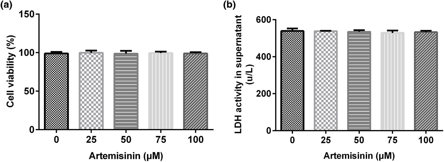
Cytotoxic effect of ART on SH-SY5Y cells. SH-SY5Y cells were treated with ART at different concentrations (0, 25, 50,75, and 100 μM) for 24 h. Different ART concentrations had no significant effect on SH-SY5Y: (a) cell viability and (b) lactate dehydrogenase activity.
3.1.1 ART protects SH-SY5Y cells from OGD/R-induced injuries
SH-SY5Y cells were pre-treated with the indicated concentrations of ART (25, 50, and 100 μM) for 2 h and then exposed to OGD/R. The results of the MTT and LDH release cell death assays indicated that OGD/R-exposure significantly repressed the viability of SH-SY5Y cells (Figure 2a) and enhanced LDH activity (Figure 2b). FCM results showed that OGD/R treatment markedly increased SH-SY5Y cell apoptosis (Figure 2c and d), and caspase-3 activity in OGD/R-injured SH-SY5Y cells was significantly increased (Figure 2e). Western blot analysis revealed that the cleaved caspase-3 protein expression and ratio of cleaved caspase-3/GAPDH were apparently increased in OGD/R-injured SH-SY5Y cells (Figure 2f and g). All the effects could be reversed by ART treatment. This reversal was dependent on ART concentration, and the reversal effect was positively correlated with ART concentration. These results indicated that ART increased the viability and reduced the apoptosis of OGD/R-exposed SH-SY5Y cells in a dose-dependent manner.

ART increases the viability and reduces apoptosis of hypoxia-glucose deprivation and reoxygenation (OGD/R)-exposed SH-SY5Y cells in a dose-dependent manner. SH-SY5Y cells were pre-treated with the indicated concentrations of ART (0, 25, 50, and 100 μM) for 2 h and then exposed to OGD/R. (a) Viability of SH-SY5Y cells was determined using an MTT assay. (b) LDH activity was measured using an LDH assay kit. (c and d) Cell apoptosis was detected via flow cytometry. (e) Caspase-3 activity was measured using a caspase-3 activity detection kit. (f) Cleaved caspase-3 protein detection was detected using western blotting. (g) Cleaved caspase-3/GAPDH ratio. **p < 0.01 vs Control; #,## p < 0.05, 0.01 vs OGD/R.
The secretion of TNF-α, IL-1β, and IL-6 in SH-SY5Y cells was detected using ELISA. The results indicated that the secretion of TNF-α, IL-1β, and IL-6 was increased in OGD/R-exposed SH-SY5Y cells (Figure 3a–c), and ART decreased the secretion of TNF-α, IL-1β, and IL-6 in a dose-dependent manner in OGD/R-exposed SH-SY5Y cells (Figure 3a–c).

Effects of ART on cell inflammation in hypoxia-glucose deprivation and reoxygenation (OGD/R)-induced SH-SY5Y cells. SH-SY5Y cells were pre-treated with the indicated concentrations of ART (0, 25, 50, and 100 μM) for 2 h and then exposed to OGD/R. (a–c) ELISA was used to determine the secretion of TNF-α, IL-1β, and IL-6 in the supernatant of SH-SY5Y cells. **p < 0.01 vs Control; #,## p < 0.05, 0.01 vs OGD/R.
The activities of SOD, CAT, and GSH-Px in SH-SY5Y cells were significantly inhibited, and the level of malondialdehyde (MAD) was significantly increased by OGD/R exposure (Figure 4a–d). In OGD/R-exposed SH-SY5Y cells, ART treatment alleviated the inhibition of SOD, CAT, and GSH-Px activities and increased MAD levels in a dose-dependent manner (Figure 4a–d).

Effects of ART on the oxidative stress in OGD/R-induced SH-SY5Y cells. SH-SY5Y cells were pre-treated with the indicated concentrations of ART (0, 25, 50, and 100 μM) for 2 h and then exposed to OGD/R. Next the activities of SOD (a), CAT (b), and GSH-Px (c) and the level of MAD (d) were determined. **p < 0.01 vs Control; #,## p < 0.05, 0.01 vs OGD/R.
Western blotting was used to assess the expression of p65 and p-p65, and RT-qPCR was performed to analyze the transcription level of p65. The results indicated that p-p65 expression and the ratio of p-p65/p65 were notably increased, and ART treatment reduced p-p65 expression and the ratio of p-p65/p65 in OGD/R-exposed SH-SY5Y cells in a dose-dependent manner (Figure 5a and b). There was no significant change in p65 transcription levels among the groups (Figure 5c).

ART inhibits the activation of NF-κB signaling pathway in OGD/R-induced SH-SY5Y cells. SH-SY5Y cells were pre-treated with the indicated concentrations of ART (0, 25, 50, and 100 μM) for 2 h and then exposed to OGD/R. (a) Protein expressions of p-p65 and p65 were measured using western blotting. (b) p-p65/p65 ratio. (c) mRNA expression of p65 was determined via RT-qPCR. **p < 0.01 vs Control; #,## p < 0.05, 0.01 vs OGD/R.
3.1.2 ART protects SH-SY5Y cells from OGD/R-induced injury by inhibiting the NF-κB signaling pathway
Findings indicated that OGD/R-exposed SH-SY5Y cell viability was significantly improved and LDH activity was reduced by treatment with ART or an NF-κB agonist (Figure 6a and b). The NF-κB agonist reversed the ART-induced increase in cell viability in OGD/R-exposed SH-SY5Y cells (Figure 6a and b). Apoptosis was distinctly decreased by treatment with ART or an NF-κB agonist, whereas apoptosis was distinctly increased by co-treatment with ART and an NF-κB agonist in OGD/R-exposed SH-SY5Y cells compared with that of ART treatment alone (Figure 6c and d). Caspase-3 activity in OGD/R-exposed SH-SY5Y cells was markedly inhibited by treatment with ART or an NF-κB agonist; however, co-treatment with ART and the NF-κB agonist markedly improved caspase-3 activity in OGD/R-exposed SH-SY5Y cells compared with that of ART treatment alone (Figure 6e). Cleaved caspase-3 protein expression and the ratio of cleaved caspase-3/GAPDH were significantly decreased by treatment with ART or an NF-κB agonist. Compared with that of the ART treatment group, cleaved caspase-3 expression and the ratio of cleaved caspase-3/GAPDH were significantly increased by co-treatment with ART and NF-κB agonist in OGD/R-exposed SH-SY5Y cells (Figure 6f and g).

NF-κB agonist reverses the effects of ART on the viability and apoptosis of SH-SY5Y cells triggered by OGD/R exposure. SH-SY5Y cells were pre-treated with 100 μM ART, an NF-κB antagonist, or 100 μM ART + NF-κB agonist for 2 h and then exposed to OGD/R. (a) SH-SY5Y cell viability was determined using an MTT assay. (b) LDH activity was measured using an LDH assay kit. (c and d) Cell apoptosis was detected via flow cytometry (e) Caspase-3 activity was measured using a caspase-3 activity detection kit. (f) Protein expression of cleaved caspase-3 was detected using western blotting. (g) Cleaved caspase-3/GAPDH ratio. **p < 0.01 vs OGD/R; ## p < 0.05, 0.01 vs OGD/R + ART.
The secretion of TNF-α, IL-1β, and IL-6 was evidently decreased in OGD/R-exposed SH-SY5Y cells by treatment with ART or an NF-κB agonist, and co-treatment with ART and the NF-κB agonist evidently increased the secretion of TNF-α, IL-1β, and IL-6 compared with that of ART treatment alone (Figure 7a–c). In OGD/R-exposed SH-SY5Y cells, the activities of SOD, CAT, and GSH-Px were markedly inhibited by treatment with ART or an NF-κB agonist, whereas the inhibitory effect was attenuated by co-treatment with ART and the NF-κB agonist (Figure 7d–f). ART or NF-κB agonist treatment significantly decreased the level of MAD in OGD/R-exposed SH-SY5Y cells; however, the level of MAD was significantly increased by co-treatment with ART or an NF-κB agonist (Figure 7g).

NF-κB agonist reverses the effects of ART on inflammation and oxidative stress of SH-SY5Y cells triggered by OGD/R exposure. SH-SY5Y cells were pre-treated with 100 μM ART, an NF-κB antagonist, or 100 μM ART + NF-κB agonist for 2 h and then exposed to OGD/R. (a–c) ELISA was used to determine the secretion of TNF-α, IL-1β, and IL-6 in the supernatant of SH-SY5Y cells. (d–g) Activities of SOD, CAT, and GSH-Px and the level of MAD were determined. **p < 0.01 vs OGD/R; ## p < 0.05, 0.01 vs OGD/R + ART.
The findings suggested that p-p65 expression and the ratio of p-p65/p65 were significantly inhibited in OGD/R-injured SH-SY5Y cells after treatment with ART or the NF-κB agonist; however, the inhibitory effect of ART on OGD/R-injured SH-SY5Y cells was reversed by co-treatment with ART and the NF-κB agonist (Figure 8a and b). The transcription level of p65 did not differ among the groups (Figure 8c). ART attenuates OGD/R injury by inhibiting the NF-κB signaling pathway.
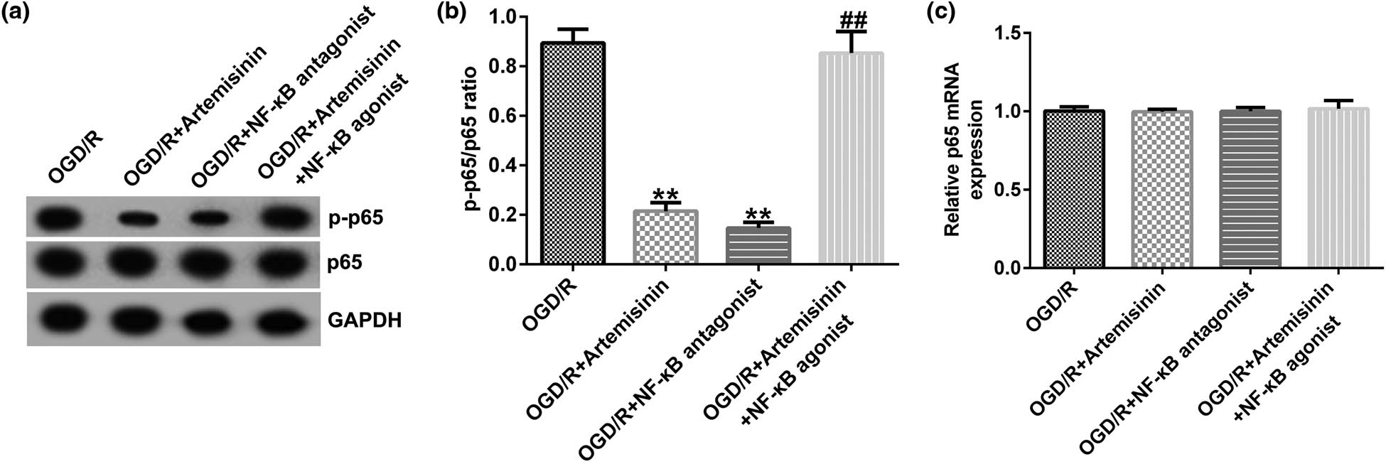
NF-κB agonist reverses the inhibitory effect of ART on the NF-κB signaling pathway in OGD/R-exposed SH-SY5Y cells. SH-SY5Y cells were pre-treated with 100 μM ART, an NF-κB antagonist, or 100 μM ART + NF-κB agonist for 2 h and then exposed to OGD/R. (a) Protein expressions of p-p65 and p65 were measured using western blotting. (b) p-p65/p65 ratio. (c) mRNA expression of p65 was determined via RT-qPCR. **p < 0.01 vs OGD/R; ## p < 0.05, 0.01 vs OGD/R + ART.
4 Discussion
Ischemic strokes cause severe disability and death, accounting for approximately 87% of all stroke cases [19,32]. Extensive studies have revealed that strokes cause complex cellular biochemical events that ultimately result in necrosis, apoptosis, or autophagy in ischemic areas [24,25,26]. A comprehensive understanding of neuronal death during ischemic brain injury facilitates the development of new therapies. The pathogenesis of ischemic stroke includes excitatory toxicity, oxidative stress, inflammatory responses, and apoptosis [33]. There is increasing evidence that ART derivatives participate in I/R injury. In this study, we investigated the effects of ART on OGD/R-exposed SH-SY5Y cells. The results indicated that ART increased the viability and decreased the apoptosis of SH-SY5Y cells exposed to OGD/R in a dose-dependent manner.
The inflammatory response caused by leukocyte infiltration after cerebral ischemia plays an important role in the occurrence and development of cerebral I/R injury [34]. The inflammatory cascade after cerebral ischemia is a dynamic process involving the interaction of various cells in the ischemic area, which can cause a second injury after cerebral ischemia. The damaged brain cells produce a large number of platelet activating factors, TNF, IL-6, and other inflammatory mediators after cerebral ischemia [35,36]. The regulatory effect of ART on pro-inflammatory cytokine expression has been widely reported. In LL37-induced rosacea-like mice, ART and its derivatives significantly inhibited the expression of pro-inflammatory factors (IL-1β, IL-6, and TNFα) and TLR2 [37]. In OGD/R-exposed SH-SY5Y cells, our results indicated that the secretion of inflammatory cytokines TNF-α, IL-1β, and IL-6 was significantly increased, and ART treatment inhibited the OGD/R-induced SH-SY5Y cell inflammatory response. The secretion of TNF-α, IL-1β, and IL-6 was inhibited by ART treatment in a dose-dependent manner.
Oxidative stress mediated by ROS is another important factor associated with cerebral I/R injury dysfunction. Antioxidant therapy helps to reduce neuronal variability and improve neurological prognosis [38,39]. Thus, we examined oxidative stress in SH-SY5Y cells following exposure to OGD/R. The levels of free radicals are difficult to measure because of their relatively short half-lives. Therefore, the activities of antioxidant enzymes, such as SOD, CAT, and GSH-Px, and lipid peroxidation by-products, such as MDA, can be used to indirectly evaluate the level of free radicals [40]. In OGD/R-induced SH-SY5Y cells, the antioxidant enzyme (SOD, CAT, and GSH-Px) activities were significantly decreased, and the MAD level was significantly increased. ART contains an endoperoxide bridge, which makes it an antioxidant. The mechanism of killing malaria parasites is related to the production of destructive ROS induced by ART [41]. Our results revealed that ART treatment could alleviate the inhibition of SOD, CAT, and GSH-Px activities and decrease MAD levels induced by OGD/R exposure in a dose-dependent manner.
NF-κB activation is involved in various cellular processes, including the regulation of cell survival, apoptosis, inflammation, and oxidative stress [42,43]. Moreover, NF-κB plays an important role in I/R injury [9,14,16,44]. NF-κB pathway is activated in cerebral I/R [45,46]. After cerebral I/R, NF-κB downregulation can alleviate cerebral edema and neurological dysfunction, and the NF-κB signaling pathway plays an important role in ventricular remodeling after myocardial infarction [16,47,48]. Matsui et al. found that an NF-κB inhibitor could reduce I/R injury in animal experiments [49]. In our study, p-p65 expression and the p-p65/p65 ratio in OGD/R-exposed SH SY5Y cells were significantly increased, and ART treatment could reduce p-p65 expression and the p-p65/p65 ratio in a dose-dependent manner. NF-κB agonist treatment could significantly increase the cell viability, decrease the apoptosis, and inhibit the inflammatory response and oxidative stress of OGD/R-exposed SH-SY5Y cells, suggesting that NF-κB is involved in the regulation of OGD/R-induced cell proliferation, apoptosis, inflammation, and oxidative stress. The NF-κB agonist and ART have similar effects on OGD/R-exposed SH-SY5Y cells. We then investigated whether the regulation of ART in OGD/R-exposed SH SY5Y cells is related to NF-κB pathway. As expected, all the effects of ART on OGD/R-exposed SH SY5Y cells could be reversed by co-treatment with an NF-κB agonist and ART.
Overall, ART protects neurons from OGD/R-induced damage in vitro by inhibiting the NF-κB signaling pathway. However, to make the effect of ART in cerebral ischemia and reperfusion injury more convincing, in vivo investigations should be performed. This study did not study the effect of ART in cerebral ischemia and reperfusion injury in animal models, and this was a limitation of the present study. We will delve into this in our next study.
5 Conclusion
We found that ART can significantly promote cell viability and inhibit cell apoptosis, inflammatory response, and oxidative stress in OGD/R induced SH SY5Y cells. The effect of ART on OGD/R-exposed SH SY5Y cells is achieved by inhibiting the NF-κB signaling pathway. These results indicate that ART may be a potential agent for the treatment of cerebral I/R injury.
-
Funding information: The present study was supported by the Joint Guidance Project of Science and Technology Plan of Qiqihar (No. LHYD-2021016), and the Science Research Foundation of Education department of Heilongjiang Province (No. 2021-KYYWF-0377).
-
Conflict of Interest: The authors declare that they have no competing interests.
-
Data availability statement: The datasets used and/or analyzed during the current study are available from the corresponding author upon reasonable request.
References
[1] Brown GD. The biosynthesis of artemisinin (Qinghaosu) and the phytochemistry of Artemisia annua L. (Qinghao). Molecules. 2010;15(11):7603–98. 10.3390/molecules15117603.Search in Google Scholar PubMed PubMed Central
[2] Wang J, Huang L, Li J, Fan Q, Long Y, Li Y, et al. Artemisinin directly targets malarial mitochondria through its specific mitochondrial activation. PLoS One. 2010;5(3):e9582. 10.1371/journal.pone.0009582.Search in Google Scholar PubMed PubMed Central
[3] Hou HP, Zhang GP, Ma LN, Su P, Zhang ZX, Dai BQ, et al. Effects and mechanism of action of artemisinin on mitochondria of plasmodium berghei. Chin J Integr Med. 2020;26(4):277–82. 10.1007/s11655-019-3164-x.Search in Google Scholar PubMed
[4] Bai L, Li H, Li J, Song J, Zhou Y, Liu B, et al. Immunosuppressive effect of artemisinin and hydroxychloroquine combination therapy on IgA nephropathy via regulating the differentiation of CD4 + T cell subsets in rats. Int Immunopharmacol. 2019;70:313–23. 10.1016/j.intimp.2019.02.056.Search in Google Scholar PubMed
[5] Lam NS, Long X, Su XZ, Lu F. Artemisinin and its derivatives in treating helminthic infections beyond schistosomiasis. Pharmacol Res. 2018;133:77–100. 10.1016/j.phrs.2018.04.025.Search in Google Scholar PubMed
[6] Efferth T, Romero MR, Wolf DG, Stamminger T, Marin JJ, Marschall M. The antiviral activities of artemisinin and artesunate. Clin Infect Dis. 2008;47(6):804–11. 10.1086/591195.Search in Google Scholar PubMed
[7] Wong YK, Xu C, Kalesh KA, He Y, Lin Q, Wong WSF, et al. Artemisinin as an anticancer drug: Recent advances in target profiling and mechanisms of action. Med Res Rev. 2017;37(6):1492–517. 10.1002/med.21446.Search in Google Scholar PubMed
[8] Das AK. Anticancer effect of anti-malarial artemisinin compounds. Ann Med Health Sci Res. 2015;5(2):93–102. 10.4103/2141-9248.153609.Search in Google Scholar PubMed PubMed Central
[9] Tran KQ, Tin AS, Firestone GL. Artemisinin triggers a G1 cell cycle arrest of human Ishikawa endometrial cancer cells and inhibits cyclin-dependent kinase-4 promoter activity and expression by disrupting nuclear factor-κB transcriptional signaling. Anticancer Drugs. 2014;25(3):270–81. 10.1097/CAD.0000000000000054.Search in Google Scholar PubMed PubMed Central
[10] Kumari K, Keshari S, Sengupta D, Sabat SC, Mishra SK. Transcriptome analysis of genes associated with breast cancer cell motility in response to Artemisinin treatment. BMC Cancer. 2017;17(1):858. 10.1186/s12885-017-3863-7.Search in Google Scholar PubMed PubMed Central
[11] Efferth T. Cancer combination therapies with artemisinin-type drugs. Biochem Pharmacol. 2017;139:56–70. 10.1016/j.bcp.2017.03.019.Search in Google Scholar PubMed
[12] Kiani BH, Kayani WK, Khayam AU, Dilshad E, Ismail H, Mirza B. Artemisinin and its derivatives: a promising cancer therapy. Mol Biol Rep. 2020;47(8):6321–36. 10.1007/s11033-020-05669-z.Search in Google Scholar PubMed
[13] From the American Association of Neurological Surgeons (AANS), American Society of Neuroradiology (ASNR), Cardiovascular and Interventional Radiology Society of Europe (CIRSE). Multisociety consensus quality improvement revised consensus statement for endovascular therapy of acute ischemic stroke. Int J Stroke. 2018;13(6):612–32. 10.1177/1747493018778713.Search in Google Scholar PubMed
[14] Wang KS, Li J, Wang Z, Mi C, Ma J, Piao LX, et al. Artemisinin inhibits inflammatory response via regulating NF-κB and MAPK signaling pathways. Immunopharmacol Immunotoxicol. 2017;39(1):28–36. 10.1080/08923973.2016.1267744.Search in Google Scholar PubMed
[15] Cheong DHJ, Tan DWS, Wong FWS, Tran T. Anti-malarial drug, artemisinin and its derivatives for the treatment of respiratory diseases. Pharmacol Res. 2020;158:104901. 10.1016/j.phrs.2020.104901.Search in Google Scholar PubMed PubMed Central
[16] Gu Y, Wang X, Wang X, Yuan M, Wu G, Hu J, et al. Artemisinin attenuates post-infarct myocardial remodeling by down-regulating the NF-κB pathway. Tohoku J Exp Med. 2012;227(3):161–70. 10.1620/tjem.227.161.Search in Google Scholar PubMed
[17] Wang F, Gao Q, Yang J, Wang C, Cao J, Sun J, et al. Artemisinin suppresses myocardial ischemia-reperfusion injury via NLRP3 inflammasome mechanism. Mol Cell Biochem. 2020;474(1–2):171–80. 10.1007/s11010-020-03842-3.Search in Google Scholar PubMed
[18] Zhao X, Fang J, Li S, Gaur U, Xing X, Wang H, et al. Artemisinin attenuated hydrogen peroxide (H2O2)-induced oxidative injury in SH-SY5Y and hippocampal neurons via the activation of AMPK pathway. Int J Mol Sci. 2019;20(11):2680. 10.3390/ijms20112680.Search in Google Scholar PubMed PubMed Central
[19] Cuartero MI, de la Parra J, García-Culebras A, Ballesteros I, Lizasoain I, Moro MÁ. The kynurenine pathway in the acute and chronic phases of cerebral ischemia. Curr Pharm Des. 2016;22(8):1060–73. 10.2174/1381612822666151214125950.Search in Google Scholar PubMed PubMed Central
[20] Wang T, Zhu L, Liu H, Yu G, Guo Y. Picroside II protects SH-SY5Y cells from autophagy and apoptosis following oxygen glucose deprivation/reoxygen injury by inhibiting JNK signal pathway. Anat Rec (Hoboken). 2019;302(12):2245–54. 10.1002/ar.24214.Search in Google Scholar PubMed
[21] Kraft P, De Meyer SF, Kleinschnitz C. Next-generation antithrombotics in ischemic stroke: preclinical perspective on ‘bleeding-free antithrombosis.’ J Cereb Blood Flow Metab. 2012;32(10):1831–40. 10.1038/jcbfm.2012.108.Search in Google Scholar PubMed PubMed Central
[22] Vivien D, Gauberti M, Montagne A, Defer G, Touzé E. Impact of tissue plasminogen activator on the neurovascular unit: from clinical data to experimental evidence. J Cereb Blood Flow Metab. 2011;31(11):2119–34. 10.1038/jcbfm.2011.127.Search in Google Scholar PubMed PubMed Central
[23] Yellon DM, Hausenloy DJ. Myocardial reperfusion injury. N Engl J Med. 2007;357(11):1121–35. 10.1056/NEJMra071667.Search in Google Scholar PubMed
[24] Zille M, Farr TD, Przesdzing I, Müller J, Sommer C, Dirnagl U, et al. Visualizing cell death in experimental focal cerebral ischemia: promises, problems, and perspectives. J Cereb Blood Flow Metab. 2012;32(2):213–31. 10.1038/jcbfm.2011.150.Search in Google Scholar PubMed PubMed Central
[25] Qin AP, Liu CF, Qin YY, Hong LZ, Xu M, Yang L, et al. Autophagy was activated in injured astrocytes and mildly decreased cell survival following glucose and oxygen deprivation and focal cerebral ischemia. Autophagy. 2010;6(6):738–53. 10.4161/auto.6.6.12573.Search in Google Scholar PubMed
[26] Bredesen DE, Rao RV, Mehlen P. Cell death in the nervous system. Nature. 2006;443(7113):796–802. 10.1038/nature05293.Search in Google Scholar PubMed PubMed Central
[27] Zhang Y, Zhang Y, Jin XF, Zhou XH, Dong XH, Yu WT, et al. The role of astragaloside IV against cerebral ischemia/reperfusion injury: suppression of apoptosis via promotion of P62-LC3-autophagy. Molecules. 2019;24(9):1838. 10.3390/molecules24091838.Search in Google Scholar PubMed PubMed Central
[28] Cui Y, Wang JQ, Shi XH, Wang YY, Liu HY, Li Z, et al. Nodal mitigates cerebral ischemia-reperfusion injury via inhibiting oxidative stress and inflammation. Eur Rev Med Pharmacol Sci. 2019;23(13):5923–33. 10.26355/eurrev_201907_18337.Search in Google Scholar PubMed
[29] Dai Y, Zhang H, Zhang J, Yan M. Isoquercetin attenuates oxidative stress and neuronal apoptosis after ischemia/reperfusion injury via Nrf2-mediated inhibition of the NOX4/ROS/NF-κB pathway. Chem Biol Interact. 2018;284:32–40. 10.1016/j.cbi.2018.02.017.Search in Google Scholar PubMed
[30] Wang J, Wang A, He H, She X, He Y, Li S, et al. Trametenolic acid B protects against cerebral ischemia and reperfusion injury through modulation of microRNA-10a and PI3K/Akt/mTOR signaling pathways. Biomed Pharmacother. 2019;112:108692. 10.1016/j.biopha.2019.108692.Search in Google Scholar PubMed
[31] Yuan X, Li J, Li Y, Deng Z, Zhou L, Long J, et al. Artemisinin, a potential option to inhibit inflammation and angiogenesis in rosacea. Biomed Pharmacother. 2019;117:109181. 10.1016/j.biopha.2019.109181.Search in Google Scholar PubMed
[32] Wu Z, Wu P, Zuo X, Yu N, Qin Y, Xu Q, et al. LncRNA-N1LR enhances neuroprotection against ischemic stroke probably by inhibiting p53 phosphorylation. Mol Neurobiol. 2017;54(10):7670–85. 10.1007/s12035-016-0246-z.Search in Google Scholar PubMed
[33] Khoshnam SE, Winlow W, Farbood Y, Moghaddam HF, Farzaneh M. Emerging roles of microRNAs in ischemic stroke: as possible therapeutic agents. J Stroke. 2017;19(2):166–87. 10.5853/jos.2016.01368.Search in Google Scholar PubMed PubMed Central
[34] Yin KJ, Deng Z, Huang H, Hamblin M, Xie C, Zhang J, et al. miR-497 regulates neuronal death in mouse brain after transient focal cerebral ischemia. Neurobiol Dis. 2010;38(1):17–26. 10.1016/j.nbd.2009.12.021.Search in Google Scholar PubMed PubMed Central
[35] Lin L, Wang X, Yu Z. Ischemia-reperfusion injury in the brain: mechanisms and potential therapeutic strategies. Biochem Pharmacol (Los Angel). 2016;5(4):213. 10.4172/2167-0501.1000213.Search in Google Scholar PubMed PubMed Central
[36] Pan J, Konstas AA, Bateman B, Ortolano GA, Pile-Spellman J. Reperfusion injury following cerebral ischemia: pathophysiology, MR imaging, and potential therapies. Neuroradiology. 2007;49(2):93–102. 10.1007/s00234-006-0183-z.Search in Google Scholar PubMed PubMed Central
[37] Kuroda S, Siesjö BK. Reperfusion damage following focal ischemia: pathophysiology and therapeutic windows. Clin Neurosci. 1997;4(4):199–212.Search in Google Scholar
[38] Yuan X, Li J, Li Y, Deng Z, Zhou L, Long J, et al. Artemisinin, a potential option to inhibit inflammation and angiogenesis in rosacea. Biomed Pharmacother. 2019;117:109181. 10.1016/j.biopha.2019.109181.Search in Google Scholar
[39] Loboda A, Damulewicz M, Pyza E, Jozkowicz A, Dulak J. Role of Nrf2/HO-1 system in development, oxidative stress response and diseases: an evolutionarily conserved mechanism. Cell Mol Life Sci. 2016;73(17):3221–47. 10.1007/s00018-016-2223-0.Search in Google Scholar PubMed PubMed Central
[40] Lu H, Wang B, Cui N, Zhang Y. Artesunate suppresses oxidative and inflammatory processes by activating Nrf2 and ROS-dependent p38 MAPK and protects against cerebral ischemia-reperfusion injury. Mol Med Rep. 2018;17(5):6639–46. 10.3892/mmr.2018.8666.Search in Google Scholar PubMed
[41] Atmaca M, Kuloglu M, Tezcan E, Ustundag B. Antioxidant enzyme and malondialdehyde levels in patients with social phobia. Psychiatry Resh. 2008;159(1–2):95–100. 10.1016/j.psychres.2002.12.004.Search in Google Scholar PubMed
[42] Mercer AE. The role of bioactivation in the pharmacology and toxicology of the artemisinin-based antimalarials. Curr Opin Drug Discov Devel. 2009;12(1):125–32.Search in Google Scholar
[43] Tóbon-Velasco JC, Cuevas E, Torres-Ramos MA. Receptor for AGEs (RAGE) as mediator of NF-kB pathway activation in neuroinflammation and oxidative stress. CNS Neurol Disord Drug Targets. 2014;13(9):1615–26. 10.2174/1871527313666140806144831.Search in Google Scholar PubMed
[44] Mitchell S, Vargas J, Hoffmann A. Signaling via the NF-κB system. Wiley Interdiscip Rev Syst Biol Med. 2016;8(3):227–41. 10.1002/wsbm.1331.Search in Google Scholar PubMed PubMed Central
[45] Xie W, Zhu T, Dong X, Nan F, Meng X, Zhou P, et al. HMGB1-triggered inflammation inhibition of notoginseng leaf triterpenes against cerebral ischemia and reperfusion injury via MAPK and NF-κB signaling pathways. Biomolecules. 2019;9(10):512. 10.3390/biom9100512.Search in Google Scholar PubMed PubMed Central
[46] Liang W, Lin C, Yuan L, Chen L, Guo P, Li P, et al. Preactivation of Notch1 in remote ischemic preconditioning reduces cerebral ischemia-reperfusion injury through crosstalk with the NF-κB pathway. J Neuroinflammation. 2019;16(1):181. 10.1186/s12974-019-1570-9.Search in Google Scholar PubMed PubMed Central
[47] Liang W, Lin C, Yuan L, Chen L, Guo P, Li P, et al. Preactivation of Notch1 in remote ischemic preconditioning reduces cerebral ischemia-reperfusion injury through crosstalk with the NF-κB pathway. J Neuroinflam. 2019;16(1):181. 10.1186/s12974-019-1570-9.Search in Google Scholar
[48] Li W, Suwanwela NC, Patumraj S. Curcumin by down-regulating NF-kB and elevating Nrf2, reduces brain edema and neurological dysfunction after cerebral I/R. Microvasc Res. 2016;106:117–27. 10.1016/j.mvr.2015.12.008.Search in Google Scholar PubMed
[49] Matsui N, Kasajima K, Hada M, Nagata T, Senga N, Yasui Y, et al. Inhibiton of NF-κB activation during ischemia reduces hepatic ischemia/reperfusion injury in rats. J Toxicol Sci. 2005;30(2):103–10. 10.2131/jts.30.103.Search in Google Scholar PubMed
© 2022 Hui Ji et al., published by De Gruyter
This work is licensed under the Creative Commons Attribution 4.0 International License.
Articles in the same Issue
- Research Articles
- AMBRA1 attenuates the proliferation of uveal melanoma cells
- A ceRNA network mediated by LINC00475 in papillary thyroid carcinoma
- Differences in complications between hepatitis B-related cirrhosis and alcohol-related cirrhosis
- Effect of gestational diabetes mellitus on lipid profile: A systematic review and meta-analysis
- Long noncoding RNA NR2F1-AS1 stimulates the tumorigenic behavior of non-small cell lung cancer cells by sponging miR-363-3p to increase SOX4
- Promising novel biomarkers and candidate small-molecule drugs for lung adenocarcinoma: Evidence from bioinformatics analysis of high-throughput data
- Plasmapheresis: Is it a potential alternative treatment for chronic urticaria?
- The biomarkers of key miRNAs and gene targets associated with extranodal NK/T-cell lymphoma
- Gene signature to predict prognostic survival of hepatocellular carcinoma
- Effects of miRNA-199a-5p on cell proliferation and apoptosis of uterine leiomyoma by targeting MED12
- Does diabetes affect paraneoplastic thrombocytosis in colorectal cancer?
- Is there any effect on imprinted genes H19, PEG3, and SNRPN during AOA?
- Leptin and PCSK9 concentrations are associated with vascular endothelial cytokines in patients with stable coronary heart disease
- Pericentric inversion of chromosome 6 and male fertility problems
- Staple line reinforcement with nebulized cyanoacrylate glue in laparoscopic sleeve gastrectomy: A propensity score-matched study
- Retrospective analysis of crescent score in clinical prognosis of IgA nephropathy
- Expression of DNM3 is associated with good outcome in colorectal cancer
- Activation of SphK2 contributes to adipocyte-induced EOC cell proliferation
- CRRT influences PICCO measurements in febrile critically ill patients
- SLCO4A1-AS1 mediates pancreatic cancer development via miR-4673/KIF21B axis
- lncRNA ACTA2-AS1 inhibits malignant phenotypes of gastric cancer cells
- circ_AKT3 knockdown suppresses cisplatin resistance in gastric cancer
- Prognostic value of nicotinamide N-methyltransferase in human cancers: Evidence from a meta-analysis and database validation
- GPC2 deficiency inhibits cell growth and metastasis in colon adenocarcinoma
- A pan-cancer analysis of the oncogenic role of Holliday junction recognition protein in human tumors
- Radiation increases COL1A1, COL3A1, and COL1A2 expression in breast cancer
- Association between preventable risk factors and metabolic syndrome
- miR-29c-5p knockdown reduces inflammation and blood–brain barrier disruption by upregulating LRP6
- Cardiac contractility modulation ameliorates myocardial metabolic remodeling in a rabbit model of chronic heart failure through activation of AMPK and PPAR-α pathway
- Quercitrin protects human bronchial epithelial cells from oxidative damage
- Smurf2 suppresses the metastasis of hepatocellular carcinoma via ubiquitin degradation of Smad2
- circRNA_0001679/miR-338-3p/DUSP16 axis aggravates acute lung injury
- Sonoclot’s usefulness in prediction of cardiopulmonary arrest prognosis: A proof of concept study
- Four drug metabolism-related subgroups of pancreatic adenocarcinoma in prognosis, immune infiltration, and gene mutation
- Decreased expression of miR-195 mediated by hypermethylation promotes osteosarcoma
- LMO3 promotes proliferation and metastasis of papillary thyroid carcinoma cells by regulating LIMK1-mediated cofilin and the β-catenin pathway
- Cx43 upregulation in HUVECs under stretch via TGF-β1 and cytoskeletal network
- Evaluation of menstrual irregularities after COVID-19 vaccination: Results of the MECOVAC survey
- Histopathologic findings on removed stomach after sleeve gastrectomy. Do they influence the outcome?
- Analysis of the expression and prognostic value of MT1-MMP, β1-integrin and YAP1 in glioma
- Optimal diagnosis of the skin cancer using a hybrid deep neural network and grasshopper optimization algorithm
- miR-223-3p alleviates TGF-β-induced epithelial-mesenchymal transition and extracellular matrix deposition by targeting SP3 in endometrial epithelial cells
- Clinical value of SIRT1 as a prognostic biomarker in esophageal squamous cell carcinoma, a systematic meta-analysis
- circ_0020123 promotes cell proliferation and migration in lung adenocarcinoma via PDZD8
- miR-22-5p regulates the self-renewal of spermatogonial stem cells by targeting EZH2
- hsa-miR-340-5p inhibits epithelial–mesenchymal transition in endometriosis by targeting MAP3K2 and inactivating MAPK/ERK signaling
- circ_0085296 inhibits the biological functions of trophoblast cells to promote the progression of preeclampsia via the miR-942-5p/THBS2 network
- TCD hemodynamics findings in the subacute phase of anterior circulation stroke patients treated with mechanical thrombectomy
- Development of a risk-stratification scoring system for predicting risk of breast cancer based on non-alcoholic fatty liver disease, non-alcoholic fatty pancreas disease, and uric acid
- Tollip promotes hepatocellular carcinoma progression via PI3K/AKT pathway
- circ_0062491 alleviates periodontitis via the miR-142-5p/IGF1 axis
- Human amniotic fluid as a source of stem cells
- lncRNA NONRATT013819.2 promotes transforming growth factor-β1-induced myofibroblastic transition of hepatic stellate cells by miR24-3p/lox
- NORAD modulates miR-30c-5p-LDHA to protect lung endothelial cells damage
- Idiopathic pulmonary fibrosis telemedicine management during COVID-19 outbreak
- Risk factors for adverse drug reactions associated with clopidogrel therapy
- Serum zinc associated with immunity and inflammatory markers in Covid-19
- The relationship between night shift work and breast cancer incidence: A systematic review and meta-analysis of observational studies
- LncRNA expression in idiopathic achalasia: New insight and preliminary exploration into pathogenesis
- Notoginsenoside R1 alleviates spinal cord injury through the miR-301a/KLF7 axis to activate Wnt/β-catenin pathway
- Moscatilin suppresses the inflammation from macrophages and T cells
- Zoledronate promotes ECM degradation and apoptosis via Wnt/β-catenin
- Epithelial-mesenchymal transition-related genes in coronary artery disease
- The effect evaluation of traditional vaginal surgery and transvaginal mesh surgery for severe pelvic organ prolapse: 5 years follow-up
- Repeated partial splenic artery embolization for hypersplenism improves platelet count
- Low expression of miR-27b in serum exosomes of non-small cell lung cancer facilitates its progression by affecting EGFR
- Exosomal hsa_circ_0000519 modulates the NSCLC cell growth and metastasis via miR-1258/RHOV axis
- miR-455-5p enhances 5-fluorouracil sensitivity in colorectal cancer cells by targeting PIK3R1 and DEPDC1
- The effect of tranexamic acid on the reduction of intraoperative and postoperative blood loss and thromboembolic risk in patients with hip fracture
- Isocitrate dehydrogenase 1 mutation in cholangiocarcinoma impairs tumor progression by sensitizing cells to ferroptosis
- Artemisinin protects against cerebral ischemia and reperfusion injury via inhibiting the NF-κB pathway
- A 16-gene signature associated with homologous recombination deficiency for prognosis prediction in patients with triple-negative breast cancer
- Lidocaine ameliorates chronic constriction injury-induced neuropathic pain through regulating M1/M2 microglia polarization
- MicroRNA 322-5p reduced neuronal inflammation via the TLR4/TRAF6/NF-κB axis in a rat epilepsy model
- miR-1273h-5p suppresses CXCL12 expression and inhibits gastric cancer cell invasion and metastasis
- Clinical characteristics of pneumonia patients of long course of illness infected with SARS-CoV-2
- circRNF20 aggravates the malignancy of retinoblastoma depending on the regulation of miR-132-3p/PAX6 axis
- Linezolid for resistant Gram-positive bacterial infections in children under 12 years: A meta-analysis
- Rack1 regulates pro-inflammatory cytokines by NF-κB in diabetic nephropathy
- Comprehensive analysis of molecular mechanism and a novel prognostic signature based on small nuclear RNA biomarkers in gastric cancer patients
- Smog and risk of maternal and fetal birth outcomes: A retrospective study in Baoding, China
- Let-7i-3p inhibits the cell cycle, proliferation, invasion, and migration of colorectal cancer cells via downregulating CCND1
- β2-Adrenergic receptor expression in subchondral bone of patients with varus knee osteoarthritis
- Possible impact of COVID-19 pandemic and lockdown on suicide behavior among patients in Southeast Serbia
- In vitro antimicrobial activity of ozonated oil in liposome eyedrop against multidrug-resistant bacteria
- Potential biomarkers for inflammatory response in acute lung injury
- A low serum uric acid concentration predicts a poor prognosis in adult patients with candidemia
- Antitumor activity of recombinant oncolytic vaccinia virus with human IL2
- ALKBH5 inhibits TNF-α-induced apoptosis of HUVECs through Bcl-2 pathway
- Risk prediction of cardiovascular disease using machine learning classifiers
- Value of ultrasonography parameters in diagnosing polycystic ovary syndrome
- Bioinformatics analysis reveals three key genes and four survival genes associated with youth-onset NSCLC
- Identification of autophagy-related biomarkers in patients with pulmonary arterial hypertension based on bioinformatics analysis
- Protective effects of glaucocalyxin A on the airway of asthmatic mice
- Overexpression of miR-100-5p inhibits papillary thyroid cancer progression via targeting FZD8
- Bioinformatics-based analysis of SUMOylation-related genes in hepatocellular carcinoma reveals a role of upregulated SAE1 in promoting cell proliferation
- Effectiveness and clinical benefits of new anti-diabetic drugs: A real life experience
- Identification of osteoporosis based on gene biomarkers using support vector machine
- Tanshinone IIA reverses oxaliplatin resistance in colorectal cancer through microRNA-30b-5p/AVEN axis
- miR-212-5p inhibits nasopharyngeal carcinoma progression by targeting METTL3
- Association of ST-T changes with all-cause mortality among patients with peripheral T-cell lymphomas
- LINC00665/miRNAs axis-mediated collagen type XI alpha 1 correlates with immune infiltration and malignant phenotypes in lung adenocarcinoma
- The perinatal factors that influence the excretion of fecal calprotectin in premature-born children
- Effect of femoral head necrosis cystic area on femoral head collapse and stress distribution in femoral head: A clinical and finite element study
- Does the use of 3D-printed cones give a chance to postpone the use of megaprostheses in patients with large bone defects in the knee joint?
- lncRNA HAGLR modulates myocardial ischemia–reperfusion injury in mice through regulating miR-133a-3p/MAPK1 axis
- Protective effect of ghrelin on intestinal I/R injury in rats
- In vivo knee kinematics of an innovative prosthesis design
- Relationship between the height of fibular head and the incidence and severity of knee osteoarthritis
- lncRNA WT1-AS attenuates hypoxia/ischemia-induced neuronal injury during cerebral ischemic stroke via miR-186-5p/XIAP axis
- Correlation of cardiac troponin T and APACHE III score with all-cause in-hospital mortality in critically ill patients with acute pulmonary embolism
- LncRNA LINC01857 reduces metastasis and angiogenesis in breast cancer cells via regulating miR-2052/CENPQ axis
- Endothelial cell-specific molecule 1 (ESM1) promoted by transcription factor SPI1 acts as an oncogene to modulate the malignant phenotype of endometrial cancer
- SELENBP1 inhibits progression of colorectal cancer by suppressing epithelial–mesenchymal transition
- Visfatin is negatively associated with coronary artery lesions in subjects with impaired fasting glucose
- Treatment and outcomes of mechanical complications of acute myocardial infarction during the Covid-19 era: A comparison with the pre-Covid-19 period. A systematic review and meta-analysis
- Neonatal stroke surveillance study protocol in the United Kingdom and Republic of Ireland
- Oncogenic role of TWF2 in human tumors: A pan-cancer analysis
- Mean corpuscular hemoglobin predicts the length of hospital stay independent of severity classification in patients with acute pancreatitis
- Association of gallstone and polymorphisms of UGT1A1*27 and UGT1A1*28 in patients with hepatitis B virus-related liver failure
- TGF-β1 upregulates Sar1a expression and induces procollagen-I secretion in hypertrophic scarring fibroblasts
- Antisense lncRNA PCNA-AS1 promotes esophageal squamous cell carcinoma progression through the miR-2467-3p/PCNA axis
- NK-cell dysfunction of acute myeloid leukemia in relation to the renin–angiotensin system and neurotransmitter genes
- The effect of dilution with glucose and prolonged injection time on dexamethasone-induced perineal irritation – A randomized controlled trial
- miR-146-5p restrains calcification of vascular smooth muscle cells by suppressing TRAF6
- Role of lncRNA MIAT/miR-361-3p/CCAR2 in prostate cancer cells
- lncRNA NORAD promotes lung cancer progression by competitively binding to miR-28-3p with E2F2
- Noninvasive diagnosis of AIH/PBC overlap syndrome based on prediction models
- lncRNA FAM230B is highly expressed in colorectal cancer and suppresses the maturation of miR-1182 to increase cell proliferation
- circ-LIMK1 regulates cisplatin resistance in lung adenocarcinoma by targeting miR-512-5p/HMGA1 axis
- LncRNA SNHG3 promoted cell proliferation, migration, and metastasis of esophageal squamous cell carcinoma via regulating miR-151a-3p/PFN2 axis
- Risk perception and affective state on work exhaustion in obstetrics during the COVID-19 pandemic
- lncRNA-AC130710/miR-129-5p/mGluR1 axis promote migration and invasion by activating PKCα-MAPK signal pathway in melanoma
- SNRPB promotes cell cycle progression in thyroid carcinoma via inhibiting p53
- Xylooligosaccharides and aerobic training regulate metabolism and behavior in rats with streptozotocin-induced type 1 diabetes
- Serpin family A member 1 is an oncogene in glioma and its translation is enhanced by NAD(P)H quinone dehydrogenase 1 through RNA-binding activity
- Silencing of CPSF7 inhibits the proliferation, migration, and invasion of lung adenocarcinoma cells by blocking the AKT/mTOR signaling pathway
- Ultrasound-guided lumbar plexus block versus transversus abdominis plane block for analgesia in children with hip dislocation: A double-blind, randomized trial
- Relationship of plasma MBP and 8-oxo-dG with brain damage in preterm
- Identification of a novel necroptosis-associated miRNA signature for predicting the prognosis in head and neck squamous cell carcinoma
- Delayed femoral vein ligation reduces operative time and blood loss during hip disarticulation in patients with extremity tumors
- The expression of ASAP3 and NOTCH3 and the clinicopathological characteristics of adult glioma patients
- Longitudinal analysis of factors related to Helicobacter pylori infection in Chinese adults
- HOXA10 enhances cell proliferation and suppresses apoptosis in esophageal cancer via activating p38/ERK signaling pathway
- Meta-analysis of early-life antibiotic use and allergic rhinitis
- Marital status and its correlation with age, race, and gender in prognosis of tonsil squamous cell carcinomas
- HPV16 E6E7 up-regulates KIF2A expression by activating JNK/c-Jun signal, is beneficial to migration and invasion of cervical cancer cells
- Amino acid profiles in the tissue and serum of patients with liver cancer
- Pain in critically ill COVID-19 patients: An Italian retrospective study
- Immunohistochemical distribution of Bcl-2 and p53 apoptotic markers in acetamiprid-induced nephrotoxicity
- Estradiol pretreatment in GnRH antagonist protocol for IVF/ICSI treatment
- Long non-coding RNAs LINC00689 inhibits the apoptosis of human nucleus pulposus cells via miR-3127-5p/ATG7 axis-mediated autophagy
- The relationship between oxygen therapy, drug therapy, and COVID-19 mortality
- Monitoring hypertensive disorders in pregnancy to prevent preeclampsia in pregnant women of advanced maternal age: Trial mimicking with retrospective data
- SETD1A promotes the proliferation and glycolysis of nasopharyngeal carcinoma cells by activating the PI3K/Akt pathway
- The role of Shunaoxin pills in the treatment of chronic cerebral hypoperfusion and its main pharmacodynamic components
- TET3 governs malignant behaviors and unfavorable prognosis of esophageal squamous cell carcinoma by activating the PI3K/AKT/GSK3β/β-catenin pathway
- Associations between morphokinetic parameters of temporary-arrest embryos and the clinical prognosis in FET cycles
- Long noncoding RNA WT1-AS regulates trophoblast proliferation, migration, and invasion via the microRNA-186-5p/CADM2 axis
- The incidence of bronchiectasis in chronic obstructive pulmonary disease
- Integrated bioinformatics analysis shows integrin alpha 3 is a prognostic biomarker for pancreatic cancer
- Inhibition of miR-21 improves pulmonary vascular responses in bronchopulmonary dysplasia by targeting the DDAH1/ADMA/NO pathway
- Comparison of hospitalized patients with severe pneumonia caused by COVID-19 and influenza A (H7N9 and H1N1): A retrospective study from a designated hospital
- lncRNA ZFAS1 promotes intervertebral disc degeneration by upregulating AAK1
- Pathological characteristics of liver injury induced by N,N-dimethylformamide: From humans to animal models
- lncRNA ELFN1-AS1 enhances the progression of colon cancer by targeting miR-4270 to upregulate AURKB
- DARS-AS1 modulates cell proliferation and migration of gastric cancer cells by regulating miR-330-3p/NAT10 axis
- Dezocine inhibits cell proliferation, migration, and invasion by targeting CRABP2 in ovarian cancer
- MGST1 alleviates the oxidative stress of trophoblast cells induced by hypoxia/reoxygenation and promotes cell proliferation, migration, and invasion by activating the PI3K/AKT/mTOR pathway
- Bifidobacterium lactis Probio-M8 ameliorated the symptoms of type 2 diabetes mellitus mice by changing ileum FXR-CYP7A1
- circRNA DENND1B inhibits tumorigenicity of clear cell renal cell carcinoma via miR-122-5p/TIMP2 axis
- EphA3 targeted by miR-3666 contributes to melanoma malignancy via activating ERK1/2 and p38 MAPK pathways
- Pacemakers and methylprednisolone pulse therapy in immune-related myocarditis concomitant with complete heart block
- miRNA-130a-3p targets sphingosine-1-phosphate receptor 1 to activate the microglial and astrocytes and to promote neural injury under the high glucose condition
- Review Articles
- Current management of cancer pain in Italy: Expert opinion paper
- Hearing loss and brain disorders: A review of multiple pathologies
- The rationale for using low-molecular weight heparin in the therapy of symptomatic COVID-19 patients
- Amyotrophic lateral sclerosis and delayed onset muscle soreness in light of the impaired blink and stretch reflexes – watch out for Piezo2
- Interleukin-35 in autoimmune dermatoses: Current concepts
- Recent discoveries in microbiota dysbiosis, cholangiocytic factors, and models for studying the pathogenesis of primary sclerosing cholangitis
- Advantages of ketamine in pediatric anesthesia
- Congenital adrenal hyperplasia. Role of dentist in early diagnosis
- Migraine management: Non-pharmacological points for patients and health care professionals
- Atherogenic index of plasma and coronary artery disease: A systematic review
- Physiological and modulatory role of thioredoxins in the cellular function
- Case Reports
- Intrauterine Bakri balloon tamponade plus cervical cerclage for the prevention and treatment of postpartum haemorrhage in late pregnancy complicated with acute aortic dissection: Case series
- A case of successful pembrolizumab monotherapy in a patient with advanced lung adenocarcinoma: Use of multiple biomarkers in combination for clinical practice
- Unusual neurological manifestations of bilateral medial medullary infarction: A case report
- Atypical symptoms of malignant hyperthermia: A rare causative mutation in the RYR1 gene
- A case report of dermatomyositis with the missed diagnosis of non-small cell lung cancer and concurrence of pulmonary tuberculosis
- A rare case of endometrial polyp complicated with uterine inversion: A case report and clinical management
- Spontaneous rupturing of splenic artery aneurysm: Another reason for fatal syncope and shock (Case report and literature review)
- Fungal infection mimicking COVID-19 infection – A case report
- Concurrent aspergillosis and cystic pulmonary metastases in a patient with tongue squamous cell carcinoma
- Paraganglioma-induced inverted takotsubo-like cardiomyopathy leading to cardiogenic shock successfully treated with extracorporeal membrane oxygenation
- Lineage switch from lymphoma to myeloid neoplasms: First case series from a single institution
- Trismus during tracheal extubation as a complication of general anaesthesia – A case report
- Simultaneous treatment of a pubovesical fistula and lymph node metastasis secondary to multimodal treatment for prostate cancer: Case report and review of the literature
- Two case reports of skin vasculitis following the COVID-19 immunization
- Ureteroiliac fistula after oncological surgery: Case report and review of the literature
- Synchronous triple primary malignant tumours in the bladder, prostate, and lung harbouring TP53 and MEK1 mutations accompanied with severe cardiovascular diseases: A case report
- Huge mucinous cystic neoplasms with adhesion to the left colon: A case report and literature review
- Commentary
- Commentary on “Clinicopathological features of programmed cell death-ligand 1 expression in patients with oral squamous cell carcinoma”
- Rapid Communication
- COVID-19 fear, post-traumatic stress, growth, and the role of resilience
- Erratum
- Erratum to “Tollip promotes hepatocellular carcinoma progression via PI3K/AKT pathway”
- Erratum to “Effect of femoral head necrosis cystic area on femoral head collapse and stress distribution in femoral head: A clinical and finite element study”
- Erratum to “lncRNA NORAD promotes lung cancer progression by competitively binding to miR-28-3p with E2F2”
- Retraction
- Expression and role of ABIN1 in sepsis: In vitro and in vivo studies
- Retraction to “miR-519d downregulates LEP expression to inhibit preeclampsia development”
- Special Issue Computational Intelligence Methodologies Meets Recurrent Cancers - Part II
- Usefulness of close surveillance for rectal cancer patients after neoadjuvant chemoradiotherapy