Reversible Fluorescent Turn-on Sensors for Fe3+ based on a Receptor Composed of Tri-oxygen Atoms of Amide Groups in Water
-
 ,
,

Abstract
Compounds Rh6G-1 and RhB-2 with a novel receptor composed of tri-oxygen atoms of amide groups were designed and synthesized as new reversible fluorescent sensors for Fe3+. The prominent features of the novel sensor Rh6G-1 include a large fluorescence turn-on response in essentially pure water at room temperature, high sensitivity, high selectivity, a limit of detection, cell membrane permeability, and low cytotoxicity. These desirable attributes enable us to successfully employ the new sensor Rh6G-1 for Fe3+ bioimaging in living cells.
1 Introduction
Iron is an essential trace element with significant roles in chemical and biological processes [1]. As iron transporting, storage, and balance are tightly regulated in an organism [2], iron deficiency or overload may induce various diseases [1,3]. Thus, the detection of iron ions is highly important. Many sophisticated analytical techniques, including atomic absorption, ICP-AES, and voltammetry have been used to detect Fe3+ ions in environment [4, 5, 6]. However, there is a high demand to develop inexpensive and real-time monitoring methods for the detection of Fe3+ in biosystems.
Recently, a number of fluorescence-enhanced Fe3+ sensors based on reversible Fe3+ coordination or Fe3+- mediated reactions have been reported [7, 8, 9, 10, 11, 12, 13, 14, 15, 16, 17, 18]. However, some of them could only function in organic solvents or aqueous-organic media. This incompatibility with water may restrict their potential bio-applications. Therefore, the design of new reversible fluorescent turn-on Fe3+ sensors with high selectivity, high sensitivity, rapid response, and good water solubility for bio-applications remains demanding.
Herein, the choice of rhodamine derivatives as the fluorophore is based on the excellent photophysical properties of rhodamine derivatives, which have been extensively employed as a sensing platform for diverse arrays of metal ions, neutral bio-molecules (e.g. NO, HClO, Cys, etc), and anions [19, 20, 21]. Furthermore, inspired by the prior report that Fe3+ strongly complexes with the tri-oxygen atoms of the amide groups in the bioactive ferrichromes [22], we further select the similar tri-oxygen atoms of the amide groups as the candidate recognition unit for Fe3+. To our best knowledge, this recognition unit has not been previously exploited in design of reversible fluorescent Fe3+ sensors. Notably, the novel recognition unit is distinct from the existing N, S, O or N, O atoms as the Fe3+ receptors [7].
In this work, we report the synthesis of new reversible fluorescent turn-on sensors Rh6G-1 for Fe3+ in essentially pure water. The sensors of Rh6G-1 and RhB-2 have been synthesized in one step by condensation of rhodamine 6G/B hydrazides with excess isocyanatobenzene (PhNCO), and find that sensors Rh6G-1demonstrate high selectivity toward Fe3+ over a wide range of tested metal ions in almost pure water. More importantly, the sensor Rh6G-1 is a reversible turn-on sensor for Fe3+, and could effectively work at cellular level.
2 Materials and Methods
2.1 Materials and instruments
Unless otherwise stated, all reagents were purchased from commercial suppliers and used without further purification. Solvents used were purified by standard methods prior to use. Twice-distilled water was used throughout all experiments. The salts used in the stock solutions of metal ions were NaCl, KCl, NiCl2·6H2O, AgNO3, MnCl2·4H2O, ZnCl2, MgCl2·6H2O, CaCl2, CdCl2·2.5H2O, CuCl2·2H2O, FeCl2·4H2O, CoCl2·6H2O, HgCl2, Pd(C2H3O2)2, PbCl2, AuCl3 and FeCl3·6H2O. Melting points of compounds were measured on a Beijing Taike XT-4 microscopy melting point apparatus, all melting points were uncorrected; Low resolution mass spectra were performed using an LCQ Advantage ion trap mass spectrometer from Thermo Finnigan or Agilent 1100 HPLC/MSD spectrometer; High resolution mass spectrometric (HRMS) analyses were measured on a Finnigan MAT 95 XP spectrometer. NMR spectra were recorded on an INOVA-400 spectrometer, using TMS as an internal standard; Electronic absorption spectra were obtained on a LabTech UV Power spectrometer; Photoluminescent spectra were recorded with a HITACHI F4600 fluorescence spectrophotometer; Cell imaging was performed with a Nikon eclipse TE300 inverted fluorescence microscopy; TLC analyses were performed on silica gel plates and column chromatography was conducted over silica gel (mesh 200–300), both of which were obtained from the Qingdao Ocean Chemicals.
2.2 HeLa cell incubation and imaging using sensor Rh6G-1
HeLa cells were grown in MEM (modified Eagle’s medium) supplemented with 10% FBS (fetal bovine serum) in an atmosphere of 5% CO2 and 95% air at 37°C. The cells were plated on 6-well plates and allowed to adhere for 24 hours. Immediately before the experiments, the cells were washed with PBS buffer, and then the cells were incubated with sensor Rh6G-1 (10 μM) and Hoechst 33258 (4.5 μM) for 30 min at 37ºC in PBS buffer (containing 1% DMSO as a cosolvent), and then washed with PBS three times. After incubating with ferric citrate (10 equiv.) for another 30 min at 37 ºC, the HeLa cells were rinsed with PBS three times, and the fluorescence images were acquired through a Nikon eclipse TE300 inverted fluorescence microscopy equipped with a cooled CCD camera.
2.3 Cytotoxicity assays
HeLa cells were grown in the modified Eagle’s medium (MEM) supplemented with 10% FBS (fetal bovine serum) in an atmosphere of 5% CO2 and 95% air at 37°C. Immediately before the experiments, the cells were placed in a 96-well plate, followed by addition of increasing concentrations of sensor Rh6G-1 (99% MEM and 1% DMSO). The final concentrations of the sensor were kept from 5 to 200 μM (n = 3). The cells were then incubated at 37°C in an atmosphere of 5% CO2 and 95% air at 37oC for 24 hours, respectively, followed by the standard MTT assays. An untreated assay with MEM (n = 3) was also conducted under the same conditions.
2.4 Synthesis of Compound Rh6G-1
The starting material, Rhodamine 6G hydrazide 1, is a known compound [23] . Rhodamine 6G hydrazide 1 (800.5 mg, 1.9 mmol) and PhNCO (1200.1 mg, 10.0 mmol) in anhydrous toluene (75 mL) were heated to reflux for 1 hour. The hot solution was cooled to room temperature, and the solvent was removed under reduced pressure. The resulting residue was purified on a silica gel column (CH2Cl2 / petroleum = 1 : 2) to produce compound Rh6G-1 as a white powder (249.6 mg, isolated yield: 19.7%). mp 157-159oC; 1H NMR (400 MHz, CDCl3): δ = 1.13-1.16 (t, 6H), 1.84 (s, 6H), 2.87-2.95 (m, 2H), 3.01-3.09 (m, 2H), 3.34 (bs, 2H), 6.26 (s, 2H), 6.38 (s, 2H), 6.95-7.01 (m, 6H), 7.12-7.16 (t, J = 7.8 Hz, 4H), 7.39-7.41 (d, J = 7.6 Hz, 1H), 7.66-7.69 (t, J = 7.4 Hz, 4H), 7.74-7.77 (d, J = 7.4 Hz, 1H), 8.09-8.11 (d, J = 7.2 Hz, 1H), 8.67 (bs, 2H); 13C NMR (100 MHz, CDCl3):14.54, 16.70, 38.17, 68.85, 96.59, 105.59, 117.57, 120.33, 123.75, 124.53, 125.05, 128.34, 129.07, 129.29, 130.39, 134.08, 137.13, 148.10, 148.57, 151.29, 153.60, 166.88; ESI-MS m/z 667.3 [M+H]+; HRMS (ESI) m/z calcd for C40H39N6O4 [M+H]+: 667.3027. Found 667.3013.
Synthesis of Compound RhB-2: The starting material, Rhodamine B hydrazide 2, is a known compound [24]. Rhodamine B hydrazide 2 (100.0 mg, 0.22 mmol) and PhNCO (157.1 mg, 1.32 mmol) in anhydrous toluene (60 mL) were heated to 60 C for 2 hours. Subsequently, the reaction mixture was heated to 110 °C and further stirred for 10 hours. The hot solution was cooled to room temperature, and the solvent was removed under reduced pressure. The resulting residue was purified on a silica gel column (petroleum ether/ CH2Cl2 / ethyl acetate = 3 : 2: 1) to produce compound RhB-2 as a white powder (111.0 mg, 72.6%). mp 170-172 oC; 1H NMR (400 MHz, CDCl3): δ = 0.981.01 (t, J = 7.0, 12H), 3.16-3.21 (q, 8H), 6.23-6.26 (dd, J = 8.8, 2.4 Hz, 2H), 6.32 (d, J = 2.4 Hz, 2H), 6.59-6.61 (d, J = 8.8 Hz, 2H), 6.92-6.96 (t, J = 7.6 Hz, 2H), 7.03-7.05 (d, J = 7.6 Hz, 4H), 7.09-7.13 (t, 4H), 7.42-7.44 (d, J = 7.6 Hz, 1H), 7.53-7.66 (t, J = 7.6 Hz, 1H), 7.72-7.60 (t, J = 7.6 Hz, 1H), 8.03-8.05 (d, J = 7.6 Hz, 1H), 8.76 (bs, 2H); 13C NMR (100 MHz, CDCl3): 12.39, 44.32, 97.75, 107.59, 120.82, 123.89, 124.56, 125.00, 128.36, 129.00, 129.47, 130.50, 133.90, 136.96, 148.21, 149.24, 151.39, 155.21, 166.92; ESI-MS m/z 695.1 [M+H]+; HRMS (ESI) m/z calcd for C42H43N6O4 [M+H]+: 695.3340. Found 695.3328.
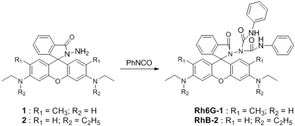
Synthesis of the compounds Rh6G-1 and RhB-2. Reagent and experimental condition: anhydrous toluene, reflux.
Ethical approval: The conducted research is not related to either human or animal use.
3 Results and discussion
3.1 UV–vis and fluorescence spectra of sensor Rh6G-1 titrated with Fe3+
Fluorescence titrations of Fe3+ to sensor Rh6G-1 (10 μM) were conducted in almost pure water (containing 1% DMSO) with excitation at 500 nm at room temperature. Sensor Rh6G-1 exhibited almost no fluorescence in water, indicating that Rh6G-1 exists predominantly as the spirocyclic form. However, upon addition of Fe3+ from FeCl3, the fluorescence intensity at around 556 nm significantly increases (Figure 1), and an 82-fold fluorescence enhancement was observed. The absorption titration studies are in good agreement with the turn-on fluorescence response (Figure 2). Treatment of Fe3+ elicited formation of an intense absorption peak at around 542 nm, indicating that sensor Rh6G-1 is in the ring-opening form in the presence of Fe3+. Notably, the sensor showed an excellent linear relationship between the fluorescence intensity at 556 nm and the concentrations of Fe3+ from 1 to 170 μM (Figure 3), suggesting that the sensor is potentially useful for quantitative determination of Fe3+. The detection limit (S/N = 3) of sensor Rh6G-1 was determined to be 1.2 μM in water (containing 1% DMSO) (Figure S1).

Fluorescence spectra of sensor Rh6G-1 (10 μM) in the presence of increasing concentrations of Fe3+ (0-50 equiv.) in water (containing 1% DMSO as a cosolvent) with excitation at 500 nm. Inset: Fluorescent intensity at 556 nm of sensor Rh6G-1 (10 μM) upon addition of Fe3+ (0-50 equiv.) excited at 500 nm, and the visual fluorescence color changes of the solutions a, b: a, sensor Rh6G-1 (10 μM); b, sensor Rh6G-1 (10 μM) + Fe3+ (50 eq).
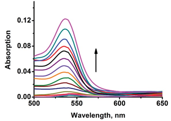
Absorption spectra of sensor Rh6G-1 (10 μM) in the presence of increasing concentrations of Fe3+ (0 - 50 equiv.) in water (containing 1% DMSO as a cosolvent).
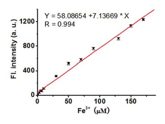
Plot of fluorescence intensity of sensor Rh6G-1 (10 μM) at 556 nm vs. Fe3+ concentration (1 - 170 μM) in water (containing 1% DMSO as a cosolvent) excited at 500 nm.
Although compound RhB-2 may also operate as a novel fluorescent Fe3+ sensor in water containing 20% CH3CN (Figure S2A), it is inferior to compound Rh6G-1 in terms of sensitivity in water containing 1% DMSO. The fluorescence intensity of compound RhB-2 increased from 2.7 in the absence of Fe3+ to 17.7 in the presence of 100 equiv. of Fe3+ in water (containing 1% DMSO) (Figure S2B), only a 6.6-fold fluorescence enhancement, which is much less than that (82-fold fluorescence enhancement) of compound Rh6G-1 under the same solvent system (water containing 1% DMSO). Thus, in this work, we focused on sensor Rh6G-1 for further studies.
3.2 Binding mode of sensor Rh6G-1 with Fe3+
Job’s plot according to the method for continuous variations [25] shows a 1:1 binding stoichiometry between sensor Rh6G-1 and Fe3+ (Figure S3A). Based on the 1: 1 binding mode, the binding constant of sensor Rh6G-1 in water (containing 1% DMSO as a cosolvent) was calculated as Log Ka = 3.34 from the results of the fluorescence titration experiments (Figure S3B) [26]. Importantly, the formation of sensor Rh6G-1/Fe3+ complex is prompt and reversible. The reversible nature of the interactions between Rh6G-1 and Fe3+ was tested with TPEN (N, N, N’, N’-Tetrakis(2-pyridylmethyl)-1, 2- ethylenediamine) which is known to bind strongly with various metal ions including Fe3+. A solution of sensor Rh6G-1 (10 μM) incubated with Fe3+ (10 equiv.) in water (containing 1% DMSO as a cosolvent) exhibited a strong fluorescence at around 556 nm. However, upon further titration with TPEN (0 - 60 equiv.), the fluorescence was gradually quenched (Figure 4), indicating the reversible character of the binding of sensor Rh6G-1 with Fe3+. To further test the reversible character, we carried out the cyclic tests for the reversibility of the fluorescent turn-on behavior with quenching by TPEN and followed by adding Fe3+ (Figure S4), the result showed that fluorescence enhancement can still be recovered after four cycles. Thus, based on these studies and the interaction mode of Fe3+ with the bioactive
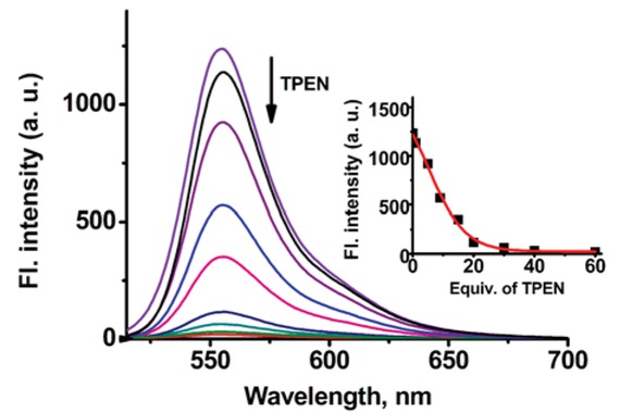
Fluorescence titration spectra of a solution of sensor Rh6G-1 (10 μM) + Fe3+ (10 equiv.) with TPEN (0 - 60 equiv.) in water (containing 1% DMSO as a cosolvent). Inset: Fluorescent intensity at 556 nm of a solution of sensor Rh6G-1 (10 μM) + Fe3+ (10 equiv.) upon addition of TPEN (0 - 60 equiv.). Excitation at 500 nm.
ferrichromes [22], a likely binding mode of sensor Rh6G-1 with Fe3+ was proposed (Scheme 2).
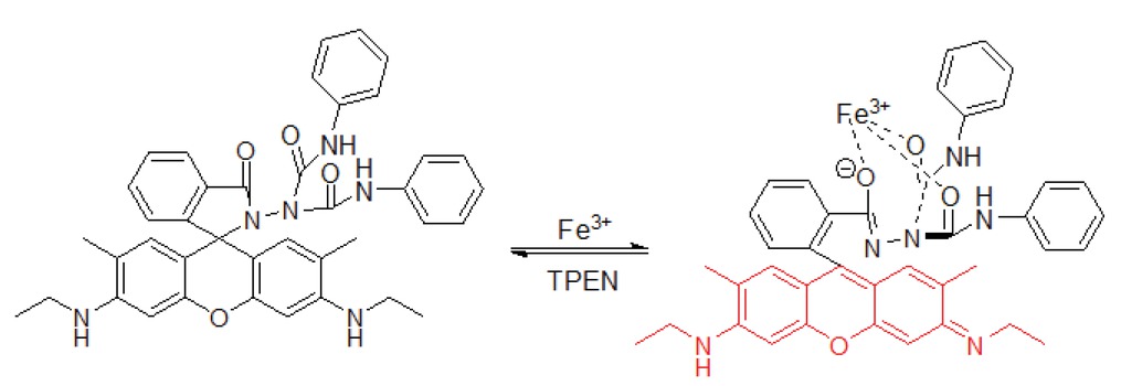
The possible binding mode of sensor Rh6G-1 with FeCl3.
To further understand the nature of interaction between sensor Rh6G-1 and Fe3+, the ESI-MS spectrum (positive ion mode) was carried out. The peak at m/z 667.3 (calcd 666.3) corresponded to [Rh6G-1+H] +, when excess Fe3+ was added, the new highest peak was obtained at m/z 740.4 corresponded to [Rh6G-1+ Fe3++H3O+] (Figure S5), which indicated that sensor Rh6G-1 have coordinated with Fe3+. The possibility reaction mechanism was depicted in Scheme 2.
3.3 Selectivity studies
We then examined the selectivity of sensor Rh6G-1 towards other metal species. The fluorescence titration experiment was carried out in water (containing 1% DMSO as a cosolvent) with a diverse array of metal species. Fluorescence data reveal that sensor Rh6G-1 selectively responds to Fe3+ over various metal species tested (Figure 5). Other metal species such as Pd2+, Ca2+, Cd2+, Co2+, Zn2+, Cu2+, Hg2+, Mg2+, Mn2+, Ag+, Au3+, Ni2+, Pb2+, Na+, and K+ displayed very slighted fluorescence variations. Notably, sensor Rh6G-1 has a high selectivity for Fe3+ over Fe2+. We further investigated the fluorescent turn-on response of sensor Rh6G-1 toward Fe3+ in the presence of other potentially competing species. The other species only exhibited minimum interference (Figure S6). This indicates that sensor Rh6G-1 is useful to detect Fe3+ in the presence of other related species. Compound RhB-2 also exhibited the high selectivity for Fe3+ in water containing 20% CH3CN (Figures S7-8).
3.4 Effect of pH
To study the practical applicability, the effect of pH on the fluorescence response of sensor Rh6G-1 to Fe3+ was investigated. As shown in Figure 6, in the absence of Fe3+, almost no change in fluorescence intensity was observed in the free sensor Rh6G-1 over a wide pH range of 4.5 - 9.0, indicating that the free sensor was stable across a wide pH range. Upon treatment with Fe3+, the maximal fluorescence signal was observed in the pH range of 5.5 - 8.5. Thus, the observation that sensor Rh6G-1 had the maximal sensing response at physiological pH, suggests that sensor Rh6G-1 was promising for biological applications.

Fluorescence intensity changes of sensor Rh6G-1 (10 μM) in response to various metal species (20 equiv.) in water (containing 1 % DMSO as a cosolvent). 1. none; 2. Pd2+; 3. Ca2+; 4. Cd2+; 5. Co2+; 6. Zn2+; 7. Cu2+; 8. Fe2+; 9. Fe3+; 10. Hg2+; 11. Mg2+; 12. Mn2+; 13, Ag+, 14, Au3+, 15. Ni2+; 16. Pb2+; 17. Na+, 18. K+. Excitation at 500 nm; emission at 556 nm.
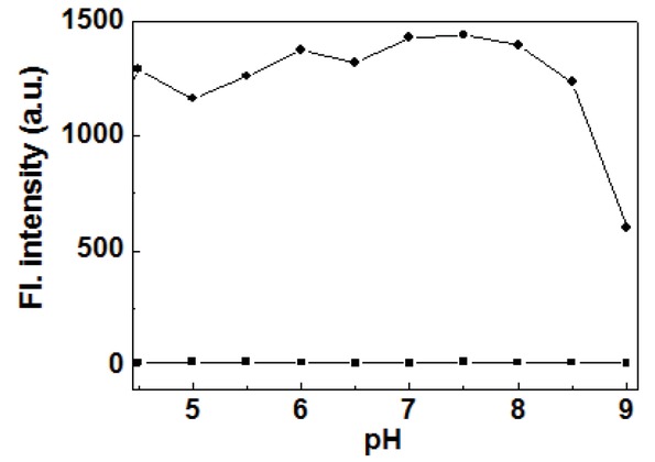
The fluorescence responses (at 556 nm) of free sensor Rh6G-1 (10 μM) (■) and Rh6G-1 (10 μM) + 20 equiv Fe3+ (●) in water (containing 1 % DMSO as a cosolvent) as a function of different pH values.
3.5 Fluorescence Image in Living Cells
The potential toxicity is a concern for any fluorescent sensors intended as molecular imaging agents. Thus, we set out to examine the potential toxicity of sensor Rh6G-1 against the representative cell line, Hela cells. The living cells were incubated with various concentrations (0 - 200 μM) of the sensor for 24 h, and then the cell viability was determined by the standard 3-(4,5-dimethylthiazol-2-yl)-2,5-diphenyltetrazolium bromide (MTT) assays [27]. Almost 90% of the cells were still alive after treatment with 50 μM sensor Rh6G-1 for 24 h (Figure 7), suggesting that sensor Rh6G-1 has low cytotoxicity [28].
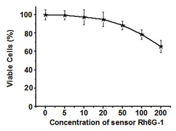
Cytotoxicity assay of sensor Rh6G-1 at different concentrations (a: 0 μΜ; b: 5 μΜ; c: 10 μΜ; d: 20 μΜ; e: 50 μΜ; f: 100 μΜ; g: 200 μΜ) for HeLa cells.
The observation that sensor Rh6G-1 can function very well in essentially pure water renders it desirable for imaging of Fe3+ in living cells. To test this possibility, sensor Rh6G-1 was incubated with the living HeLa cells. As shown in Figure 8b, the cells treated with only sensor Rh6G-1 exhibited almost no fluorescence. However, the cells pre-loaded with sensor Rh6G-1 and further incubated with Fe3+ displayed intense orange fluorescence (Figure 8e), consistent with the emission profiles of sensor Rh6G-1 incubated with Fe3+ (Figure 1). Furthermore, the nuclear staining with Hoechst 33258 revealed that sensor Rh6G-1 associates with the cytoplasm of HeLa cells (Figures 8d-f).
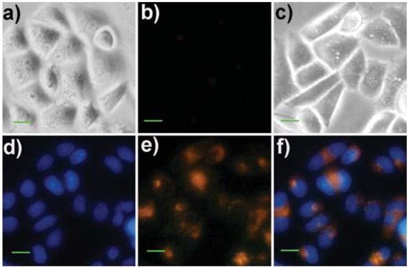
Bright-field and fluorescence images of HeLa cells. (a) Brightfield image of the cells treated with sensor Rh6G-1 (10 μM) for 30 min; (b) Fluorescence image of panel (a) from the red channel; (c) Bright-field image of the cells pre-treated with sensor Rh6G-1 (10 μM) and Hoechst 33258 (4.5 μM) for 30 min, and then incubated with Fe3+ (10 equiv.) for 30 min; (d) Fluorescence image of panel (c) from the blue channel; (e) Fluorescence image of panel (c) from the green/red channel; (f) Overlay of panels (d) and (e). Scale bar: 30 μm.
4 Conclusions
In conclusion, compounds Rh6G-1 and RhB-2 containing tri-oxygen atoms of the amide groups were designed and synthesized as new reversible fluorescent sensors for Fe3+. The favorable features of the novel sensor Rh6G-1 include a big fluorescent turn-on response in almost pure water at room temperature, high sensitivity, high selectivity, cell membrane permeability, and low cytotoxicity. These desirable attributes enable us to successfully employ the new sensor Rh6G-1 for Fe3+ bioimaging in living cells. We expect that the new Fe3+ ligand composed of tri-oxygen atoms of the amide groups will be useful for development of a wide variety of reversible fluorescent Fe3+ sensors based on distinct dyes.
Acknowledgments
This work was financially supported by Hunan Provincial Key Lab of Dark Tea and Jin-hua (2016TP1022), NSFC (21502048), the Natural Science Foundation of Hunan Province (2016JJ3102), Science and Technology Innovative Research Team in Higher Educational Institutions of Hunan Province. There is no any conflict of interest in connection with this articleand no portion of it has been published or under consideration of publication by another journal
Conflict of interest: Authors declare no conflict of interest.
Supplemental Material: The online version of this article offers supplementary material (https://doi.org/10.1515/chem-2018-0140).
References
[1] Touati D., Iron and oxidative stress in bacteria, Arch. Biochem. Biophys., 2000, 373, 1-6.10.1006/abbi.1999.1518Search in Google Scholar PubMed
[2] Beutler E., Felitti V., Gelbart T., Ho N., Genetics of iron storage and hemochromatosis, Drug Metab. Dispos., 2001, 29, 495-499.Search in Google Scholar
[3] Andrews N.C., Disorders of iron metabolism, N. Engl. J. Med., 1999, 341, 1986-1195.10.1038/npg.els.0006072Search in Google Scholar
[4] Baytak S., Balaban A., Türker A.R., Erk B., Atomic absorption spectrometric determination of Fe(III) and Cr(III) in various samples after preconcentration by solid phase extraction with pyridine-2-carbaldehyde thiosemicarbazone, J. Anal. Chem., 2006, 61, 476-482.10.1134/S106193480605008XSearch in Google Scholar
[5] Vladescu L., Costache M., Badea I., Preparation, characterization,and metal-sorption studies of a mordant yellow 10-loaded wool, a new stable chelating material based on bleached wool, Acta Chrom., 2004, 14, 187-197.Search in Google Scholar
[6] Mokgalaka N.S., Wondimu T., McCrindle R.I., Slurry nebulization ICP-OES for the determination of Cu, Fe, Mg, Mn and Zn, Bull. Chem. Soc. Ethiop., 2008, 22, 313-321.10.4314/bcse.v22i3.61195Search in Google Scholar
[7] Chen X., Pradhan T., Wang F., Kim J.S., Yoon J., Fluorescent chemosensors based on spiroring-opening of xanthenes and related derivatives, Chem. Rev., 2012, 112, 1910-1956.10.1021/cr200201zSearch in Google Scholar PubMed
[8] Bricks J.L., Kovalchuk A., Trieflinger C., Nofz M., Büschel M., Tolmachev A.I., et al., On the development of sensor molecules that display FeIII-amplified fluorescence, J. Am. Chem. Soc., 2005, 127, 13522-13529.10.1021/ja050652tSearch in Google Scholar PubMed
[9] Xiang Y., Tong A., A new rhodamine-based chemosensor exhibiting selective FeIII-amplified fluorescence, Org. Lett., 2006, 8, 1549-1552.10.1021/ol060001hSearch in Google Scholar PubMed
[10] Mao J., Wang L., Dou W., Tang X., Yan Y., Liu W., Tuning the Selectivity of Two Chemosensors to Fe(III) and Cr(III), Org. Lett., 2007, 9, 4567-4570.10.1021/ol7020687Search in Google Scholar PubMed
[11] Qin J., Yang Z., Wang G., Li C., FRET-based rhodamine-coumarin conjugate as a Fe3+ selective ratiometric fluorescent sensor in aqueous media, Tetrahedron Lett., 2015, 56, 5024-5029.10.1016/j.tetlet.2015.07.023Search in Google Scholar
[12] Zhao M., Deng Z., Tang J., Zhou X., Chen Z., Li X., et al., 2-(1-Pyrenyl) benzimidazole as a ratiometric and “turn-on” fluorescent probe for iron(III) ions in aqueous solution, Analyst, 2016, 141, 2308-2312.10.1039/C5AN02565FSearch in Google Scholar PubMed
[13] Ma S., Yang Z., She M., Sun W., Yin B., Liu P., et al., Design and synthesis of functionalized rhodamine based probes for specific intracellular fluorescence imaging of Fe3+ Dyes Pigm., 2015, 115, 120-126.10.1016/j.dyepig.2014.12.014Search in Google Scholar
[14] Hu Z.Q., Du M., Zhang L.F., Guo F.Y., Liu M.D., Li M., A novel colorimetric and fluorescent chemosensor for cyanide ion in aqueous media based on a rhodamine derivative in the presence of Fe3+ ion, Sens. Actuators, B, 2014, 192, 439-443.10.1016/j.snb.2013.10.138Search in Google Scholar
[15] Zhou F., Leng T.H., Liu Y.J., Wang C.Y., Shi P., Zhu W.H., Water-soluble rhodamine-based chemosensor for Fe3+ with high sensitivity, selectivity and anti-interference capacity and its imaging application in living cells, Dyes Pigm.., 2017, 142, 429-436.10.1016/j.dyepig.2017.03.057Search in Google Scholar
[16] Wang K., Lei Y., Zhang S., Zheng W., Chen J., Chen S., et al., Fluorescent probe for Fe(III) with high selectivity and its application in living cells, Sens. Actuators, B, 2017, 252, 1140-1145.10.1016/j.snb.2017.07.184Search in Google Scholar
[17] Yan F., Zheng T., Shi D., Zou Y., Wang Y., Fu M., et al., Rhodamine-aminopyridine based fluorescent sensors for Fe3+ in water: Synthesis, quantum chemical interpretation and living cell application, Sens. Actuators, B, 2015, 215, 598-606.10.1016/j.snb.2015.03.096Search in Google Scholar
[18] Lee M.H., Giap T.V., Kim S.H., Lee Y.H., Kang C., Kim J.S., A novel strategy to selectively detect Fe(III) in aqueous media driven by hydrolysis of a rhodamine 6G Schiff base, Chem. Commun., 2010, 46, 1407-1409.10.1039/B921526CSearch in Google Scholar PubMed
[19] Kim H.N., Lee M.H., Kim H.J., Kim J.S., Yoon J., A new trend in rhodamine-based chemosensors: application of spirolactam ring-opening to sensing ions, Chem. Soc. Rev., 2008, 37, 1465-1472.10.1039/b802497aSearch in Google Scholar PubMed
[20] Beija M., Afonso C.A.M., Martinho J.M.G., Synthesis and applications of Rhodamine derivatives as fluorescent probes, Chem. Soc. Rev., 2009, 38, 2410-2433.10.1039/b901612kSearch in Google Scholar PubMed
[21] Chen X., Wang F., Hyun J.Y., Wei T., Qiang J., Ren X., et al., Recent progress in the development of fluorescent, luminescent and colorimetric probes for detection of reactive oxygen and nitrogen species, Chem. Soc. Rev., 2016, 45, 2976-3016.10.1039/C6CS00192KSearch in Google Scholar PubMed
[22] Nudelman R., Ardon O., Hadar Y., Chen Y., Libman J., Shanzer A., Modular fluorescent-labeled siderophore analogues, J. Med. Chem., 1998, 41, 1671-1678.10.1021/jm970581bSearch in Google Scholar PubMed
[23] Yang Y.K., Yook K.J., Tae J., A rhodamine-based fluorescent and colorimetric chemodosimeter for the rapid detection of Hg2+ ions in aqueous media, J. Am. Chem. Soc., 2005, 127, 16760-16761.10.1021/ja054855tSearch in Google Scholar
[24] Xiao X.F., Guo X.Q., Zhao Y.B., Development of a novel rhodamine type fluorescent probe to determine peroxynitrite, Talanta, 2002, 57, 883-890.10.1016/S0039-9140(02)00120-0Search in Google Scholar
[25] Vosburgh W.C., Cooper G.R., Complex ions. I. The identification of complex ions in solution by spectrophotometric measurements, J. Am. Chem. Soc., 1941, 63, 437-442.10.1021/ja01847a025Search in Google Scholar
[26] Ho I.T., Lee G.H., Chung W.S., Synthesis of upper-Rim allyl-and p-methoxyphenylazocalix[4]arenes and their efficiencies in chromogenic sensing of Hg2+ Ion, J. Org. Chem., 2007, 72, 2434-2442.10.1021/jo062280sSearch in Google Scholar
[27] Mosmann T., Rapid colorimetric assay for cellular growth and survival: Application to proliferation and cytotoxicity assays, J. Immunol. Methods, 1983, 65, 55-63.10.1016/0022-1759(83)90303-4Search in Google Scholar
[28] Wang J., Long L., Xiao X., A Fast-responsive Fluorescent Probe for Sulfite and Its Bioimaging, Luminescence, 2016, 32, 775-781.10.1002/bio.3023Search in Google Scholar PubMed
© 2018 Jiaoliang Wang et al., published by De Gruyter
This work is licensed under the Creative Commons Attribution-NonCommercial-NoDerivatives 4.0 License.
Articles in the same Issue
- Regular Articles
- The effect of CuO modification for a TiO2 nanotube confined CeO2 catalyst on the catalytic combustion of butane
- The preparation and antibacterial activity of cellulose/ZnO composite: a review
- Linde Type A and nano magnetite/NaA zeolites: cytotoxicity and doxorubicin loading efficiency
- Performance and thermal decomposition analysis of foaming agent NPL-10 for use in heavy oil recovery by steam injection
- Spectroscopic (FT-IR, FT-Raman, UV, 1H and 13C NMR) insights, electronic profiling and DFT computations on ({(E)-[3-(1H-imidazol-1-yl)-1-phenylpropylidene] amino}oxy)(4-nitrophenyl)methanone, an imidazole-bearing anti-Candida agent
- A Simplistic Preliminary Assessment of Ginstling-Brounstein Model for Solid Spherical Particles in the Context of a Diffusion-Controlled Synthesis
- M-Polynomials And Topological Indices Of Zigzag And Rhombic Benzenoid Systems
- Photochemical Transformation of some 3-benzyloxy-2-(benzo[b]thiophen-2-yl)-4Hchromen-4-ones: A Remote Substituent Effect
- Dynamic Changes of Secondary Metabolites and Antioxidant Activity of Ligustrum lucidum During Fruit Growth
- Studies on the flammability of polypropylene/ammonium polyphosphate and montmorillonite by using the cone calorimeter test
- DSC, FT-IR, NIR, NIR-PCA and NIR-ANOVA for determination of chemical stability of diuretic drugs: impact of excipients
- Antioxidant and Hepatoprotective Effects of Methanolic Extracts of Zilla spinosa and Hammada elegans Against Carbon Tetrachlorideinduced Hepatotoxicity in Rats
- Prunus cerasifera Ehrh. fabricated ZnO nano falcates and its photocatalytic and dose dependent in vitro bio-activity
- Organic biocides hosted in layered double hydroxides: enhancing antimicrobial activity
- Experimental study on the regulation of the cholinergic pathway in renal macrophages by microRNA-132 to alleviate inflammatory response
- Synthesis, characterization, in-vitro antimicrobial properties, molecular docking and DFT studies of 3-{(E)-[(4,6-dimethylpyrimidin-2-yl)imino]methyl} naphthalen-2-ol and Heteroleptic Mn(II), Co(II), Ni(II) and Zn(II) complexes
- M-Polynomials and Topological Indices of Dominating David Derived Networks
- Human Health Risk Assessment of Trace Metals in Surface Water Due to Leachate from the Municipal Dumpsite by Pollution Index: A Case Study from Ndawuse River, Abuja, Nigeria
- Analysis of Bowel Diseases from Blood Serum by Autofluorescence and Atomic Force Microscopy Techniques
- Hydrographic parameters and distribution of dissolved Cu, Ni, Zn and nutrients near Jeddah desalination plant
- Relationships between diatoms and environmental variables in industrial water biotopes of Trzuskawica S.A. (Poland)
- Optimum Conversion of Major Ginsenoside Rb1 to Minor Ginsenoside Rg3(S) by Pulsed Electric Field-Assisted Acid Hydrolysis Treatment
- Antioxidant, Anti-microbial Properties and Chemical Composition of Cumin Essential Oils Extracted by Three Methods
- Regulatory mechanism of ulinastatin on autophagy of macrophages and renal tubular epithelial cells
- Investigation of the sustained-release mechanism of hydroxypropyl methyl cellulose skeleton type Acipimox tablets
- Bio-accumulation of Polycyclic Aromatic Hydrocarbons in the Grey Mangrove (Avicennia marina) along Arabian Gulf, Saudi Coast
- Dynamic Change of Secondary Metabolites and spectrum-effect relationship of Malus halliana Koehne flowers during blooming
- Lipids constituents from Gardenia aqualla Stapf & Hutch
- Effect of using microwaves for catalysts preparation on the catalytic acetalization of glycerol with furfural to obtain fuel additives
- Effect of Humic Acid on the Degradation of Methylene Blue by Peroxymonosulfate
- Serum containing drugs of Gua Lou Xie Bai decoction (GLXB-D) can inhibit TGF-β1-Induced Epithelial to Mesenchymal Transition (EMT) in A549 Cells
- Antiulcer Activity of Different Extracts of Anvillea garcinii and Isolation of Two New Secondary Metabolites
- Analysis of Metabolites in Cabernet Sauvignon and Shiraz Dry Red Wines from Shanxi by 1H NMR Spectroscopy Combined with Pattern Recognition Analysis
- Can water temperature impact litter decomposition under pollution of copper and zinc mixture
- Released from ZrO2/SiO2 coating resveratrol inhibits senescence and oxidative stress of human adipose-derived stem cells (ASC)
- Validated thin-layer chromatographic method for alternative and simultaneous determination of two anti-gout agents in their fixed dose combinations
- Fast removal of pollutants from vehicle emissions during cold-start stage
- Review Article
- Catalytic activities of heterogeneous catalysts obtained by copolymerization of metal-containing 2-(acetoacetoxy)ethyl methacrylate
- Antibiotic Residue in the Aquatic Environment: Status in Africa
- Regular Articles
- Mercury fractionation in gypsum using temperature desorption and mass spectrometric detection
- Phytosynthetic Ag doped ZnO nanoparticles: Semiconducting green remediators
- Epithelial–Mesenchymal Transition Induced by SMAD4 Activation in Invasive Growth Hormone-Secreting Adenomas
- Physicochemical properties of stabilized sewage sludge admixtures by modified steel slag
- In Vitro Cytotoxic and Antiproliferative Activity of Cydonia oblonga flower petals, leaf and fruit pellet ethanolic extracts. Docking simulation of the active flavonoids on anti-apoptotic protein Bcl-2
- Synthesis and Characterization of Pd exchanged MMT Clay for Mizoroki-Heck Reaction
- A new selective, and sensitive method for the determination of lixivaptan, a vasopressin 2 (V2)-receptor antagonist, in mouse plasma and its application in a pharmacokinetic study
- Anti-EGFL7 antibodies inhibit rat prolactinoma MMQ cells proliferation and PRL secretion
- Density functional theory calculations, vibration spectral analysis and molecular docking of the antimicrobial agent 6-(1,3-benzodioxol-5-ylmethyl)-5-ethyl-2-{[2-(morpholin-4-yl)ethyl] sulfanyl}pyrimidin-4(3H)-one
- Effect of Nano Zeolite on the Transformation of Cadmium Speciation and Its Uptake by Tobacco in Cadmium-contaminated Soil
- Effects and Mechanisms of Jinniu Capsule on Methamphetamine-Induced Conditioned Place Preference in Rats
- Calculating the Degree-based Topological Indices of Dendrimers
- Efficient optimization and mineralization of UV absorbers: A comparative investigation with Fenton and UV/H2O2
- Metabolites of Tryptophane and Phenylalanine as Markers of Small Bowel Ischemia-Reperfusion Injury
- Adsorption and determination of polycyclic aromatic hydrocarbons in water through the aggregation of graphene oxide
- The role of NR2C2 in the prolactinomas
- Chromium removal from industrial wastewater using Phyllostachys pubescens biomass loaded Cu-S nanospheres
- Hydrotalcite Anchored Ruthenium Catalyst for CO2 Hydrogenation Reaction
- Preparation of Calcium Fluoride using Phosphogypsum by Orthogonal Experiment
- The mechanism of antibacterial activity of corylifolinin against three clinical bacteria from Psoralen corylifolia L
- 2-formyl-3,6-bis(hydroxymethyl)phenyl benzoate in Electrochemical Dry Cell
- Electro-photocatalytic degradation of amoxicillin using calcium titanate
- Effect of Malus halliana Koehne Polysaccharides on Functional Constipation
- Structural Properties and Nonlinear Optical Responses of Halogenated Compounds: A DFT Investigation on Molecular Modelling
- DMFDMA catalyzed synthesis of 2-((Dimethylamino)methylene)-3,4-dihydro-9-arylacridin-1(2H)-ones and their derivatives: in-vitro antifungal, antibacterial and antioxidant evaluations
- Production of Methanol as a Fuel Energy from CO2 Present in Polluted Seawater - A Photocatalytic Outlook
- Study of different extraction methods on finger print and fatty acid of raw beef fat using fourier transform infrared and gas chromatography-mass spectrometry
- Determination of trace fluoroquinolones in water solutions and in medicinal preparations by conventional and synchronous fluorescence spectrometry
- Extraction and determination of flavonoids in Carthamus tinctorius
- Therapeutic Application of Zinc and Vanadium Complexes against Diabetes Mellitus a Coronary Disease: A review
- Study of calcined eggshell as potential catalyst for biodiesel formation using used cooking oil
- Manganese oxalates - structure-based Insights
- Topological Indices of H-Naphtalenic Nanosheet
- Long-Term Dissolution of Glass Fibers in Water Described by Dissolving Cylinder Zero-Order Kinetic Model: Mass Loss and Radius Reduction
- Topological study of the para-line graphs of certain pentacene via topological indices
- A brief insight into the prediction of water vapor transmissibility in highly impermeable hybrid nanocomposites based on bromobutyl/epichlorohydrin rubber blends
- Comparative sulfite assay by voltammetry using Pt electrodes, photometry and titrimetry: Application to cider, vinegar and sugar analysis
- MicroRNA delivery mediated by PEGylated polyethylenimine for prostate cancer therapy
- Reversible Fluorescent Turn-on Sensors for Fe3+ based on a Receptor Composed of Tri-oxygen Atoms of Amide Groups in Water
- Sonocatalytic degradation of methyl orange in aqueous solution using Fe-doped TiO2 nanoparticles under mechanical agitation
- Hydrotalcite Anchored Ruthenium Catalyst for CO2 Hydrogenation Reaction
- Production and Analysis of Recycled Ammonium Perrhenate from CMSX-4 superalloys
- Topical Issue on Agriculture
- New phosphorus biofertilizers from renewable raw materials in the aspect of cadmium and lead contents in soil and plants
- Survey of content of cadmium, calcium, chromium, copper, iron, lead, magnesium, manganese, mercury, sodium and zinc in chamomile and green tea leaves by electrothermal or flame atomizer atomic absorption spectrometry
- Biogas digestate – benefits and risks for soil fertility and crop quality – an evaluation of grain maize response
- A numerical analysis of heat transfer in a cross-current heat exchanger with controlled and newly designed air flows
- Freshwater green macroalgae as a biosorbent of Cr(III) ions
- The main influencing factors of soil mechanical characteristics of the gravity erosion environment in the dry-hot valley of Jinsha river
- Free amino acids in Viola tricolor in relation to different habitat conditions
- The influence of filler amount on selected properties of new experimental resin dental composite
- Effect of poultry wastewater irrigation on nitrogen, phosphorus and carbon contents in farmland soil
- Response of spring wheat to NPK and S fertilization. The content and uptake of macronutrients and the value of ionic ratios
- The Effect of Macroalgal Extracts and Near Infrared Radiation on Germination of Soybean Seedlings: Preliminary Research Results
- Content of Zn, Cd and Pb in purple moor-grass in soils heavily contaminated with heavy metals around a zinc and lead ore tailing landfill
- Topical Issue on Research for Natural Bioactive Products
- Synthesis of (±)-3,4-dimethoxybenzyl-4-methyloctanoate as a novel internal standard for capsinoid determination by HPLC-ESI-MS/MS(QTOF)
- Repellent activity of monoterpenoid esters with neurotransmitter amino acids against yellow fever mosquito, Aedes aegypti
- Effect of Flammulina velutipes (golden needle mushroom, eno-kitake) polysaccharides on constipation
- Bioassay-directed fractionation of a blood coagulation factor Xa inhibitor, betulinic acid from Lycopus lucidus
- Antifungal and repellent activities of the essential oils from three aromatic herbs from western Himalaya
- Chemical composition and microbiological evaluation of essential oil from Hyssopus officinalis L. with white and pink flowers
- Bioassay-guided isolation and identification of Aedes aegypti larvicidal and biting deterrent compounds from Veratrum lobelianum
- α-Terpineol, a natural monoterpene: A review of its biological properties
- Utility of essential oils for development of host-based lures for Xyleborus glabratus (Coleoptera: Curculionidae: Scolytinae), vector of laurel wilt
- Phenolic composition and antioxidant potential of different organs of Kazakh Crataegus almaatensis Pojark: A comparison with the European Crataegus oxyacantha L. flowers
- Isolation of eudesmane type sesquiterpene ketone from Prangos heyniae H.Duman & M.F.Watson essential oil and mosquitocidal activity of the essential oils
- Comparative analysis of the polyphenols profiles and the antioxidant and cytotoxicity properties of various blue honeysuckle varieties
- Special Issue on ICCESEN 2017
- Modelling world energy security data from multinomial distribution by generalized linear model under different cumulative link functions
- Pine Cone and Boron Compounds Effect as Reinforcement on Mechanical and Flammability Properties of Polyester Composites
- Artificial Neural Network Modelling for Prediction of SNR Effected by Probe Properties on Ultrasonic Inspection of Austenitic Stainless Steel Weldments
- Calculation and 3D analyses of ERR in the band crack front contained in a rectangular plate made of multilayered material
- Improvement of fuel properties of biodiesel with bioadditive ethyl levulinate
- Properties of AlSi9Cu3 metal matrix micro and nano composites produced via stir casting
- Investigation of Antibacterial Properties of Ag Doped TiO2 Nanofibers Prepared by Electrospinning Process
- Modeling of Total Phenolic contents in Various Tea samples by Experimental Design Methods
- Nickel doping effect on the structural and optical properties of indium sulfide thin films by SILAR
- The effect mechanism of Ginnalin A as a homeopathic agent on various cancer cell lines
- Excitation functions of proton induced reactions of some radioisotopes used in medicine
- Oxide ionic conductivity and microstructures of Pr and Sm co-doped CeO2-based systems
- Rapid Synthesis of Metallic Reinforced in Situ Intermetallic Composites in Ti-Al-Nb System via Resistive Sintering
- Oxidation Behavior of NiCr/YSZ Thermal Barrier Coatings (TBCs)
- Clustering Analysis of Normal Strength Concretes Produced with Different Aggregate Types
- Magnetic Nano-Sized Solid Acid Catalyst Bearing Sulfonic Acid Groups for Biodiesel Synthesis
- The biological activities of Arabis alpina L. subsp. brevifolia (DC.) Cullen against food pathogens
- Humidity properties of Schiff base polymers
- Free Vibration Analysis of Fiber Metal Laminated Straight Beam
- Comparative study of in vitro antioxidant, acetylcholinesterase and butyrylcholinesterase activity of alfalfa (Medicago sativa L.) collected during different growth stages
- Isothermal Oxidation Behavior of Gadolinium Zirconate (Gd2Zr2O7) Thermal Barrier Coatings (TBCs) produced by Electron Beam Physical Vapor Deposition (EB-PVD) technique
- Optimization of Adsorption Parameters for Ultra-Fine Calcite Using a Box-Behnken Experimental Design
- The Microstructural Investigation of Vermiculite-Infiltrated Electron Beam Physical Vapor Deposition Thermal Barrier Coatings
- Modelling Porosity Permeability of Ceramic Tiles using Fuzzy Taguchi Method
- Experimental and theoretical study of a novel naphthoquinone Schiff base
- Physicochemical properties of heat treated sille stone for ceramic industry
- Sand Dune Characterization for Preparing Metallurgical Grade Silicon
- Catalytic Applications of Large Pore Sulfonic Acid-Functionalized SBA-15 Mesoporous Silica for Esterification
- One-photon Absorption Characterizations, Dipole Polarizabilities and Second Hyperpolarizabilities of Chlorophyll a and Crocin
- The Optical and Crystallite Characterization of Bilayer TiO2 Films Coated on Different ITO layers
- Topical Issue on Bond Activation
- Metal-mediated reactions towards the synthesis of a novel deaminolysed bisurea, dicarbamolyamine
- The structure of ortho-(trifluoromethyl)phenol in comparison to its homologues – A combined experimental and theoretical study
- Heterogeneous catalysis with encapsulated haem and other synthetic porphyrins: Harnessing the power of porphyrins for oxidation reactions
- Recent Advances on Mechanistic Studies on C–H Activation Catalyzed by Base Metals
- Reactions of the organoplatinum complex [Pt(cod) (neoSi)Cl] (neoSi = trimethylsilylmethyl) with the non-coordinating anions SbF6– and BPh4–
- Erratum
- Investigation on Two Compounds of O, O’-dithiophosphate Derivatives as Corrosion Inhibitors for Q235 Steel in Hydrochloric Acid Solution