Prognostic role of oxytocin receptor in colon adenocarcinoma
-
Junjie Sun


Abstract
The oxytocin receptor (OXTR) is directly involved in the pathological mechanisms of multiple cancers, including breast cancer, prostate cancer, and ovarian cancer; however, the role of OXTR in the modulation of colon adenocarcinoma (COAD) growth, metastasis, and clinical prognosis remains to be elucidated. This study used systematic bioinformatics analysis to explore the effects of OXTR on modulating COAD growth and prognosis in patients with COAD. Compared with normal tissues, OXTR mRNA level was higher in COAD tissues, which was associated with tumor progression. Elevated mRNA level of OXTR also indicated a poor prognosis in COAD patients. Furthermore, high mRNA level of OXTR was significantly associated with pathways involved in cell cycle regulation and signal transduction pathways, including the hedgehog, mTOR, TGF-β, and Wnt signaling pathways. OXTR expression was significantly correlated with the infiltration level of type 2T helper cell, central memory CD8 T cell, CD56 bright natural killer cell, activated CD8 T cell, activated B cell, and Type 1T helper cell. Moreover, silencing OXTR inhibited cell proliferation, migration, and invasion, and arrested the cell cycle. In conclusion, high mRNA level of OXTR indicates poor prognosis.
1 Introduction
Colon adenocarcinoma (COAD), widely accepted as an illness that starts in the rectum or colon, ranks as the third most common cancer and the fourth leading cause of cancer-related death in the world [1,2,3]. Metastasis is usually present in most COAD patients at the first diagnosis, and this trend has been more severe over the last two decades due to changes in eating habits and living conditions [4]. Currently, the selective treatment for COAD is based on two common methods, surgery and chemotherapy, which are mainly administered based on the tumor stage [3]. The stage of COAD is mainly described by the TNM (tumor-node-metastasis) system initiated by the American Joint Committee on Cancer (AJCC) and the Union for International Cancer Control (UICC) [5]. Due to environmental pollution, poor dietary patterns, obesity, and dysbiosis of gut microbiota, the incidence of COAD, especially among the younger population, has largely increased in recent decades, which adds a heavy burden to the health care system and our society [6,7]. Thus, understanding the pathological mechanism for COAD is very urgent.
The processes of cell proliferation and related metastasis in COAD patients are very complex and mainly include the activation of oncogenes and the loss of function of tumor suppressor genes, leading to overactivation of signal transduction networks, including the TGF-β, Wnt/β-catenin, Smad, Notch, Hippo-YAP, MAPK, HIF-1, mTOR, and PI3K/Akt pathways, to promote cancer cell proliferation and metastasis [8,9,10,11,12,13,14,15,16].
The oxytocin receptor (OXTR) is a molecule that is responsible for the recognition of the hormone and neurotransmitter oxytocin. Previous studies show some evidence that OXTR-mediated signaling is implicated in multiple biological and pathological processes ranging from the involvement of OXTR in reproductive and social behavior to its promotion of multiple cancers [17,18,19]. The OXTR-oxytocin axis has been implicated in preventing the emergence of breast cancer [20]. Coupling of OXTR with Gi proteins enables the recognition of oxytocin to induce migration and metastasis in prostate cancer [21]. However, the role of the OXTR signaling network in COAD remains to be elucidated. Thus, this study aims to explore the role of OXTR in modulating the proliferation, metastasis, and prognosis of COAD patients by using bioinformatics analysis, which will be helpful for the diagnosis and treatment of COAD.
2 Materials and methods
2.1 Analysis of OXTR expression in various tumor tissues in The Cancer Genome Atlas (TCGA)
The expression levels of OXTR in various tumor tissues and adjacent normal tissues were analyzed in Tumor Immune Estimation Resource (TIMER, https://cistrome.shinyapps.io/timer), and the relationships between OXTR expression levels in various tumors and overall survival (OS) were analyzed in GEPIA2 (http://gepia2.cancer-pku.cn) [22]. The cutoff of the high OXTR group was 75%, and the cutoff of the low OXTR group was 25%.
2.2 Evaluation of OXTR expression at transcriptional level in COAD patients
RNA-sequence data of COAD patients were downloaded from TCGA and Gene Expression Omnibus (GEO: GSE9348, GSE32323, GSE38026, GSE44076, and GSE115313) database. After converting the “count” value from TCGA into logarithm, GraphPad Prism 8 was used to analyze the mRNA level of OXTR in COAD tissue and non-tumor tissue. Normalized values downloaded from GEO were used to analyze the mRNA levels of OXTR in COAD tissues and non-tumor tissues using GraphPad Prism 8.
2.3 OS rate assessment by Kaplan–Meier (KM) curves
According to the median expression value of OXTR at the mRNA level, COAD patients were divided into two groups, the high OXTR mRNA group and the low OXTR mRNA group. The OS in COAD patients was assessed by KM curves. Patients who were alive or disease-free for ≥5 years were evaluated as OS good. Patients who died of the disease or relapsed within 2 years were evaluated as OS poor.
2.4 ROC curve analysis
We used SPSS software (version 21.0) to draw the ROC curve and calculated the area under the curve (AUC) value to evaluate the ability of OXTR to identify COAD patients.
2.5 Gene set enrichment analysis
The RNA-sequence data of COAD patients from TCGA were converted into logarithms, the patients were divided into high expression groups and low expression groups according to the median value of OXTR, and then GSEA 3.0 software was used for GSEA analysis, including Gene Ontology (GO) and Kyoto Encyclopedia of Genes and Genomes (KEGG) enrichment analyses [23,24]. Pearson’s correlation analysis was used for ranking genes.
2.6 Construction of protein–protein interaction (PPI) and gene co-expression networks
We used the cBioPortal database to analyze the genes co-expressed with OXTR. Genes with Spearman correlation coefficient with OXTR expression >0.3 or <−0.3 were uploaded to Cytoscape software (version 3.7.1) to map the gene co-expression network.
We screened genes whose absolute value of Spearman correlation coefficient with OXTR expression was greater than 0.3 and uploaded them to the STRING website to analyze PPI, as described previously [25]. The threshold value is P value < 0.05. We removed protein nodes that do not interact with other proteins and input PPI pairs into Cytoscape software (Version 3.7.1) to build PPI network, and the top ten hub genes were identified according to the Cytoscape plug-in (degrees ranking of cytoHubba).
2.7 Analysis of immune infiltration
Gene expression RNA-sequence (TOIL RSEM fpkm, n = 10,535) and phenotype (Curated clinical data, n = 12,591) of TCGA Pan-Cancer (PANCAN) were downloaded from Xena (http://xena.ucsc.edu) [26]. The gencode.v36.annotation.gtf file was downloaded from GENCODE (https://www.gencodegenes.org) to convert TCGA Pan-Cancer expression TPM from ensembl gene id to Symbol, and merge the expression and survival data. According to the data of marker genes of immune cells described by Charoentong et al. [27], the GSVA package (version 1.36.3) in R software (version 4.0.2) was used to perform single-sample gene set enrichment analysis to identify immune infiltration. Finally, the relationship between the immune infiltration and the level of OXTR in TCGA-COAD (n = 329) samples was demonstrated.
2.8 Cell culture
All cell lines used in this study were obtained from Xiamen Immocell Biotechnology Co., Ltd (Xiamen, China). The human COAD cell lines HCT-8 (catalog number: IM-H099) and RKO (catalog number: IM-H409) were cultured in RPMI-1640 Medium (ATCC, Catalog No. 30-2001) with 10% horse serum and Eagle’s Minimum Essential Medium with 10% fetal bovine serum (FBS), respectively, and SW620 (catalog number: IM-H112) and SW480 (catalog number: IM-H111) were cultured in Leibovitz’s L-15 Medium (ATCC, Catalog No. 30-2008) with 10% FBS. Human normal colon epithelial cell line NCM-460 was cultured in DMEM (Gibco, Detroit, MI, USA) with 10% FBS.
2.9 Quantitative PCR (qPCR)
Cells were lysed with an RNA isolation kit (Sigma, catalog number: 83913-1EA) to extract total RNA which was reverse transcribed to cDNA using a HiScript II 1st Strand cDNA Synthesis kit (VAZYME, catalog number: R101-01/02, Nanjing, Jiangsu, China) according to the manufacturer’s instructions. qPCR analysis was performed using the obtained cDNA, the iQ5 Real-Time PCR Detection System (Bio-Rad Laboratories, Inc.), and a ChamQ SYBR qPCR Master Mix kit (Vazyme Biotech Co., Ltd). The relative expression levels of genes were normalized to the 18S rRNA levels using the 2−ΔΔCq method. The primers used for qPCR are shown as follows. 18S forward primer: 5′-CGACGACCCATTCGAACGTCT-3′; 18S reverse primer: 5′-CTCTCCGGAATCGAACCCTGA-3′; OXTR forward primer: 5′-TCATCGTGTGCTGGACGCCTTT-3′; OXTR reverse primer: 5′-CGTGAACAGCATGTAGATCCAGG-3′. The data are expressed as the mean value ± standard deviation (SD) of three independent experiments.
2.10 Western blotting
Cells were lysed in RIPA buffer (Beyotime, catalog number: P0013C) to extract protein which was quantitated using a BCA protein concentration determination kit (Beyotime, catalog number: P0012S). 20 µg of protein was separated by 10% SDS-PAGE. Proteins were transferred to polyvinylidene fluoride membranes (Millipore, catalog number: IPVH00010), which were blocked with 5% skim milk and then incubated with OXTR antibody (23045-1-AP, 1:1,000, Proteintech, Wuhan, China) or GAPDH antibody (10494-1-AP, 1:20,000, Proteintech, Wuhan, China) for 2 h at 25°C, followed by incubation with HRP-conjugated goat anti-rabbit IgG (SA00001-2, 1:10,000, Proteintech, Wuhan, China) for 1 h at 25°C. The membranes were visualized using a typically enhanced chemiluminescent kit (Thermo Fisher Scientific). ImageJ v1.48 (National Institutes of Health) was used for densitometry. Three independent experiments were performed.
2.11 MTT assay
After seeded into 96-well plates with 1.0 × 10⁴ cells per well, the cells were transfected with small interfering RNA (siRNA) of OXTR (siOXTR) or the negative control of siOXTR (siNC). After 24, 48, or 72 h, 20 µL of MTT (5 mg/mL) per well was added and the cells were incubated at 37°C for 4 h. After the culture supernatant was carefully aspirated, 150 µL of DMSO per well was added, and the cell culture plate was shaken for 10 min to dissolve the crystals. Subsequently, the light absorption value of each well was measured at 490 nm on an enzyme-linked immunosorbent detector. The cell growth curve was plotted with time as the abscissa and absorbance as the ordinate. The data are represented in terms of mean value ± SD for sextuple wells.
2.12 Cell cycle assay
Cells seeded at 1.2 × 10⁶ cells per well were transfected with siRNA as described above. After 24 h, the cells were harvested and fixed in 70% ethanol at 4°C overnight. After permeabilized by 0.2% Triton X-100 containing 10 µg/mL RNase at 37°C, the cells were stained with propidium iodide (PI, 20 µg/mL) and analyzed using a flow cytometer. The experiments were performed thrice independently.
2.13 Transwell assay
Migration and invasion were measured without and with Transwell plates, respectively. A total of 1 × 10⁵ A-498 cells transfected with siOXTR or siNC in serum-free medium were plated in the upper chambers of the Transwell plates (8-µm pore size; Corning, Inc., Corning, NY, USA), and 10% FBS medium was added to the lower chambers of the Transwell plates. After incubation for 24 h at 37°C, the migrated and invasive cells were stained with 0.5% crystal violet. Stained cells were counted in six randomly-selected fields. The experiments were performed thrice independently.
2.14 Statistical analysis
All assays were performed independently at least 3 times. Statistical analysis of experimental data was performed using SPSS software 22.0 (IBM Corp.). Mann–Whitney test was performed for non-parametric data between two groups. Student’s t test (unpaired) for nonparametric and parametric data between two groups, one-way ANOVA followed by Tukey’s post-hoc test were used to identify the significant differences among multiple groups. Log-rank test was used for KM survival analysis. p < 0.05 was considered to indicate a statistically significant difference.
3 Results
3.1 The expression pattern of OXTR in various tumor tissues and its relationship with patients’ OS were analyzed
The transcriptional levels of OXTR in some adjacent normal tissues and tumor tissues were analyzed using TIMER. The results showed that the expression of OXTR mRNA in COAD tissues was significantly higher than that in adjacent normal tissues (P < 0.001) (Figure 1a). Moreover, the KM curve analyzed in GEPIA2 indicated that in COAD, LGG, LUAD, MESO, SARC, and STAD (the squares marked by the red edge), patients with high levels of OXTR had poorer OS than those with low levels of OXTR (Figure 1b).
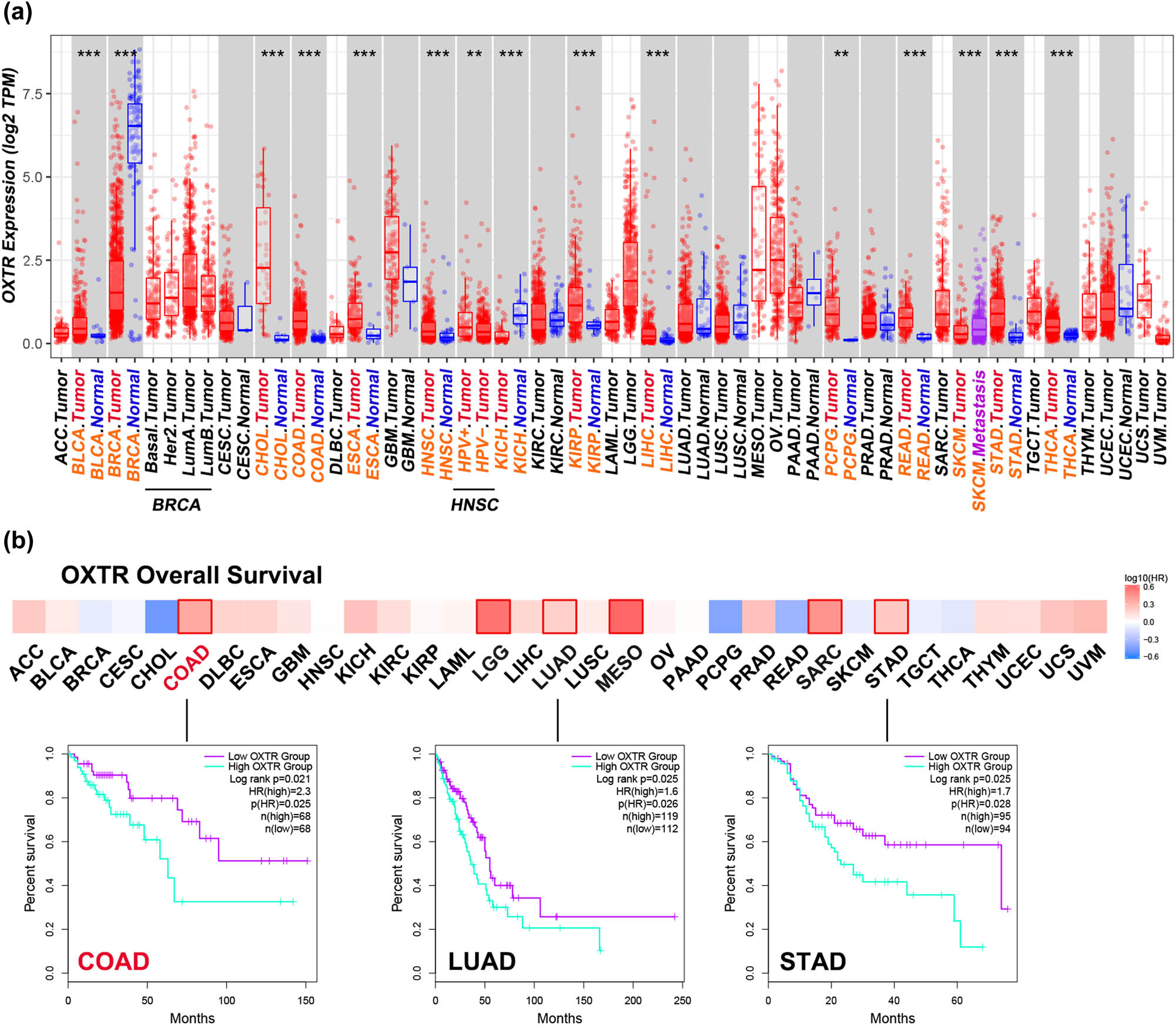
Analysis of OXTR expression in various tumor tissues in TCGA. (a) Transcriptional levels of OXTR in different tumor tissues and adjacent normal tissues. (b) The relationships between OXTR expression levels in various tumors and OS were analyzed by KM curve. COAD: low OXTR group: n = 68, high OXTR group: n = 68; LUAD: low OXTR group: n = 112, high OXTR group: n = 119; STAD: low OXTR group: n = 94, high OXTR group: n = 95. ACC: adrenocortical carcinoma; BLCA: bladder urothelial carcinoma; BRCA: breast invasive carcinoma; CESC: cervical squamous cell carcinoma and endocervical adenocarcinoma; CHOL: cholangiocarcinoma; COAD: colon adenocarcinoma; DLBC: lymphoid neoplasm diffuse large B-cell lymphoma; ESCA: esophageal carcinoma; GBM: glioblastoma multiforme; HNSC: head and neck squamous cell carcinoma; HPV: human papillomavirus; KICH: kidney chromophobe; KIRC: kidney renal clear cell carcinoma; KIRP: kidney renal papillary cell carcinoma; LAML: acute myeloid leukemia; LGG: brain lower grade glioma; LIHC: liver hepatocellular carcinoma; LUAD: lung adenocarcinoma; LUSC: lung squamous cell carcinoma; MESO: mesothelioma; OV: ovarian serous cystadenocarcinoma; PAAD: pancreatic adenocarcinoma; PCPG: pheochromocytoma and paraganglioma; PRAD: prostate adenocarcinoma; READ: rectum adenocarcinoma; SARC: sarcoma; SKCM: skin cutaneous melanoma; STAD: stomach adenocarcinoma; TGCT: testicular germ cell tumors; THCA: thyroid carcinoma; THYM: thymoma; UCEC: uterine corpus endometrial carcinoma; UCS: uterine carcinosarcoma; UVM: uveal melanoma; LumA: luminal A; LumB: luminal B; Her2: human epidermal growth factor receptor-2. **: p < 0.01; ***: p < 0.001.
3.2 OXTR mRNA level is high in COAD patients from six datasets
To further determine the expression pattern of OXTR in COAD tissues, the OXTR mRNA levels in COAD samples from TCGA, GSE9348, GSE32323, GSE38026, GSE44076, and GSE115313 database were analyzed. The result shows that OXTR transcription levels in COAD tissues are higher than that in normal tissues (Figure 2).
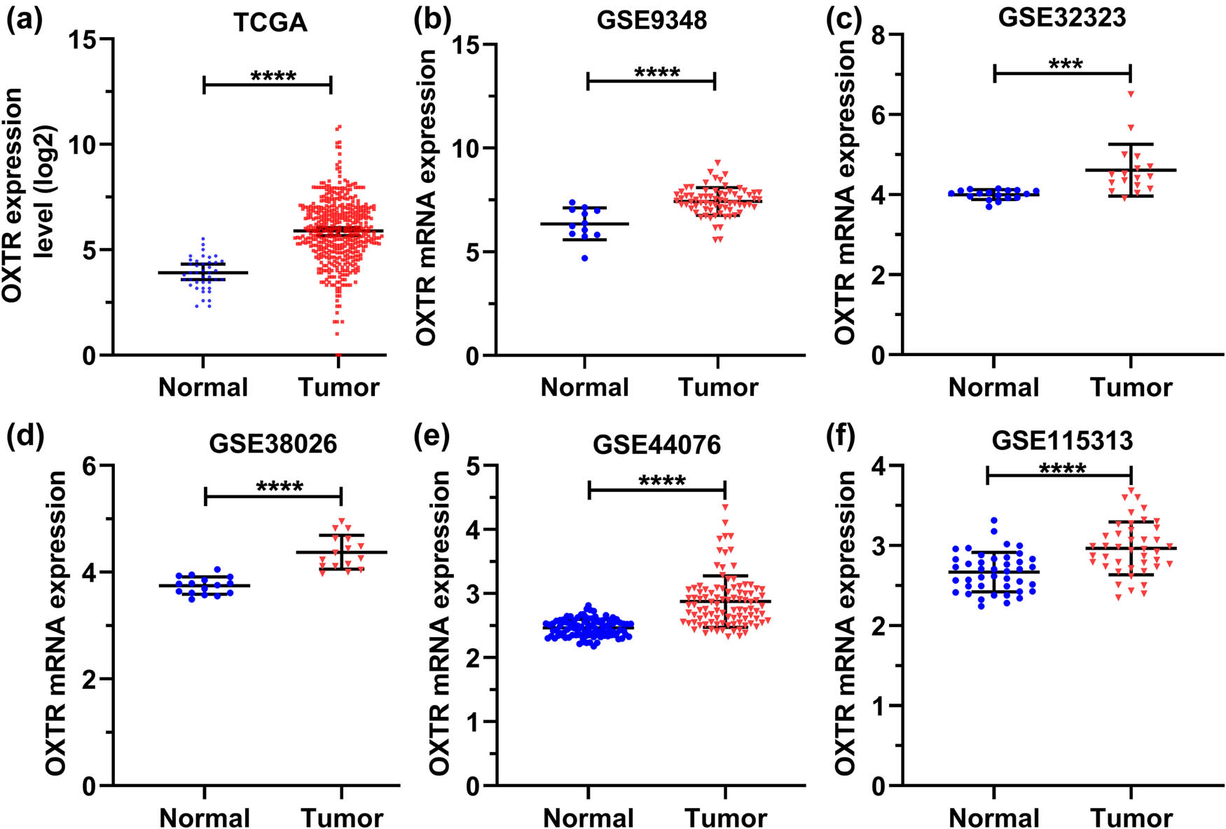
OXTR mRNA level is high in COAD patients from six datasets. (a–f) GraphPad Prism 8 was used to analyze the expression of OXTR in COAD samples from TCGA (a), GSE9348 (b), GSE32323 (c), GSE38026 (d), GSE44076 (e), and GSE115313 (f) databases. ***: p < 0.001; ****: p < 0.0001.
3.3 The level of OXTR is related to the progression of COAD
In order to investigate the role of upregulated OXTR mRNA in COAD patients, we further analyzed the relationship between OXTR mRNA levels and tumor progression. Patients (n = 428) were divided into groups with low or high OXTR expression using the survminer package in R language according to the survival data of COAD patients and the expression data of OXTR to investigate the clinical significance of OXTR expression. There were no significant differences between the two groups in gender, colon polyp history, hypermutation, tumor type, pathologic M, pathologic N, pathologic T, disease free survival (DFS), and progression-free interval (PFI) (all P > 0.05, Table 1). However, OXTR levels were strongly correlated with age, TNM stage, living status, and disease status (all p < 0.05, Table 1). Overall, these findings suggested that the level of OXTR is related to the progression of COAD.
Association between OXTR expression and the clinical parameters in patients with COAD in TCGA
| OXTR expression | p-value | |||
|---|---|---|---|---|
| Total | High | Low | ||
| (N = 428) | (N = 296) | (N = 132) | ||
| Age (year) | 0.00566 | |||
| >60 | 296 (69.2%) | 192 (64.9%) | 104 (78.8%) | |
| ≤60 | 132 (30.8%) | 104 (35.1%) | 28 (21.2%) | |
| Gender | 1 | |||
| Male | 228 (53.3%) | 158 (53.4%) | 70 (53.0%) | |
| Female | 200 (46.7%) | 138 (46.6%) | 62 (47.0%) | |
| TNM stage | 0.0309 | |||
| I | 73 (17.1%) | 43 (14.5%) | 30 (22.7%) | |
| II | 168 (39.3%) | 119 (40.2%) | 49 (37.1%) | |
| III | 126 (29.4%) | 84 (28.4%) | 42 (31.8%) | |
| IV | 61 (14.3%) | 50 (16.9%) | 11 (8.3%) | |
| History of colon polyps | 0.0642 | |||
| Yes | 127 (29.7%) | 78 (26.4%) | 49 (37.1%) | |
| No | 239 (55.8%) | 171 (57.8%) | 68 (51.5%) | |
| Unknown | 62 (14.5%) | 47 (15.9%) | 15 (11.4%) | |
| Hypermutation | 0.762 | |||
| FALSE | 111 (25.9%) | 75 (25.3%) | 36 (27.3%) | |
| Unknown | 317 (74.1%) | 221 (74.7%) | 96 (72.7%) | |
| Type | 0.81 | |||
| Colon adenocarcinoma | 363 (84.8%) | 253 (85.5%) | 110 (83.3%) | |
| Colon mucinous adenocarcinoma | 60 (14.0%) | 40 (13.5%) | 20 (15.2%) | |
| Unknown | 5 (1.2%) | 3 (1.0%) | 2 (1.5%) | |
| Pathologic M | 0.102 | |||
| M0 | 320 (74.8%) | 212 (71.6%) | 108 (81.8%) | |
| M1 | 61 (14.3%) | 50 (16.9%) | 11 (8.3%) | |
| MX | 43 (10.0%) | 31 (10.5%) | 12 (9.1%) | |
| Unknown | 4 (0.9%) | 3 (1.0%) | 1 (0.8%) | |
| Pathologic N | 0.0909 | |||
| N0 | 249 (58.2%) | 169 (57.1%) | 80 (60.6%) | |
| N1 | 102 (23.8%) | 66 (22.3%) | 36 (27.3%) | |
| N2 | 77 (18.0%) | 61 (20.6%) | 16 (12.1%) | |
| Pathologic T | 0.0552 | |||
| T1 | 10 (2.3%) | 7 (2.4%) | 3 (2.3%) | |
| T2 | 72 (16.8%) | 42 (14.2%) | 30 (22.7%) | |
| T3 | 296 (69.2%) | 205 (69.3%) | 91 (68.9%) | |
| T4 | 49 (11.4%) | 41 (13.9%) | 8 (6.1%) | |
| Unknown | 1 (0.2%) | 1 (0.3%) | 0 (0%) | |
| Residual tumor | 0.818 | |||
| R0 | 314 (73.4%) | 215 (72.6%) | 99 (75.0%) | |
| R1–R2 | 25 (5.8%) | 17 (5.7%) | 8 (6.1%) | |
| Unknown | 89 (20.8%) | 64 (21.6%) | 25 (18.9%) | |
| Living status | 0.0318 | |||
| Alive | 334 (78.0%) | 222 (75.0%) | 112 (84.8%) | |
| Dead | 94 (22.0%) | 74 (25.0%) | 20 (15.2%) | |
| Disease status | 0.0082 | |||
| No | 159 (37.1%) | 97 (32.8%) | 62 (47.0%) | |
| Yes | 22 (5.1%) | 19 (6.4%) | 3 (2.3%) | |
| Unknown | 247 (57.7%) | 180 (60.8%) | 67 (50.8%) | |
| DFS | 0.112 | |||
| No | 355 (82.9%) | 238 (80.4%) | 117 (88.6%) | |
| Yes | 58 (13.6%) | 46 (15.5%) | 12 (9.1%) | |
| Unknown | 15 (3.5%) | 12 (4.1%) | 3 (2.3%) | |
| PFI | 0.0513 | |||
| No | 312 (72.9%) | 207 (69.9%) | 105 (79.5%) | |
| Yes | 116 (27.1%) | 89 (30.1%) | 27 (20.5%) | |
DFS: disease free survival; PFI: progression-free interval.
Bold values prompts p < 0.05.
3.4 The ROC curve shows that OXTR could distinguish COAD tissues from normal tissues
To confirm that OXTR mRNA level is associated with tumor progression and poor prognosis in patients, we utilized ROC curves. We found that OXTR could discriminate COAD tissues from normal tissues with an AUC of 0.8900 (95% CI: 0.8569–0.9232; p < 0.0001) (Figure 3a). Similarly, it also effectively distinguished COAD tissues from normal tissues in 42 paired samples with an AUC of 0.9210 (95% CI: 0.8559–0.9862; p < 0.0001) (Figure 3b). However, we found that OXTR cannot effectively distinguish COAD subgroups, including TNM stage (TNM stages I + II versus TNM stages III + IV, AUC = 0.5639, p = 0.0078, Figure 3c), T stage (stages T1 + T2 versus stages T3 + T4, AUC = 0.5930, p = 0.0090, Figure 3d), OS (OS-good versus OS-poor, AUC = 0.5769, p = 0.0058, Figure 3e), and living status (living versus deceased, AUC = 0.5703, p = 0.0474, Figure 3f). These results confirm that OXTR can be used as a biomarker of COAD, but it cannot effectively distinguish COAD tissues in different states.
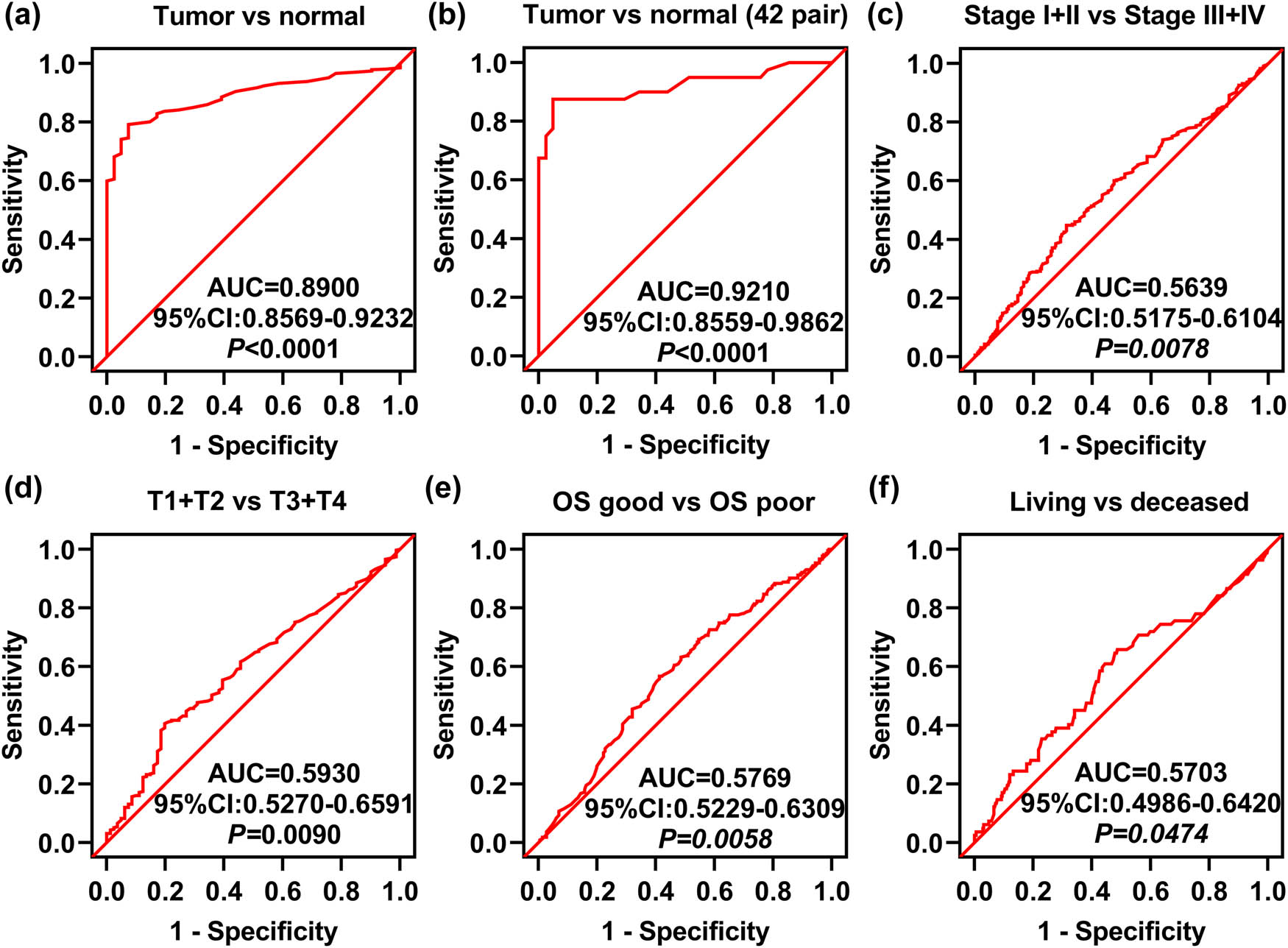
ROC curve analysis of OXTR expression related to the indicated clinical parameters in COAD patients. (a) The ROC curve analysis of COAD tissue and unpaired normal tissue. (b) The ROC curve analysis of COAD tissues and paired normal tissues. (c–f) ROC curve analysis of TNM stage (c), T stage (d), OS (e), and living status (f). AUC: area under the curve; OS: overall survival.
3.5 COAD patients with high levels of OXTR are more likely to show short OS time
We used KM survival analysis to verify the relationship between OS time and OXTR mRNA level in COAD patients. Based on the median value of OXTR mRNA level in the TCGA data set, we divided 268 patients with COAD into high OXTR mRNA group and low OXTR mRNA group. Patients with high OXTR expression have shorter OS time than those with low OXTR expression (Figure 4a, p = 0.0083). Moreover, male (p = 0.0099) patients with high OXTR mRNA level showed shorter OS, and age > 60 years (p = 0.0417) and M0 stage (p = 0.0393) patients with high OXTR expression are more likely to exhibit a short OS time, but there was no significant correlation between OS time and OXTR level in females (p = 0.1869), age ≤ 60 years (p = 0.0965), T1 + T2 stage (p = 0.1015), T3 + T4 stage (p = 0.0503), N0 stage (p = 0.1375), stages I + II (p = 0.3625), and stages III + IV (p = 0.0901) patients (Figure 4b–k). All these data reveal that high levels of OXTR are associated with patients exhibiting a short OS time.
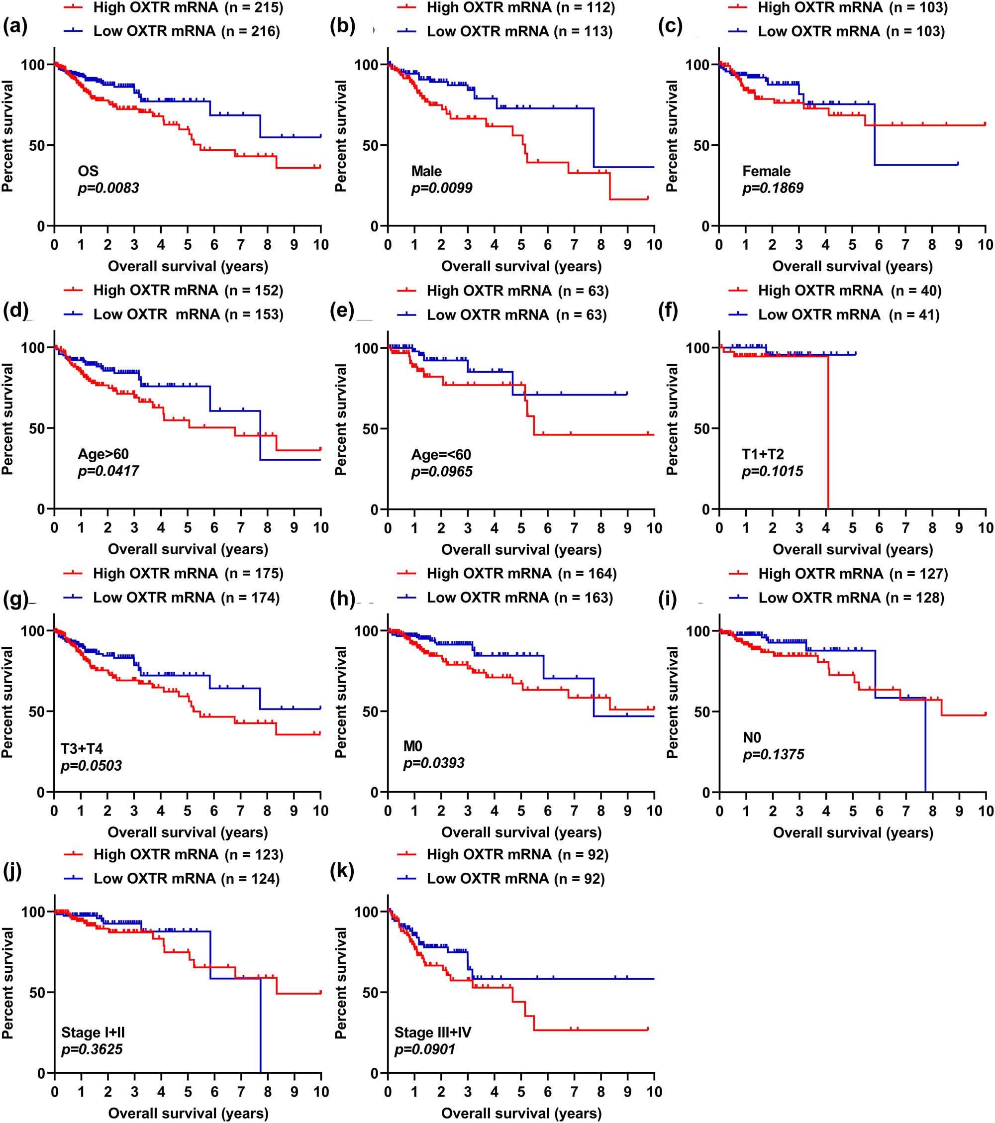
COAD patients with high levels of OXTR are more likely to show short OS time. (a) The correlation analysis between OXTR mRNA level and OS in COAD patients was performed by the KM curve. (b–k) KM curve analysis of male (b), female (c), age > 60 years (d), age ≤ 60 years (e), stages T1 + T2 (f), stages T3 + T4 (g), no metastasis (h), N0 stage (i), stages I + II (j), and stages III + IV (k) patients with COAD.
3.6 OXTR is involved in cell cycle regulation
To explore the association between OXTR expression and genes involved in cell cycle regulation, we performed GO enrichment analysis and found that high OXTR expression was associated with cell cycle regulation (Figure 5a). Furthermore, the expression of OXTR was strongly associated with genes involved in cell cycle regulation, including CCND1, RAD51D, MTBP, METTL3, ADAM17, CDK2, DDL39B, and EZH2 (Figure 5b). These findings show that high levels of OXTR are involved in COAD cell proliferation.
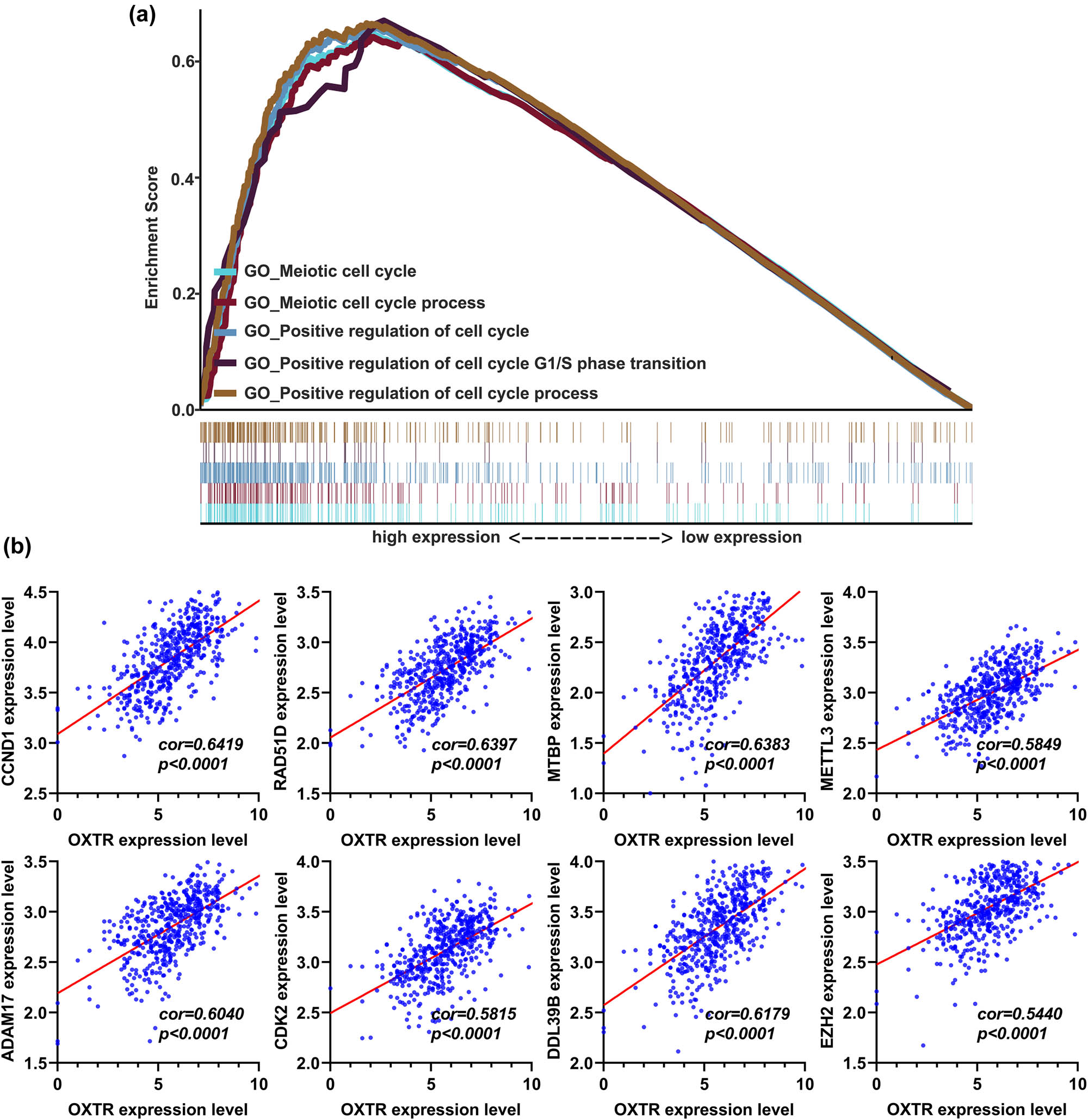
GO enrichment analysis of the correlation between OXTR mRNA level and genes related to cell cycle regulation. (a) Gene set enrichment analysis of the correlation between OXTR expression and mRNA levels of genes involved in cell cycle regulation. (b) Correlation analysis between OXTR expression and mRNA levels of genes involved in cell cycle regulation. GO, gene ontology.
3.7 OXTR is involved in regulating four signaling pathways closely related to the occurrence and development of tumors
We studied the relationship between OXTR levels and signaling pathways through KEGG enrichment analysis, finding that OXTR level was positively associated with four main well-studied signaling pathways in COAD, including the hedgehog, mTOR, TGF-β, and Wnt signaling pathways (Figure 6a). Moreover, OXTR level was positively associated with colorectal cancer (Figure 6a). In addition, OXTR levels were positively correlated with the levels of genes related to these pathways, including BRAF, BTRC, CSNK2A2, GSK3B, RBL1, RHEB, SMAD5, and TSC1 (Figure 6b).

KEGG enrichment analysis of the correlation of OXTR mRNA level with colorectal cancer and four main signaling pathways in colorectal cancer. (a) Gene set enrichment analysis of the correlation between OXTR expression level and the expression of genes related to four signaling pathways in colorectal cancer. (b) Correlation analysis between the OXTR expression level and mRNA levels of genes related to four signaling pathways in COAD. KEGG: kyoto encyclopedia of genes and genomes.
3.8 PPI and gene co-expression network analysis
Genes co-expressed with OXTR were screened using cBioPortal database. The Spearman correlation coefficient of a total of 146 genes was greater than 0.3 (Figure 7a). Subsequently, these genes were used for PPI network. After the removal of proteins that did not interact with other proteins, a total of 34 genes were found in the PPI network, including 10 nodes (DYNC2H1, LCN2, FN1, ABL2, FOXO3, NOTCH4, WTIP, ACTA2, CDH2, and NES) (Figure 7b). Unfortunately, no genes encoding proteins that interact with OXTR have been found (Figure 7b).
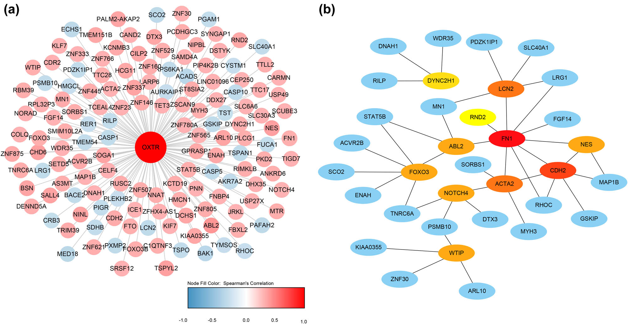
PPI and gene co-expression network analysis. (a) Hub genes of the PPI network. (b) Gene co-expression networks. Blue indicates genes that are negatively related to OXTR, and red indicates genes that are positively related to OXTR. The darker the color, the stronger the correlation.
3.9 Correlations between OXTR expression and immune infiltration in COAD
After analyzing the immune infiltration in COAD tissues, we found that OXTR level is positively correlated with the infiltration of type 2T helper cell, central memory CD8 T cell, and CD57 bright natural killer cell, while negatively correlated with the infiltration of activated CD8 T cell, activated B cell, and Type 1T helper cell (Figure 8).
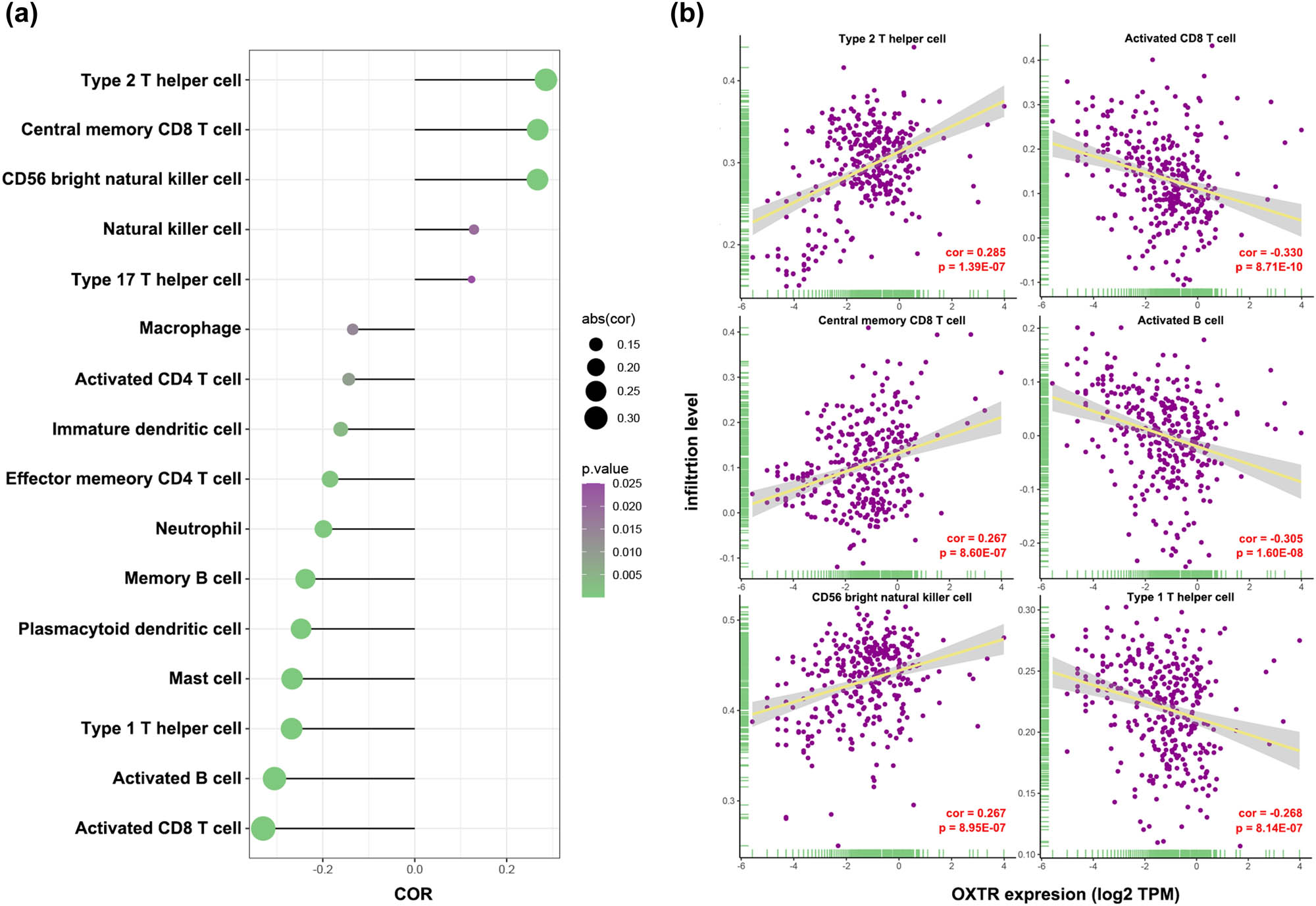
Correlations between OXTR expression and immune infiltration in COAD. (a) Rod diagram of the relationship between OXTR levels and infiltration levels of various immune cells. (b) Scatter plot of the association between OXTR levels and infiltration levels of type 2T helper cell, central memory CD8 T cell, CD57 bright natural killer cell, activated CD8 T cell, activated B cell, and Type 1T helper cell.
3.10 Silencing OXTR inhibits cell proliferation, migration, and invasion
In order to explore the effect of upregulated OXTR in COAD tissues on the biological processes of COAD cells, we silenced the expression of OXTR in HCT-8 and SW480 cells. We tested the expression of OXTR in human normal colon epithelial cell line (NCM-460) and COAD cell lines (HCT-8, SW480, SW620, and RKO), and found that the expression level of OXTR in COAD cell lines was higher than that in NCM-460 cells. Among them, the expression level of OXTR was the highest in HCT-8 cells, followed by that in SW480 cells (Figure 9a and b). Therefore, subsequent cell experiments were performed with HCT-8 and SW480 cells. We designed three siRNA of OXTR (siOXTR-1, siOXTR-2, and siOXTR-3), and their sequences are shown in Table 2. After they were transfected into cells for 48 h, qPCR was performed and it was found that siOXTR-3 had the most significant effect on reducing the OXTR mRNA level in cells (Figure 9c). Therefore, siOXTR-3 was used in subsequent experiments. In addition, transfection of siOXTR into cells also reduced the level of OXTR protein in the cells (Figure 9d). Moreover, decreasing OXTR inhibited cell proliferation, migration, and invasion, and arrested the cell cycle (Figure 9e–h). These findings indicate that the upregulated OXTR in COAD tissue may play a role in promoting cell proliferation, migration, and invasion.
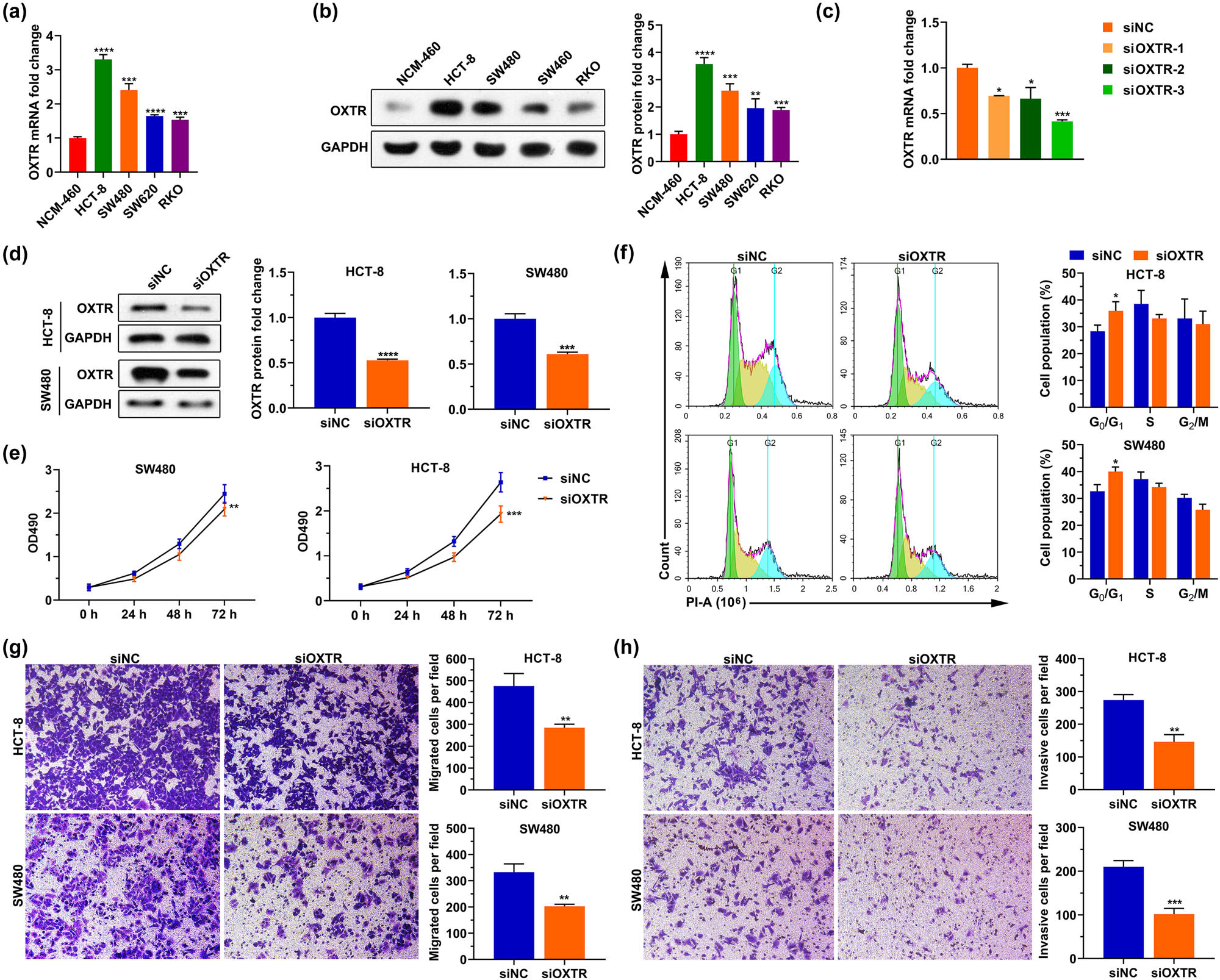
Silencing OXTR inhibits cell proliferation, migration, and invasion. (a and b) OXTR mRNA (a) and protein (b) levels in human normal colonic epithelial cell line (NCM-460) and COAD cell lines (HCT-8, SW480, SW620, and RKO). (c and d) After HCT-8 and SW480 cells were transfected with siOXTR, qPCR (c) and western blotting (d) assays were used to analyze the levels of OXTR mRNA and protein in the cells, respectively. (e‒h) After silencing OXTR, MTT assay, PI staining, and transwell assay were used to detect cell proliferation (e), cell cycle (f), migration (g), and invasion (h). siOXTR: small interfering RNA of OXTR; siNC: the negative control of siOXTR; PI: propidium iodide. *: p < 0.05, **: p < 0.01, ***: p < 0.001, ****: p < 0.0001.
The sequences of siRNAs
| Name | Sequence (5′-3′) |
|---|---|
| siNC | UUCUCCGAACGUGUCACGUTT |
| siOXTR-1 | AUCUUGAAGCUGAUAAGGCCG |
| siOXTR-2 | AGAUCUUGAAGCUGAUAAGGC |
| siOXTR-3 | UGAUGAAAGUCAUCUUGACCG |
siNC: the negative control of siRNA. siOXTR: the siRNA of OXTR.
4 Discussion
COAD is generally believed to be an illness derived from the accumulation of genetic and epigenetic mutations in epithelial cells. OXTR coupling with Gi and Gh proteins enables association with oxytocin to relay signals to trigger reproductive and social behavior [17,18]. Several previous studies have demonstrated that the activation of OXTR-mediated signaling promotes or prevents tumorigenesis and metastasis in multiple cancers, including breast cancer, non-small-cell lung cancer, prostate cancer, and ovarian cancer [20,21,28,29]. In the present study, we explored the role of OXTR in modulating progress and metastasis of COAD by bioinformatics analysis. Our results found that the mRNA OXTR upregulation was associated with growth and distant metastasis of COAD, and high mRNA level of OXTR indicated a poor prognosis in COAD patients. In addition, we also found that downregulating the expression of OXTR in COAD cells reduced the ability of cells to proliferate, migrate and invade, and blocked cell cycle progression.
Moreover, studies have shown that excessive activation of TGF-β, Wnt/β-catenin, Smad, Notch, MAPK, HIF-1, and mTOR signals in COAD led to the development of COAD [8,10,11,12]. GO and KEGG enrichment analyses also indicated that high OXTR expression was associated with loss of cell cycle regulation and significantly associated with four main signaling transduction pathways, including the hedgehog, mTOR, TGF-β, and Wnt signaling pathways. Therefore, we hypothesized that OXTR may be involved in overactivation of the mTOR, TGF-β, and Wnt signaling pathways to promote tumor progression, which needs further study. However, through PPI network analysis, no genes encoding proteins that interact with OXTR were found, indicating that further experimental studies are needed to understand the proteins interacting with OXTR.
This study lacks animal experiments and test results of OXTR protein in COAD tissue, so it is impossible to directly verify the influence of OXTR on COAD tumor growth, which is the limitation of this study. In addition, we found that OXTR levels did not have a significant effect on OS in females with COAD, but have a significant effect on OS in males with COAD. The reason why OXTR level had no significant effect on the OS of females with COAD may be that some female factors affected the effect of OXTR, or the sample size of females with COAD was not large enough, which still needs further investigation.
In conclusion, our findings clarified that the mRNA level of OXTR was elevated in COAD tissues and distant metastasis-prone COAD patients. Our results also suggest that COAD patients with high levels of OXTR have poorer OS than patients with low levels of OXTR. OXTR level was positively associated with hedgehog, mTOR, TGF-β, and Wnt signaling pathways, and regulation of cell cycle. OXTR expression was significantly correlated with the infiltration level of activated CD8 T cell and activated B cell. Moreover, knockdown of OXTR suppressed the proliferation, migration, and invasion of COAD cells, and blocked the cells in the G0/G1 phase. OXTR might be a potential therapeutic target for COAD.
Acknowledgements
We are very grateful for the funding support of the Scientific Research Project of Health and Family Planning Commission of Wuxi [Grant number Q201617].
-
Funding information: This work was supported by the Scientific Research Project of Health and Family Planning Commission of Wuxi (Grant number Q201617).
-
Author contributions: Zhenyu Xu, Junjie Sun, and Dong Hua designed this study. Zhenyu Xu, Junjie Sun, Yang Chen, Ting Zhang, Yan Qin, and Dong Hua performed bioinformatics analysis and in vitro experiments. Zhenyu Xu and Junjie Sun drafted the manuscript. Yang Chen, Ting Zhang, Yan Qin, and Dong Hua revised this manuscript. All authors approved the version to be published, and agreed to be accountable for all aspects of the work.
-
Conflict of interest: All authors have no financial interest or conflict of interest in association with this work.
-
Data availability statement: All relevant data and materials are presented in the manuscript. For more information, please contact the corresponding author.
References
[1] Torre LA, Bray F, Siegel RL, Ferlay J, Lortet-Tieulent J, Jemal A. Global cancer statistics, 2012. CA Cancer J Clin. 2015;65(2):87–108. 10.3322/caac.21262.Suche in Google Scholar PubMed
[2] Jemal A, Bray F, Center MM, Ferlay J, Ward E, Forman D. Global cancer statistics. CA Cancer J Clin. 2011;61(2):69–90. 10.3322/caac.20107.Suche in Google Scholar PubMed
[3] Khan K, Wale A, Brown G, Chau I. Colorectal cancer with liver metastases: neoadjuvant chemotherapy, surgical resection first or palliation alone? World J Gastroenterol. 2014;20(35):12391–406. 10.3748/wjg.v20.i35.12391.Suche in Google Scholar PubMed PubMed Central
[4] Diener-West M, Dobbins TW, Phillips TL, Nelson DF. Identification of an optimal subgroup for treatment evaluation of patients with brain metastases using RTOG study 7916. Int J Radiat Oncol Biol Phys. 1989;16(3):669–73. 10.1016/0360-3016(89)90483-5.Suche in Google Scholar PubMed
[5] Edge SB, Compton CC. The American Joint Committee on Cancer: the 7th edition of the AJCC cancer staging manual and the future of TNM. Ann Surg Oncol. 2010;17(6):1471–4. 10.1245/s10434-010-0985-4.Suche in Google Scholar PubMed
[6] Siegel RL, Jemal A, Ward EM. Increase in incidence of colorectal cancer among young men and women in the United States. Cancer Epidemiol Biomarkers Prev. 2009;18(6):1695–8. 10.1158/1055-9965.EPI-09-0186.Suche in Google Scholar PubMed
[7] Center MM, Jemal A, Ward E. International trends in colorectal cancer incidence rates. Cancer Epidemiol Biomarkers Prev. 2009;18(6):1688–94. 10.1158/1055-9965.EPI-09-0090.Suche in Google Scholar PubMed
[8] Li X-L, Zhou J, Chen Z-R, Chng W-J. P53 mutations in colorectal cancer - molecular pathogenesis and pharmacological reactivation. World J Gastroenterol. 2015;21(1):84–93. 10.3748/wjg.v21.i1.84.Suche in Google Scholar PubMed PubMed Central
[9] Lao VV, Grady WM. Epigenetics and colorectal cancer. Nat Rev Gastroenterol Hepatol. 2011;8(12):686–700. 10.1038/nrgastro.2011.173.Suche in Google Scholar PubMed PubMed Central
[10] Ying J, Li H, Yu J, Ng KM, Poon FF, Wong SCC, et al. WNT5A exhibits tumor-suppressive activity through antagonizing the Wnt/beta-catenin signaling, and is frequently methylated in colorectal cancer. Clin Cancer Res. 2008;14(1):55–61. 10.1158/1078-0432.CCR-07-1644.Suche in Google Scholar PubMed
[11] Hlubek F, Brabletz T, Budczies J, Pfeiffer S, Jung A, Kirchner T. Heterogeneous expression of Wnt/beta-catenin target genes within colorectal cancer. Int J Cancer. 2007;121(9):1941–8. 10.1002/ijc.22916.Suche in Google Scholar PubMed
[12] Zhou S, Buckhaults P, Zawel L, Bunz F, Riggins G, Dai JL, et al. Targeted deletion of Smad4 shows it is required for transforming growth factor beta and activin signaling in colorectal cancer cells. Proc Natl Acad Sci U S A. 1998;95(5):2412–6. 10.1073/pnas.95.5.2412.Suche in Google Scholar PubMed PubMed Central
[13] Kazanjian A, Shroyer NF. NOTCH signaling and ATOH1 in colorectal cancers. Curr Colorectal Cancer Rep. 2011;7(2):121–7. 10.1007/s11888-011-0090-5.Suche in Google Scholar PubMed PubMed Central
[14] Wang X, Sun D, Tai J, Chen S, Yu M, Ren D, et al. TFAP2C promotes stemness and chemotherapeutic resistance in colorectal cancer via inactivating Hippo signaling pathway. J Exp Clin Cancer Res. 2018;37(1):27. 10.1186/s13046-018-0683-9.Suche in Google Scholar PubMed PubMed Central
[15] Saw CLL, Kong A-NT. Nuclear factor-erythroid 2-related factor 2 as a chemopreventive target in colorectal cancer. Expert Opin Ther Targets. 2011;15(3):281–95. 10.1517/14728222.2011.553602.Suche in Google Scholar PubMed PubMed Central
[16] Hanada N, Takahata T, Zhou Q, Ye X, Sun R, Itoh J, et al. Methylation of the KEAP1 gene promoter region in human colorectal cancer. BMC Cancer. 2012;12:66. 10.1186/1471-2407-12-66.Suche in Google Scholar PubMed PubMed Central
[17] Prichard ZM, Mackinnon AJ, Jorm AF, Easteal S. AVPR1A and OXTR polymorphisms are associated with sexual and reproductive behavioral phenotypes in humans. Mutation in brief no. 981. Online. Hum Mutat. 2007;28(11):1150. 10.1002/humu.9510.Suche in Google Scholar PubMed
[18] Jurek B, Neumann ID. The oxytocin receptor: from intracellular signaling to behavior. Physiol Rev. 2018;98(3):1805–908. 10.1152/physrev.00031.2017.Suche in Google Scholar PubMed
[19] Cassoni P, Sapino A, Marrocco T, Chini B, Bussolati G. Oxytocin and oxytocin receptors in cancer cells and proliferation. J Neuroendocrinol. 2004;16(4):362–4. 10.1111/j.0953-8194.2004.01165.x.Suche in Google Scholar PubMed
[20] Ariana M, Pornour M, Mehr SS, Vaseghi H, Ganji SM, Alivand MR, et al. Preventive effects of oxytocin and oxytocin receptor in breast cancer pathogenesis. Per Med. 2019;16(1):25–34. 10.2217/pme-2018-0009.Suche in Google Scholar PubMed
[21] Zhong M, Boseman ML, Millena AC, Khan SA. Oxytocin induces the migration of prostate cancer cells: involvement of the Gi-coupled signaling pathway. Mol Cancer Res. 2010;8(8):1164–72. 10.1158/1541-7786.MCR-09-0329.Suche in Google Scholar PubMed PubMed Central
[22] Tang Z, Li C, Kang B, Gao G, Li C, Zhang Z. GEPIA: a web server for cancer and normal gene expression profiling and interactive analyses. Nucleic Acids Res. 2017;45(W1):W98–102. 10.1093/nar/gkx247.Suche in Google Scholar PubMed PubMed Central
[23] Subramanian A, Tamayo P, Mootha VK, Mukherjee S, Ebert BL, Gillette MA, et al. Gene set enrichment analysis: a knowledge-based approach for interpreting genome-wide expression profiles. Proc Natl Acad Sci U S A. 2005;102(43):15545–50. 10.1073/pnas.0506580102.Suche in Google Scholar PubMed PubMed Central
[24] Liberzon A, Subramanian A, Pinchback R, Thorvaldsdóttir H, Tamayo P, Mesirov JP. Molecular signatures database (MSigDB) 3.0. Bioinformatics. 2011;27(12):1739–40. 10.1093/bioinformatics/btr260.Suche in Google Scholar PubMed PubMed Central
[25] Yao X, Hu W, Zhang J, Huang C, Zhao H, Yao X. Application of cAMP-dependent catalytic subunit β (PRKACB) low expression in predicting worse overall survival: a potential therapeutic target for colorectal carcinoma. J Cancer. 2020;11(16):4841–50. 10.7150/jca.46156.Suche in Google Scholar PubMed PubMed Central
[26] Goldman M, Craft B, Swatloski T, Cline M, Morozova O, Diekhans M, et al. The UCSC cancer genomics browser: update 2015. Nucleic Acids Res. 2015;43(Database issue):D812–7. 10.1093/nar/gku1073.Suche in Google Scholar PubMed PubMed Central
[27] Charoentong P, Finotello F, Angelova M, Mayer C, Efremova M, Rieder D, et al. Pan-cancer immunogenomic analyses reveal genotype-immunophenotype relationships and predictors of response to checkpoint blockade. Cell Rep. 2017;18(1):248–62. 10.1016/j.celrep.2016.12.019.Suche in Google Scholar PubMed
[28] Kuzumaki N, Suzuki A, Narita M, Hosoya T, Nagasawa A, Imai S, et al. Multiple analyses of G-protein coupled receptor (GPCR) expression in the development of gefitinib-resistance in transforming non-small-cell lung cancer. PLoS One. 2012;7(10):e44368. 10.1371/journal.pone.0044368.Suche in Google Scholar PubMed PubMed Central
[29] Ji H, Liu N, Yin Y, Wang X, Chen X, Li J, et al. Oxytocin inhibits ovarian cancer metastasis by repressing the expression of MMP-2 and VEGF. J Cancer. 2018;9(8):1379–84. 10.7150/jca.23769.Suche in Google Scholar PubMed PubMed Central
© 2021 Junjie Sun et al., published by De Gruyter
This work is licensed under the Creative Commons Attribution 4.0 International License.
Artikel in diesem Heft
- Research Articles
- Identification of ZG16B as a prognostic biomarker in breast cancer
- Behçet’s disease with latent Mycobacterium tuberculosis infection
- Erratum
- Erratum to “Suffering from Cerebral Small Vessel Disease with and without Metabolic Syndrome”
- Research Articles
- GPR37 promotes the malignancy of lung adenocarcinoma via TGF-β/Smad pathway
- Expression and role of ABIN1 in sepsis: In vitro and in vivo studies
- Additional baricitinib loading dose improves clinical outcome in COVID-19
- The co-treatment of rosuvastatin with dapagliflozin synergistically inhibited apoptosis via activating the PI3K/AKt/mTOR signaling pathway in myocardial ischemia/reperfusion injury rats
- SLC12A8 plays a key role in bladder cancer progression and EMT
- LncRNA ATXN8OS enhances tamoxifen resistance in breast cancer
- Case Report
- Serratia marcescens as a cause of unfavorable outcome in the twin pregnancy
- Spleno-adrenal fusion mimicking an adrenal metastasis of a renal cell carcinoma: A case report and embryological background
- Research Articles
- TRIM25 contributes to the malignancy of acute myeloid leukemia and is negatively regulated by microRNA-137
- CircRNA circ_0004370 promotes cell proliferation, migration, and invasion and inhibits cell apoptosis of esophageal cancer via miR-1301-3p/COL1A1 axis
- LncRNA XIST regulates atherosclerosis progression in ox-LDL-induced HUVECs
- Potential role of IFN-γ and IL-5 in sepsis prediction of preterm neonates
- Rapid Communication
- COVID-19 vaccine: Call for employees in international transportation industries and international travelers as the first priority in global distribution
- Case Report
- Rare squamous cell carcinoma of the kidney with concurrent xanthogranulomatous pyelonephritis: A case report and review of the literature
- An infertile female delivered a baby after removal of primary renal carcinoid tumor
- Research Articles
- Hypertension, BMI, and cardiovascular and cerebrovascular diseases
- Case Report
- Coexistence of bilateral macular edema and pale optic disc in the patient with Cohen syndrome
- Research Articles
- Correlation between kinematic sagittal parameters of the cervical lordosis or head posture and disc degeneration in patients with posterior neck pain
- Review Articles
- Hepatoid adenocarcinoma of the lung: An analysis of the Surveillance, Epidemiology, and End Results (SEER) database
- Research Articles
- Thermography in the diagnosis of carpal tunnel syndrome
- Pemetrexed-based first-line chemotherapy had particularly prominent objective response rate for advanced NSCLC: A network meta-analysis
- Comparison of single and double autologous stem cell transplantation in multiple myeloma patients
- The influence of smoking in minimally invasive spinal fusion surgery
- Impact of body mass index on left atrial dimension in HOCM patients
- Expression and clinical significance of CMTM1 in hepatocellular carcinoma
- miR-142-5p promotes cervical cancer progression by targeting LMX1A through Wnt/β-catenin pathway
- Comparison of multiple flatfoot indicators in 5–8-year-old children
- Early MRI imaging and follow-up study in cerebral amyloid angiopathy
- Intestinal fatty acid-binding protein as a biomarker for the diagnosis of strangulated intestinal obstruction: A meta-analysis
- miR-128-3p inhibits apoptosis and inflammation in LPS-induced sepsis by targeting TGFBR2
- Dynamic perfusion CT – A promising tool to diagnose pancreatic ductal adenocarcinoma
- Biomechanical evaluation of self-cinching stitch techniques in rotator cuff repair: The single-loop and double-loop knot stitches
- Review Articles
- The ambiguous role of mannose-binding lectin (MBL) in human immunity
- Case Report
- Membranous nephropathy with pulmonary cryptococcosis with improved 1-year follow-up results: A case report
- Fertility problems in males carrying an inversion of chromosome 10
- Acute myeloid leukemia with leukemic pleural effusion and high levels of pleural adenosine deaminase: A case report and review of literature
- Metastatic renal Ewing’s sarcoma in adult woman: Case report and review of the literature
- Burkitt-like lymphoma with 11q aberration in a patient with AIDS and a patient without AIDS: Two cases reports and literature review
- Skull hemophilia pseudotumor: A case report
- Judicious use of low-dosage corticosteroids for non-severe COVID-19: A case report
- Adult-onset citrullinaemia type II with liver cirrhosis: A rare cause of hyperammonaemia
- Clinicopathologic features of Good’s syndrome: Two cases and literature review
- Fatal immune-related hepatitis with intrahepatic cholestasis and pneumonia associated with camrelizumab: A case report and literature review
- Research Articles
- Effects of hydroxyethyl starch and gelatin on the risk of acute kidney injury following orthotopic liver transplantation: A multicenter retrospective comparative clinical study
- Significance of nucleic acid positive anal swab in COVID-19 patients
- circAPLP2 promotes colorectal cancer progression by upregulating HELLS by targeting miR-335-5p
- Ratios between circulating myeloid cells and lymphocytes are associated with mortality in severe COVID-19 patients
- Risk factors of left atrial appendage thrombus in patients with non-valvular atrial fibrillation
- Clinical features of hypertensive patients with COVID-19 compared with a normotensive group: Single-center experience in China
- Surgical myocardial revascularization outcomes in Kawasaki disease: systematic review and meta-analysis
- Decreased chromobox homologue 7 expression is associated with epithelial–mesenchymal transition and poor prognosis in cervical cancer
- FGF16 regulated by miR-520b enhances the cell proliferation of lung cancer
- Platelet-rich fibrin: Basics of biological actions and protocol modifications
- Accurate diagnosis of prostate cancer using logistic regression
- miR-377 inhibition enhances the survival of trophoblast cells via upregulation of FNDC5 in gestational diabetes mellitus
- Prognostic significance of TRIM28 expression in patients with breast carcinoma
- Integrative bioinformatics analysis of KPNA2 in six major human cancers
- Exosomal-mediated transfer of OIP5-AS1 enhanced cell chemoresistance to trastuzumab in breast cancer via up-regulating HMGB3 by sponging miR-381-3p
- A four-lncRNA signature for predicting prognosis of recurrence patients with gastric cancer
- Knockdown of circ_0003204 alleviates oxidative low-density lipoprotein-induced human umbilical vein endothelial cells injury: Circulating RNAs could explain atherosclerosis disease progression
- Propofol postpones colorectal cancer development through circ_0026344/miR-645/Akt/mTOR signal pathway
- Knockdown of lncRNA TapSAKI alleviates LPS-induced injury in HK-2 cells through the miR-205/IRF3 pathway
- COVID-19 severity in relation to sociodemographics and vitamin D use
- Clinical analysis of 11 cases of nocardiosis
- Cis-regulatory elements in conserved non-coding sequences of nuclear receptor genes indicate for crosstalk between endocrine systems
- Four long noncoding RNAs act as biomarkers in lung adenocarcinoma
- Real-world evidence of cytomegalovirus reactivation in non-Hodgkin lymphomas treated with bendamustine-containing regimens
- Relation between IL-8 level and obstructive sleep apnea syndrome
- circAGFG1 sponges miR-28-5p to promote non-small-cell lung cancer progression through modulating HIF-1α level
- Nomogram prediction model for renal anaemia in IgA nephropathy patients
- Effect of antibiotic use on the efficacy of nivolumab in the treatment of advanced/metastatic non-small cell lung cancer: A meta-analysis
- NDRG2 inhibition facilitates angiogenesis of hepatocellular carcinoma
- A nomogram for predicting metabolic steatohepatitis: The combination of NAMPT, RALGDS, GADD45B, FOSL2, RTP3, and RASD1
- Clinical and prognostic features of MMP-2 and VEGF in AEG patients
- The value of miR-510 in the prognosis and development of colon cancer
- Functional implications of PABPC1 in the development of ovarian cancer
- Prognostic value of preoperative inflammation-based predictors in patients with bladder carcinoma after radical cystectomy
- Sublingual immunotherapy increases Treg/Th17 ratio in allergic rhinitis
- Prediction of improvement after anterior cruciate ligament reconstruction
- Effluent Osteopontin levels reflect the peritoneal solute transport rate
- circ_0038467 promotes PM2.5-induced bronchial epithelial cell dysfunction
- Significance of miR-141 and miR-340 in cervical squamous cell carcinoma
- Association between hair cortisol concentration and metabolic syndrome
- Microvessel density as a prognostic indicator of prostate cancer: A systematic review and meta-analysis
- Characteristics of BCR–ABL gene variants in patients of chronic myeloid leukemia
- Knee alterations in rheumatoid arthritis: Comparison of US and MRI
- Long non-coding RNA TUG1 aggravates cerebral ischemia and reperfusion injury by sponging miR-493-3p/miR-410-3p
- lncRNA MALAT1 regulated ATAD2 to facilitate retinoblastoma progression via miR-655-3p
- Development and validation of a nomogram for predicting severity in patients with hemorrhagic fever with renal syndrome: A retrospective study
- Analysis of COVID-19 outbreak origin in China in 2019 using differentiation method for unusual epidemiological events
- Laparoscopic versus open major liver resection for hepatocellular carcinoma: A case-matched analysis of short- and long-term outcomes
- Travelers’ vaccines and their adverse events in Nara, Japan
- Association between Tfh and PGA in children with Henoch–Schönlein purpura
- Can exchange transfusion be replaced by double-LED phototherapy?
- circ_0005962 functions as an oncogene to aggravate NSCLC progression
- Circular RNA VANGL1 knockdown suppressed viability, promoted apoptosis, and increased doxorubicin sensitivity through targeting miR-145-5p to regulate SOX4 in bladder cancer cells
- Serum intact fibroblast growth factor 23 in healthy paediatric population
- Algorithm of rational approach to reconstruction in Fournier’s disease
- A meta-analysis of exosome in the treatment of spinal cord injury
- Src-1 and SP2 promote the proliferation and epithelial–mesenchymal transition of nasopharyngeal carcinoma
- Dexmedetomidine may decrease the bupivacaine toxicity to heart
- Hypoxia stimulates the migration and invasion of osteosarcoma via up-regulating the NUSAP1 expression
- Long noncoding RNA XIST knockdown relieves the injury of microglia cells after spinal cord injury by sponging miR-219-5p
- External fixation via the anterior inferior iliac spine for proximal femoral fractures in young patients
- miR-128-3p reduced acute lung injury induced by sepsis via targeting PEL12
- HAGLR promotes neuron differentiation through the miR-130a-3p-MeCP2 axis
- Phosphoglycerate mutase 2 is elevated in serum of patients with heart failure and correlates with the disease severity and patient’s prognosis
- Cell population data in identifying active tuberculosis and community-acquired pneumonia
- Prognostic value of microRNA-4521 in non-small cell lung cancer and its regulatory effect on tumor progression
- Mean platelet volume and red blood cell distribution width is associated with prognosis in premature neonates with sepsis
- 3D-printed porous scaffold promotes osteogenic differentiation of hADMSCs
- Association of gene polymorphisms with women urinary incontinence
- Influence of COVID-19 pandemic on stress levels of urologic patients
- miR-496 inhibits proliferation via LYN and AKT pathway in gastric cancer
- miR-519d downregulates LEP expression to inhibit preeclampsia development
- Comparison of single- and triple-port VATS for lung cancer: A meta-analysis
- Fluorescent light energy modulates healing in skin grafted mouse model
- Silencing CDK6-AS1 inhibits LPS-induced inflammatory damage in HK-2 cells
- Predictive effect of DCE-MRI and DWI in brain metastases from NSCLC
- Severe postoperative hyperbilirubinemia in congenital heart disease
- Baicalin improves podocyte injury in rats with diabetic nephropathy by inhibiting PI3K/Akt/mTOR signaling pathway
- Clinical factors predicting ureteral stent failure in patients with external ureteral compression
- Novel H2S donor proglumide-ADT-OH protects HUVECs from ox-LDL-induced injury through NF-κB and JAK/SATA pathway
- Triple-Endobutton and clavicular hook: A propensity score matching analysis
- Long noncoding RNA MIAT inhibits the progression of diabetic nephropathy and the activation of NF-κB pathway in high glucose-treated renal tubular epithelial cells by the miR-182-5p/GPRC5A axis
- Serum exosomal miR-122-5p, GAS, and PGR in the non-invasive diagnosis of CAG
- miR-513b-5p inhibits the proliferation and promotes apoptosis of retinoblastoma cells by targeting TRIB1
- Fer exacerbates renal fibrosis and can be targeted by miR-29c-3p
- The diagnostic and prognostic value of miR-92a in gastric cancer: A systematic review and meta-analysis
- Prognostic value of α2δ1 in hypopharyngeal carcinoma: A retrospective study
- No significant benefit of moderate-dose vitamin C on severe COVID-19 cases
- circ_0000467 promotes the proliferation, metastasis, and angiogenesis in colorectal cancer cells through regulating KLF12 expression by sponging miR-4766-5p
- Downregulation of RAB7 and Caveolin-1 increases MMP-2 activity in renal tubular epithelial cells under hypoxic conditions
- Educational program for orthopedic surgeons’ influences for osteoporosis
- Expression and function analysis of CRABP2 and FABP5, and their ratio in esophageal squamous cell carcinoma
- GJA1 promotes hepatocellular carcinoma progression by mediating TGF-β-induced activation and the epithelial–mesenchymal transition of hepatic stellate cells
- lncRNA-ZFAS1 promotes the progression of endometrial carcinoma by targeting miR-34b to regulate VEGFA expression
- Anticoagulation is the answer in treating noncritical COVID-19 patients
- Effect of late-onset hemorrhagic cystitis on PFS after haplo-PBSCT
- Comparison of Dako HercepTest and Ventana PATHWAY anti-HER2 (4B5) tests and their correlation with silver in situ hybridization in lung adenocarcinoma
- VSTM1 regulates monocyte/macrophage function via the NF-κB signaling pathway
- Comparison of vaginal birth outcomes in midwifery-led versus physician-led setting: A propensity score-matched analysis
- Treatment of osteoporosis with teriparatide: The Slovenian experience
- New targets of morphine postconditioning protection of the myocardium in ischemia/reperfusion injury: Involvement of HSP90/Akt and C5a/NF-κB
- Superenhancer–transcription factor regulatory network in malignant tumors
- β-Cell function is associated with osteosarcopenia in middle-aged and older nonobese patients with type 2 diabetes: A cross-sectional study
- Clinical features of atypical tuberculosis mimicking bacterial pneumonia
- Proteoglycan-depleted regions of annular injury promote nerve ingrowth in a rabbit disc degeneration model
- Effect of electromagnetic field on abortion: A systematic review and meta-analysis
- miR-150-5p affects AS plaque with ASMC proliferation and migration by STAT1
- MALAT1 promotes malignant pleural mesothelioma by sponging miR-141-3p
- Effects of remifentanil and propofol on distant organ lung injury in an ischemia–reperfusion model
- miR-654-5p promotes gastric cancer progression via the GPRIN1/NF-κB pathway
- Identification of LIG1 and LIG3 as prognostic biomarkers in breast cancer
- MitoQ inhibits hepatic stellate cell activation and liver fibrosis by enhancing PINK1/parkin-mediated mitophagy
- Dissecting role of founder mutation p.V727M in GNE in Indian HIBM cohort
- circATP2A2 promotes osteosarcoma progression by upregulating MYH9
- Prognostic role of oxytocin receptor in colon adenocarcinoma
- Review Articles
- The function of non-coding RNAs in idiopathic pulmonary fibrosis
- Efficacy and safety of therapeutic plasma exchange in stiff person syndrome
- Role of cesarean section in the development of neonatal gut microbiota: A systematic review
- Small cell lung cancer transformation during antitumor therapies: A systematic review
- Research progress of gut microbiota and frailty syndrome
- Recommendations for outpatient activity in COVID-19 pandemic
- Rapid Communication
- Disparity in clinical characteristics between 2019 novel coronavirus pneumonia and leptospirosis
- Use of microspheres in embolization for unruptured renal angiomyolipomas
- COVID-19 cases with delayed absorption of lung lesion
- A triple combination of treatments on moderate COVID-19
- Social networks and eating disorders during the Covid-19 pandemic
- Letter
- COVID-19, WHO guidelines, pedagogy, and respite
- Inflammatory factors in alveolar lavage fluid from severe COVID-19 pneumonia: PCT and IL-6 in epithelial lining fluid
- COVID-19: Lessons from Norway tragedy must be considered in vaccine rollout planning in least developed/developing countries
- What is the role of plasma cell in the lamina propria of terminal ileum in Good’s syndrome patient?
- Case Report
- Rivaroxaban triggered multifocal intratumoral hemorrhage of the cabozantinib-treated diffuse brain metastases: A case report and review of literature
- CTU findings of duplex kidney in kidney: A rare duplicated renal malformation
- Synchronous primary malignancy of colon cancer and mantle cell lymphoma: A case report
- Sonazoid-enhanced ultrasonography and pathologic characters of CD68 positive cell in primary hepatic perivascular epithelioid cell tumors: A case report and literature review
- Persistent SARS-CoV-2-positive over 4 months in a COVID-19 patient with CHB
- Pulmonary parenchymal involvement caused by Tropheryma whipplei
- Mediastinal mixed germ cell tumor: A case report and literature review
- Ovarian female adnexal tumor of probable Wolffian origin – Case report
- Rare paratesticular aggressive angiomyxoma mimicking an epididymal tumor in an 82-year-old man: Case report
- Perimenopausal giant hydatidiform mole complicated with preeclampsia and hyperthyroidism: A case report and literature review
- Primary orbital ganglioneuroblastoma: A case report
- Primary aortic intimal sarcoma masquerading as intramural hematoma
- Sustained false-positive results for hepatitis A virus immunoglobulin M: A case report and literature review
- Peritoneal loose body presenting as a hepatic mass: A case report and review of the literature
- Chondroblastoma of mandibular condyle: Case report and literature review
- Trauma-induced complete pacemaker lead fracture 8 months prior to hospitalization: A case report
- Primary intradural extramedullary extraosseous Ewing’s sarcoma/peripheral primitive neuroectodermal tumor (PIEES/PNET) of the thoracolumbar spine: A case report and literature review
- Computer-assisted preoperative planning of reduction of and osteosynthesis of scapular fracture: A case report
- High quality of 58-month life in lung cancer patient with brain metastases sequentially treated with gefitinib and osimertinib
- Rapid response of locally advanced oral squamous cell carcinoma to apatinib: A case report
- Retrieval of intrarenal coiled and ruptured guidewire by retrograde intrarenal surgery: A case report and literature review
- Usage of intermingled skin allografts and autografts in a senior patient with major burn injury
- Retraction
- Retraction on “Dihydromyricetin attenuates inflammation through TLR4/NF-kappa B pathway”
- Special Issue Computational Intelligence Methodologies Meets Recurrent Cancers - Part I
- An artificial immune system with bootstrap sampling for the diagnosis of recurrent endometrial cancers
- Breast cancer recurrence prediction with ensemble methods and cost-sensitive learning