3D-printed porous scaffold promotes osteogenic differentiation of hADMSCs
-
Xuebin He
 ,
Ying Qiao
,
Ying Qiao

Abstract
Objective
To explore the role of a three-dimensional (3D)-printed porous titanium alloy scaffold (3D scaffold) in the osteogenic differentiation of human adipose-derived mesenchymal stem cells (hADMSCs) and the underlying mechanism.
Methods
hADMSCs were divided into control and 3D scaffold groups. The osteogenic differentiation of hADMSCs and expression of osteogenic makers were estimated. Based on the information from published articles, five candidate circular RNAs were selected, and among them, hsa_circ_0019142 showed the most promising results. Finally, control group cells were overexpressed or silenced with the hsa_circ_0019142. Then, Alizarin red S (ARS) staining, calcium content analysis and estimation of alkaline phosphatase (ALP), osteocalcin (OCN), runt-related transcription factor 2 (RUNX2), and collagen-1 (COL1) were performed to evaluate the role of hsa_circ_0019142 on osteogenic differentiation.
Results
Osteogenic differentiation of the hADMSCs was significantly higher in the 3D scaffold group than in the control group, as evidenced by ARS staining, increased calcium concentration, and elevated expression of above four osteogenic factors. qPCR revealed that the expression of hsa_circ_0019142 was significantly higher in the 3D scaffold group. Overexpression of hsa_circ_0019142 promoted the osteogenic differentiation of hADMSCs, while knockdown of hsa_circ_0019142 caused the opposite results.
Conclusion
The 3D-printed scaffold promoted osteogenic differentiation of hADMSCs by upregulating hsa_circ_0019142.
1 Introduction
Replacement and regeneration of bone defects that occur following trauma, osteoarthritis, tumors, metabolic disorders, and osteoporosis poses significant challenges [1]. Bone grafting (autologous and allografting) plays an important role in the field of orthopedics; however, the chance of disease transmission (in allografting) and donor-related issues, such as impaired mobility and unavailability of a suitable donor, limit the widespread application of grafting [2,3]. Regenerative medicine aims to address this issue, specifically through the integration of biological, medical, and engineering principles [4]. The focus is on the maintenance and improvement in specific organ functions through the replacement and/or repair of specific organs [4,5]. The conventional two-dimensional (2D) cell culture does not allow interaction between the cells and extracellular matrix [6]. Thus, three-dimensional (3D) scaffolds composed of stem cells and other biomolecules are gaining popularity [7].
Bone tissue engineering, using titanium and its alloys such as Ti6Al4V, has been widely used in the fields of dentistry and orthopedics [8]. However, the elastic modulus of dense titanium is far greater than that of natural bones (Ti: 110 GPa and Ti6Al4V: 125 GPa vs cancellous bone: 1.5–11.2 GPa and cortical bone: 7–20 GPa), leading to increased bone resorption and implant failure [9]. Hence, compared to dense titanium, porous titanium appears to be far more promising owing to its mechanical properties; the porous nature of the scaffold favors circulation of body fluids, including blood, and transportation of nutrients and oxygen [3]. Besides these benefits, the pores also allow bone regeneration by providing space for the same, better bonding between the tissue and metal, and more efficient transfer of the load, thus avoiding stress shielding effects and prolonging the life of the implant in the long term [3,8,9]. Recently published reports indicated the potential of 3D scaffolds in many medical conditions including wound healing, cardiovascular diseases, and bone regeneration [10,11,12].
Circular RNAs (circRNAs) are abundant in the cells and are evolutionally conserved [13]. There is a growing consensus that circRNAs are not only the by-products of splicing, but also a new class of non-coding RNAs (ncRNAs). NcRNAs, including the housekeeping RNAs such as rRNA, tRNA, and small nucleolar RNA, are considered as a class of RNAs that do not encode proteins. Many of the circRNAs act as microRNA (miRNA) sponges, and thus, play an important role in gene expression [14]. Current evidence revealed circRNAs as potential biomarkers with high prognostic and diagnostic values for different diseases, such as cardiovascular diseases, neurological disorders, and malignancies [15]. Additionally, circRNAs involved in bone regeneration were recently found to be also involved in the regulation of osteogenic differentiation [16,17].
3D printing technology is a burgeoning industry that aims to provide personalized and precise solutions according to the requirements of clinical patients and experimental studies. In this study, a 3D porous titanium alloy scaffold (3D scaffold) was manufactured by 3D printing and implanted with human adipose-derived mesenchymal stem cells (hADMSCs) to evaluate the effects of the 3D scaffold on osteogenic differentiation of the hADMSCs, and the circRNAs responsible for this process were also investigated.
2 Materials and methods
2.1 3D printing and scaffold preparation
First, Ti6Al4V films (diameter of 14 mm and thickness of 1 mm) were ultrasonically cleaned in acetone, ethanol, and deionized water (dH2O) separately before use. Next the scaffold was prepared using a selective laser melting 3D printing machine (EOSINT M280, EOS Ltd., Munich, Germany). Before incubating the cells, the scaffolds were ultrasonically cleaned with acetone, ethanol, and dH2O separately and finally dried using gaseous nitrogen.
2.2 Cell culture
The hADMSCs were purchased from the Cell Bank of the Chinese Academy of Sciences. The cells were cultured in alpha-MEM medium (Life Technologies, USA), supplemented with 10% of fetal bovine serum, and maintained in a humidified chamber in 5% of CO2 at 37°C. The medium was replaced with fresh medium every three days. The cells were separated into two groups, the control and test (3D scaffold) groups. Cells of the control group were seeded in the 12-well plate directly, while cells of the test group were seeded in the 3D scaffold that was placed in the 12-well plate. The cells of both groups were cultured in osteogenesis induction medium, supplemented with 100 nM of dexamethasone (Sigma-Aldrich, Saint Louis, MO), 0.2 mM of ascorbic acid (Sigma-Aldrich), 10 mM of β-glycerophosphate (Sigma-Aldrich), and 50 ng/mL of bone morphogenetic protein-2 (BMP-2, R&D Systems, MN, USA), for 14 days.
2.3 Cell collection
To compare the effects of the 3D scaffold on osteogenic differentiation of the cells from the two different groups (the control and 3D scaffold groups), the cells in the plate and 3D scaffold were digested with trypsin. The cell density and living cell rate were determined using Countstar® BioTech (Countstar, Shanghai, China). Then, equivalent living cells collected from the two groups were used for further evaluation.
2.4 Mineralization assay
The collected cells were seeded in 12-well culture plates overnight, fixed with 4% of paraformaldehyde for 30 min and then incubated in a solution of 0.1% Alizarin red S (ARS; Sigma-Aldrich) at pH of 4.2 in the dark for 20 min to stain the calcified nodules. To evaluate the effects of hsa_circ_0019142 on calcium deposition, the cells were seeded in 12-well culture plates and cultured in osteogenic medium for 14 days. Then, the cells were fixed and stained, as described previously. Calcium concentration of each sample was determined using a calcium assay kit (MAK022, Sigma-Aldrich), following the manufacturers’ instructions.
2.5 Transfection assay
The hADMSCs were transfected with either the overexpression vector of hsa_circ_0019142 or with the negative control vector (NC; GenePharma, Shanghai, China) using Lipofectamine® 2000 Transfection Reagent (Life Technologies, USA), according to the manufacturer’s instructions. For knockdown of hsa_circ_0019142 in hADMSCs, cells were transfected with si-hsa_circ_0019142 (5′-ACA AAC GGT TGA ACT GGC AAT-3′) and si-NC (5′-UUC UCC GAA CGU GUC ACG UTT-3′), using Lipofectamine® 2000 Transfection Reagent following the manufacturer’s instructions.
2.6 Quantitative polymerase chain reaction (qPCR)
RNA was isolated using the TRIzol RNA reagent (Invitrogen, Carlsbad, CA), according to the manufacturer’s instructions. The RNase-Free DNase Set (Qiagen) was used to eliminate any contamination of the genomic DNA. For the synthesis of complementary DNA , total RNA (1 µg) was reverse transcribed using random primers and the reverse transcriptase enzyme (Applied Biosystems, Foster City, CA). Subsequently, qPCR was performed using the Power SYBR Green PCR Mastermix (Applied Biosystems, Foster City, CA, USA) on an Applied Biosystems 7500 Fast Real-time PCR System. The reaction was conducted in triplicate for each gene. The human gene-specific primers are listed in Table 1.
The primers used for qPCR
| circRNAs/genes | Forward (5′ → 3′) | Reverse (5′ → 3′) |
|---|---|---|
| hsa_circ_0131461 | CAAGAGGAATCCAGCAAAGC | CATGGAGAGAGGACCTGAGC |
| hsa_circ_0026827 | CTCGGCACTTAGCATCATCA | TCCATGTTTCTGCTCCTGTG |
| hsa_circ_0011173 | ACCTCTTAAAGGGCGAGAGC | GAGGTTCTTGTTGGCCTGAG |
| hsa_circ_0019142 | TTTCCAGGAAGCAGCAGGAT | ATACGCTGGACTCAGGTTGG |
| hsa_circ_0019236 | ATATAAAATCCGGCCCCTCA | CTTCAGCTTCCTCTGCTTCC |
| ALP | GTGAACCGCAACTGGTACTC | GAGCTGCGTAGCGATGTCC |
| OCN | CACTCCTCGCCCTATTGGC | CCCTCCTGCTTGGACACAAAG |
| RUNX2 | TGGTTACTGTCATGGCGGGTA | TCTCAGATCGTTGAACCTTGCTA |
| COL1 | GAGGGCCAAGACGAAGACATC | CAGATCACGTCATCGCACAAC |
| β-Actin | CATGTACGTTGCTATCCAGGC | CTCCTTAATGTCACGCACGAT |
2.7 Western blot
Cell lysates were prepared using the cell lysis buffer (Sigma-Aldrich) and centrifuged (13,800×g for 5 min at 4°C). Total protein concentration was measured using the Bradford protein assay kit from Hangzhou MultiSciences (Lianke) Biotech Co., Ltd. (Hangzhou, China), according to the manufacturer’s instructions. Protein (20 μg) was separated on 12% SDS-PAGE gels and then transferred to polyvinylidene fluoride membranes. The membranes were incubated with the respective monoclonal antibodies at 4°C overnight and then incubated with the secondary antibody at room temperature for 1 h; β-actin was used as a loading control.
2.8 Statistical analysis
Data are presented as mean values ± standard deviation. One-way ANOVA and Student–Newman–Keuls post hoc test were performed for data analysis. The SPSS 17.0 (Chicago, IL, USA) was used to analyze the data. P < 0.05 was considered statistically significant.
3 Results
3.1 3D-printed titanium alloy scaffold promoted osteogenic differentiation of the hADMSCs
A 3D scaffold with a dense porous structure was successfully printed (Figure 1a). The 3D scaffold was seeded with cells and then cultured in the induction medium for osteogenic differentiation. ARS is an anthraquinone dye, which has been widely used to assess calcium-rich deposits in cell cultures. Both the control and 3D scaffold groups were stained with ARS (Figure 2a). Next we measured the calcium concentration in the cells; it was significantly higher (P < 0.001) in the 3D scaffold group than in the control group (Figure 2b).
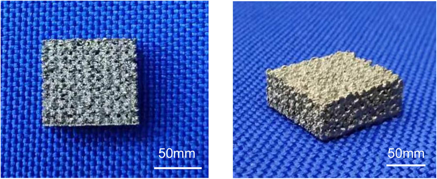
The 3D-printed porous titanium alloy scaffold (parameters: 100 mm × 90 mm × 45 mm). 3D, three-dimensional.
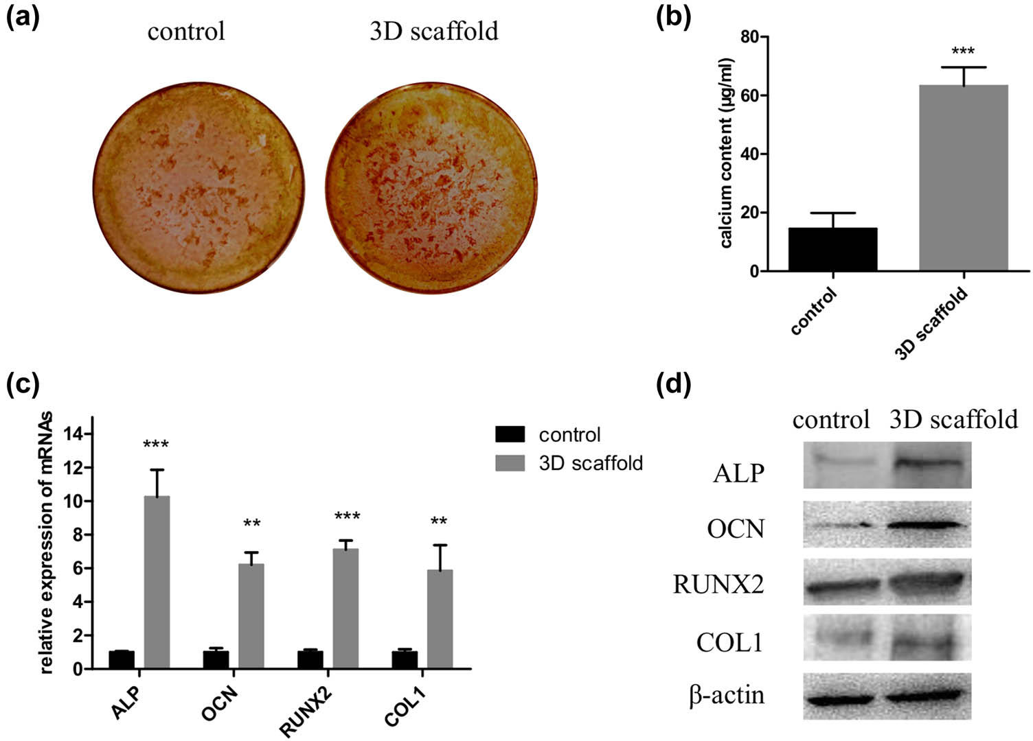
The 3D scaffold promotes osteogenic differentiation of hADMSCs. Cells were seeded in a 2D culture plate (control) and the 3D scaffold (test) to induce osteogenesis; then, they were digested and collected for ARS staining (a), calcium concentration analyses (b), and detection of relative mRNA (c) and protein levels of ALP, OCN, RUNX2, and COL1 (d) (n = 3 in each experiment). 3D, three-dimensional; 2D, two-dimensional; ARS, Alizarin red S. **P < 0.01 and ***P < 0.001.
3.2 Estimation of the osteogenic activity of the 3D scaffold
To assess the osteogenic activity of the two cell groups, we measured the mRNAs of the osteogenesis markers, including runt-related transcription factor 2 (RUNX2), collagen protein 1 (COL1), osteocalcin (OCN), and alkaline phosphatase (ALP), using qPCR. Expression of the mRNAs was significantly increased in the 3D scaffold group compared to the control group (P < 0.001 for ALP and RUNX2 and P < 0.01 for OCN and COL1) (Figure 2c). Next western blot analysis demonstrated that the expression of the corresponding proteins were visibly higher in the 3D scaffold group than in the control group (Figure 2d).
3.3 Expression levels of the circRNAs in the hADMSCs that were cultured in the 3D scaffold
qPCR revealed that among the five candidate circRNAs, hsa_circ_0131461 and hsa_circ_0019142 showed significantly increased expression levels and hsa_circ_0026827 showed a significantly reduced expression level in the 3D scaffold group (P < 0.05, P < 0.001, and P < 0.01, respectively) (Figure 3). Since the fold change (FC) of hsa_circ_0019142, among the FCs of the three circRNAs, was the highest (P < 0.001), hsa_circ_0019142 was chosen for further study.
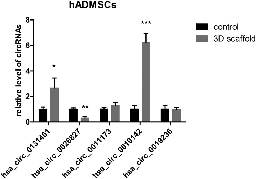
Relative expression of circRNAs in the 3D scaffold group. Cells were seeded in a 2D culture plate (control) and the 3D scaffold (test) to induce osteogenesis; then, they were digested and collected to analyze the relative levels of the five candidate circRNAs (n = 3). 3D, three dimensional; 2D, two dimensional. *P < 0.05, **P < 0.01, and ***P < 0.001.
3.4 Overexpression of hsa_circ_0019142 promoted osteogenic differentiation of the hADMSCs
After the hADMSCs were transfected with the overexpression vector of hsa_circ_0019142, successful transfection was confirmed by qPCR. Expression of hsa_circ_0019142 was found to be significantly higher (P < 0.01) in the cells transfected with hsa_circ_0019142 than in those transfected with the NC vector (Figure 4a). Next the cells were seeded in 12-well culture plates to induce osteogenesis. ARS staining revealed greater osteogenic activity in the hsa_circ_0019142 overexpressed cells than that in the NC cells (Figure 4b). Similarly, the calcium concentration was found to be significantly higher (P < 0.01) in the hsa_circ_0019142 overexpressed cells than in the NC cells (Figure 4c). The expression of the different osteogenesis markers (ALP, OCN, RUNX2, and COL1) was significantly elevated in the hsa_circ_0019142 overexpressed cells than in the NC cells (P < 0.05 for ALP and COL1, P < 0.01 for OCN, and P < 0.001 for RUNX2) (Figure 4d). This was further confirmed by the similar results obtained from the western blot analysis (Figure 4e).
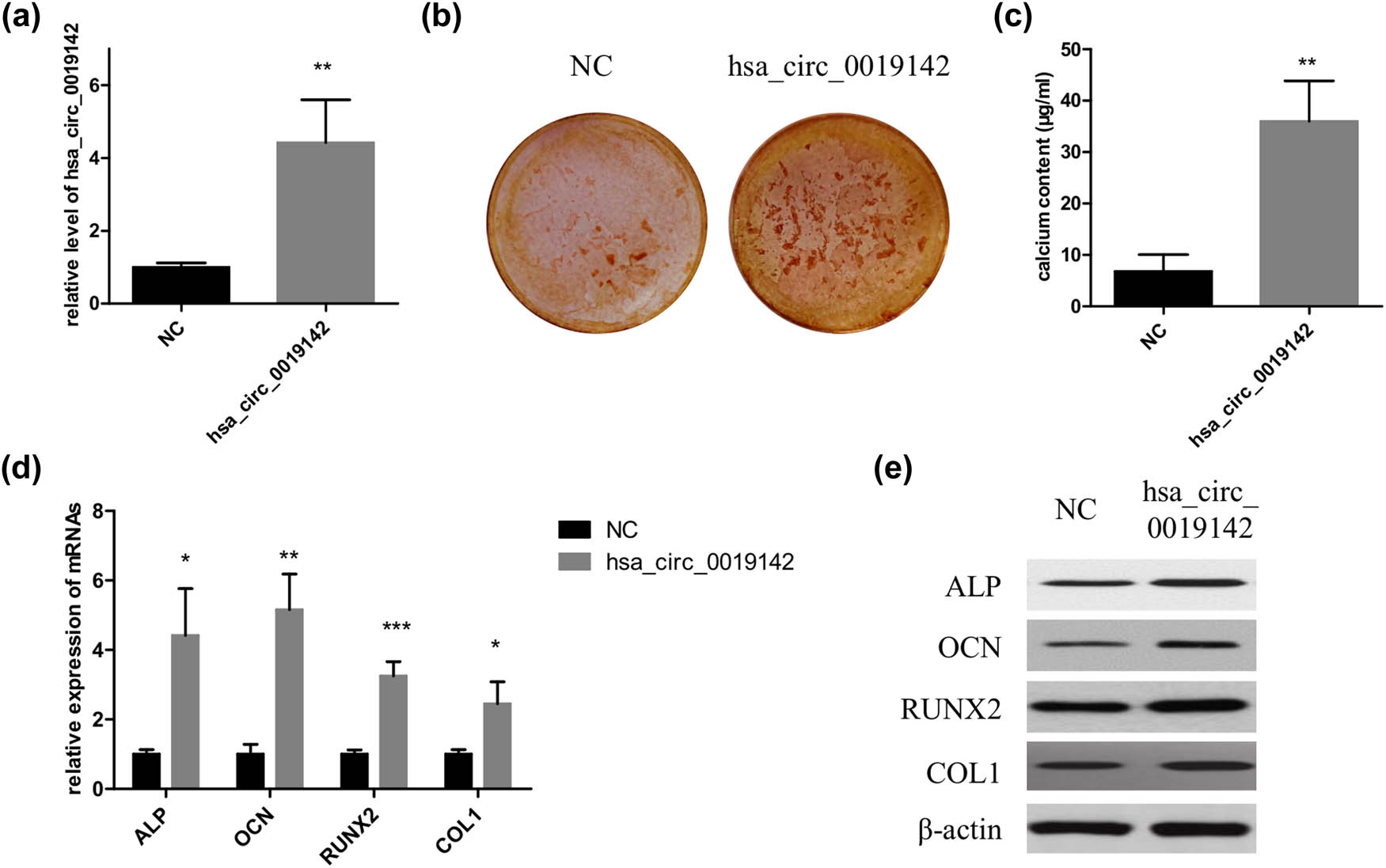
Overexpression of hsa_circ_0019142 promotes osteogenic differentiation of hADMSCs. (a) qPCR verification of the level of hsa_circ_0019142 in cells overexpressing it. After the induction of osteogenesis in the hsa_circ_0019142 overexpressed cells in the culture plate, the cells were used for ARS staining (b), calcium concentration analyses (c), and detection of relative mRNA (d) and protein levels of ALP, OCN, RUNX2, and COL1 (e) (n = 3 in each experiment). NC, negative control; ARS, Alizarin red S. *P < 0.05, **P < 0.01, and ***P < 0.001.
3.5 Knockdown of hsa_circ_0019142 inhibited osteogenic differentiation of the hADMSCs
qPCR result in Figure 5a indicated that the expression level of hsa_circ_0019142 was successfully silenced in hADMSCs. After the induction of osteogenic differentiation, ARS staining results indicated that the mineralization in si-hsa_circ_0019142 group was significantly lower (P < 0.01) than that in si-NC group (Figure 5b and c). Besides, the mRNA expression levels of ALP, OCN, RUNX2, and COL1 were all obviously reduced after the hsa_circ_0019142 was silenced in hADMSCs (Figure 5d), and the protein levels of the four markers were also decreased (Figure 5e).
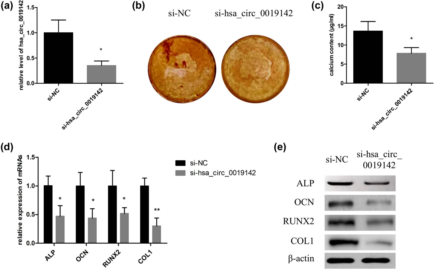
Knockdown of hsa_circ_0019142 inhibits osteogenic differentiation of hADMSCs. (a) qPCR verification of the level of hsa_circ_0019142 in cells silencing it. After the induction of osteogenesis in the hsa_circ_0019142 silenced cells in the culture plate, the cells were used for ARS staining (b), calcium concentration analyses (c), and detection of relative mRNA (d) and protein levels of ALP, OCN, RUNX2, and COL1 (e) (n = 3 in each experiment). NC, negative control; ARS, Alizarin red S. *P < 0.05 and **P < 0.01.
4 Discussion
A vast majority of the world’s population suffers from diseases that can be successfully managed, and even cured in some cases, through the transplantation of organs or tissues, but the lack of availability of tissues/organs remains one of the greatest challenges [18]. Hence, searching for an alternative to bone grafting has taken a prominent seat in the field of regenerative medicine. In the present study, osteogenic differentiation of the hADMSCs in the 3D scaffold and the underlying mechanism were studied for the first time, aiming to provide a potential solution for bone regeneration.
Besides being multipotent, the hADMSCs have self-renewal properties; they can differentiate into adipocytes [19], osteoblasts [19], and chondrocytes [20]. The hADMSCs are advantageous compared to other stem cells because they are easily accessible and can be obtained in large amounts through subcutaneous liposuction [21] without ethical or political issues as these mesenchymal stem cells are obtained from autologous fat [22]. Therefore, hADMSCs were used in the present study. Stem cell differentiation is an important strategy in regenerative medicine. BMPs are involved in multiple processes, including the regulation of osteogenesis. BMP-2 is known to enhance the osteogenic differentiation of mesenchymal stem cells obtained from various locations; hence it was supplied in the osteogenic differentiation medium to assist in osteogenesis.
Our results indicated that osteogenic mineralization of the hADMSCs was successfully induced after treatment of both the 2D plate and 3D scaffold with BMP-2. Moreover, hADMSCs in the 3D scaffold group possessed stronger osteogenic differentiation potentials and showed increased calcium concentration and higher expression of the osteogenesis-related factors, such as RUNX2, COL1, OCN, and ALP, than that of the cells in the control group. This indicated a significant advantage of the 3D scaffold in osteogenic induction, and this could be attributed to the unique titanium alloy material or porous structure of the scaffold [23]. Notably, it was difficult to compare the results of ARS staining of the two groups directly. Hence, the cells were collected after osteogenic induction, which are also required for further analyses of mRNA and protein levels of the markers. Additionally, the cell number and living cell rate were calculated after the cells were collected from the two groups. Only equivalent living cells were used for further evaluation, ensuring the reasonability in the comparisons and good repeatability in the study.
Recently, a review on the prospects of circRNAs in osteogenesis demonstrated that different circRNAs were involved in the modulation (upregulation or downregulation) of different signaling pathways and proteins that were associated with osteogenesis [24], highlighting the important role of circRNAs in osteogenesis. The relationship between the circRNAs and osteoblastic differentiation of stem cells has also been evidenced in previous studies. Zhang et al. identified 3938 upregulated and 1505 downregulated circRNAs after the osteoblastic differentiation of human bone marrow stem cells [25]. Peng et al. explored the role of hsa_circRNA_33287 in the osteogenic differentiation of maxillary sinus membrane stem cells [26]. The overexpression and silencing of hsa_circRNA_33287 increased and decreased the expression levels of the key osteogenesis markers (including RUNX2, osterix, and ALP), respectively. Hence, circRNAs might also play a role in the osteogenic differentiation prompted by the 3D scaffold. To obtain the candidate circRNAs, we focused on two important circRNA expression profiles, which were identified from the osteogenic differentiation of bone marrow stem cells [25] and MC3T3-E1 cells [27], presenting plenty of potential circRNAs that were involved in osteogenic differentiation. Five circRNAs, among the top eight differentially expressed circRNAs in each profile, were then randomly selected as candidate circRNAs.
qPCR results indicated that three of the five candidates significantly changed in the 3D scaffold group, and hsa_circ_0019142, which showed the highest FC, was chosen for further confirmation studies. The results revealed that calcium concentration and expression levels of RUNX2, COL1, OCN, and ALP were significantly increased in the hsa_circ_0019142 group indicating an enhanced osteogenic differentiation of the hADMSCs due to the overexpression of hsa_circ_0019142, while knockdown of hsa_circ_0019142 led to the opposite results. Peng et al. also showed that hsa_circRNA_33287 acted as a molecular sponge for miR-214-3p, regulating RUNX3 expression by targeting its 3′-UTR region and thus promoting ectopic bone formation [26]. MiRNA sponging is one of the important mechanisms by which circRNA exerts its functions [13,14,15]. Hence, the promoted effect of hsa_circ_0019142 on the osteogenic differentiation of the hADMSCs might also be dependent on the direct regulation of the objective miRNA and target genes by circRNA. However, this finding still requires further study.
In conclusion, the present study proved that hADMSCs, which were seeded in the 3D-printed porous titanium alloy scaffold, possessed higher osteogenic differentiation potentials due to the upregulation of hsa_circ_0019142.
Acknowledgements
We are grateful for the technology supports in 3D printing from Medvance medical technology (Shanghai) Co., Ltd.
-
Funding information: This study is supported by Talent Introduction Project of Shanghai Pudong Hospital (YJRCJJ201703).
-
Conflict of interest: Authors declare that they do not have any financial, personal, or professional conflicts of interest. The authors also declare that there are no conflicts of interest regarding the publication of this paper with any people and organization.
-
Data availability statement: The datasets generated during and/or analysed during the current study are available from the corresponding author on reasonable request.
References
[1] Bose S, Roy M, Bandyopadhyay A. Recent advances in bone tissue engineering scaffolds. Trends Biotechnol. 2012;30:546–54. 10.1016/j.tibtech.2012.07.005.Search in Google Scholar PubMed PubMed Central
[2] Mehta M, Schmidt-Bleek K, Duda GN, Mooney DJ. Biomaterial delivery of morphogens to mimic the natural healing cascade in bone. Adv Drug Deliv Rev. 2012;64:1257–76. 10.1016/j.addr.2012.05.006.Search in Google Scholar PubMed PubMed Central
[3] Ran Q, Yang W, Hu Y, Shen X, Yu Y, Xiang Y, et al. Osteogenesis of 3D printed porous Ti6Al4V implants with different pore sizes. J Mech Behav Biomed Mater. 2018;84:1–11. 10.1016/j.jmbbm.2018.04.010.Search in Google Scholar PubMed
[4] Helder MN, Knippenberg M, Klein-Nulend J, Wuisman PI. Stem cells from adipose tissue allow challenging new concepts for regenerative medicine. Tissue Eng. 2007;13:1799–808. 10.1089/ten.2006.0165.Search in Google Scholar PubMed
[5] Nerem RM. Regenerative medicine: the emergence of an industry. J R Soc Interface. 2010;7(Suppl 6):S771–5. 10.1098/rsif.2010.0348.focus.Search in Google Scholar PubMed PubMed Central
[6] Pampaloni F, Reynaud EG, Stelzer EH. The third dimension bridges the gap between cell culture and live tissue. Nat Rev Mol Cell Biol. 2007;8:839–45. 10.1038/nrm2236.Search in Google Scholar PubMed
[7] Mason C, Dunnill P. A brief definition of regenerative medicine. Regen Med. 2008;3:1–5. 10.2217/17460751.3.1.1.Search in Google Scholar PubMed
[8] Amin Yavari S, van der Stok J, Chai YC, Wauthle R, Tahmasebi Birgani Z, Habibovic P, et al. Bone regeneration performance of surface-treated porous titanium. Biomaterials. 2014;35:6172–81. 10.1016/j.biomaterials.2014.04.054.Search in Google Scholar PubMed
[9] Chen Y, Frith JE, Dehghan-Manshadi A, Attar H, Kent D, Soro NDM, et al. Mechanical properties and biocompatibility of porous titanium scaffolds for bone tissue engineering. J Mech Behav Biomed Mater. 2017;75:169–74. 10.1016/j.jmbbm.2017.07.015.Search in Google Scholar PubMed
[10] Nie C, Yang D, Morris SF. Local delivery of adipose-derived stem cells via acellular dermal matrix as a scaffold: a new promising strategy to accelerate wound healing. Med Hypotheses. 2009;72:679–82. 10.1016/j.mehy.2008.10.033.Search in Google Scholar PubMed
[11] Ryan AJ, Brougham CM, Garciarena CD, Kerrigan SW, O’Brien FJ. Towards 3D in vitro models for the study of cardiovascular tissues and disease. Drug Discov Today. 2016;21:1437–45. 10.1016/j.drudis.2016.04.014.Search in Google Scholar PubMed
[12] Yin S, Zhang W, Zhang Z, Jiang X. Recent advances in scaffold design and material for vascularized tissue-engineered bone regeneration. Adv Healthc Mater. 2019;8:e1801433. 10.1002/adhm.201801433.Search in Google Scholar PubMed
[13] Liu J, Liu T, Wang X, He A. Circles reshaping the RNA world: from waste to treasure. Mol Cancer. 2017;16:58. 10.1186/s12943-017-0630-y.Search in Google Scholar PubMed PubMed Central
[14] Hansen TB, Jensen TI, Clausen BH, Bramsen JB, Finsen B, Damgaard CK, et al. Natural RNA circles function as efficient microRNA sponges. Nature. 2013;495:384–8. 10.1038/nature11993.Search in Google Scholar PubMed
[15] Qu S, Yang X, Li X, Wang J, Gao Y, Shang R, et al. Circular RNA: a new star of noncoding RNAs. Cancer Lett. 2015;365:141–8. 10.1016/j.canlet.2015.06.003.Search in Google Scholar PubMed
[16] Ji F, Pan J, Shen Z, Yang Z, Wang J, Bai X, et al. The circular RNA circRNA124534 promotes osteogenic differentiation of human dental pulp stem cells through modulation of the mir-496/beta-catenin pathway. Front Cell Dev Biol. 2020;8:230. 10.3389/fcell.2020.00230.Search in Google Scholar PubMed PubMed Central
[17] Li X, Zheng Y, Zheng Y, Huang Y, Zhang Y, Jia L, et al. Circular RNA CDR1as regulates osteoblastic differentiation of periodontal ligament stem cells via the miR-7/GDF5/SMAD and p38 MAPK signaling pathway. Stem Cell Res Ther. 2018;9:232. 10.1186/s13287-018-0976-0.Search in Google Scholar PubMed PubMed Central
[18] Sheehy E, Conrad SL, Brigham LE, Luskin R, Weber P, Eakin M, et al. Estimating the number of potential organ donors in the United States. N Engl J Med. 2003;349:667–74. 10.1056/NEJMsa021271.Search in Google Scholar PubMed
[19] Halvorsen YD, Franklin D, Bond AL, Hitt DC, Auchter C, Boskey AL, et al. Extracellular matrix mineralization and osteoblast gene expression by human adipose tissue-derived stromal cells. Tissue Eng. 2001;7:729–41. 10.1089/107632701753337681.Search in Google Scholar PubMed
[20] Estes BT, Wu AW, Guilak F. Potent induction of chondrocytic differentiation of human adipose-derived adult stem cells by bone morphogenetic protein 6. Arthritis Rheum. 2006;54:1222–32. 10.1002/art.21779.Search in Google Scholar PubMed
[21] Baer PC, Geiger H. Adipose-derived mesenchymal stromal/stem cells: tissue localization, characterization, and heterogeneity. Stem Cell Int. 2012;2012:812693. 10.1155/2012/812693.Search in Google Scholar PubMed PubMed Central
[22] Cawthorn WP, Scheller EL, MacDougald OA. Adipose tissue stem cells: the great WAT hope. Trends Endocrinol Metab. 2012;23:270–7. 10.1016/j.tem.2012.01.003.Search in Google Scholar PubMed PubMed Central
[23] Prasopthum A, Cooper M, Shakesheff KM, Yang J. Three-dimensional printed scaffolds with controlled micro-/nanoporous surface topography direct chondrogenic and osteogenic differentiation of mesenchymal stem cells. ACS Appl Mater Interfaces. 2019;11:18896–906. 10.1021/acsami.9b01472.Search in Google Scholar PubMed
[24] Huang X, Cen X, Zhang B, Liao Y, Zhu G, Liu J, et al. Prospect of circular RNA in osteogenesis: A novel orchestrator of signaling pathways. J Cell Physiol. 2019;234:21450–9. 10.1002/jcp.28866.Search in Google Scholar PubMed
[25] Zhang M, Jia L, Zheng Y. circRNA expression profiles in human bone marrow stem cells undergoing osteoblast differentiation. Stem Cell Rev Rep. 2019;15:126–38. 10.1007/s12015-018-9841-x.Search in Google Scholar PubMed
[26] Peng W, Zhu S, Chen J, Wang J, Rong Q, Chen S. Hsa_circRNA_33287 promotes the osteogenic differentiation of maxillary sinus membrane stem cells via miR-214-3p/Runx3. Biomed Pharmacother. 2019;109:1709–17. 10.1016/j.biopha.2018.10.159.Search in Google Scholar PubMed
[27] Qian DY, Yan GB, Bai B, Chen Y, Zhang SJ, Yao YC, et al. Differential circRNA expression profiles during the BMP2-induced osteogenic differentiation of MC3T3-E1 cells. Biomed Pharmacother. 2017;90:492–9. 10.1016/j.biopha.2017.03.051.Search in Google Scholar PubMed
© 2021 Xuebin He et al., published by De Gruyter
This work is licensed under the Creative Commons Attribution 4.0 International License.
Articles in the same Issue
- Research Articles
- Identification of ZG16B as a prognostic biomarker in breast cancer
- Behçet’s disease with latent Mycobacterium tuberculosis infection
- Erratum
- Erratum to “Suffering from Cerebral Small Vessel Disease with and without Metabolic Syndrome”
- Research Articles
- GPR37 promotes the malignancy of lung adenocarcinoma via TGF-β/Smad pathway
- Expression and role of ABIN1 in sepsis: In vitro and in vivo studies
- Additional baricitinib loading dose improves clinical outcome in COVID-19
- The co-treatment of rosuvastatin with dapagliflozin synergistically inhibited apoptosis via activating the PI3K/AKt/mTOR signaling pathway in myocardial ischemia/reperfusion injury rats
- SLC12A8 plays a key role in bladder cancer progression and EMT
- LncRNA ATXN8OS enhances tamoxifen resistance in breast cancer
- Case Report
- Serratia marcescens as a cause of unfavorable outcome in the twin pregnancy
- Spleno-adrenal fusion mimicking an adrenal metastasis of a renal cell carcinoma: A case report and embryological background
- Research Articles
- TRIM25 contributes to the malignancy of acute myeloid leukemia and is negatively regulated by microRNA-137
- CircRNA circ_0004370 promotes cell proliferation, migration, and invasion and inhibits cell apoptosis of esophageal cancer via miR-1301-3p/COL1A1 axis
- LncRNA XIST regulates atherosclerosis progression in ox-LDL-induced HUVECs
- Potential role of IFN-γ and IL-5 in sepsis prediction of preterm neonates
- Rapid Communication
- COVID-19 vaccine: Call for employees in international transportation industries and international travelers as the first priority in global distribution
- Case Report
- Rare squamous cell carcinoma of the kidney with concurrent xanthogranulomatous pyelonephritis: A case report and review of the literature
- An infertile female delivered a baby after removal of primary renal carcinoid tumor
- Research Articles
- Hypertension, BMI, and cardiovascular and cerebrovascular diseases
- Case Report
- Coexistence of bilateral macular edema and pale optic disc in the patient with Cohen syndrome
- Research Articles
- Correlation between kinematic sagittal parameters of the cervical lordosis or head posture and disc degeneration in patients with posterior neck pain
- Review Articles
- Hepatoid adenocarcinoma of the lung: An analysis of the Surveillance, Epidemiology, and End Results (SEER) database
- Research Articles
- Thermography in the diagnosis of carpal tunnel syndrome
- Pemetrexed-based first-line chemotherapy had particularly prominent objective response rate for advanced NSCLC: A network meta-analysis
- Comparison of single and double autologous stem cell transplantation in multiple myeloma patients
- The influence of smoking in minimally invasive spinal fusion surgery
- Impact of body mass index on left atrial dimension in HOCM patients
- Expression and clinical significance of CMTM1 in hepatocellular carcinoma
- miR-142-5p promotes cervical cancer progression by targeting LMX1A through Wnt/β-catenin pathway
- Comparison of multiple flatfoot indicators in 5–8-year-old children
- Early MRI imaging and follow-up study in cerebral amyloid angiopathy
- Intestinal fatty acid-binding protein as a biomarker for the diagnosis of strangulated intestinal obstruction: A meta-analysis
- miR-128-3p inhibits apoptosis and inflammation in LPS-induced sepsis by targeting TGFBR2
- Dynamic perfusion CT – A promising tool to diagnose pancreatic ductal adenocarcinoma
- Biomechanical evaluation of self-cinching stitch techniques in rotator cuff repair: The single-loop and double-loop knot stitches
- Review Articles
- The ambiguous role of mannose-binding lectin (MBL) in human immunity
- Case Report
- Membranous nephropathy with pulmonary cryptococcosis with improved 1-year follow-up results: A case report
- Fertility problems in males carrying an inversion of chromosome 10
- Acute myeloid leukemia with leukemic pleural effusion and high levels of pleural adenosine deaminase: A case report and review of literature
- Metastatic renal Ewing’s sarcoma in adult woman: Case report and review of the literature
- Burkitt-like lymphoma with 11q aberration in a patient with AIDS and a patient without AIDS: Two cases reports and literature review
- Skull hemophilia pseudotumor: A case report
- Judicious use of low-dosage corticosteroids for non-severe COVID-19: A case report
- Adult-onset citrullinaemia type II with liver cirrhosis: A rare cause of hyperammonaemia
- Clinicopathologic features of Good’s syndrome: Two cases and literature review
- Fatal immune-related hepatitis with intrahepatic cholestasis and pneumonia associated with camrelizumab: A case report and literature review
- Research Articles
- Effects of hydroxyethyl starch and gelatin on the risk of acute kidney injury following orthotopic liver transplantation: A multicenter retrospective comparative clinical study
- Significance of nucleic acid positive anal swab in COVID-19 patients
- circAPLP2 promotes colorectal cancer progression by upregulating HELLS by targeting miR-335-5p
- Ratios between circulating myeloid cells and lymphocytes are associated with mortality in severe COVID-19 patients
- Risk factors of left atrial appendage thrombus in patients with non-valvular atrial fibrillation
- Clinical features of hypertensive patients with COVID-19 compared with a normotensive group: Single-center experience in China
- Surgical myocardial revascularization outcomes in Kawasaki disease: systematic review and meta-analysis
- Decreased chromobox homologue 7 expression is associated with epithelial–mesenchymal transition and poor prognosis in cervical cancer
- FGF16 regulated by miR-520b enhances the cell proliferation of lung cancer
- Platelet-rich fibrin: Basics of biological actions and protocol modifications
- Accurate diagnosis of prostate cancer using logistic regression
- miR-377 inhibition enhances the survival of trophoblast cells via upregulation of FNDC5 in gestational diabetes mellitus
- Prognostic significance of TRIM28 expression in patients with breast carcinoma
- Integrative bioinformatics analysis of KPNA2 in six major human cancers
- Exosomal-mediated transfer of OIP5-AS1 enhanced cell chemoresistance to trastuzumab in breast cancer via up-regulating HMGB3 by sponging miR-381-3p
- A four-lncRNA signature for predicting prognosis of recurrence patients with gastric cancer
- Knockdown of circ_0003204 alleviates oxidative low-density lipoprotein-induced human umbilical vein endothelial cells injury: Circulating RNAs could explain atherosclerosis disease progression
- Propofol postpones colorectal cancer development through circ_0026344/miR-645/Akt/mTOR signal pathway
- Knockdown of lncRNA TapSAKI alleviates LPS-induced injury in HK-2 cells through the miR-205/IRF3 pathway
- COVID-19 severity in relation to sociodemographics and vitamin D use
- Clinical analysis of 11 cases of nocardiosis
- Cis-regulatory elements in conserved non-coding sequences of nuclear receptor genes indicate for crosstalk between endocrine systems
- Four long noncoding RNAs act as biomarkers in lung adenocarcinoma
- Real-world evidence of cytomegalovirus reactivation in non-Hodgkin lymphomas treated with bendamustine-containing regimens
- Relation between IL-8 level and obstructive sleep apnea syndrome
- circAGFG1 sponges miR-28-5p to promote non-small-cell lung cancer progression through modulating HIF-1α level
- Nomogram prediction model for renal anaemia in IgA nephropathy patients
- Effect of antibiotic use on the efficacy of nivolumab in the treatment of advanced/metastatic non-small cell lung cancer: A meta-analysis
- NDRG2 inhibition facilitates angiogenesis of hepatocellular carcinoma
- A nomogram for predicting metabolic steatohepatitis: The combination of NAMPT, RALGDS, GADD45B, FOSL2, RTP3, and RASD1
- Clinical and prognostic features of MMP-2 and VEGF in AEG patients
- The value of miR-510 in the prognosis and development of colon cancer
- Functional implications of PABPC1 in the development of ovarian cancer
- Prognostic value of preoperative inflammation-based predictors in patients with bladder carcinoma after radical cystectomy
- Sublingual immunotherapy increases Treg/Th17 ratio in allergic rhinitis
- Prediction of improvement after anterior cruciate ligament reconstruction
- Effluent Osteopontin levels reflect the peritoneal solute transport rate
- circ_0038467 promotes PM2.5-induced bronchial epithelial cell dysfunction
- Significance of miR-141 and miR-340 in cervical squamous cell carcinoma
- Association between hair cortisol concentration and metabolic syndrome
- Microvessel density as a prognostic indicator of prostate cancer: A systematic review and meta-analysis
- Characteristics of BCR–ABL gene variants in patients of chronic myeloid leukemia
- Knee alterations in rheumatoid arthritis: Comparison of US and MRI
- Long non-coding RNA TUG1 aggravates cerebral ischemia and reperfusion injury by sponging miR-493-3p/miR-410-3p
- lncRNA MALAT1 regulated ATAD2 to facilitate retinoblastoma progression via miR-655-3p
- Development and validation of a nomogram for predicting severity in patients with hemorrhagic fever with renal syndrome: A retrospective study
- Analysis of COVID-19 outbreak origin in China in 2019 using differentiation method for unusual epidemiological events
- Laparoscopic versus open major liver resection for hepatocellular carcinoma: A case-matched analysis of short- and long-term outcomes
- Travelers’ vaccines and their adverse events in Nara, Japan
- Association between Tfh and PGA in children with Henoch–Schönlein purpura
- Can exchange transfusion be replaced by double-LED phototherapy?
- circ_0005962 functions as an oncogene to aggravate NSCLC progression
- Circular RNA VANGL1 knockdown suppressed viability, promoted apoptosis, and increased doxorubicin sensitivity through targeting miR-145-5p to regulate SOX4 in bladder cancer cells
- Serum intact fibroblast growth factor 23 in healthy paediatric population
- Algorithm of rational approach to reconstruction in Fournier’s disease
- A meta-analysis of exosome in the treatment of spinal cord injury
- Src-1 and SP2 promote the proliferation and epithelial–mesenchymal transition of nasopharyngeal carcinoma
- Dexmedetomidine may decrease the bupivacaine toxicity to heart
- Hypoxia stimulates the migration and invasion of osteosarcoma via up-regulating the NUSAP1 expression
- Long noncoding RNA XIST knockdown relieves the injury of microglia cells after spinal cord injury by sponging miR-219-5p
- External fixation via the anterior inferior iliac spine for proximal femoral fractures in young patients
- miR-128-3p reduced acute lung injury induced by sepsis via targeting PEL12
- HAGLR promotes neuron differentiation through the miR-130a-3p-MeCP2 axis
- Phosphoglycerate mutase 2 is elevated in serum of patients with heart failure and correlates with the disease severity and patient’s prognosis
- Cell population data in identifying active tuberculosis and community-acquired pneumonia
- Prognostic value of microRNA-4521 in non-small cell lung cancer and its regulatory effect on tumor progression
- Mean platelet volume and red blood cell distribution width is associated with prognosis in premature neonates with sepsis
- 3D-printed porous scaffold promotes osteogenic differentiation of hADMSCs
- Association of gene polymorphisms with women urinary incontinence
- Influence of COVID-19 pandemic on stress levels of urologic patients
- miR-496 inhibits proliferation via LYN and AKT pathway in gastric cancer
- miR-519d downregulates LEP expression to inhibit preeclampsia development
- Comparison of single- and triple-port VATS for lung cancer: A meta-analysis
- Fluorescent light energy modulates healing in skin grafted mouse model
- Silencing CDK6-AS1 inhibits LPS-induced inflammatory damage in HK-2 cells
- Predictive effect of DCE-MRI and DWI in brain metastases from NSCLC
- Severe postoperative hyperbilirubinemia in congenital heart disease
- Baicalin improves podocyte injury in rats with diabetic nephropathy by inhibiting PI3K/Akt/mTOR signaling pathway
- Clinical factors predicting ureteral stent failure in patients with external ureteral compression
- Novel H2S donor proglumide-ADT-OH protects HUVECs from ox-LDL-induced injury through NF-κB and JAK/SATA pathway
- Triple-Endobutton and clavicular hook: A propensity score matching analysis
- Long noncoding RNA MIAT inhibits the progression of diabetic nephropathy and the activation of NF-κB pathway in high glucose-treated renal tubular epithelial cells by the miR-182-5p/GPRC5A axis
- Serum exosomal miR-122-5p, GAS, and PGR in the non-invasive diagnosis of CAG
- miR-513b-5p inhibits the proliferation and promotes apoptosis of retinoblastoma cells by targeting TRIB1
- Fer exacerbates renal fibrosis and can be targeted by miR-29c-3p
- The diagnostic and prognostic value of miR-92a in gastric cancer: A systematic review and meta-analysis
- Prognostic value of α2δ1 in hypopharyngeal carcinoma: A retrospective study
- No significant benefit of moderate-dose vitamin C on severe COVID-19 cases
- circ_0000467 promotes the proliferation, metastasis, and angiogenesis in colorectal cancer cells through regulating KLF12 expression by sponging miR-4766-5p
- Downregulation of RAB7 and Caveolin-1 increases MMP-2 activity in renal tubular epithelial cells under hypoxic conditions
- Educational program for orthopedic surgeons’ influences for osteoporosis
- Expression and function analysis of CRABP2 and FABP5, and their ratio in esophageal squamous cell carcinoma
- GJA1 promotes hepatocellular carcinoma progression by mediating TGF-β-induced activation and the epithelial–mesenchymal transition of hepatic stellate cells
- lncRNA-ZFAS1 promotes the progression of endometrial carcinoma by targeting miR-34b to regulate VEGFA expression
- Anticoagulation is the answer in treating noncritical COVID-19 patients
- Effect of late-onset hemorrhagic cystitis on PFS after haplo-PBSCT
- Comparison of Dako HercepTest and Ventana PATHWAY anti-HER2 (4B5) tests and their correlation with silver in situ hybridization in lung adenocarcinoma
- VSTM1 regulates monocyte/macrophage function via the NF-κB signaling pathway
- Comparison of vaginal birth outcomes in midwifery-led versus physician-led setting: A propensity score-matched analysis
- Treatment of osteoporosis with teriparatide: The Slovenian experience
- New targets of morphine postconditioning protection of the myocardium in ischemia/reperfusion injury: Involvement of HSP90/Akt and C5a/NF-κB
- Superenhancer–transcription factor regulatory network in malignant tumors
- β-Cell function is associated with osteosarcopenia in middle-aged and older nonobese patients with type 2 diabetes: A cross-sectional study
- Clinical features of atypical tuberculosis mimicking bacterial pneumonia
- Proteoglycan-depleted regions of annular injury promote nerve ingrowth in a rabbit disc degeneration model
- Effect of electromagnetic field on abortion: A systematic review and meta-analysis
- miR-150-5p affects AS plaque with ASMC proliferation and migration by STAT1
- MALAT1 promotes malignant pleural mesothelioma by sponging miR-141-3p
- Effects of remifentanil and propofol on distant organ lung injury in an ischemia–reperfusion model
- miR-654-5p promotes gastric cancer progression via the GPRIN1/NF-κB pathway
- Identification of LIG1 and LIG3 as prognostic biomarkers in breast cancer
- MitoQ inhibits hepatic stellate cell activation and liver fibrosis by enhancing PINK1/parkin-mediated mitophagy
- Dissecting role of founder mutation p.V727M in GNE in Indian HIBM cohort
- circATP2A2 promotes osteosarcoma progression by upregulating MYH9
- Prognostic role of oxytocin receptor in colon adenocarcinoma
- Review Articles
- The function of non-coding RNAs in idiopathic pulmonary fibrosis
- Efficacy and safety of therapeutic plasma exchange in stiff person syndrome
- Role of cesarean section in the development of neonatal gut microbiota: A systematic review
- Small cell lung cancer transformation during antitumor therapies: A systematic review
- Research progress of gut microbiota and frailty syndrome
- Recommendations for outpatient activity in COVID-19 pandemic
- Rapid Communication
- Disparity in clinical characteristics between 2019 novel coronavirus pneumonia and leptospirosis
- Use of microspheres in embolization for unruptured renal angiomyolipomas
- COVID-19 cases with delayed absorption of lung lesion
- A triple combination of treatments on moderate COVID-19
- Social networks and eating disorders during the Covid-19 pandemic
- Letter
- COVID-19, WHO guidelines, pedagogy, and respite
- Inflammatory factors in alveolar lavage fluid from severe COVID-19 pneumonia: PCT and IL-6 in epithelial lining fluid
- COVID-19: Lessons from Norway tragedy must be considered in vaccine rollout planning in least developed/developing countries
- What is the role of plasma cell in the lamina propria of terminal ileum in Good’s syndrome patient?
- Case Report
- Rivaroxaban triggered multifocal intratumoral hemorrhage of the cabozantinib-treated diffuse brain metastases: A case report and review of literature
- CTU findings of duplex kidney in kidney: A rare duplicated renal malformation
- Synchronous primary malignancy of colon cancer and mantle cell lymphoma: A case report
- Sonazoid-enhanced ultrasonography and pathologic characters of CD68 positive cell in primary hepatic perivascular epithelioid cell tumors: A case report and literature review
- Persistent SARS-CoV-2-positive over 4 months in a COVID-19 patient with CHB
- Pulmonary parenchymal involvement caused by Tropheryma whipplei
- Mediastinal mixed germ cell tumor: A case report and literature review
- Ovarian female adnexal tumor of probable Wolffian origin – Case report
- Rare paratesticular aggressive angiomyxoma mimicking an epididymal tumor in an 82-year-old man: Case report
- Perimenopausal giant hydatidiform mole complicated with preeclampsia and hyperthyroidism: A case report and literature review
- Primary orbital ganglioneuroblastoma: A case report
- Primary aortic intimal sarcoma masquerading as intramural hematoma
- Sustained false-positive results for hepatitis A virus immunoglobulin M: A case report and literature review
- Peritoneal loose body presenting as a hepatic mass: A case report and review of the literature
- Chondroblastoma of mandibular condyle: Case report and literature review
- Trauma-induced complete pacemaker lead fracture 8 months prior to hospitalization: A case report
- Primary intradural extramedullary extraosseous Ewing’s sarcoma/peripheral primitive neuroectodermal tumor (PIEES/PNET) of the thoracolumbar spine: A case report and literature review
- Computer-assisted preoperative planning of reduction of and osteosynthesis of scapular fracture: A case report
- High quality of 58-month life in lung cancer patient with brain metastases sequentially treated with gefitinib and osimertinib
- Rapid response of locally advanced oral squamous cell carcinoma to apatinib: A case report
- Retrieval of intrarenal coiled and ruptured guidewire by retrograde intrarenal surgery: A case report and literature review
- Usage of intermingled skin allografts and autografts in a senior patient with major burn injury
- Retraction
- Retraction on “Dihydromyricetin attenuates inflammation through TLR4/NF-kappa B pathway”
- Special Issue Computational Intelligence Methodologies Meets Recurrent Cancers - Part I
- An artificial immune system with bootstrap sampling for the diagnosis of recurrent endometrial cancers
- Breast cancer recurrence prediction with ensemble methods and cost-sensitive learning