Functional implications of PABPC1 in the development of ovarian cancer
-
Cong Feng


Abstract
This research aimed to probe the expression characteristics of poly(A)-binding protein cytoplasmic 1 (PABPC1) and its role on the phenotype of ovarian cancer (OC) cells and to further investigate the possible underlying mechanism. The expression of PABPC1 was analyzed according to the data from gene expression omnibus, The Cancer Genome Atlas (TCGA) and Oncomine databases and the RNA sequencing data set from TCGA were downloaded for evaluating the prognostic values. We revealed that compared with the healthy samples, PABPC1 was upregulated in OC samples. High expression of PABPC1 had a connection with a shorter survival for patients with OC. Loss and gain of function assays revealed that silencing PABPC1 significantly suppressed the viability, invasion and migration of SK-OV-3 cells, while PABPC1 overexpression in A2780 cells showed the reverse outcomes. Moreover, Western blot demonstrated that silencing PABPC1 notably inactivated the epithelial–mesenchymal transition (EMT) process, while upregulation of PABPC1 promoted the mitigation of epithelial phenotype and the acquisition of mesenchymal phenotype. Taken together, PABPC1 was upregulated in OC cells and served as a carcinogene to promote the OC cell growth and invasion partly by modulating the EMT process, which implied that PABPC1 might be considered as a useful biomarker for OC therapeutics.
1 Introduction
Ovarian cancer (OC), known as a common malignancy in gynecology, has the highest mortality among the malignant tumors in female reproductive system and seriously threatens women’s life and health [1,2]. The American Cancer Society estimated approximately 22,530 new cases of OC and 13,980 OC-related deaths in the United States in 2019 [3]. This carcinoma is frequently diagnosed at an advanced stage due to its asymptomatic development and the lack of effective diagnostic approach at an early stage [4,5]. Although patients with OC usually respond well to the first-line chemotherapy based on platinum compounds and taxanes, most of the patients with OC still relapse [6]. According to the platinum-free interval cutoff of 6 months, the first recurrence is usually classified as platinum sensitive and platinum resistant [7]. After this first recurrence, the patients are rarely cured, and the general approach used in this recurrence can guide the approach to subsequent recurrences [8]. Platinum-sensitive secondary recurrence permits the use of platinum-based chemotherapy and a further cytopenia surgery; however, the prognosis of these people is usually poor [9]. Nevertheless, in this scenario, the introduction of new targeted therapies changed the prognosis of patients with both platinum-sensitive and platinum-resistant recurrence. [9]. Therefore, it is of important significance for the treatment of OC to elucidate the potential mechanism of action of OC process.
Poly(A)-binding protein (PABP) family is usually considered as a protective barrier of the mRNA poly(A) tail to mediate multiple aspects of translation and stability of mRNA [10,11]. PABP cytoplasmic 1 (PABPC1) is widely distributed in the cytoplasm of eukaryotes, binds to the poly(A) tail and interacts with specific sequences in the mRNA, allowing it to play a vital role in many kinds of cellular activities such as participating in the initiation of translation and in the regulation of mRNA decay [12,13]. Years of research have demonstrated that PABPC1 was abnormally expressed in numerous tumors and was involved in carcinogenicity [14,15,16,17]. PABPC1 was found to be highly expressed in hepatocellular carcinoma and induced cell viability by promoting entry into the S phase [14]. It was also reported that PABPC1 plays a carcinogenic role by inhibiting the expression of miR-34c in gastric carcinoma [15]. However, low expression of PABPC1 could promote tumor cells growth and lead to a poor prognosis in esophageal cancer [16,17]. Herein the question that puzzled us is what role PABPC1 plays in OC, which has rarely been reported.
In view of this, aiming at investigating the role of PABPC1 in OC, we downloaded and analyzed the data from the online public databases and assessed the relationship between PABPC1 and survival rate. Aside from this, we detected the effects of PABPC1 on the phenotype of OC in vitro using the SK-OV-3 and A2780 cell lines. Furthermore, since accumulating studies have shown that the role of epithelial–mesenchymal transition (EMT) in OC is closely linked to the invasion and metastasis of tumor cells [18,19,20], the relationship between PABPC1 and EMT-related markers was also investigated. This work provided evidences in vitro supporting the possibility of PABPC1 serving as a carcinogene to enhance OC cells’ proliferative, invasive and migratory potential by modulating the EMT process.
2 Materials and methods
2.1 Data acquisition
Initially, we analyzed the expression of PABPC1 using the data set GSE54388 which was downloaded from Gene Expression Omnibus (GEO, https://www.ncbi.nlm.nih.gov/geo/) database. The RNA sequencing data sets were acquired from The Cancer Genome Atlas (TCGA, https://cancergenome.nih.gov) and normal samples were downloaded from Genotype-Tissue Expression (GTEx, https://gtexportal.org/home/) database. Different data sets including Bonome Ovarian data set and Yoshihara Ovarian data set from Oncomine (https://www.oncomine.org) database were also used to analyze the PABPC1 expression pattern. A total of 374 cases of OC in TCGA cohort were divided into high and low PABPC1 expression groups according to the median expression of PABPC1 for evaluating the correlation between PABPC1 and the overall survival rate.
2.2 Cell lines
Human OC cell lines SK-OV-3 and A2780 were obtained from the Shanghai Cell Bank, Chinese Academy of Medical Sciences (Shanghai, China), and OVCAR-3 was acquired from the American Type Culture Collection (Rockville, VA, USA). From Procell Technology (Wuhan, Hubei, China), normal OC cell line IOSE80 was purchased. All cells were incubated at 37°C under the 5% CO2 atmosphere in Roswell Park Memorial Institute-1640 medium (Procell) supplemented with 10% fetal bovine serum (FBS) and 1% penicillin–streptomycin solution.
2.3 RNA interference and transfection
Small interfering RNAs (siRNAs) targeting PABPC1 (si-PABPC1#1: 5′-CATCGACAATAAAGCACTAT-3′; si-PABPC1#2: 5′-CTAGCCAAATTGCTCAACTA-3′) and their control siRNA (si-con: 5′-CGAACTCACTGGTCTGACC-3′) were designed to downregulate PABPC1 expression. Plasmid pcDNA3.1-PABPC1 and empty vector pcDNA3.1 were conducted to overexpress PABPC1. All siRNA sequences and plasmid vectors were constructed by the GenePharma Corporation (Shanghai, China).
Lipofectamine™ 2000 transfection reagent (Thermo Fisher Scientific, Waltham, MA, USA) served to carry out transfection according to the manufacturer’s protocols. Upon transfection for 24 h, the transfection efficacy was detected.
2.4 RNA extraction and real-time quantitative polymerase chain reaction (RT-qPCR)
Using the TRIzol reagent (Invitrogen, Carlsbad, CA, USA), total RNAs from cells were extracted and then reverse transcribed by PrimeScript RT Reagent kit (TaKaRa, Tokyo, Japan). The mRNA level of PABPC1 was tested by RT-qPCR with the SYBR Premix Ex Taq (TaKaRa) on the Applied Biosystems 7500 Thermocycler (Thermo Fisher Scientific) according to the manufacturer’s protocols. Glyceraldehyde-3-phosphate dehydrogenase (GAPDH) served as the control. Primer sequences for RT-qPCR were:
PABPC1 forward: 5′-CAGAGAATGGCAAGTGTACGAGC-3′,
PABPC1 reverse: 5′-GCTAGGAGGATAGTATGCAGCAC-3′;
GAPDH forward: 5′-TGTGTCCGTCGTGGATCTGA-3′,
GAPDH reverse: 5′-CCTGCTTCACCACCTTCTTGA-3′.
2.5 Western blot
After transfection for 24 h, proteins were extracted from the cells using the radio immunoprecipitation assay lysis buffer (Beyotime, Nantong, China) on ice. Protein concentration was measured via the bicinchoninic acid protein assay kit (Beyotime). Equal amount of protein (20 μg) was separated by 10% sodium dodecyl sulfate–polyacrylamide gel electrophoresis and transferred onto polyvinylidene fluoride membranes, which were then blocked for an hour with 5% skimmed milk powder. Next the membranes were incubated with the primary antibodies including anti-PABPC1 (ab233280), anti-E-cadherin (ab1416), anti-N-cadherin (ab202030), anti-snail (ab229701) and anti-vimentin (ab193555) overnight at 4°C. Additionally, anti-PABPC1 and all EMT-related protein primary antibodies were purchased from Abcam Trading Company Ltd (Shanghai, China). GAPDH rabbit monoclonal antibody (AF1186; Beyotime) was regarded as an internal control. Subsequently, these membranes were washed with Tris-buffered saline Tween thrice for 5 min and were incubated with the horseradish peroxidase-conjugated secondary antibodies for 1 h at room temperature. The protein luminescence effect was evaluated via an enhanced chemiluminescence plus with a bioimaging system. Image J software served to scan the gray values.
2.6 Cell proliferation assay
After transfection for 24 h, cells at a density of 1,000 cells/well were routinely incubated in 96-well plates. After incubation for 0, 24, 48 and 72 h, 10 μL of cell counting kit 8 (CCK-8; Dojindo, Tabaru, Japan) reagent was supplied to each well. Next the optical density (OD) values were recorded 2 h later using a microplate reader at 450 nm wavelength.
2.7 Plate clone formation assay
To prepare cell suspension, trypsin was first used to digest the cells of logarithmic growth. Then at 400 cells/dish, the cell suspension was plated into 60 mm dishes containing 5 mL preculture medium and kept at an atmosphere of 5% CO2 at 37°C for 2 weeks. After washing twice with phosphate-buffered saline (PBS), fixing with 4% paraformaldehyde for 30 min and staining with 0.1% crystal violet for 30 min, the number of colonies was counted.
2.8 Transwell assay
To evaluate the OC cells’ invasion and migration, 24-well Transwell chambers (Genetimes, Shanghai, China) were adopted. The invasion and migration assay procedures were similar, except that the latter did not perform gelatinization. Briefly, the transfected cells (1 × 10⁵ cells/well) were incubated in 100 μL serum-free RPMI-1640 in the upper chambers, and the lower chambers were supplemented with 500 μL RPMI-1640 plus 10% FBS. Next using the cotton swabs, the residual cells on upper chambers were wiped off when cultured for 24 h. After that, the cells that migrated or invaded to the lower chamber were washed with PBS, fixed and stained for 30 min. Upon being washed with PBS again, the migrated or invaded cells were photographed and counted under a microscope with five random selected fields.
2.9 Statistical analyses
Survival curve was plotted utilizing Kaplan–Meier method with log-rank tests for comparison. Comparison of two groups was performed utilizing Student’s t test, and one-way analysis of variance served to compare the differences among three or more groups, following a Dunnett post hoc test. All statistical analyses were conducted with GraphPad Prism version 6.0 (GraphPad Software, La Jolla, CA, USA) or SPSS version 21.0 (SPSS, Chicago, IL, USA) software. The results of the in vitro experiments were expressed as mean value ± standard deviation from triplicate experiments. A P < 0.05 was indicated as a statistically significant difference.
-
Ethical statement: Ethical approval was not required. No human and animal assays were used in this study.
3 Results
3.1 PABPC1 is upregulated in OC and has a connection with poor survival
At first, the expression of PABPC1 was evaluated by bioinformatic analysis using GEO data set GSE54388. As shown in Figure 1a, a significant upregulation was observed in serious OC samples in comparison with normal healthy samples (P = 0.0003). Then we analyzed the expression of PABPC1 from TCGA which included 378 OC tissues. Since no normal tissues were observed in the OC-related RNA-Seq data set in TCGA, we downloaded 88 normal ovarian tissue samples from GTEx database as controls. It was shown that PABPC1 expression in OC samples was higher than that in the normal samples (P < 0.001; Figure 1b). Moreover, we found that PABPC1 displayed a higher expression in OC tissues based on the Bonome Ovarian data set and Yoshihara Ovarian data set from Oncomine database when compared with nontumor tissues (P < 0.01; Figure 1c and d). To validate the observations from bioinformatic analyses, we perform RT-qPCR to examine mRNA expression of PABPC1 in three OC cell lines. A significant upregulation of PABPC1 was shown in all OC cell lines compared to the normal cell line IOSE80 (P < 0.01; Figure 1e), which was in agreement with the outcomes by bioinformatic analysis.
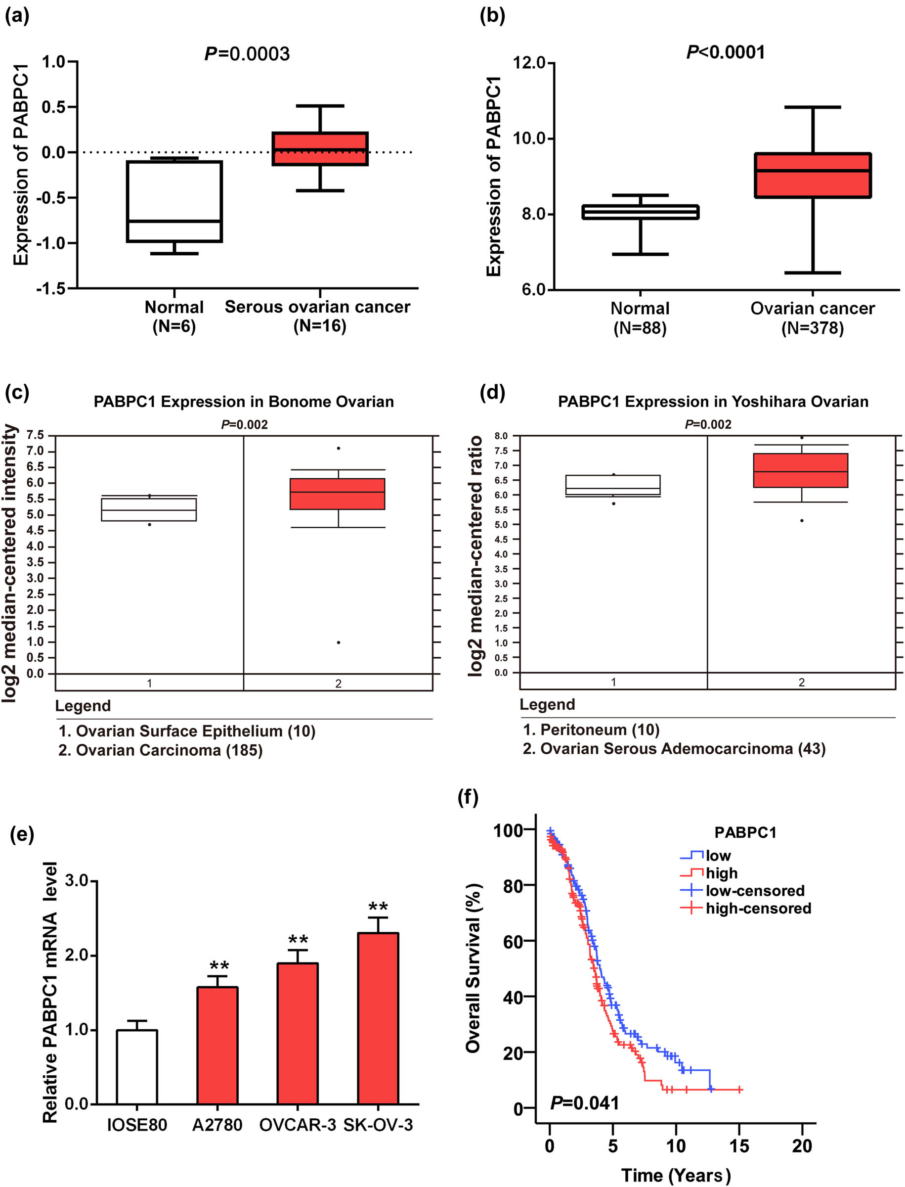
Expression of PABPC1 and Kaplan–Meier curves. High expression of PABPC1 was observed in GSE54388 from GEO database (a), TCGA database and GTEx database (b), and Oncomine database (c and d). (e) mRNA expression of PABPC1 in OC cell lines (A2780, OVCAR-3, SK-OV-3) and normal cell line (IOSE80) was examined using RT-qPCR analysis. **P < 0.01 vs IOSE80 cell line. (f) Kaplan–Meier method was utilized to plot the overall survival curve based upon the TCGA database (N = 374).
The correlation between high/low PABPC1 expression and the overall survival of patients with OC was probed through Kaplan–Meier methods. The curve chart revealed that patients with high PABPC1 levels exhibited a worse overall survival compared with those with low PABPC1 level (P = 0.041; Figure 1f).
3.2 Detection gain and loss function of PABPC1 efficiency
Based on the previous RT-qPCR assay, we found that, among OC-related cell lines, mRNA expression of PABPC1 was highest in SK-OV-3 cells and lowest in A2780 cells. To make the following tests more optimized, SK-OV-3 was chose to perform the loss of function assay and A2780 was chose as the targeted cells for the next overexpression experiments.
Efficiency of overexpression and knockdown was measured on the mRNA and protein expression aspects. Upon transfection with si-PABPC1#1 and si-PABPC1#2, the results showed that the mRNA and protein levels of PABPC1 were remarkably reduced in SK-OV-3 cells in contrasts with si-con group (P < 0.01; Figure 2a and b). By the way, the sequence of si-PABPC1#1 was selected to carry out the subsequent loss-of-function assay due to its better efficiency than si-PABPC1#2 group. Furthermore, PABPC1 expressions were notably enhanced in A2780 cells after transfecting with pcDNA3.1-PABPC1 in comparison with vector group (P < 0.01; Figure 2c and d).
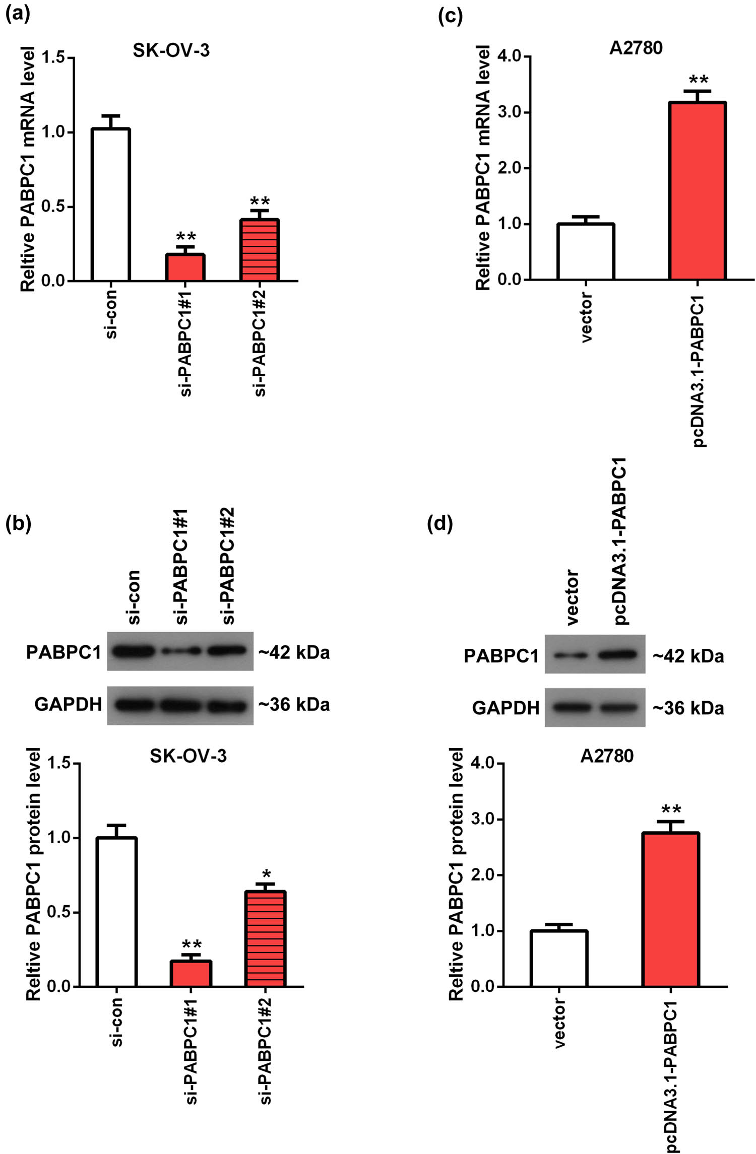
Detection of knockdown and overexpression of PABPC1 efficacy. The mRNA (a and c) and protein (b and d) levels of PABPC1 in SK-OV-3 (a and b) and A2780 (c and d) cells were tested using RT-qPCR (a and c) and Western blot (b and d) analyses. SK-OV-3 cells were transfected with si-PABPC1#1, si-PABPC1#2 and si-con. A2780 cells were transfected with pcDNA3.1-PABPC1 and pcDNA3.1 empty vector. **P < 0.01 vs si-con or vector group.
3.3 PABPC1 expression impacted OC cell proliferative ability
To examine the action of PABPC1 on OC cell proliferation, CCK-8 and plate clone formation assays were performed. According to the results of CCK-8 assay, the OD values of SK-OV-3 cells were notably reduced after downregulation of PABPC1 by comparison with control at 48 and 72 h (P < 0.01; Figure 3a). In the overexpression tests, the opposite trend appeared, which is based on the evidence that the OD values were remarkably higher in pcDNA3.1-PABPC1 group in comparison with the vector group (P < 0.01; Figure 3b).
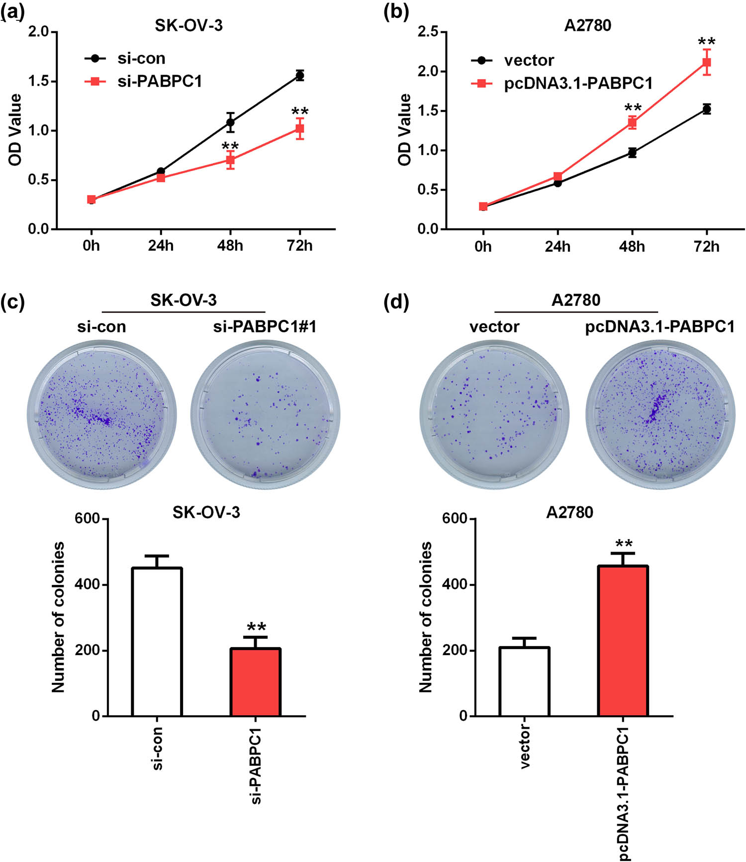
Influences of knockdown and overexpression of PABPC1 on proliferation. The SK-OV-3 cells proliferative (a) and clonogenic (c) capacities were detected utilizing CCK-8 (a) and clone formation (c) assays after depletion of PABPC1. The A2780 cells proliferative (b) and clonogenic (d) capacities were detected utilizing CCK-8 (b) and clone formation (d) assays after upregulation of PABPC1. **P < 0.01 vs si-con or vector group.
A similar trend was observed in clone formation assays. As shown in Figure 3c, the visible colonies in si-PABPC1 group was less than that in si-con group in SK-OV-3 cells (P < 0.01; Figure 3c). On the contrary, upregulation of PABPC1 in A2780 cells presented a better cloning efficiency when compared with the control (P < 0.01; Figure 3d).
3.4 PABPC1 expression affected OC cell invasiveness and motility
The action of PABPC1 on the abilities of invasion and migration of OC cells was investigated using Transwell assays. Results of invasion assays revealed that the invaded SK-OV-3 cells in si-PABPC1 group (87.67 ± 17.01) showed a nearly twofold decrease compared with the control (155 ± 38.04; P < 0.01; Figure 4a and b). Similarly, the migrated SK-OV-3 cells in si-PABPC1 group (161.67 ± 30.53) were markedly decreased compared with the si-con group (298 ± 25.63; P < 0.01; Figure 4a and b). On the other hand, after PABPC1overexpression (156.67 ± 24.03), the invaded number of A2780 cells was more than twice as many as that of the vector group (64.33 ± 18.56; P < 0.01; Figure 4c and d). Moreover, the migrated A2780 cells in pcDNA3.1-PABPC1 group (301.33 ± 37.82) were also significantly increased when compared with the vector group (165.67 ± 29.09; P < 0.01; Figure 4c and d).
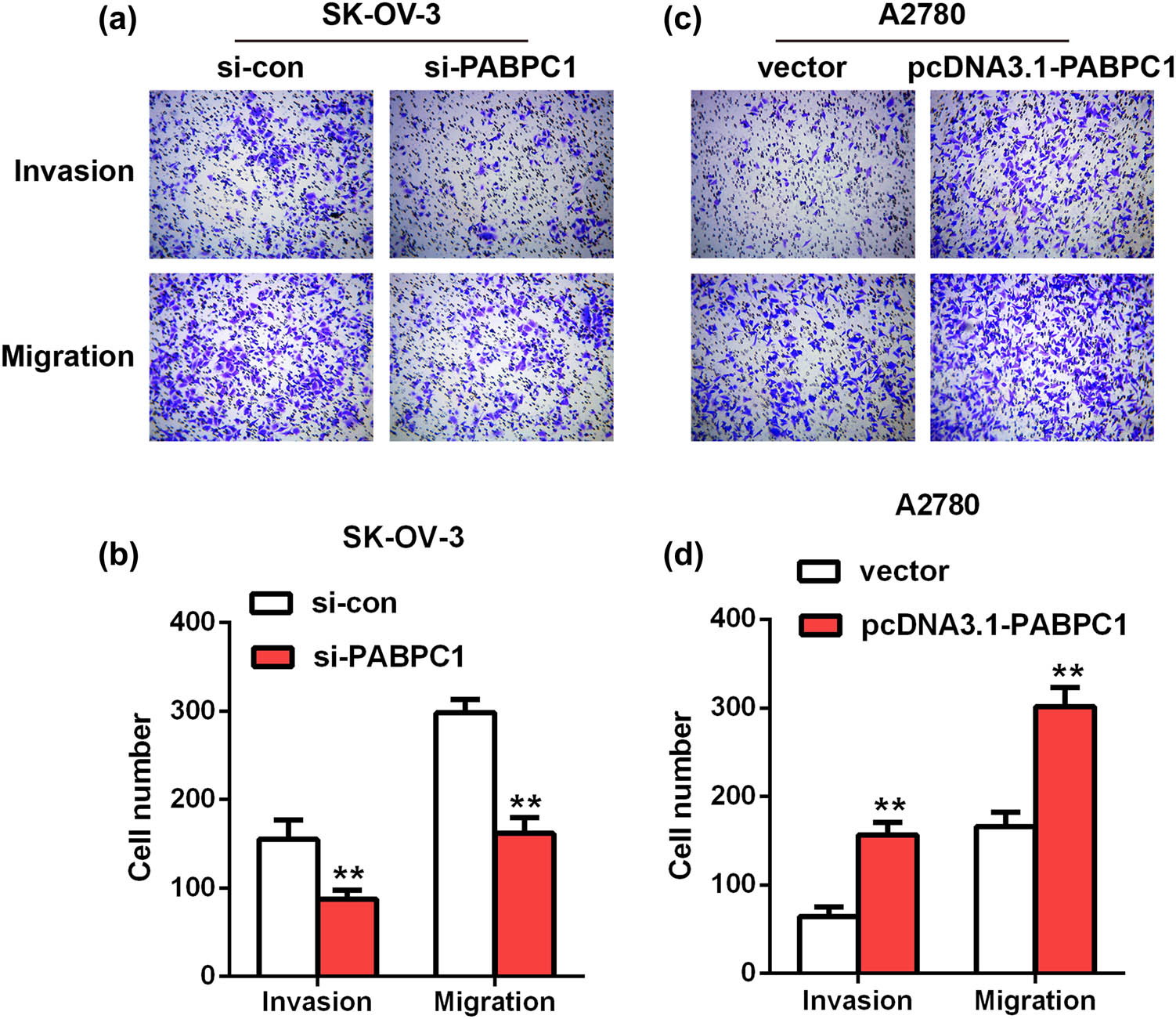
Impacts of downregulation and upregulation of PABPC1 on invasion and migration. The impacts of PABPC1 knockdown on SK-OV-3 cells (a and b) and the impacts of PABPC1 overexpression on A2780 cells (c and d) invasion and migration were measured by Transwell assays.**P < 0.01 vs si-con or vector group.
3.5 PABPC1 expression was associated with EMT process in OC cells
Our previous functional experiments indicated that PABPC1 could promote OC cells’ movement and invasion. Considering that the EMT is frequently used to explain how cancer cells acquire aggressiveness [21], we investigated the action of PABPC1 on the EMT-related markers using Western blot analysis. The outcomes illustrated that the protein expression of epithelial marker E-cadherin was notably elevated after depletion of PABPC1 in SK-OV-3 cells compared with the control (P < 0.01; Figure 5a), whereas the levels of mesenchymal markers N-cadherin and vimentin and transcription factor snail were all significantly downregulated (P < 0.01; Figure 5a). For overexpression of PABPC1, the levels of these markers in A2780 showed the reverse results, as with restrained E-cadherin levels and elevated the levels of N-cadherin, vimentin and snail (P < 0.01; Figure 5b).
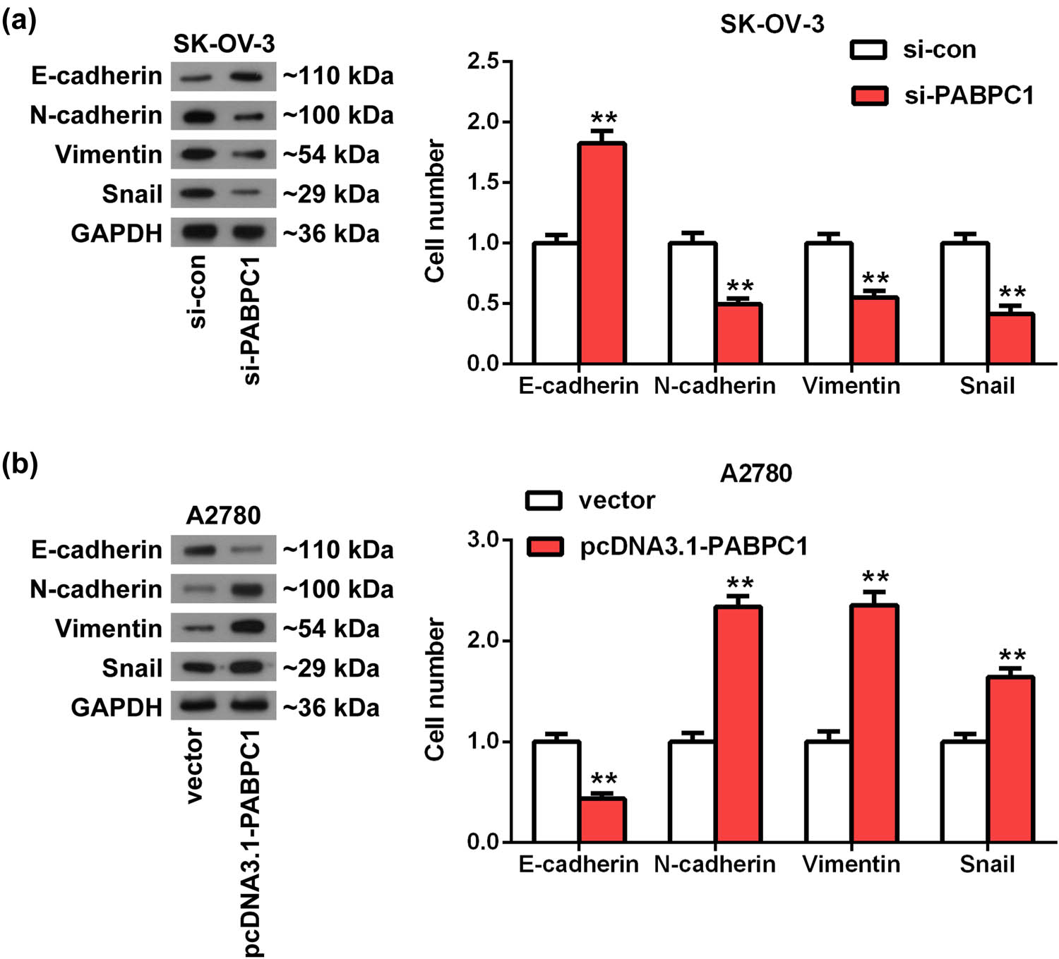
PABPC1 regulated the EMT process. (a and b) Western blot was employed to evaluate the effects of PABPC1 knockdown (a) and PABPC1 overexpression (b) on the protein expression of EMT-related markers in SK-OV-3 (a) and A2780 (b) cells. **P < 0.01 vs si-con or vector group.
4 Discussion
Although cancer registry statistics revealed an overall decline trend in OC incidence and mortality in Europe, North America and other regions over the past three to four decades, the 5-year relative survival is still less than satisfactory [22]. In recent years, the major treatment of OC remains cytoreductive surgery and platinum-based chemotherapy. Despite most patients of OC initially being platinum sensitive, the majority of them will develop platinum resistance upon several recurrences, which presents a low response to the second-line chemotherapy [23]. Moreover, OC encompasses several heterogeneous subtypes with different clinical phenotypes, molecular features and prognosis [24]. Targeted therapy is expected to be a more effective and less toxic therapeutic strategy for OC. Therefore, with the expectation of providing a new possible view for OC-targeted therapy, we investigated the functional role of PABPC1 on OC cells as well as the potential effect on EMT. Our data showed that PABPC1 serves as an oncogene to facilitate cell proliferative capacity and invasive and migratory abilities by regulating the EMT process in OC.
In this study, we first revealed the upregulation of PABPC1 in OC tissues using bioinformatic analysis and found that high expression of PABPC1 was associated with the poor prognosis in patients with OC, implying that PABPC1 might present a tumorigenic effect on the progression of OC. Furthermore, functional experiments were implemented to determine the action of PABPC1 on the cell viability and aggressiveness using the A2780 and SK-OV-3 cell lines. The results revealed that depletion of PABPC1 dramatically inhibited SK-OV-3 cell viability and invasiveness, while upregulation of PABPC1 in A2780 cells exhibited the opposite results. Noticeably, previous research have also suggested the involvement of PABPC1 in the growth and metastasis of numerous tumors. As reported, highly expressed PABPC1 was observed in gastric carcinoma tissues and could predict poor survival, while downregulation of PABPC1 could induce apoptosis [15]. Similarly, in metastatic duodenal cancer, PABPC1 expression was upregulated in tumor cells, and overexpression of PABPC1 enhanced cell proliferative capacity, colony numbers and metastasis ability [16]. All these data provided evidences supporting that PABPC1 may act as an oncogene involved in the carcinogenesis of OC.
The transition from epithelial cells to motile mesenchymal cells is known as EMT [25], and it is characterized by reduced adhesion, cytoskeletal recombination and increased mobility [26]. EMT has been explored for decades in the development of mammals and is considered to be an important mechanism for tumor progression and metastasis [27]. It can prevent the spread of tumor cells in the early stage or eradicate the existing metastatic cells in advanced stage [28,29]. EMT pathway as a new therapeutic approach for cancer has attracted more and more attention in cancers. In OC, previous studies have reported that microRNA-1271 inhibited the EMT process [30], whereas miR-9 promoted the EMT [31]. Moreover, overexpression of TEL1 in SK-OV-3 cells could reverse the EMT process [32] and knockdown of HOXB-AS3-inhibited epithelial OC cell EMT [33]. These reports prompted us to probe whether PABPC1 is connected with the EMT process in OC. To gain insight into this underlying mechanism of action, we detected EMT relative protein levels including E-cadherin, N-cadherin, vimentin and snail. Western blot revealed that reducing PABPC1 levels in SK-OV-3 cells could significantly suppress the EMT action, as evidenced by raising E-cadherin levels and reducing N-cadherin, vimentin and snail levels, whereas increasing PABPC1 expression got the reverse outcomes. These results suggested that PABPC1 as a cancer-promoting gene facilitated the growth and invasiveness of OC cells partly by regulating the EMT. As we know, elucidating the molecular mechanism of the regulation process of EMT and exploring the treatment targeting molecule of EMT are the key issues in the study of the EMT mechanism in malignant tumors [34,35]. Therefore, further investigations will be needed on the correlation of PABPC1 expression and EMT in OC.
The present study has limitations. One limitation of this study is that we only analyzed PABPC1 expression in OC and its relationship with the overall survival using the online data sets. Hence, the prognostic significance of PABPC1 in relevant clinical specimens has to be assessed to confirm its potential involvement. Another limitation is that the functional experiments in this study were obtained from human OC cell lines in vitro. The in vivo functions of PABPC1 in OC development in the pathogenesis of OC need to be further explored.
In summary, this study demonstrated that PABPC1 was significantly upregulated in OC cells and had a connection with poor survival rate for patients with OC. Functionally, our work suggested that PABPC1 plays a promotional effect on the cells’ proliferation, invasion and migration in OC partly through regulating the EMT process. These data imply that PABPC1 may serve as a biological carcinogene in the development of OC, which provides a new insight of research and therapeutic interventions for OC.
-
Funding information: No funding.
-
Author contributions: C. F. and Y. -H. H. were in charge of project development, data collection, data analysis and manuscript writing; N. Q. contributed to data collection, data analysis and figure preparation; J. L., Q. -H. S., and Y. L. were involved in data collection and data analysis; and L. -L. Y. contributed to the design of the work and manuscript revision.
-
Conflict of interest: The authors have no conflicts of interest to declare.
-
Data availability statement: The data were available from the corresponding author on reasonable request.
References
[1] Torre LA, Trabert B, DeSantis CE, Miller KD, Samimi G, Runowicz CD, et al. Ovarian cancer statistics, 2018. CA Cancer J Clin. 2018;68:284–96.10.3322/caac.21456Search in Google Scholar PubMed PubMed Central
[2] Kalampokas E, Young H, Bednarek A, Habib M, Parkin DE, Gurumurthy M, et al. Surgical outcomes and morbidity after radical surgery for ovarian cancer in aberdeen royal infirmary, the northeast of scotland gynaecologic oncology centre. Anticancer Res. 2018;38:923–8.10.26226/morressier.599bdc7dd462b80296ca14abSearch in Google Scholar
[3] Siegel RL, Miller KD, Jemal A. Cancer statistics, 2019. CA Cancer J Clin. 2019;69:7–34.10.3322/caac.21551Search in Google Scholar PubMed
[4] Dong X, Men X, Zhang W, Lei P. Advances in tumor markers of ovarian cancer for early diagnosis. Indian J Cancer. 2014;51(Suppl 3):e72–6.10.4103/0019-509X.154049Search in Google Scholar PubMed
[5] Bristow RE, Chang J, Ziogas A, Randall LM, Anton-Culver H. High-volume ovarian cancer care: survival impact and disparities in access for advanced-stage disease. Gynecol Oncol. 2014;132:403–10.10.1016/j.ygyno.2013.12.017Search in Google Scholar PubMed
[6] Kujawa KA, Lisowska KM. Ovarian cancer – from biology to clinic. Postepy higieny i medycyny doswiadczalnej (Online). 2015;69:1275–90.10.5604/17322693.1184451Search in Google Scholar PubMed
[7] Fung-Kee-Fung M, Oliver T, Elit L, Oza A, Hirte HW, Bryson P. Optimal chemotherapy treatment for women with recurrent ovarian cancer. Curr Oncol. 2007;14:195–208.10.3747/co.2007.148Search in Google Scholar PubMed PubMed Central
[8] Armstrong DK, Alvarez RD, Bakkum-Gamez JN, Barroilhet L, Behbakht K, Berchuck A, et al. NCCN guidelines insights: ovarian cancer, version 1.2019. J Natl Compr Canc Netw. 2019;17:896–909.10.6004/jnccn.2019.0039Search in Google Scholar PubMed
[9] Garzon S, Laganà AS, Casarin J, Raffaelli R, Cromi A, Franchi M, et al. Secondary and tertiary ovarian cancer recurrence: what is the best management? Gland Surg. 2020;9:1118–29.10.21037/gs-20-325Search in Google Scholar PubMed PubMed Central
[10] Gray NK, Hrabalkova L, Scanlon JP, Smith RW. Poly(A)-binding proteins and mRNA localization: who rules the roost? Biochem Soc Trans. 2015;43:1277–84.10.1042/BST20150171Search in Google Scholar PubMed
[11] Ozturk S, Uysal F. Poly(A)-binding proteins are required for translational regulation in vertebrate oocytes and early embryos. Reprod Fertil Dev. 2017;29:1890–901.10.1071/RD16283Search in Google Scholar PubMed
[12] Goss DJ, Kleiman FE. Poly(A) binding proteins: are they all created equal? Wiley Interdiscip Rev RNA. 2013;4:167–79.10.1002/wrna.1151Search in Google Scholar PubMed PubMed Central
[13] Kuhn U, Wahle E. Structure and function of poly(A) binding proteins. Biochim Biophys Acta. 2004;1678:67–84.10.1016/j.bbaexp.2004.03.008Search in Google Scholar PubMed
[14] Zhang H, Sheng C, Yin Y, Wen S, Yang G, Cheng Z, et al. PABPC1 interacts with AGO2 and is responsible for the microRNA mediated gene silencing in high grade hepatocellular carcinoma. Cancer Lett. 2015;367:49–57.10.1016/j.canlet.2015.07.010Search in Google Scholar PubMed
[15] Zhu J, Ding H, Wang X, Lu Q. PABPC1 exerts carcinogenesis in gastric carcinoma by targeting miR-34c. Int J Clin Exp Pathol. 2015;8:3794–802.Search in Google Scholar
[16] Ohshima K, Kanto K, Hatakeyama K, Ide T, Wakabayashi-Nakao K, Watanabe Y, et al. Exosome-mediated extracellular release of polyadenylate-binding protein 1 in human metastatic duodenal cancer cells. Proteomics. 2014;14:2297–306.10.1002/pmic.201300477Search in Google Scholar PubMed
[17] Takashima N, Ishiguro H, Kuwabara Y, Kimura M, Haruki N, Ando T, et al. Expression and prognostic roles of PABPC1 in esophageal cancer: correlation with tumor progression and postoperative survival. Oncol Rep. 2006;15:667–71.10.3892/or.15.3.667Search in Google Scholar
[18] Lu DH, Yang J, Gao LK, Min J, Tang JM, Hu M, et al. Lysine demethylase 2A promotes the progression of ovarian cancer by regulating the PI3K pathway and reversing epithelialmesenchymal transition. Oncol Rep. 2019;41:917–27.10.3892/or.2018.6888Search in Google Scholar
[19] Chen Y, Wang DD, Wu YP, Su D, Zhou TY, Gai RH, et al. MDM2 promotes epithelial-mesenchymal transition and metastasis of ovarian cancer SKOV3 cells. Br J Cancer. 2017;117:1192–201.10.1038/bjc.2017.265Search in Google Scholar PubMed PubMed Central
[20] Fang D, Chen H, Zhu JY, Wang W, Teng Y, Ding HF, et al. Epithelial-mesenchymal transition of ovarian cancer cells is sustained by Rac1 through simultaneous activation of MEK1/2 and Src signaling pathways. Oncogene. 2017;36:1546–58.10.1038/onc.2016.323Search in Google Scholar PubMed PubMed Central
[21] Loret N, Denys H, Tummers P, Berx G. The role of epithelial-to-mesenchymal plasticity in ovarian cancer progression and therapy resistance. Cancers (Basel). 2019;11:838.10.3390/cancers11060838Search in Google Scholar PubMed PubMed Central
[22] Eisenhauer EA. Real-world evidence in the treatment of ovarian cancer. Ann Oncol. 2017;28:viii61–5.10.1093/annonc/mdx443Search in Google Scholar PubMed
[23] Guan LY, Lu Y. New developments in molecular targeted therapy of ovarian cancer. Discov Med. 2018;26:219–29.10.21820/23987073.2018.12.26Search in Google Scholar
[24] Li L, Bi X, Sun H, Liu S, Yu M, Zhang Y, et al. Characterization of Ovarian cancer cells and tissues by Fourier transform infrared spectroscopy. J Ovarian Res. 2018;11:64.10.1186/s13048-018-0434-8Search in Google Scholar PubMed PubMed Central
[25] Lamouille S, Xu J, Derynck R. Molecular mechanisms of epithelial-mesenchymal transition. Nat Rev Mol Cell Biol. 2014;15:178–96.10.1038/nrm3758Search in Google Scholar PubMed PubMed Central
[26] Roshan MK, Soltani A, Soleimani A, Kahkhaie KR, Afshari AR, Soukhtanloo M. Role of AKT and mTOR signaling pathways in the induction of epithelial-mesenchymal transition (EMT) process. Biochimie. 2019;165:229–34.10.1016/j.biochi.2019.08.003Search in Google Scholar PubMed
[27] Singh M, Yelle N, Venugopal C, Singh SK. EMT: mechanisms and therapeutic implications. Pharmacol Ther. 2018;182:80–94.10.1016/j.pharmthera.2017.08.009Search in Google Scholar PubMed
[28] Chen T, You Y, Jiang H, Wang ZZ. Epithelial-mesenchymal transition (EMT): a biological process in the development, stem cell differentiation, and tumorigenesis. 2017;232:3261–72.10.1002/jcp.25797Search in Google Scholar PubMed PubMed Central
[29] Thompson EW, Newgreen DF, Tarin D. Carcinoma invasion and metastasis: a role for epithelial-mesenchymal transition? Cancer Res. 2005;65:5991–5. discussion 5995.10.1158/0008-5472.CAN-05-0616Search in Google Scholar PubMed
[30] Chen Y, Wang L, Zhou J. Effects of microRNA-1271 on ovarian cancer via inhibition of epithelial-mesenchymal transition and cisplatin resistance. J Obs Gynaecol Res. 2019;45:2243–54.10.1111/jog.14079Search in Google Scholar PubMed
[31] Sui X, Jiao YN, Yang LH, Liu J. miR-9 accelerates epithelial-mesenchymal transition of ovarian cancer cells via inhibiting E-cadherin. Eur Rev Med Pharmacol Sci. 2019;23:209–16.Search in Google Scholar
[32] Duan H, Yan Z, Chen W, Wu Y, Han J, Guo H, et al. TET1 inhibits EMT of ovarian cancer cells through activating Wnt/beta-catenin signaling inhibitors DKK1 and SFRP2. Gynecol Oncol. 2017;147:408–17.10.1016/j.ygyno.2017.08.010Search in Google Scholar PubMed
[33] Zhuang XH, Liu Y, Li JL. Overexpression of long noncoding RNA HOXB-AS3 indicates an unfavorable prognosis and promotes tumorigenesis in epithelial ovarian cancer via Wnt/beta-catenin signaling pathway Biosci Rep. 2019;39:BSR20190906.10.1042/BSR20190906Search in Google Scholar PubMed PubMed Central
[34] Friedman JR, Richbart SD, Merritt JC, Brown KC, Denning KL, Tirona MT, et al. Capsaicinoids: Multiple effects on angiogenesis, invasion and metastasis in human cancers. Biomed Pharmacother. 2019;118:109317.10.1016/j.biopha.2019.109317Search in Google Scholar PubMed PubMed Central
[35] Felipe Lima J, Nofech-Mozes S, Bayani J, Bartlett JM. EMT in breast carcinoma: a review. J Clin Med. 2016;5:65.10.3390/jcm5070065Search in Google Scholar PubMed PubMed Central
© 2021 Cong Feng et al., published by De Gruyter
This work is licensed under the Creative Commons Attribution 4.0 International License.
Articles in the same Issue
- Research Articles
- Identification of ZG16B as a prognostic biomarker in breast cancer
- Behçet’s disease with latent Mycobacterium tuberculosis infection
- Erratum
- Erratum to “Suffering from Cerebral Small Vessel Disease with and without Metabolic Syndrome”
- Research Articles
- GPR37 promotes the malignancy of lung adenocarcinoma via TGF-β/Smad pathway
- Expression and role of ABIN1 in sepsis: In vitro and in vivo studies
- Additional baricitinib loading dose improves clinical outcome in COVID-19
- The co-treatment of rosuvastatin with dapagliflozin synergistically inhibited apoptosis via activating the PI3K/AKt/mTOR signaling pathway in myocardial ischemia/reperfusion injury rats
- SLC12A8 plays a key role in bladder cancer progression and EMT
- LncRNA ATXN8OS enhances tamoxifen resistance in breast cancer
- Case Report
- Serratia marcescens as a cause of unfavorable outcome in the twin pregnancy
- Spleno-adrenal fusion mimicking an adrenal metastasis of a renal cell carcinoma: A case report and embryological background
- Research Articles
- TRIM25 contributes to the malignancy of acute myeloid leukemia and is negatively regulated by microRNA-137
- CircRNA circ_0004370 promotes cell proliferation, migration, and invasion and inhibits cell apoptosis of esophageal cancer via miR-1301-3p/COL1A1 axis
- LncRNA XIST regulates atherosclerosis progression in ox-LDL-induced HUVECs
- Potential role of IFN-γ and IL-5 in sepsis prediction of preterm neonates
- Rapid Communication
- COVID-19 vaccine: Call for employees in international transportation industries and international travelers as the first priority in global distribution
- Case Report
- Rare squamous cell carcinoma of the kidney with concurrent xanthogranulomatous pyelonephritis: A case report and review of the literature
- An infertile female delivered a baby after removal of primary renal carcinoid tumor
- Research Articles
- Hypertension, BMI, and cardiovascular and cerebrovascular diseases
- Case Report
- Coexistence of bilateral macular edema and pale optic disc in the patient with Cohen syndrome
- Research Articles
- Correlation between kinematic sagittal parameters of the cervical lordosis or head posture and disc degeneration in patients with posterior neck pain
- Review Articles
- Hepatoid adenocarcinoma of the lung: An analysis of the Surveillance, Epidemiology, and End Results (SEER) database
- Research Articles
- Thermography in the diagnosis of carpal tunnel syndrome
- Pemetrexed-based first-line chemotherapy had particularly prominent objective response rate for advanced NSCLC: A network meta-analysis
- Comparison of single and double autologous stem cell transplantation in multiple myeloma patients
- The influence of smoking in minimally invasive spinal fusion surgery
- Impact of body mass index on left atrial dimension in HOCM patients
- Expression and clinical significance of CMTM1 in hepatocellular carcinoma
- miR-142-5p promotes cervical cancer progression by targeting LMX1A through Wnt/β-catenin pathway
- Comparison of multiple flatfoot indicators in 5–8-year-old children
- Early MRI imaging and follow-up study in cerebral amyloid angiopathy
- Intestinal fatty acid-binding protein as a biomarker for the diagnosis of strangulated intestinal obstruction: A meta-analysis
- miR-128-3p inhibits apoptosis and inflammation in LPS-induced sepsis by targeting TGFBR2
- Dynamic perfusion CT – A promising tool to diagnose pancreatic ductal adenocarcinoma
- Biomechanical evaluation of self-cinching stitch techniques in rotator cuff repair: The single-loop and double-loop knot stitches
- Review Articles
- The ambiguous role of mannose-binding lectin (MBL) in human immunity
- Case Report
- Membranous nephropathy with pulmonary cryptococcosis with improved 1-year follow-up results: A case report
- Fertility problems in males carrying an inversion of chromosome 10
- Acute myeloid leukemia with leukemic pleural effusion and high levels of pleural adenosine deaminase: A case report and review of literature
- Metastatic renal Ewing’s sarcoma in adult woman: Case report and review of the literature
- Burkitt-like lymphoma with 11q aberration in a patient with AIDS and a patient without AIDS: Two cases reports and literature review
- Skull hemophilia pseudotumor: A case report
- Judicious use of low-dosage corticosteroids for non-severe COVID-19: A case report
- Adult-onset citrullinaemia type II with liver cirrhosis: A rare cause of hyperammonaemia
- Clinicopathologic features of Good’s syndrome: Two cases and literature review
- Fatal immune-related hepatitis with intrahepatic cholestasis and pneumonia associated with camrelizumab: A case report and literature review
- Research Articles
- Effects of hydroxyethyl starch and gelatin on the risk of acute kidney injury following orthotopic liver transplantation: A multicenter retrospective comparative clinical study
- Significance of nucleic acid positive anal swab in COVID-19 patients
- circAPLP2 promotes colorectal cancer progression by upregulating HELLS by targeting miR-335-5p
- Ratios between circulating myeloid cells and lymphocytes are associated with mortality in severe COVID-19 patients
- Risk factors of left atrial appendage thrombus in patients with non-valvular atrial fibrillation
- Clinical features of hypertensive patients with COVID-19 compared with a normotensive group: Single-center experience in China
- Surgical myocardial revascularization outcomes in Kawasaki disease: systematic review and meta-analysis
- Decreased chromobox homologue 7 expression is associated with epithelial–mesenchymal transition and poor prognosis in cervical cancer
- FGF16 regulated by miR-520b enhances the cell proliferation of lung cancer
- Platelet-rich fibrin: Basics of biological actions and protocol modifications
- Accurate diagnosis of prostate cancer using logistic regression
- miR-377 inhibition enhances the survival of trophoblast cells via upregulation of FNDC5 in gestational diabetes mellitus
- Prognostic significance of TRIM28 expression in patients with breast carcinoma
- Integrative bioinformatics analysis of KPNA2 in six major human cancers
- Exosomal-mediated transfer of OIP5-AS1 enhanced cell chemoresistance to trastuzumab in breast cancer via up-regulating HMGB3 by sponging miR-381-3p
- A four-lncRNA signature for predicting prognosis of recurrence patients with gastric cancer
- Knockdown of circ_0003204 alleviates oxidative low-density lipoprotein-induced human umbilical vein endothelial cells injury: Circulating RNAs could explain atherosclerosis disease progression
- Propofol postpones colorectal cancer development through circ_0026344/miR-645/Akt/mTOR signal pathway
- Knockdown of lncRNA TapSAKI alleviates LPS-induced injury in HK-2 cells through the miR-205/IRF3 pathway
- COVID-19 severity in relation to sociodemographics and vitamin D use
- Clinical analysis of 11 cases of nocardiosis
- Cis-regulatory elements in conserved non-coding sequences of nuclear receptor genes indicate for crosstalk between endocrine systems
- Four long noncoding RNAs act as biomarkers in lung adenocarcinoma
- Real-world evidence of cytomegalovirus reactivation in non-Hodgkin lymphomas treated with bendamustine-containing regimens
- Relation between IL-8 level and obstructive sleep apnea syndrome
- circAGFG1 sponges miR-28-5p to promote non-small-cell lung cancer progression through modulating HIF-1α level
- Nomogram prediction model for renal anaemia in IgA nephropathy patients
- Effect of antibiotic use on the efficacy of nivolumab in the treatment of advanced/metastatic non-small cell lung cancer: A meta-analysis
- NDRG2 inhibition facilitates angiogenesis of hepatocellular carcinoma
- A nomogram for predicting metabolic steatohepatitis: The combination of NAMPT, RALGDS, GADD45B, FOSL2, RTP3, and RASD1
- Clinical and prognostic features of MMP-2 and VEGF in AEG patients
- The value of miR-510 in the prognosis and development of colon cancer
- Functional implications of PABPC1 in the development of ovarian cancer
- Prognostic value of preoperative inflammation-based predictors in patients with bladder carcinoma after radical cystectomy
- Sublingual immunotherapy increases Treg/Th17 ratio in allergic rhinitis
- Prediction of improvement after anterior cruciate ligament reconstruction
- Effluent Osteopontin levels reflect the peritoneal solute transport rate
- circ_0038467 promotes PM2.5-induced bronchial epithelial cell dysfunction
- Significance of miR-141 and miR-340 in cervical squamous cell carcinoma
- Association between hair cortisol concentration and metabolic syndrome
- Microvessel density as a prognostic indicator of prostate cancer: A systematic review and meta-analysis
- Characteristics of BCR–ABL gene variants in patients of chronic myeloid leukemia
- Knee alterations in rheumatoid arthritis: Comparison of US and MRI
- Long non-coding RNA TUG1 aggravates cerebral ischemia and reperfusion injury by sponging miR-493-3p/miR-410-3p
- lncRNA MALAT1 regulated ATAD2 to facilitate retinoblastoma progression via miR-655-3p
- Development and validation of a nomogram for predicting severity in patients with hemorrhagic fever with renal syndrome: A retrospective study
- Analysis of COVID-19 outbreak origin in China in 2019 using differentiation method for unusual epidemiological events
- Laparoscopic versus open major liver resection for hepatocellular carcinoma: A case-matched analysis of short- and long-term outcomes
- Travelers’ vaccines and their adverse events in Nara, Japan
- Association between Tfh and PGA in children with Henoch–Schönlein purpura
- Can exchange transfusion be replaced by double-LED phototherapy?
- circ_0005962 functions as an oncogene to aggravate NSCLC progression
- Circular RNA VANGL1 knockdown suppressed viability, promoted apoptosis, and increased doxorubicin sensitivity through targeting miR-145-5p to regulate SOX4 in bladder cancer cells
- Serum intact fibroblast growth factor 23 in healthy paediatric population
- Algorithm of rational approach to reconstruction in Fournier’s disease
- A meta-analysis of exosome in the treatment of spinal cord injury
- Src-1 and SP2 promote the proliferation and epithelial–mesenchymal transition of nasopharyngeal carcinoma
- Dexmedetomidine may decrease the bupivacaine toxicity to heart
- Hypoxia stimulates the migration and invasion of osteosarcoma via up-regulating the NUSAP1 expression
- Long noncoding RNA XIST knockdown relieves the injury of microglia cells after spinal cord injury by sponging miR-219-5p
- External fixation via the anterior inferior iliac spine for proximal femoral fractures in young patients
- miR-128-3p reduced acute lung injury induced by sepsis via targeting PEL12
- HAGLR promotes neuron differentiation through the miR-130a-3p-MeCP2 axis
- Phosphoglycerate mutase 2 is elevated in serum of patients with heart failure and correlates with the disease severity and patient’s prognosis
- Cell population data in identifying active tuberculosis and community-acquired pneumonia
- Prognostic value of microRNA-4521 in non-small cell lung cancer and its regulatory effect on tumor progression
- Mean platelet volume and red blood cell distribution width is associated with prognosis in premature neonates with sepsis
- 3D-printed porous scaffold promotes osteogenic differentiation of hADMSCs
- Association of gene polymorphisms with women urinary incontinence
- Influence of COVID-19 pandemic on stress levels of urologic patients
- miR-496 inhibits proliferation via LYN and AKT pathway in gastric cancer
- miR-519d downregulates LEP expression to inhibit preeclampsia development
- Comparison of single- and triple-port VATS for lung cancer: A meta-analysis
- Fluorescent light energy modulates healing in skin grafted mouse model
- Silencing CDK6-AS1 inhibits LPS-induced inflammatory damage in HK-2 cells
- Predictive effect of DCE-MRI and DWI in brain metastases from NSCLC
- Severe postoperative hyperbilirubinemia in congenital heart disease
- Baicalin improves podocyte injury in rats with diabetic nephropathy by inhibiting PI3K/Akt/mTOR signaling pathway
- Clinical factors predicting ureteral stent failure in patients with external ureteral compression
- Novel H2S donor proglumide-ADT-OH protects HUVECs from ox-LDL-induced injury through NF-κB and JAK/SATA pathway
- Triple-Endobutton and clavicular hook: A propensity score matching analysis
- Long noncoding RNA MIAT inhibits the progression of diabetic nephropathy and the activation of NF-κB pathway in high glucose-treated renal tubular epithelial cells by the miR-182-5p/GPRC5A axis
- Serum exosomal miR-122-5p, GAS, and PGR in the non-invasive diagnosis of CAG
- miR-513b-5p inhibits the proliferation and promotes apoptosis of retinoblastoma cells by targeting TRIB1
- Fer exacerbates renal fibrosis and can be targeted by miR-29c-3p
- The diagnostic and prognostic value of miR-92a in gastric cancer: A systematic review and meta-analysis
- Prognostic value of α2δ1 in hypopharyngeal carcinoma: A retrospective study
- No significant benefit of moderate-dose vitamin C on severe COVID-19 cases
- circ_0000467 promotes the proliferation, metastasis, and angiogenesis in colorectal cancer cells through regulating KLF12 expression by sponging miR-4766-5p
- Downregulation of RAB7 and Caveolin-1 increases MMP-2 activity in renal tubular epithelial cells under hypoxic conditions
- Educational program for orthopedic surgeons’ influences for osteoporosis
- Expression and function analysis of CRABP2 and FABP5, and their ratio in esophageal squamous cell carcinoma
- GJA1 promotes hepatocellular carcinoma progression by mediating TGF-β-induced activation and the epithelial–mesenchymal transition of hepatic stellate cells
- lncRNA-ZFAS1 promotes the progression of endometrial carcinoma by targeting miR-34b to regulate VEGFA expression
- Anticoagulation is the answer in treating noncritical COVID-19 patients
- Effect of late-onset hemorrhagic cystitis on PFS after haplo-PBSCT
- Comparison of Dako HercepTest and Ventana PATHWAY anti-HER2 (4B5) tests and their correlation with silver in situ hybridization in lung adenocarcinoma
- VSTM1 regulates monocyte/macrophage function via the NF-κB signaling pathway
- Comparison of vaginal birth outcomes in midwifery-led versus physician-led setting: A propensity score-matched analysis
- Treatment of osteoporosis with teriparatide: The Slovenian experience
- New targets of morphine postconditioning protection of the myocardium in ischemia/reperfusion injury: Involvement of HSP90/Akt and C5a/NF-κB
- Superenhancer–transcription factor regulatory network in malignant tumors
- β-Cell function is associated with osteosarcopenia in middle-aged and older nonobese patients with type 2 diabetes: A cross-sectional study
- Clinical features of atypical tuberculosis mimicking bacterial pneumonia
- Proteoglycan-depleted regions of annular injury promote nerve ingrowth in a rabbit disc degeneration model
- Effect of electromagnetic field on abortion: A systematic review and meta-analysis
- miR-150-5p affects AS plaque with ASMC proliferation and migration by STAT1
- MALAT1 promotes malignant pleural mesothelioma by sponging miR-141-3p
- Effects of remifentanil and propofol on distant organ lung injury in an ischemia–reperfusion model
- miR-654-5p promotes gastric cancer progression via the GPRIN1/NF-κB pathway
- Identification of LIG1 and LIG3 as prognostic biomarkers in breast cancer
- MitoQ inhibits hepatic stellate cell activation and liver fibrosis by enhancing PINK1/parkin-mediated mitophagy
- Dissecting role of founder mutation p.V727M in GNE in Indian HIBM cohort
- circATP2A2 promotes osteosarcoma progression by upregulating MYH9
- Prognostic role of oxytocin receptor in colon adenocarcinoma
- Review Articles
- The function of non-coding RNAs in idiopathic pulmonary fibrosis
- Efficacy and safety of therapeutic plasma exchange in stiff person syndrome
- Role of cesarean section in the development of neonatal gut microbiota: A systematic review
- Small cell lung cancer transformation during antitumor therapies: A systematic review
- Research progress of gut microbiota and frailty syndrome
- Recommendations for outpatient activity in COVID-19 pandemic
- Rapid Communication
- Disparity in clinical characteristics between 2019 novel coronavirus pneumonia and leptospirosis
- Use of microspheres in embolization for unruptured renal angiomyolipomas
- COVID-19 cases with delayed absorption of lung lesion
- A triple combination of treatments on moderate COVID-19
- Social networks and eating disorders during the Covid-19 pandemic
- Letter
- COVID-19, WHO guidelines, pedagogy, and respite
- Inflammatory factors in alveolar lavage fluid from severe COVID-19 pneumonia: PCT and IL-6 in epithelial lining fluid
- COVID-19: Lessons from Norway tragedy must be considered in vaccine rollout planning in least developed/developing countries
- What is the role of plasma cell in the lamina propria of terminal ileum in Good’s syndrome patient?
- Case Report
- Rivaroxaban triggered multifocal intratumoral hemorrhage of the cabozantinib-treated diffuse brain metastases: A case report and review of literature
- CTU findings of duplex kidney in kidney: A rare duplicated renal malformation
- Synchronous primary malignancy of colon cancer and mantle cell lymphoma: A case report
- Sonazoid-enhanced ultrasonography and pathologic characters of CD68 positive cell in primary hepatic perivascular epithelioid cell tumors: A case report and literature review
- Persistent SARS-CoV-2-positive over 4 months in a COVID-19 patient with CHB
- Pulmonary parenchymal involvement caused by Tropheryma whipplei
- Mediastinal mixed germ cell tumor: A case report and literature review
- Ovarian female adnexal tumor of probable Wolffian origin – Case report
- Rare paratesticular aggressive angiomyxoma mimicking an epididymal tumor in an 82-year-old man: Case report
- Perimenopausal giant hydatidiform mole complicated with preeclampsia and hyperthyroidism: A case report and literature review
- Primary orbital ganglioneuroblastoma: A case report
- Primary aortic intimal sarcoma masquerading as intramural hematoma
- Sustained false-positive results for hepatitis A virus immunoglobulin M: A case report and literature review
- Peritoneal loose body presenting as a hepatic mass: A case report and review of the literature
- Chondroblastoma of mandibular condyle: Case report and literature review
- Trauma-induced complete pacemaker lead fracture 8 months prior to hospitalization: A case report
- Primary intradural extramedullary extraosseous Ewing’s sarcoma/peripheral primitive neuroectodermal tumor (PIEES/PNET) of the thoracolumbar spine: A case report and literature review
- Computer-assisted preoperative planning of reduction of and osteosynthesis of scapular fracture: A case report
- High quality of 58-month life in lung cancer patient with brain metastases sequentially treated with gefitinib and osimertinib
- Rapid response of locally advanced oral squamous cell carcinoma to apatinib: A case report
- Retrieval of intrarenal coiled and ruptured guidewire by retrograde intrarenal surgery: A case report and literature review
- Usage of intermingled skin allografts and autografts in a senior patient with major burn injury
- Retraction
- Retraction on “Dihydromyricetin attenuates inflammation through TLR4/NF-kappa B pathway”
- Special Issue Computational Intelligence Methodologies Meets Recurrent Cancers - Part I
- An artificial immune system with bootstrap sampling for the diagnosis of recurrent endometrial cancers
- Breast cancer recurrence prediction with ensemble methods and cost-sensitive learning