miR-496 inhibits proliferation via LYN and AKT pathway in gastric cancer
-
Rui Su
 and
Jun Zhang
and
Jun Zhang

Abstract
MicroRNAs (miRNAs) operate as tumor suppressor or carcinogen to regulate cell proliferation, metastasis, invasion, differentiation, apoptosis, and metabolic process. In the present research, we investigated the effect and mechanism of miR-496 in human gastric cancer cells. miR-496 was downregulated in two gastric cancer cell lines, AGS and MKN45, compared with normal gastric epithelial cell line GES-1. miR-496 mimics inhibited the proliferation of AGS cells after the transfection for 48 and 72 h. The migration and invasion of AGS cells were also inhibited by the transfection of miR-496 mimics. miR-496 mimics induced the apoptosis through upregulating the levels of Bax and Active Caspase 3 and downregulating the levels of Bcl-2 and Total Caspase 3. Bioinformatics analysis showed that there was a binding site between miR-496 and Lyn kinase (LYN). miR-496 mimics could inhibit the expression of LYN in AGS cells. LYN overexpression blocked the inhibition of tumor cell growth, as well as the inhibition of AKT/mTOR signaling pathway induced by miR-496. In conclusion, miR-496 inhibited the proliferation through the AKT/mTOR signaling pathway via targeting LYN in gastric cancer cells. Our research provides a new potential target for clinical diagnosis and targeted treatment for gastric cancer.
1 Introduction
Gastric cancer is one of the most common human cancers, ranking third in the cancer-related deaths [1]. Studies show that both genetic and epigenetic factors are involved in the pathogenesis of human tumors, including gastric cancer [2]: however, due to the low sensitivity and specificity of tumor markers for gastric cancer, gastric cancer markers are relatively limited [3]. Therefore, the discovery of new markers and medicine for the diagnosis and treatment of tumor is an urgent problem [4,5]. miRNAs are non-coding RNAs with 21–25 bp [6]. They can reduce mRNA level by inhibiting mRNA translation or binding the 3ʹ-UTR region of mRNA, thus silencing their homologous target genes, playing an important role in cell processes and maintaining normal physiological environment [6]. Special miRNA operates as a tumor suppressor or carcinogen, so it is considered as a biomarker for early diagnosis and prognosis of cancer [7,8,9]. These miRNAs also regulate cell proliferation, metastasis, invasion, differentiation, apoptosis, and metabolic process [7,8,9].
miR-496 is an RNA gene and is affiliated with the miRNA class. It is located on human chromosome 14 and has unknown biological functions [10]. Limited studies have shown that low expression of miR-496 is related to aging process [11,12], and plays an important role in the formation and differentiation of bone cells [13]. miR-496 overexpression inhibits breast cancer cell proliferation through MBD2-dependence [14]. At present, there are few reports on miR-496, and its biological function in tumorigenesis is still unclear. In addition, miR-496 has not been reported in the proliferation and metastasis of gastric cancer cells. Therefore, we investigated the role and mechanism of miR-496 in vitro to provide new approach for clinical diagnosis and treatment of gastric cancer.
2 Materials and methods
2.1 Cell culture and transfection
Human normal gastric epithelial cell line GES-1 and gastric cancer cell lines AGS and MKN45 were all purchased from the cell bank of The Chinese Academy of Sciences (Shanghai, China). DMEM with 10% FBS was used for cell culture. miR-496 mimics, miRNA mimics negative control, and Lyn kinase (LYN) overexpression plasmid were purchased from GenePharma (Shanghai, China) and transfected into AGS cells using Lipofectamine 2000 (Invitrogen, CA, USA) according to the manufacturer’s protocol.
2.2 Fluorescence quantitative PCR (qPCR)
After the transfection for 24 h, total RNA was extracted from AGS cells by TRIzol reagent (Invitrogen, USA) and reverse transcripted to cDNA using M-MLV Reverse Transcriptase and TaqMan™ MicroRNA Reverse Transcription Kit (Invitrogen, USA). QPCR was performed to detect the level of miR-496 and LYN using the qPCR kit according to the manufacturer’s protocol (Invitrogen, USA). The relative expression of miR-496 and LYN was analyzed using 2−ΔΔCt method.
2.3 Western blot
After the transfection for 48 h, whole-cell protein was extracted using a RIPA lysis buffer. The protein concentrations were detected by bicinchoninic acid method. Then, the protein extracts were separated using SDS-PAGE and transferred to a polyvinylidene fluoride membrane. After blocking with skimmed milk, the protein was incubated with the primary antibodies for 1 h, followed by secondary antibodies for 1 h. Primary antibodies, including anti-Bcl-2, anti-Bax, anti-total Caspase 3, anti-cleaved Caspase 3, anti-GAPDH, anti-Vimentin, anti-Cyclin D1, anti-Snail2, anti-p-AKT (Ser473), anti-p-mTOR (Ser2448), and anti-p70S6K (1:1,000), were purchased from Cell Signaling Technology Inc. (Danvers, MA, USA).
2.4 CCK8 assay
The proliferation of AGS cells was detected by Cell Counting Kit-8 (CCK8). After transfection, cells were transferred into a 96-well plate (1,000 cells/well). CCK8 reagent was added to the cells at a series of time points after the transfection (0, 24, 48, and 72 h). Cell viability was represented by OD value at 450 nm.
2.5 Clonogenic assay
After the transfection, about 500 cells were transferred into a 6 cm dish and cultured at 37°C for 2 weeks. Then, the colonies were fixed with methanol for 20 min and stained by 0.1% of crystal violet for 30 min. Visible colonies were counted separately by two researchers.
2.6 Transwell
For cell migration, AGS cells were transferred into the upper chamber of the transwell inserts after transfection (2 × 10⁵ cells). Serum-free medium was added into the lower chamber of the transwell inserts. After incubation for 48 h, AGS cells were fixed with methanol for 10 min and stained with 0.1% of crystal violet for 10 min. The migrated cells were counted in five random fields. For cell invasion, transwell inserts were pre-coated with Matrigel.
2.7 Flow cytometry analysis
After transfection for 48 h, the apoptosis of AGS cells was detected using Annexin-V and PI (BD Biosciences, Franklin Lakes, NJ, USA) following the manufacturer’s protocol. The percentage of apoptotic cells was analyzed on a FACScalibur flow cytometer using CellQuest Pro software (Becton, Dickinson and Company, USA).
2.8 Statistical analysis
A one-way analysis followed by post hoc Bonferroni test was performed to analyze the differences between groups. All data in the present study were analyzed and plotted with GraphPad Prism 8 (GraphPad Software Inc., San Diego, California, USA). All experiments were performed in triplicate. P < 0.05 was considered statistically significant.
3 Results
3.1 miR-496 inhibits the proliferation in gastric cancer cells
We first detected the expression of miR-496 in gastric cancer cell lines AGS and MKN45 by qPCR with the normal gastric epithelial cell line GES-1 as a control. miR-496 was downregulated in AGS and MKN45 compared with GES-1 cells (P < 0.05, Figure 1a). Then, the miR-496 mimics or miRNA mimics negative control was transfected into the AGS cells to generate miR-496 overexpressed (miR-496) or negative control (NC) cells, respectively. The level of miR-496 increased significantly in the miR-496 group compared with the NC group (P < 0.05, Figure 1b). The proliferation was detected using CCK8 and clonogenic assay. As shown in Figure 1c, the OD450 declined significantly after the transfection of miR-496 mimics for 48 and 72 h compared with the NC cells (P < 0.05). The colony numbers also markedly declined after transfection and incubated for 2 weeks compared with the NC cells (P < 0.05, Figure 1d). These data indicated that miR-496 could inhibit the proliferation of human gastric cancer cells.
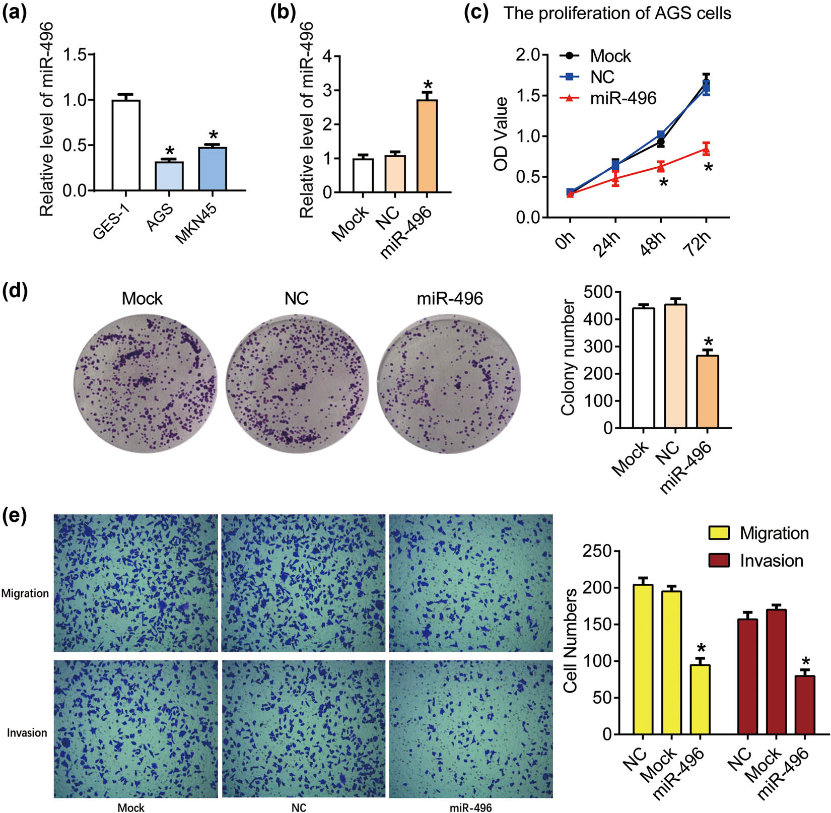
miR-496 inhibited the proliferation and metastasis in gastric cancer cells. (a) QPCR was performed to detect the level of miR-496 in AGS cells. (b) The level of miR-496 in AGS cells transfected with miR-496 mimics was detected by qPCR. Relative level of miR-496 was analyzed using 2−ΔΔCt method and normalized to mock group. (c) The proliferation of AGS cells was determined using CCK8 assay. OD value (450 nm) was measured every 24 h. (d) Clonogenic assay was used to detect the proliferation of AGS cells. Colony number was counted 2 weeks after the culture. (e) The migration and invasion of AGS cells were detected by transwell assay after the transfection for 24 h. NC = negative control. *P < 0.05.
3.2 miR-496 inhibits the migration and invasion in gastric cancer cells
Next the migration and invasion of the AGS cells with the transfection of miR-496 mimics were detected using transwell assay. As shown in Figure 1e, the migration cell numbers decreased after the transfection of miR-496 mimics (P < 0.05); the invasion cell numbers also declined after the transfection of miR-496 mimics compared with the miRNA mimics negative control (P < 0.05). These results suggested that miR-496 could inhibit the metastasis in gastric cancer cells.
3.3 miR-496 promotes the apoptosis in gastric cancer cells
Then, we detected the apoptosis of the AGS cells using flow cytometry. As shown in Figure 2a, the percentage of apoptosis cells in AGS cells transfected with miR-496 mimics (9.50 ± 0.75%) increased compared with the NC cells (4.03 ± 0.50%) (P < 0.05). In order to analyze the molecular mechanism by which miR-496 promotes tumor cell apoptosis, western blot was used to detect the expression of critical apoptotic factors in each group, including Bax, Active Caspase 3, Total Caspase 3, and Bcl-2. As shown in Figure 2b–f, miR-496 mimics upregulated the expression of Bax and Active Caspase 3, but downregulated the expression of Total Caspase 3 and Bcl-2 (P < 0.05). These results proved that miR-496 promoted the apoptosis through regulating apoptotic factors in gastric cancer cells.
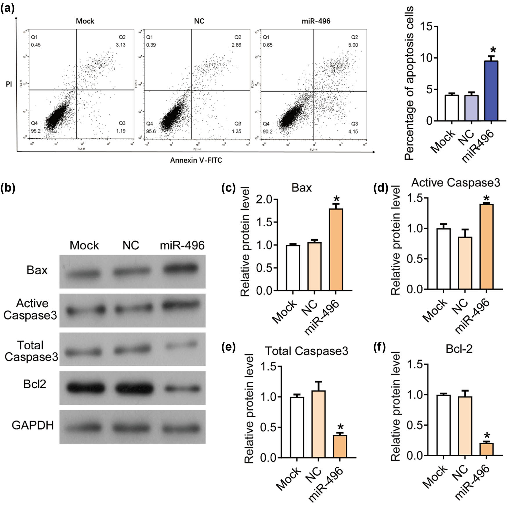
miR-496 promoted the apoptosis in gastric cancer cells. (a) Flow cytometry was performed to detect the apoptosis in AGS cells. (b) The expression levels of apoptosis-related proteins, (c) Bax, (d) active Caspase 3, (e) total Caspase 3, and (f) Bcl-2 were detected by western blot. The relative protein levels were normalized to mock group. NC = negative control. *P < 0.05.
3.4 miR-496 inhibits the expression of LYN in gastric cancer cells
Next we predicted the potential binding targets of miR-496 using the online analysis tool TargetScan (http://www.targetscan.org/vert_71/) [15]. According to the analysis on targetScan, there was a binding site between miR-496 and 3ʹ-UTR of LYN (Figure 3a). After transfected with miR-496 mimics, the mRNA and protein level of LYN significantly declined compared with the NC cells according to the results of qPCR (Figure 3b) and western blot (Figure 3c). Thus, we hypothesized that LYN was the downstream direct target of miR-496 in gastric cancer cells.
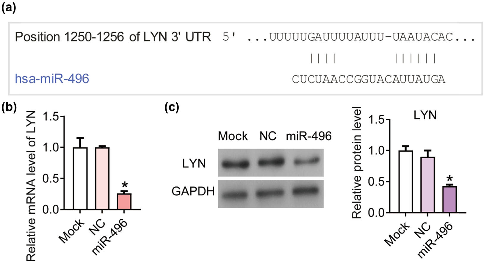
miR-496 inhibited the expression of LYN in gastric cancer cells. (a) According to the analysis on targetScan, there was a binding site between miR-496 and 3ʹ-UTR of LYN. (b) QPCR was performed to detect the level of LYN in AGS cells transfected with miR-496 mimics. Relative level of LYN was analyzed using 2−ΔΔCt method and normalized to mock group. (c) The expression levels of LYN were detected by western blot. The relative protein levels were normalized to mock group. NC = negative control. *P < 0.05.
3.5 LYN blocks the inhibition of tumor cell growth induced by miR-496 in gastric cancer cells
In order to elucidate whether miR-496 exerts tumor suppressive effect in gastric cancer through LYN, we transfected miR-496 mimics and LYN overexpression plasmid (miR-496 & LYN) in AGS cell line at the same time (Figure 4a), and detected the cell proliferation and apoptosis with miR-496 mimics as a control. As shown in Figure 4b, OD450 of miR-496 & LYN group increased markedly compared with the miR-496 group (P < 0.05). The percentage of apoptosis cells in miR-496 & LYN group (2.20 ± 0.36%) declined compared with the miR-496 group (8.31 ± 0.35%) (Figure 4c, P < 0.05). These data indicated that miR-496 might inhibit the growth of gastric cancer cells by suppressing the expression of LYN.
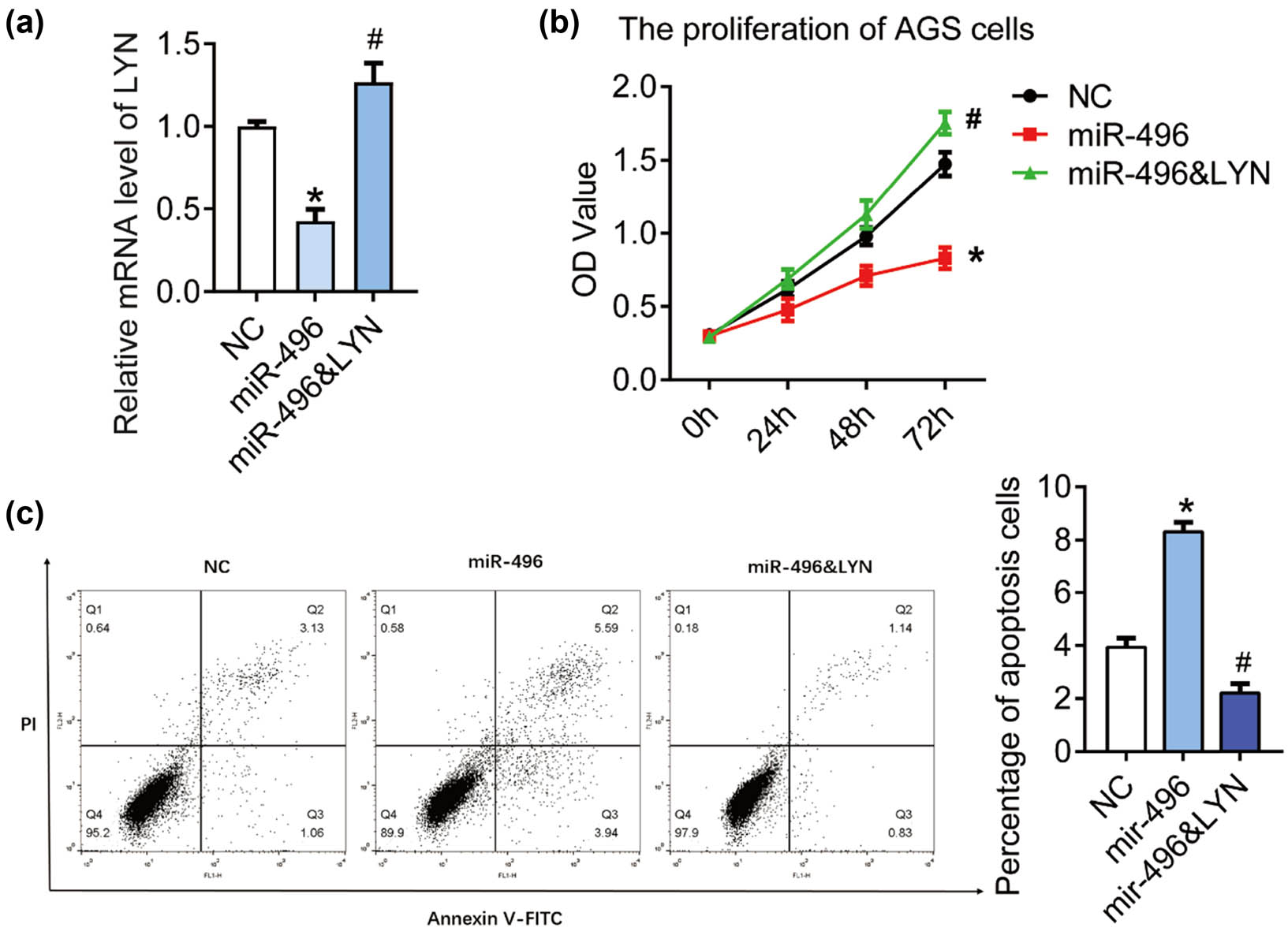
LYN blocked cell apoptosis induced by miR-496 in gastric cancer cells. (a) QPCR was performed to detect the level of LYN in AGS cells transfected with miR-496 mimics or miR-496 mimics + LYN overexpression. (b) The proliferation was detected by CCK8 assay. OD value (450 nm) was measured every 24 h. (c) Flow cytometry was performed to detect the apoptosis in AGS cells. NC = negative control. *P < 0.05; # P < 0.05.
3.6 miR-496 inhibits the AKT/mTOR signaling pathway via LYN in gastric cancer cells
Finally, we explored the molecular mechanism by which miR-496/LYN regulated the growth of gastric cancer. AKT/mTOR signaling pathway is widely involved in the origin and development of solid tumors and plays a key role in the proliferation and apoptosis of tumor cells [16,17]. In our previous studies, we found that the AKT/mTOR pathway was downregulated by LYN knockdown in AGS cells, including decreased levels of p-AKT, p-mTOR, and downstream effector p70. The AKT pathway activator IGF-1 could reverse the inhibitory effects of LYN knockdown on the proliferation, migration, and invasion in AGS cells.
Thus, in this research, we detected the expression of protein related to AKT/mTOR signaling pathway in each group using western blot (Figure 5a). The phosphorylation levels of AKT (Figure 5b) and mTOR (Figure 5c) and the levels of Cyclin D1 (Figure 5d) and P70 (Figure 5e) were significantly inhibited by the transfection of miR-496 mimics and rescued by the transfection of LYN overexpression plasmid. LYN overexpression blocked the inhibition of AKT/mTOR signaling pathway induced by miR-496 mimics. Out results indicated that miR-496/LYN inhibited the tumor growth by suppressing the AKT/mTOR signaling pathway.
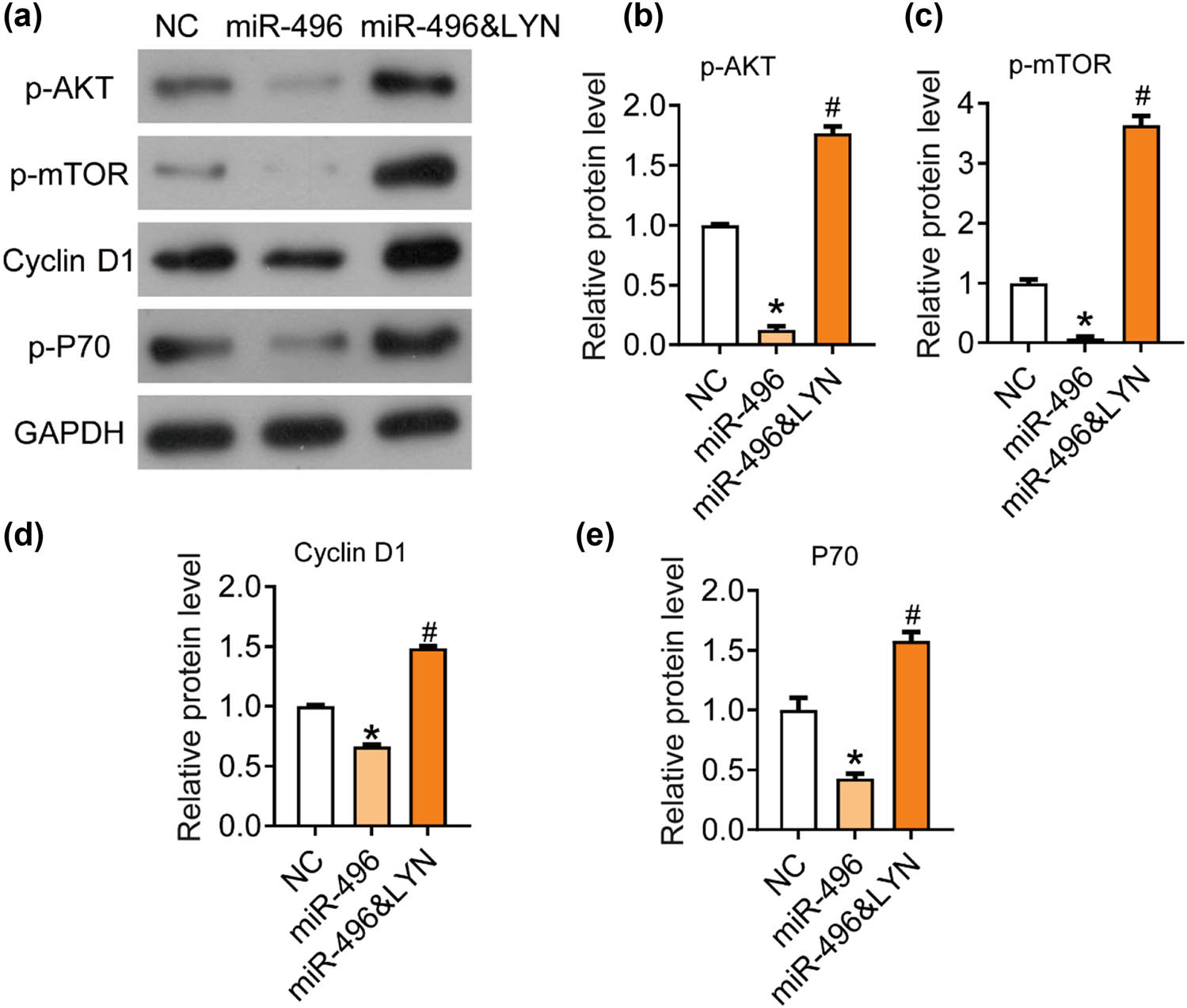
miR-496 inhibited the AKT/mTOR signaling pathway by targeting LYN in gastric cancer cells. (a) The expression levels of AKT/mTOR pathway-related proteins, (b) p-AKT, (c) p-mTOR, (d) Cyclin D1, (e) P70 were detected by western blot. The relative protein levels were normalized to NC group. NC = negative control. *P < 0.05; # P < 0.05.
4 Discussion
In recent years, the role of miR-496 in human tumors has been gradually revealed. lncRNA DANCR was found to be involved in the progression of lung cancer through sponging miR-496 and then regulating the level of mTOR [10]. miR-496 can also suppress gene expression and the proliferation in breast cancer cell lines MCF-10A, MCF-7, and MDA-MB-231 [14]. In another report, miR496 is downregulated by Human papillomavirus, notably type 16 and inhibit the post-transcriptional control of the transcription factor E2F2 in oropharyngeal cancer [18]. In the present study, we found that miR-496 was downregulated in AGS and MKN45 compared with the normal gastric epithelial cell line GES-1. At present, the expression of miR-496 in gastric cancer has not been reported. Our results suggest that miR-496 may play a role in the progression of gastric cancer, and is expected to be a clinical diagnostic indicator of gastric cancer, which still needs further experiments and clinical verification. Then, we proved that miR-496 could inhibit the proliferation, migration, and invasion, and induce the apoptosis in gastric cancer cells. Our data proved that miR-496 was involved in the progression of human gastric cancer.
Studies have shown that miRNA, which is combined with RNA-induced silencing complex, can recognize and bind 3ʹ-UTR of the target genes by incomplete or complete matching, and achieve post transcriptional regulation of target gene expression level [7,19,20]. In mammals, miRNA usually binds to the 3ʹ-UTR of the target gene through incomplete pairing, which affects the translation of target mRNA [20,21]. In addition, an miRNA can regulate multiple mRNA, and different miRNAs can also coordinate to regulate one mRNA molecule [20]. It is predicted that about one-third of the protein coding genes in the human cells are regulated by miRNA [22]. miRNAs and their target molecules form a complex regulatory network to control cell activity [22]. In the present research, we analyzed the potential target of miR-496 on targetScan. A binding site was found between miR-496 and the 3ʹ-UTR of LYN. QPCR result showed that miR-496 mimics significantly inhibited the expression of LYN in the AGS cells suggesting that LYN was a downstream target of miR-496 in gastric cancer. Then, we detected the proliferation and apoptosis in both miR-496 and LYN overexpressed cells with the only miR-496 overexpressed cells as a control. The results proved that LYN blocked the increase in cell proliferation and decrease in apoptosis induced by miR-496 in the AGS cells. LYN is a member of the Src family tyrosine kinases and operates as a pro-oncogene in the progression of human tumor [23,24]. Our previous studies showed that knockdown of LYN inhibited both proliferation and metastasis and resulted in the activation of the mitochondrial apoptotic pathway in AGS cells.
The progression of gastric cancer involves many signaling pathways [25,26,27,28,29]. Our previous study found that LYN can promote cell proliferation and metastasis in the AGS cells by activating AKT/mTOR signaling pathway. AKT/mTOR can regulate and stimulate cell growth by aggregating and integrating stimulating signals from nutrition, growth factors, energy, and environmental stresses to the cell, and is the hub of many important signal transduction pathways in the cell, involving in various biological functions such as gene transcription, protein translation, ribosome synthesis, and apoptosis [30]. Studies in recent years show that AKT/mTOR signaling pathway is closely related to the occurrence, development, and treatment of human tumors [31]. Therefore, we further studied the effect of miR-496/LYN on AKT/mTOR signaling pathway. miR-496 could significantly inhibit the phosphorylation of AKT and mTOR, as well as the expression of P70 and Cyclin D1, which were blocked by LYN overexpression. Therefore, we hypothesize that miR-496/LYN/AKT/P70 may be a cell growth regulatory pathway in gastric cancer cells. However, the role of miR-496 and LYN in gastric cancer still remains unclear and needs further exploration.
In conclusion, miR-496 inhibits the proliferation and metastasis and induces the apoptosis through targeting LYN and inhibiting the AKT/mTOR signaling pathway in gastric cancer. Our research provides a new potential target for clinical diagnosis and targeted treatment of gastric cancer.
Abbreviations
- LYN
-
Lyn kinase
- miRNAs
-
microRNAs
- CCK8
-
cell counting kit-8
- NC
-
negative control
-
Funding information: The present study was supported by the Beijing Municipal Science & Technology Commission (No. D171100006517003).
-
Conflict of interest: The authors declare no conflict of interest.
-
Data availability statement: The data that support the findings of this study are available from the corresponding author upon reasonable request.
Reference
[1] Wei L, Sun J, Zhang N, Zheng Y, Wang X, Lv L, et al. Noncoding RNAs in gastric cancer: implications for drug resistance. Mol Cancer. 2020;19(1):62.10.1186/s12943-020-01185-7Search in Google Scholar PubMed PubMed Central
[2] Piazuelo MB, Correa P. Gastric cancer: overview. Gastroenterol Clin North Am. 2013;44(3):192–201.10.25100/cm.v44i3.1263Search in Google Scholar
[3] Gotoda T, Yamamoto H, Soetikno RM. Endoscopic submucosal dissection of early gastric cancer. J Gastroenterol. 2006;41(10):929–42.10.1007/s00535-006-1954-3Search in Google Scholar PubMed
[4] Pejin B, Iodice C, Bogdanović G, Kojić V, Tešević V. Stictic acid inhibits cell growth of human colon adenocarcinoma HT-29 cells. Arab J Chem. 2017;10:S1240–2.10.1016/j.arabjc.2013.03.003Search in Google Scholar
[5] Pejin B, Kojic V, Bogdanovic G. An insight into the cytotoxic activity of phytol at in vitro conditions. Nat Product Res. 2014;28(22):2053–6.10.1080/14786419.2014.921686Search in Google Scholar PubMed
[6] Xue M, Zhuo Y, Shan B. MicroRNAs, long noncoding RNAs, and their functions in human disease. Methods Mol Biol. 2017;1617:1–25.10.1007/978-1-4939-7046-9_1Search in Google Scholar PubMed
[7] Yan C, Zhang W, Shi X, Zheng J, Jin X, Huo J. miR-760 suppresses non-small cell lung cancer proliferation and metastasis by targeting ROS1. Environ Sci Pollut Res Int. 2018;25(19):18385–91.10.1007/s11356-017-1138-0Search in Google Scholar PubMed
[8] Mario A, Croce CM. Downregulation of miR-15a and miR-16-1 at 13q14 in chronic lymphocytic leukemia. Clin Chem. 2020;4:4.Search in Google Scholar
[9] Gu C, Zhang M, Sun W, Dong C. Upregulation of miR-324-5p inhibits proliferation and invasion of colorectal cancer cells by targeting ELAVL1. Oncol Res. 2019;27:515.10.3727/096504018X15166183598572Search in Google Scholar PubMed PubMed Central
[10] Lu QC, Rui ZH, Guo ZL, Xie W, Shan S, Ren T. lncRNA-DANCR contributes to lung adenocarcinoma progression by sponging miR-496 to modulate mTOR expression. J Cell Mol Med. 22:1527–37.10.1111/jcmm.13420Search in Google Scholar PubMed PubMed Central
[11] Rubie C, Kölsch K, Halajda B, Eichler H, Wagenpfeil S, Roemer K, et al. microRNA-496 – a new, potentially aging-relevant regulator of mTOR. Cell Cycle. 15(8):1108–16.10.1080/15384101.2016.1158360Search in Google Scholar PubMed PubMed Central
[12] Hooten NN, Abdelmohsen K, Gorospe M, Ejiogu N, Zonderman AB, Evans MK. microRNA expression patterns reveal differential expression of target genes with age. PLoS One. 2010;5:e10724.10.1371/journal.pone.0010724Search in Google Scholar PubMed PubMed Central
[13] Huang J, Chen L. IL-1β inhibits osteogenesis of human bone marrow-derived mesenchymal stem cells by activating FoxD3/microRNA-496 to repress wnt signaling. Genesis. 2017;55(7):e23040.10.1002/dvg.23040Search in Google Scholar PubMed
[14] Alvarado S, Wyglinski J, Suderman M, Andrews SA, Szyf M. Methylated DNA binding domain protein 2 (MBD2) coordinately silences gene expression through activation of the microRNA hsa-mir-496 promoter in breast cancer cell line. PLoS One. 2013;8(10):e74009.10.1371/journal.pone.0074009Search in Google Scholar PubMed PubMed Central
[15] Agarwal V, Bell GW, Nam JW, Bartel DP. Predicting effective microRNA target sites in mammalian mRNAs. eLife. 2015;12(4):e05005.10.7554/eLife.05005Search in Google Scholar PubMed PubMed Central
[16] Tapia O, Riquelme I, Leal P, Sandoval A, Aedo S, Weber H, et al. The PI3K/AKT/mTOR pathway is activated in gastric cancer with potential prognostic and predictive significance. Arch Pathol Anat Physiol Klin Med. 2014;465(1):25–33.10.1007/s00428-014-1588-4Search in Google Scholar PubMed
[17] Owusu-Brackett N, Shariati M, Meric-Bernstam F. Role of PI3K/AKT/mTOR in cancer signaling: applications in precision medicine. Predictive Biomarkers in Oncology. Cham: Springer; 2019.10.1007/978-3-319-95228-4_20Search in Google Scholar
[18] Dayna M, Xiaoying Z, Monteiro MT, Barbara R, Samantha K, Meredith H, et al. Human papillomavirus 16 E6 modulates the expression of miR-496 in oropharyngeal cancer. Virology. 2018;521:149–57.10.1016/j.virol.2018.05.022Search in Google Scholar PubMed
[19] Chang L, Yuan Z, Shi H, Bian Y, Guo R. miR-145 targets the SOX11 3ʹ-UTR to suppress endometrial cancer growth. Am J Cancer Res. 2017;7(11):2305–17.Search in Google Scholar
[20] Cambronne XA, Shen R, Auer PL, Goodman RH. Capturing microRNA targets using an RNA-induced silencing complex (RISC)-trap approach. Proc Natl Acad Sci USA. 2012;109(50):20473–8.10.1073/pnas.1218887109Search in Google Scholar PubMed PubMed Central
[21] Vishnoi A, Rani S. miRNA biogenesis and regulation of diseases: an overview. New York: Springer; 2017.10.1007/978-1-4939-6524-3_1Search in Google Scholar PubMed
[22] Lewis BP, Burge CB, Bartel DP. Conserved seed pairing, often flanked by adenosines, indicates that thousands of human genes are microRNA targets. Cell. 2005;120(1):0–20.10.1016/j.cell.2004.12.035Search in Google Scholar PubMed
[23] Takata M, Sabe H, Hata A, Inazu T, Homma Y, Nukada T, et al. Tyrosine kinases Lyn and Syk regulate B cell receptor-coupled Ca2+ mobilization through distinct pathways. Embo J. 1994;13(6):1341–9.10.1002/j.1460-2075.1994.tb06387.xSearch in Google Scholar PubMed PubMed Central
[24] Croucher D, Hochgrafe F, Zhang L, Liu L, Lyons R, Rickwood D, et al. A novel signaling pathway in basal breast cancer involving Lyn and the atypical kinase SgK269/PEAK1. Cancer Res. 2013;73(10):1435–7.10.1158/0008-5472.CAN-12-1472Search in Google Scholar
[25] Zhang HY, Zhang J, Hao CZ, Zhou Y, Wang J, Cheng MS, et al. LC-0882 targets PAK4 and inhibits PAK4-related signaling pathways to suppress the proliferation and invasion of gastric cancer cells. Am J Transl Res. 2017;9(6):2736.Search in Google Scholar
[26] Wei GH, Wang X. lncRNA MEG3 inhibit proliferation and metastasis of gastric cancer via p53 signaling pathway. Eur Rev Med Pharmacol Sci. 2017;21(17):3850–6.Search in Google Scholar
[27] Ma DH, Li BS, Liu JJ, Xiao YF, Yong X, Wang SM, et al. miR-93-5p/IFNAR1 axis promotes gastric cancer metastasis through activating the STAT3 signaling pathway. Cancer Lett. 2017;408:23–32.10.1016/j.canlet.2017.08.017Search in Google Scholar PubMed
[28] Pan T, Chen W, Yuan X, Shen J, Qin C, Wang L. miR-944 inhibits metastasis of gastric cancer by preventing the epithelial-mesenchymal transition via MACC1/Met/AKT signaling. Febs Open Bio. 2017;7:7.10.1002/2211-5463.12215Search in Google Scholar PubMed PubMed Central
[29] Pejin B, Jovanović KK, Mojović M, Savić AG. New and highly potent antitumor natural products from marine-derived fungi: covering the period from 2003 to 2012. Curr Top Med Chem. 2013;13(21):2745–66.10.2174/15680266113136660197Search in Google Scholar PubMed
[30] Lopiccolo J, Blumenthal GM, Bernstein WB, Dennis PA. Targeting the PI3K/Akt/mTOR pathway: effective combinations and clinical considerations. Drug Resist Updat. 2008;11(1–2):1–50.10.1016/j.drup.2007.11.003Search in Google Scholar PubMed PubMed Central
[31] Lim HJ, Crowe P, Yang J-L. Current clinical regulation of PI3K/PTEN/Akt/mTOR signalling in treatment of human cancer. J Cancer Res Clin Oncol. 2015;141(4):671–89.10.1007/s00432-014-1803-3Search in Google Scholar PubMed
© 2021 Rui Su et al., published by De Gruyter
This work is licensed under the Creative Commons Attribution 4.0 International License.
Articles in the same Issue
- Research Articles
- Identification of ZG16B as a prognostic biomarker in breast cancer
- Behçet’s disease with latent Mycobacterium tuberculosis infection
- Erratum
- Erratum to “Suffering from Cerebral Small Vessel Disease with and without Metabolic Syndrome”
- Research Articles
- GPR37 promotes the malignancy of lung adenocarcinoma via TGF-β/Smad pathway
- Expression and role of ABIN1 in sepsis: In vitro and in vivo studies
- Additional baricitinib loading dose improves clinical outcome in COVID-19
- The co-treatment of rosuvastatin with dapagliflozin synergistically inhibited apoptosis via activating the PI3K/AKt/mTOR signaling pathway in myocardial ischemia/reperfusion injury rats
- SLC12A8 plays a key role in bladder cancer progression and EMT
- LncRNA ATXN8OS enhances tamoxifen resistance in breast cancer
- Case Report
- Serratia marcescens as a cause of unfavorable outcome in the twin pregnancy
- Spleno-adrenal fusion mimicking an adrenal metastasis of a renal cell carcinoma: A case report and embryological background
- Research Articles
- TRIM25 contributes to the malignancy of acute myeloid leukemia and is negatively regulated by microRNA-137
- CircRNA circ_0004370 promotes cell proliferation, migration, and invasion and inhibits cell apoptosis of esophageal cancer via miR-1301-3p/COL1A1 axis
- LncRNA XIST regulates atherosclerosis progression in ox-LDL-induced HUVECs
- Potential role of IFN-γ and IL-5 in sepsis prediction of preterm neonates
- Rapid Communication
- COVID-19 vaccine: Call for employees in international transportation industries and international travelers as the first priority in global distribution
- Case Report
- Rare squamous cell carcinoma of the kidney with concurrent xanthogranulomatous pyelonephritis: A case report and review of the literature
- An infertile female delivered a baby after removal of primary renal carcinoid tumor
- Research Articles
- Hypertension, BMI, and cardiovascular and cerebrovascular diseases
- Case Report
- Coexistence of bilateral macular edema and pale optic disc in the patient with Cohen syndrome
- Research Articles
- Correlation between kinematic sagittal parameters of the cervical lordosis or head posture and disc degeneration in patients with posterior neck pain
- Review Articles
- Hepatoid adenocarcinoma of the lung: An analysis of the Surveillance, Epidemiology, and End Results (SEER) database
- Research Articles
- Thermography in the diagnosis of carpal tunnel syndrome
- Pemetrexed-based first-line chemotherapy had particularly prominent objective response rate for advanced NSCLC: A network meta-analysis
- Comparison of single and double autologous stem cell transplantation in multiple myeloma patients
- The influence of smoking in minimally invasive spinal fusion surgery
- Impact of body mass index on left atrial dimension in HOCM patients
- Expression and clinical significance of CMTM1 in hepatocellular carcinoma
- miR-142-5p promotes cervical cancer progression by targeting LMX1A through Wnt/β-catenin pathway
- Comparison of multiple flatfoot indicators in 5–8-year-old children
- Early MRI imaging and follow-up study in cerebral amyloid angiopathy
- Intestinal fatty acid-binding protein as a biomarker for the diagnosis of strangulated intestinal obstruction: A meta-analysis
- miR-128-3p inhibits apoptosis and inflammation in LPS-induced sepsis by targeting TGFBR2
- Dynamic perfusion CT – A promising tool to diagnose pancreatic ductal adenocarcinoma
- Biomechanical evaluation of self-cinching stitch techniques in rotator cuff repair: The single-loop and double-loop knot stitches
- Review Articles
- The ambiguous role of mannose-binding lectin (MBL) in human immunity
- Case Report
- Membranous nephropathy with pulmonary cryptococcosis with improved 1-year follow-up results: A case report
- Fertility problems in males carrying an inversion of chromosome 10
- Acute myeloid leukemia with leukemic pleural effusion and high levels of pleural adenosine deaminase: A case report and review of literature
- Metastatic renal Ewing’s sarcoma in adult woman: Case report and review of the literature
- Burkitt-like lymphoma with 11q aberration in a patient with AIDS and a patient without AIDS: Two cases reports and literature review
- Skull hemophilia pseudotumor: A case report
- Judicious use of low-dosage corticosteroids for non-severe COVID-19: A case report
- Adult-onset citrullinaemia type II with liver cirrhosis: A rare cause of hyperammonaemia
- Clinicopathologic features of Good’s syndrome: Two cases and literature review
- Fatal immune-related hepatitis with intrahepatic cholestasis and pneumonia associated with camrelizumab: A case report and literature review
- Research Articles
- Effects of hydroxyethyl starch and gelatin on the risk of acute kidney injury following orthotopic liver transplantation: A multicenter retrospective comparative clinical study
- Significance of nucleic acid positive anal swab in COVID-19 patients
- circAPLP2 promotes colorectal cancer progression by upregulating HELLS by targeting miR-335-5p
- Ratios between circulating myeloid cells and lymphocytes are associated with mortality in severe COVID-19 patients
- Risk factors of left atrial appendage thrombus in patients with non-valvular atrial fibrillation
- Clinical features of hypertensive patients with COVID-19 compared with a normotensive group: Single-center experience in China
- Surgical myocardial revascularization outcomes in Kawasaki disease: systematic review and meta-analysis
- Decreased chromobox homologue 7 expression is associated with epithelial–mesenchymal transition and poor prognosis in cervical cancer
- FGF16 regulated by miR-520b enhances the cell proliferation of lung cancer
- Platelet-rich fibrin: Basics of biological actions and protocol modifications
- Accurate diagnosis of prostate cancer using logistic regression
- miR-377 inhibition enhances the survival of trophoblast cells via upregulation of FNDC5 in gestational diabetes mellitus
- Prognostic significance of TRIM28 expression in patients with breast carcinoma
- Integrative bioinformatics analysis of KPNA2 in six major human cancers
- Exosomal-mediated transfer of OIP5-AS1 enhanced cell chemoresistance to trastuzumab in breast cancer via up-regulating HMGB3 by sponging miR-381-3p
- A four-lncRNA signature for predicting prognosis of recurrence patients with gastric cancer
- Knockdown of circ_0003204 alleviates oxidative low-density lipoprotein-induced human umbilical vein endothelial cells injury: Circulating RNAs could explain atherosclerosis disease progression
- Propofol postpones colorectal cancer development through circ_0026344/miR-645/Akt/mTOR signal pathway
- Knockdown of lncRNA TapSAKI alleviates LPS-induced injury in HK-2 cells through the miR-205/IRF3 pathway
- COVID-19 severity in relation to sociodemographics and vitamin D use
- Clinical analysis of 11 cases of nocardiosis
- Cis-regulatory elements in conserved non-coding sequences of nuclear receptor genes indicate for crosstalk between endocrine systems
- Four long noncoding RNAs act as biomarkers in lung adenocarcinoma
- Real-world evidence of cytomegalovirus reactivation in non-Hodgkin lymphomas treated with bendamustine-containing regimens
- Relation between IL-8 level and obstructive sleep apnea syndrome
- circAGFG1 sponges miR-28-5p to promote non-small-cell lung cancer progression through modulating HIF-1α level
- Nomogram prediction model for renal anaemia in IgA nephropathy patients
- Effect of antibiotic use on the efficacy of nivolumab in the treatment of advanced/metastatic non-small cell lung cancer: A meta-analysis
- NDRG2 inhibition facilitates angiogenesis of hepatocellular carcinoma
- A nomogram for predicting metabolic steatohepatitis: The combination of NAMPT, RALGDS, GADD45B, FOSL2, RTP3, and RASD1
- Clinical and prognostic features of MMP-2 and VEGF in AEG patients
- The value of miR-510 in the prognosis and development of colon cancer
- Functional implications of PABPC1 in the development of ovarian cancer
- Prognostic value of preoperative inflammation-based predictors in patients with bladder carcinoma after radical cystectomy
- Sublingual immunotherapy increases Treg/Th17 ratio in allergic rhinitis
- Prediction of improvement after anterior cruciate ligament reconstruction
- Effluent Osteopontin levels reflect the peritoneal solute transport rate
- circ_0038467 promotes PM2.5-induced bronchial epithelial cell dysfunction
- Significance of miR-141 and miR-340 in cervical squamous cell carcinoma
- Association between hair cortisol concentration and metabolic syndrome
- Microvessel density as a prognostic indicator of prostate cancer: A systematic review and meta-analysis
- Characteristics of BCR–ABL gene variants in patients of chronic myeloid leukemia
- Knee alterations in rheumatoid arthritis: Comparison of US and MRI
- Long non-coding RNA TUG1 aggravates cerebral ischemia and reperfusion injury by sponging miR-493-3p/miR-410-3p
- lncRNA MALAT1 regulated ATAD2 to facilitate retinoblastoma progression via miR-655-3p
- Development and validation of a nomogram for predicting severity in patients with hemorrhagic fever with renal syndrome: A retrospective study
- Analysis of COVID-19 outbreak origin in China in 2019 using differentiation method for unusual epidemiological events
- Laparoscopic versus open major liver resection for hepatocellular carcinoma: A case-matched analysis of short- and long-term outcomes
- Travelers’ vaccines and their adverse events in Nara, Japan
- Association between Tfh and PGA in children with Henoch–Schönlein purpura
- Can exchange transfusion be replaced by double-LED phototherapy?
- circ_0005962 functions as an oncogene to aggravate NSCLC progression
- Circular RNA VANGL1 knockdown suppressed viability, promoted apoptosis, and increased doxorubicin sensitivity through targeting miR-145-5p to regulate SOX4 in bladder cancer cells
- Serum intact fibroblast growth factor 23 in healthy paediatric population
- Algorithm of rational approach to reconstruction in Fournier’s disease
- A meta-analysis of exosome in the treatment of spinal cord injury
- Src-1 and SP2 promote the proliferation and epithelial–mesenchymal transition of nasopharyngeal carcinoma
- Dexmedetomidine may decrease the bupivacaine toxicity to heart
- Hypoxia stimulates the migration and invasion of osteosarcoma via up-regulating the NUSAP1 expression
- Long noncoding RNA XIST knockdown relieves the injury of microglia cells after spinal cord injury by sponging miR-219-5p
- External fixation via the anterior inferior iliac spine for proximal femoral fractures in young patients
- miR-128-3p reduced acute lung injury induced by sepsis via targeting PEL12
- HAGLR promotes neuron differentiation through the miR-130a-3p-MeCP2 axis
- Phosphoglycerate mutase 2 is elevated in serum of patients with heart failure and correlates with the disease severity and patient’s prognosis
- Cell population data in identifying active tuberculosis and community-acquired pneumonia
- Prognostic value of microRNA-4521 in non-small cell lung cancer and its regulatory effect on tumor progression
- Mean platelet volume and red blood cell distribution width is associated with prognosis in premature neonates with sepsis
- 3D-printed porous scaffold promotes osteogenic differentiation of hADMSCs
- Association of gene polymorphisms with women urinary incontinence
- Influence of COVID-19 pandemic on stress levels of urologic patients
- miR-496 inhibits proliferation via LYN and AKT pathway in gastric cancer
- miR-519d downregulates LEP expression to inhibit preeclampsia development
- Comparison of single- and triple-port VATS for lung cancer: A meta-analysis
- Fluorescent light energy modulates healing in skin grafted mouse model
- Silencing CDK6-AS1 inhibits LPS-induced inflammatory damage in HK-2 cells
- Predictive effect of DCE-MRI and DWI in brain metastases from NSCLC
- Severe postoperative hyperbilirubinemia in congenital heart disease
- Baicalin improves podocyte injury in rats with diabetic nephropathy by inhibiting PI3K/Akt/mTOR signaling pathway
- Clinical factors predicting ureteral stent failure in patients with external ureteral compression
- Novel H2S donor proglumide-ADT-OH protects HUVECs from ox-LDL-induced injury through NF-κB and JAK/SATA pathway
- Triple-Endobutton and clavicular hook: A propensity score matching analysis
- Long noncoding RNA MIAT inhibits the progression of diabetic nephropathy and the activation of NF-κB pathway in high glucose-treated renal tubular epithelial cells by the miR-182-5p/GPRC5A axis
- Serum exosomal miR-122-5p, GAS, and PGR in the non-invasive diagnosis of CAG
- miR-513b-5p inhibits the proliferation and promotes apoptosis of retinoblastoma cells by targeting TRIB1
- Fer exacerbates renal fibrosis and can be targeted by miR-29c-3p
- The diagnostic and prognostic value of miR-92a in gastric cancer: A systematic review and meta-analysis
- Prognostic value of α2δ1 in hypopharyngeal carcinoma: A retrospective study
- No significant benefit of moderate-dose vitamin C on severe COVID-19 cases
- circ_0000467 promotes the proliferation, metastasis, and angiogenesis in colorectal cancer cells through regulating KLF12 expression by sponging miR-4766-5p
- Downregulation of RAB7 and Caveolin-1 increases MMP-2 activity in renal tubular epithelial cells under hypoxic conditions
- Educational program for orthopedic surgeons’ influences for osteoporosis
- Expression and function analysis of CRABP2 and FABP5, and their ratio in esophageal squamous cell carcinoma
- GJA1 promotes hepatocellular carcinoma progression by mediating TGF-β-induced activation and the epithelial–mesenchymal transition of hepatic stellate cells
- lncRNA-ZFAS1 promotes the progression of endometrial carcinoma by targeting miR-34b to regulate VEGFA expression
- Anticoagulation is the answer in treating noncritical COVID-19 patients
- Effect of late-onset hemorrhagic cystitis on PFS after haplo-PBSCT
- Comparison of Dako HercepTest and Ventana PATHWAY anti-HER2 (4B5) tests and their correlation with silver in situ hybridization in lung adenocarcinoma
- VSTM1 regulates monocyte/macrophage function via the NF-κB signaling pathway
- Comparison of vaginal birth outcomes in midwifery-led versus physician-led setting: A propensity score-matched analysis
- Treatment of osteoporosis with teriparatide: The Slovenian experience
- New targets of morphine postconditioning protection of the myocardium in ischemia/reperfusion injury: Involvement of HSP90/Akt and C5a/NF-κB
- Superenhancer–transcription factor regulatory network in malignant tumors
- β-Cell function is associated with osteosarcopenia in middle-aged and older nonobese patients with type 2 diabetes: A cross-sectional study
- Clinical features of atypical tuberculosis mimicking bacterial pneumonia
- Proteoglycan-depleted regions of annular injury promote nerve ingrowth in a rabbit disc degeneration model
- Effect of electromagnetic field on abortion: A systematic review and meta-analysis
- miR-150-5p affects AS plaque with ASMC proliferation and migration by STAT1
- MALAT1 promotes malignant pleural mesothelioma by sponging miR-141-3p
- Effects of remifentanil and propofol on distant organ lung injury in an ischemia–reperfusion model
- miR-654-5p promotes gastric cancer progression via the GPRIN1/NF-κB pathway
- Identification of LIG1 and LIG3 as prognostic biomarkers in breast cancer
- MitoQ inhibits hepatic stellate cell activation and liver fibrosis by enhancing PINK1/parkin-mediated mitophagy
- Dissecting role of founder mutation p.V727M in GNE in Indian HIBM cohort
- circATP2A2 promotes osteosarcoma progression by upregulating MYH9
- Prognostic role of oxytocin receptor in colon adenocarcinoma
- Review Articles
- The function of non-coding RNAs in idiopathic pulmonary fibrosis
- Efficacy and safety of therapeutic plasma exchange in stiff person syndrome
- Role of cesarean section in the development of neonatal gut microbiota: A systematic review
- Small cell lung cancer transformation during antitumor therapies: A systematic review
- Research progress of gut microbiota and frailty syndrome
- Recommendations for outpatient activity in COVID-19 pandemic
- Rapid Communication
- Disparity in clinical characteristics between 2019 novel coronavirus pneumonia and leptospirosis
- Use of microspheres in embolization for unruptured renal angiomyolipomas
- COVID-19 cases with delayed absorption of lung lesion
- A triple combination of treatments on moderate COVID-19
- Social networks and eating disorders during the Covid-19 pandemic
- Letter
- COVID-19, WHO guidelines, pedagogy, and respite
- Inflammatory factors in alveolar lavage fluid from severe COVID-19 pneumonia: PCT and IL-6 in epithelial lining fluid
- COVID-19: Lessons from Norway tragedy must be considered in vaccine rollout planning in least developed/developing countries
- What is the role of plasma cell in the lamina propria of terminal ileum in Good’s syndrome patient?
- Case Report
- Rivaroxaban triggered multifocal intratumoral hemorrhage of the cabozantinib-treated diffuse brain metastases: A case report and review of literature
- CTU findings of duplex kidney in kidney: A rare duplicated renal malformation
- Synchronous primary malignancy of colon cancer and mantle cell lymphoma: A case report
- Sonazoid-enhanced ultrasonography and pathologic characters of CD68 positive cell in primary hepatic perivascular epithelioid cell tumors: A case report and literature review
- Persistent SARS-CoV-2-positive over 4 months in a COVID-19 patient with CHB
- Pulmonary parenchymal involvement caused by Tropheryma whipplei
- Mediastinal mixed germ cell tumor: A case report and literature review
- Ovarian female adnexal tumor of probable Wolffian origin – Case report
- Rare paratesticular aggressive angiomyxoma mimicking an epididymal tumor in an 82-year-old man: Case report
- Perimenopausal giant hydatidiform mole complicated with preeclampsia and hyperthyroidism: A case report and literature review
- Primary orbital ganglioneuroblastoma: A case report
- Primary aortic intimal sarcoma masquerading as intramural hematoma
- Sustained false-positive results for hepatitis A virus immunoglobulin M: A case report and literature review
- Peritoneal loose body presenting as a hepatic mass: A case report and review of the literature
- Chondroblastoma of mandibular condyle: Case report and literature review
- Trauma-induced complete pacemaker lead fracture 8 months prior to hospitalization: A case report
- Primary intradural extramedullary extraosseous Ewing’s sarcoma/peripheral primitive neuroectodermal tumor (PIEES/PNET) of the thoracolumbar spine: A case report and literature review
- Computer-assisted preoperative planning of reduction of and osteosynthesis of scapular fracture: A case report
- High quality of 58-month life in lung cancer patient with brain metastases sequentially treated with gefitinib and osimertinib
- Rapid response of locally advanced oral squamous cell carcinoma to apatinib: A case report
- Retrieval of intrarenal coiled and ruptured guidewire by retrograde intrarenal surgery: A case report and literature review
- Usage of intermingled skin allografts and autografts in a senior patient with major burn injury
- Retraction
- Retraction on “Dihydromyricetin attenuates inflammation through TLR4/NF-kappa B pathway”
- Special Issue Computational Intelligence Methodologies Meets Recurrent Cancers - Part I
- An artificial immune system with bootstrap sampling for the diagnosis of recurrent endometrial cancers
- Breast cancer recurrence prediction with ensemble methods and cost-sensitive learning