Significance of miR-141 and miR-340 in cervical squamous cell carcinoma
-
Wenting Li
 ,
Bo Yang
,
Bo Yang

Abstract
Background
We investigated the expression and clinical significance of miR-141 and miR-340 in cervical squamous cell carcinoma (CSCC).
Methods
Expression of miR-141 and miR-340 in CSCC, high-grade squamous intraepithelial lesion (HSIL), and normal cervical squamous epithelium were detected by qRT-PCR. PTEN was assessed by immunohistochemistry. Their relationship with clinicopathological features was analyzed.
Results
The changes of miR-141 and miR-340 were different in CSCC, HSIL, and normal squamous epithelium (P = 0.030). miR-141 expression was statistically significant in gross type, differentiation, uterine corpus invasion, nerve invasion, vagina invasion, and FIGO stage in CSCC (P < 0.05). miR-340 expression was related to tumor size, differentiation, nerve invasion, lymph node metastasis, and FIGO stage in CSCC (P < 0.05). miR-141 and miR-340 expressions were statistically significant in different ages (P < 0.05) in HSIL. The AUC of miR-141 in CSCC diagnosis and that of miR-340 in HSIL diagnosis were 0.893 and 0.764, respectively. The sensitivity and the specificity of miR-141 for diagnosis of CSCC were 95.0% and 60.8%, respectively, while those of miR-340 for diagnosis of HSIL were 90.0 and 48.6%, respectively. miR-141 and miR-340 expressions are associated with PTEN expression (P = 0.002 and P < 0.001).
Conclusion
miR-141 and miR-340 may be associated with their target gene PTEN and involved in the carcinogenesis of cervical squamous epithelium.
1 Introduction
Cervical cancer is one of the most common malignancies in women. The latest statistics show there are more than half a million new cases and more than 300,000 deaths every year worldwide and about 100,000 new cases of cervical cancer and 30,000 deaths in China [1]. According to the 2018 data, the global incidence of cervical cancer is 13 per 100,000 people. Among the histological types of cervical cancer, cervical squamous cell carcinoma (CSCC) accounts for about 90% [2].
The most involved molecular mechanisms of CSCC development include mutations in tumor suppressor genes (such as p53, p16, and PTEN), genetic susceptibility, chromosomal translocation, and single nucleotide polymorphism [3]. The study of microRNA (miRNA) in the occurrence of malignant tumors has opened up new directions for elucidating the molecular mechanism of the cancerization from cervical epithelial lesions to CSCC. miRNA is a type of highly conserved noncoding small RNA (18–25 nucleotides) that can bind to the 3′ untranslated region of the corresponding mRNA, thus inhibiting translation or promoting degradation of the corresponding mRNA and silencing gene expression after transcription [4]. miRNAs have various types, and they are involved in the regulation of nearly one-third of protein-coding genes. They also have the characteristics of multiple targets and tissue specificity. miRNA can regulate multiple target genes, and one target gene can be regulated by multiple miRNAs. Currently, it is believed that miRNA regulates a variety of tumor-related genes and participates in tumorigenesis through regulating oncogenes and tumor suppressor genes [5]. Some miRNAs play a role similar to tumor suppressor genes, such as miR-125, miR-15a, miR-143, miR-145, and miR-340, while some function as oncogenes, such as miR-21, miR-17, miR-18a, miR141, miR-155, and miR-19a.
The tumor suppressor gene PTEN/MMAC1/TEP1 (phosphatase and tensin homolog deleted on chromosome Ten/mutated in multiple advanced cancer/TGF beta regulated and epithelial cell-enriched phosphatase1) is located on human chromosome 10q23.3, with a total length of 200 kb. It has nine exosomes and eight introns. It is the first identified tumor suppressor gene with the dual specific phosphatase activity, and its structural and functional abnormalities are commonly found in many human tumors [6]. Our previous study [7] found that the PTEN gene was a predictive target gene for multiple miRNAs in endometrial cancer. We wonder if PTEN plays the same role in cervical cancer.
In this study, we first detected miR-141 and miR-340 expressions in CSCC by RT-PCR as well as PTEN expression by immunohistochemistry and then analyzed their relationship with clinicopathological parameters. Furthermore, we explored the mechanism and significance of miR-141 and miR-340 in the transformation process from cervical precancerous lesions (intraepithelial lesions) to cancerous malignancy. Our findings may provide important clues for exploring the key molecules of squamous intraepithelial lesion progression to CSCC and may lay the foundation for the risk prediction of CSCC, as well as the study of the prognosis, and cancerization of cervical intraepithelial lesions.
2 Materials and methods
2.1 Tissues
In total, 104 patients with CSCC and 20 patients with high-grade squamous intraepithelial lesions (HSILs) who were admitted to Affiliated Tumor Hospital of Xinjiang Medical University were enrolled in this study. Surgical treatment was the primary therapeutic option in all cases. The cancer tissue specimens (n = 104) were obtained by surgical resection. The inclusion criteria were as follows: (1) patients were with CSCC or HSILs of the first onset diagnosed with cytology and/or histological biopsy and/or postoperative pathology. (2) Patients did not receive treatment before surgery, including chemotherapy, radiotherapy, and endocrine therapy. (3) Patients had no history of other malignant tumors or genetic diseases. (4) Patients were with qualified tissue specimens that were processed by standard specifications. The exclusion criteria were as follows: (1) Patients with an unclear diagnosis of CSCC or HSILs. (2) Patients with histological tumor types other than CSCC and HSILs. (3) Patients who had received radiotherapy, chemotherapy, and targeted drug therapy before surgery. (4) Patients with the history of other malignant tumors and genetic diseases. (5) Patients with unqualified tissue specimens. For control, ten subjects who underwent total hysterectomy because of uterine fibroids were enrolled. Normal cervical squamous tissues were collected from these ten control subjects. One hundred and four CSCC tissue samples were used for the detection of PTEN expression. Genomic DNA was extracted from tissue samples of 20 CSCCs, 20 high-grade squamous intraepithelial lesions (HSILs), and 10 normal cervical squamous tissues (controls). The clinical data of ethnic group, age, lymph node metastases, grade of cervical carcinomas, FIGO stage, histopathological type, gross type, tumor size, differentiation, invasion depth, myometrial invasion, uterine corpus invasion, vascular invasion, nerve invasion, and vagina invasion were collected (Table 1). The 2009 International Federation of Gynecology and Obstetrics (FIGO) guidelines and the 2014 World Health Organization criteria [2] were used for the classification of clinical staging and histopathological type of cervical carcinoma. The local Ethics Committee approved this study.
Clinicopathological characteristics of subjects included in the study
| Clinicopathological features | CSCC (n = 104) | HSILs (n = 20) | Control (n = 10) | |
|---|---|---|---|---|
| Age (year) | ≤50 | 71 | 12 | 5 |
| >50 | 33 | 8 | 5 | |
| Ethnic group | Han | 57 | 10 | 5 |
| Uygur | 47 | 10 | 5 | |
| Gross type | Exogenous/nipples | 36 | NA | NA |
| Endogenous/infiltrated | 68 | NA | NA | |
| Tumor size | ≤4 cm | 87 | NA | NA |
| >4 cm | 17 | NA | NA | |
| Differentiation | Low grade | 62 | NA | NA |
| High grade | 42 | NA | NA | |
| Invasion depth | Less than full thickness | 64 | NA | NA |
| Reach full thickness | 40 | NA | NA | |
| Uterine corpus invasion | Non-invasive | 85 | NA | NA |
| Invasive | 19 | NA | NA | |
| Vascular tumor thrombus | No tumor thrombus | 61 | NA | NA |
| Have tumor thrombus | 43 | NA | NA | |
| Nerve invasion | Non-invasive | 96 | NA | NA |
| Invasive | 8 | NA | NA | |
| Vagina invasion | Non-invasive | 99 | NA | NA |
| Invasive | 5 | NA | NA | |
| Lymph node metastasis | Non-metastasis | 81 | NA | NA |
| Metastasis | 23 | NA | NA | |
| FIGO stage | Stage I | 76 | NA | NA |
| Stage II | 28 | NA | NA | |
Note: CSCC, cervical squamous cell carcinoma; HSILs, high-grade squamous intraepithelial lesions; NA, not applicable.
2.2 qRT-PCR
Total RNA was extracted using the RNeasy FFPE kit (Qiagen, Beijing, China). The Nanodrop-2000 spectrophotometer (UV-2800H, UNICO, USA) was used to determine the RNA quality and concentration. Next, RNA was reverse transcribed into cDNA. The reaction system was (15 μL in total): 10 ng RNA sample (5 μL × 2 ng/mL), 3 μL reverse transcription primer, 1.5 μL 10 × RT buffer, 0.15 μL dNTPs (100 mmol/L), 1 μL MuhiscribeTM reverse transcriptase, 0.19 μL 20 U/μL RNase inhibitor, and 4.16 μL ribozyme-free water. The reaction conditions were as follows: 16°C for 30 min, 42°C for 30 min, and 85°C for 5 min. The single-tube TaqMan miRNA assays were used to detect and quantify mature miRNAs. U6 small nuclear RNA (Ambion, Austin, TX, USA) was used as an internal normalization control. The relative quantity of each miRNA was calculated by the comparative CT (2−ΔΔCt) method, in which ΔΔCt was calculated as follows:
2.3 Immunohistochemical staining
Immunohistochemistry was performed using the PV-6000 kit (Beijing Zhong Shan-Golden Bridge Biological Technology CO., Ltd, Beijing, China) according to the instructions. The antibody was anti-PTEN (Cat# 138G6, 1:500, Cell Signaling, USA). The color development was performed with DAB. After counterstaining with hematoxylin, the sections were observed under the microscope. The relative PTEN expression level was presented as the immunoreactive score, which was evaluated according to the positive staining percentage and staining intensity. The positive staining percentage was defined as: 0–1%, 0 point; 1–10%, 1 point; 11–33%, 2 points; 33–66%, 3 points; and ≥66% positive cells, 4 points. The staining intensity was evaluated as follows: negative staining, 0 point; weak staining, 1 point; moderate staining, 2 points; strong staining, 3 points. The immunoreactive score was 0–12. When the immunoreactive score was ≤3, it was considered as PTEN loss [8]. The ones without incubation of primary antibodies were used as negative controls.
2.4 Statistical analysis
SPSS (version 17.0; SPSS Inc., IL, USA) was used for statistical analyses. Differences between the variables were statistically evaluated using the Student’s t-test and Chi-square test. P (two tailed) <0.05 indicates a statistically significant difference.
3 Results
3.1 Expression of miR-141 and miR-340 in cervical cancer
miR-141 expression was mostly upregulated in CSCC. It showed a gradual downward trend in miR-141 expression in CSCC (4.76 ± 0.37), HSIL (−0.15 ± 0.71), and normal squamous epithelium (−0.26 ± 0.49). Statistical analysis showed that miR-141 expression between CSCC and normal squamous epithelial tissue was statistically significant (P = 0.035). At the same time, the difference in expression of miR-141 between HSIL and normal squamous epithelial tissues was also statistically significant (P = 0.008). However, miR-141 expression between HSIL and CSCC was not statistically significant (P = 0.378) (Table 2).
miR-141 in CSCC, HSIL, and normal squamous epithelium
| miR-141 | log2 relative quantity (mean ± SE) | t value | P |
|---|---|---|---|
| Normal | −0.26 ± 0.49 | ||
| HSIL | −0.15 ± 0.71 | 8.020 | 0.008 |
| CSCC | 4.76 ± 0.37 | 4.361 | 0.035 |
| CSCC compared with HSIL, t = 0.787, P = 0.378 | |||
Note: CSCC, cervical squamous cell carcinoma; HSIL, high-grade intraepithelial lesions.
Unlike miR-141, miR-340 expression was mostly downregulated in CSCC. miR-340 expression showed a gradual upward trend in CSCC (−1.48 ± 0.23), HSIL (1.00 ± 0.38), and normal squamous epithelium (3.58 ± 0.99). Statistical analysis showed that miR-340 expression in CSCC was significantly lower than that in the normal squamous cell epithelial tissue (P = 0.014). Meanwhile, the difference in miR-340 expression between HSIL and normal squamous epithelial tissue was also statistically significant (P = 0.041). However, no statistically significant difference in miR-340 expression was found between HSIL and CSCC (P = 0.860; Table 3).
miR-340 in cervical squamous cell carcinoma, high-grade intraepithelial lesions, and normal squamous epithelium
| miR-340 | log2 relative quantity (mean ± SE) | t value | P |
|---|---|---|---|
| Normal | 3.58 ± 0.99 | ||
| HSIL | 1.00 ± 0.38 | 4.567 | 0.041 |
| CSCC | −1.48 ± 0.23 | 6.398 | 0.014 |
| CSCC compared with HSIL, t = 0.031, P = 0.860 | |||
Note: CSCC, cervical squamous cell carcinoma; HSIL, high-grade intraepithelial lesions.
3.2 Relationship between miR-141/miR-340 expression and clinicopathological features of CSCC patients
In CSCC, miR-141 expression was closely related to gross type (P = 0.039), differentiation (P = 0.037), uterine corpus invasion (P = 0.001), nerve invasion (P = 0.010), vagina invasion (P = 0.038), and FIGO stage (P < 0.001) (Table 4). However, although miR-141 had a certain correlation with lymph node metastasis, there was no significant difference (P = 0.063). In addition, miR-141 was not significantly related to age, ethnic group, tumor size, invasion depth, and vascular invasion (all P > 0.05).
Relationship between miR-141/miR-340 and clinicopathological features of CSCC
| CSCC clinicopathological features | log2 relative quantity of miR-141 (mean ± SE) | log2 relative quantity of miR-340 (mean ± SE) | |
|---|---|---|---|
| Age | ≤50 | 4.79 ± 0.47 | −1.80 ± 0.27 |
| >50 | 4.69 ± 0.58 | −0.72 ± 0.39 | |
| t | 0.876 | 0.031 | |
| P | 0.353 | 0.861 | |
| Ethnic group | Han | 3.17 ± 0.43 | −1.27 ± 0.32 |
| Uygur | 6.35 ± 0.44 | −1.68 ± 0.34 | |
| t | 0.167 | 0.336 | |
| P | 0.684 | 0.565 | |
| Gross type | Exogenous and papillary | 4.01 ± 0.45 | −2.03 ± 0.45 |
| Endogenous and invasion | 5.16 ± 0.50 | −1.17 ± 0.26 | |
| t | 4.456 | 1.841 | |
| P | 0.039 | 0.180 | |
| Tumor size | ≤2.5 cm | 5.19 ± 0.39 | −1.45 ± 0.24 |
| >2.5 cm | 3.46 ± 0.81 | −1.56 ± 0.60 | |
| t | 2.424 | 6.082 | |
| P | 0.125 | 0.017 | |
| Differentiation | Low grade | 4.66 ± 0.42 | −1.18 ± 0.35 |
| High grade | 4.88 ± 0.64 | −1.84 ± 0.29 | |
| t | 4.539 | 8.367 | |
| P | 0.037 | 0.005 | |
| Invasion depth | Not reaching full thickness | 5.59 ± 0.55 | −2.23 ± 0.32 |
| Reaching full thickness | 3.93 ± 0.45 | −0.72 ± 0.28 | |
| t | 2.745 | 0.008 | |
| P | 0.103 | 0.930 | |
| Uterine corpus invasion | No | 4.97 ± 0.42 | −1.35 ± 0.24 |
| Yes | 3.58 ± 0.18 | −2.21 ± 0.75 | |
| t | 12.818 | 2.552 | |
| P | 0.001 | 0.116 | |
| Vascular invasion | No | 4.70 ± 0.47 | −1.88 ± 0.29 |
| Yes | 4.86 ± 0.59 | −0.73 ± 0.34 | |
| t | 0.014 | 0.054 | |
| P | 0.905 | 0.817 | |
| Nerve invasion | No | 4.89 ± 0.40 | −1.44 ± 0.23 |
| Yes | 3.56 ± 0.28 | −1.75 ± 1.10 | |
| t | 7.005 | 8.061 | |
| P | 0.010 | 0.006 | |
| Vagina invasion | No | 4.83 ± 0.40 | −1.57 ± 0.24 |
| Yes | 4.11 ± 0.46 | −0.64 ± 0.77 | |
| t | 4.552 | 0.299 | |
| P | 0.038 | 0.587 | |
| Lymph node metastasis | No | 4.81 ± 0.44 | −1.61 ± 0.23 |
| Yes | 4.52 ± 0.49 | −0.95 ± 0.71 | |
| t | 3.588 | 12.027 | |
| P | 0.063 | 0.001 | |
| FIGO stage | I–II stage | 5.08 ± 0.48 | −1.44 ± 0.25 |
| III–IV stage | 3.80 ± 0.22 | −1.58 ± 0.57 | |
| t | 18.076 | 5.186 | |
| P | 0.000 | 0.026 | |
Note: CSCC, cervical squamous cell carcinoma.
miR-340 expression was significantly related to the gross size (P = 0.017), differentiation (P = 0.005), nerve invasion (P = 0.006), lymph node metastasis (P = 0.001), and FIGO stage (P = 0.026; Table 4). However, there were no statistically significant difference in groups of age, ethnic group, gross type, invasion depth, uterine corpus invasion, vascular invasion, and vagina invasion (all P > 0.05).
3.3 Correlation and significance of miR-141/miR-340 expression in the diagnosis of CSCC and HSIL
Pearson correlation analysis showed that miR-141 and miR-340 expression levels were negatively correlated in CSCC (R = −0.480), but the difference was not significant (P = 0.092). In addition, in HSIL, miR-141 and miR-340 expression levels were also negatively correlated (R = −0.466); however, the difference was also not statistically significant (P = 0.178; Table 5).
Correlation of miR-141/miR-340 in CSCC and HSIL
| miR-340, R(P) | ||
|---|---|---|
| CSCC | miR-141 | −0.480(0.092) |
| HSIL | miR-141 | −0.446(0.178) |
Note: CSCC, cervical squamous cell carcinoma; HSIL, high-grade intraepithelial lesions.
At the optimal cut-off point, the sensitivity for the diagnosis of CSCC with miR-141 was 95.0%, and the specificity was 60.8%. The area under the curve was 0.893 (Figure 1a). However, the sensitivity of miR-340 for the diagnosis of HSIL was 90.0%, and the specificity was 48.6%; and the area under the curve was 0.764 (Figure 1b; Table 6).
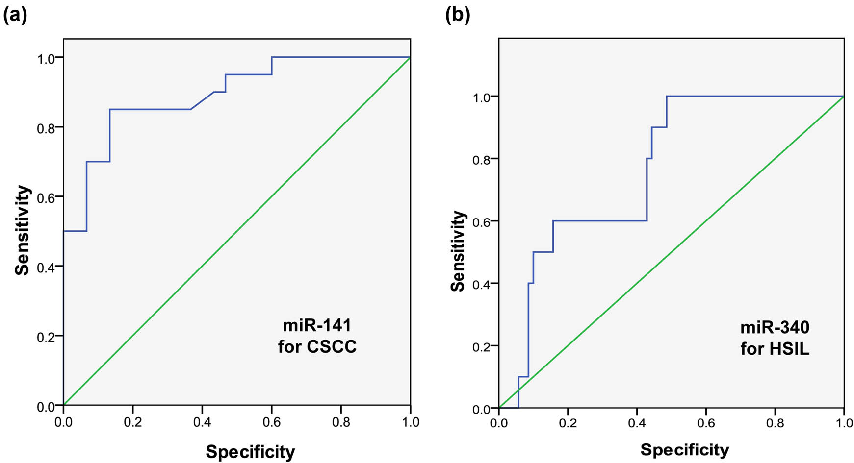
ROC curves of miR-141 for CSCC diagnosis (a) and miR-340 for HSIL diagnosis (b).
The significance of miR-141 in CSCC and miR-340 in HSIL
| miRNA | Diagnosis | Area under curve | Standard error | P | 95% CI | |
|---|---|---|---|---|---|---|
| Lower limit | Upper limit | |||||
| miR-141 | CSCC | 0.893 | 0.033 | 0.000 | 0.828 | 0.959 |
| miR-340 | HSIL | 0.764 | 0.053 | 0.000 | 0.660 | 0.869 |
Note: CSCC, cervical squamous cell carcinoma; HSIL, high-grade intraepithelial lesions.
3.4 PTEN expression in CSCC
As the target gene of miR-141 and miR-340, PTEN was expressed in the cytoplasm and the nucleus of CSCC (Figure 2). The positive rate of PTEN in CSCC was 42.3% (44/104).
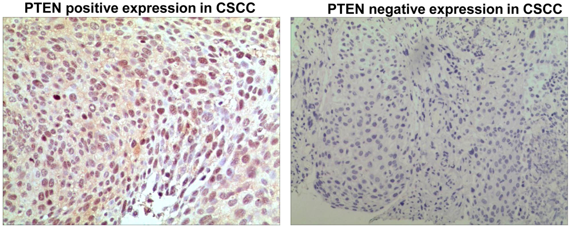
PTEN expression in CSCC. Magnification: ×10.
3.5 Relationship between PTEN expression and clinicopathological features of CSCC
In 104 cases of CSCC tissues, we found that PTEN expression was different in different age groups. However, there was no significant difference (P = 0.083). Moreover, there was no significant correlation between PTEN and other clinicopathological parameters, including ethnic group (P = 0.464), gross type (P = 0.691), tumor size (P = 0.996), differentiation (P = 0.752), invasion depth (P = 0.641), uterine corpus invasion (P = 0.628), vascular invasion (P = 0.136), nerve invasion (P = 0.865), vagina invasion (P = 0.554), lymph node metastasis (P = 0.582), and FIGO stage (P = 0.597; Table 7).
Relationship between PTEN and clinicopathological features of CSCC
| PTEN | |||
|---|---|---|---|
| CSCC clinicopathological features | Loss of expression | Positive | |
| Age | ≤50 | 34 | 38 |
| >50 | 21 | 11 | |
| χ 2 | 3.011 | ||
| P | 0.083 | ||
| Ethnic group | Han | 32 | 25 |
| Uygur | 23 | 24 | |
| χ 2 | 0.537 | ||
| P | 0.464 | ||
| Gross type | Exogenous and papillary | 20 | 16 |
| Endogenous and invasion | 35 | 33 | |
| χ 2 | 0.158 | ||
| P | 0.691 | ||
| Tumor size | ≤4 cm | 46 | 41 |
| >4 cm | 9 | 8 | |
| χ 2 | 0.000 | ||
| P | 0.996 | ||
| Differentiation | Low grade | 32 | 30 |
| High grade | 23 | 19 | |
| χ 2 | 0.100 | ||
| P | 0.752 | ||
| Invasion depth | Not reaching full thickness | 35 | 29 |
| Reaching full thickness | 20 | 20 | |
| χ 2 | 0.217 | ||
| P | 0.641 | ||
| Uterine corpus invasion | No | 44 | 41 |
| Yes | 11 | 8 | |
| χ 2 | 0.234 | ||
| P | 0.628 | ||
| Vascular invasion | No | 36 | 25 |
| Yes | 19 | 24 | |
| χ 2 | 2.226 | ||
| P | 0.136 | ||
| Nerve invasion | No | 51 | 45 |
| Yes | 4 | 4 | |
| χ 2 | 0.029 | ||
| P | 0.865 | ||
| Vagina invasion | No | 53 | 46 |
| Yes | 2 | 3 | |
| χ 2 | 0.350 | ||
| P | 0.554 | ||
| Lymph node metastasis | No | 44 | 37 |
| Yes | 11 | 12 | |
| χ 2 | 0.303 | ||
| P | 0.582 | ||
| FIGO stage | I–II stage | 39 | 37 |
| III–IV stage | 16 | 12 | |
| χ 2 | 0.279 | ||
| P | 0.597 | ||
Note: CSCC, cervical squamous cell carcinoma.
3.6 Relationship between PTEN and miR-141/miR-340 in CSCC
In CSCC, the expression of miR-141 between patients with loss of PTEN expression (4.19 ± 0.62) and those with PTEN positive expression (5.33 ± 0.37) was statistically significant (P = 0.002). The expression of miR-340 between patients with loss of PTEN expression (−1.50 ± 0.23) and those with PTEN positive expression (−1.45 ± 0.41) was also statistically significant (P < 0.001; Table 8).
Relationship between PTEN and miR-141/miR-340 levels in CSCC
| miR-141 log2 relative quantity (mean ± SE) | miR-340 log2 relative quantity (mean ± SE) | ||
|---|---|---|---|
| PTEN | Loss of expression | 4.19 ± 0.62 | −1.50 ± 0.23 |
| Positive | 5.33 ± 0.37 | −1.45 ± 0.41 | |
| t | 10.249 | 26.595 | |
| P | 0.002 | <0.001 | |
4 Discussion
Cervical cancer is one of the most common female malignant tumors in developing countries and is also one of the main causes of deaths in females [9]. Currently, the pathogenesis of CSCC has been extensively studied. However, the role of miRNA in the occurrence of malignant tumors has opened up new directions for elucidating the molecular mechanism of cervical cancer. Moreover, various abnormalities of miRNAs have been found in cervical cancer [10].
MicroRNA are small, single-stranded, noncoding RNAs that can act as oncogenes or tumor suppressor genes in the progression of tumors. Studies have shown that miRNA is differentially expressed in cervical cancer tissue, cervical intraepithelial neoplasia, and normal cervical tissue [11]. Moreover, miRNA is related to the occurrence, metastasis, and invasion of cervical cancer, which may be used as markers for the treatment and prognosis. Thus, miRNAs provide potential new targets for targeted tumor therapy. It is found that the relative expression of miR-141 mRNA in bladder cancer tissue was significantly higher than that in normal tissue adjacent to cancer [12]. However, the expression levels of miR-141 and miR-340 as well as their target gene PTEN in CSCC have been less studied.
miR-141, as a member of miR-200 family, is located at 12p13.3 and plays an important role in regulating tumor cell proliferation, migration, differentiation, and apoptosis [13]. Studies have shown that miR-141 is abnormally expressed in colorectal cancer, non-small-cell lung cancer, and gastric cancer and is increased in ovarian cancer, breast cancer, prostate cancer, renal cell cancer, and bladder cancer [14,15]. miR-340 is located at 5q35.3 and regulates the cell cycle by regulating various target proteins, which in turn affects tumor invasion and metastasis [16,17,18]. It functions as a tumor suppressor and is downregulated in solid tumors such as breast cancer, prostate cancer, gastric cancer, osteosarcoma, and melanoma [19,20]. In this study, we found that miR-141 expression was mostly upregulated in CSCC, and it showed a downward trend in tissues from CSCC to HSIL and normal squamous epithelium. In contrast, miR-340 expression was mostly downregulated in CSCC and had an upward trend in the tissues from CSCC to HSIL and normal squamous epithelium. These results indicate that miR-141 and miR-340 may play important regulatory roles in the development of normal cervical squamous epithelium into HSIL and even CSCC.
We found that miR-141 was closely related to the gross type, differentiation, uterine corpus invasion, nerve invasion, vagina invasion, and FIGO stage in CSCC. At the same time, miR-340 was closely related to the tumor size, differentiation, nerve invasion, lymph node metastasis, and FIGO stage in CSCC. These results indicate that miR-141 and miR-340 are involved in the progression of CSCC and can be used as indicators for the progress evaluation of CSCC, but the specific mechanism is yet to be further studied.
In addition, we also found that miR-141 and miR-340 showed a negative correlation in CSCC and HSIL. But the difference was not statistically significant. These results suggest that miR-141 and miR-340 may have opposite regulatory functions in the process from HSIL to CSCC. However, this conclusion still needs to be verified by further expanding the sample size. The loss expression of PTEN, the common target gene of miR-141 and miR-340, is the molecular mechanism involved in various malignant tumors [21,22,23,24]. Our results showed that the PTEN-positive expression rate in CSCC was 42.3%. In addition, PTEN expression may be related to age, but not to the ethnic group, gross type, tumor size, differentiation, invasion depth, uterine corpus invasion, vascular invasion, nerve invasion, vagina invasion, lymph node metastasis, and FIGO stage. Therefore, we speculate that miR-141 and miR-340 might have not only common but also different regulatory effects on the pathogenesis from normal cervical squamous epithelium to HSILs and invasive CSCC.
5 Conclusion
In conclusion, miR-141 and miR-340 participated in the process of abnormal hyperplasia of cervical epithelium to HSIL and in the development of CSCC. However, the underlying mechanisms may be different. We found that miR-141 and miR-340 had higher sensitivity and specificity for the diagnosis of CSCC and the diagnosis of HSIL, respectively, suggesting that the combination of miR-141 and miR-340 could be used for the diagnosis of CSCC and HSIL. However, the regulatory mechanism of miR-141 and miR-340 on target gene PTEN still needs further research and verification in cell models or animal models. Moreover, our research has good clinical application prospects. In clinical practice, the detection of miR-141 and miR-340, as well as the immunohistochemical detection of PTEN, can be applied to the early diagnosis of cervical intraepithelial lesions and CSCC, which would provide a basis for diagnosis, treatment, and prognosis of CSCC and squamous intraepithelial lesions.
-
Funding information: This study was supported by Natural Science Foundation of Shenzhen University General Hospital (SUGH2019QD013) and Natural Science Foundation of Xinjiang Uygur Autonomous Region (2016D01C364).
-
Conflict of interest: The authors report no conflict of interest.
-
Data availability statement: All data generated during this study are presented within the manuscript.
References
[1] Torre LA, Bray F, Siegel RL, Ferlay J, Lortet-Tieulent J, Jemal A. Global cancer statistics, 2012. CA Cancer J Clin. 2015;65(2):87–108.10.3322/caac.21262Search in Google Scholar PubMed
[2] Kurman RJ, Carcangiu ML, Simon HC, Young RH. WHO classification of tumors of female reproductive organs lyon. France: IARC Press; 2014.Search in Google Scholar
[3] Bajpai D, Banerjee A, Pathak S, Thakur B, Jain SK, Singh N. Single nucleotide polymorphisms in the DNA repair genes in HPV-positive cervical cancer. Eur J Cancer Prev. 2016;25(3):224–31.10.1097/CEJ.0000000000000159Search in Google Scholar PubMed
[4] Zeng K, Xianglan MO, Liu F, Xiaoxia HU. Differential expression of microRNAs in cervical cancer and cervical precancerous lesions. Cancer Res Prev Treat. 2014;10(3):144.Search in Google Scholar
[5] Wang F, Li B, Xie X. The roles and clinical significance of microRNAs in cervical cancer. Histol Histopathol. 2016;31(2):131–9.Search in Google Scholar
[6] Lee H, Choi HJ, Kang CS, Lee HJ, Lee WS, Park CS. Expression of miRNAs and PTEN in endometrial specimens ranging from histologically normal to hyperplasia and endometrial adenocarcinoma. Mod Pathol. 2012;25(11):1508–15.10.1038/modpathol.2012.111Search in Google Scholar PubMed
[7] Li W, Wang Y, Fang X, Zhou M, Li Y, Dong Y, et al. Differential expression and clinical significance of DNA methyltransferase 3B (DNMT3B), phosphatase and tensin homolog (PTEN) and human MutL homologs 1 (hMLH1) in endometrial carcinomas. Med Sci Monit. 2017;23:938–47.10.12659/MSM.902267Search in Google Scholar
[8] Dong Y, Si JW, Li WT, Liang L, Zhao J, Zhou M, et al. miR-200a/miR-141 and miR-205 upregulation might be associated with hormone receptor status and prognosis in endometrial carcinomas. Int J Clin Exp Pathol. 2015;8(3):2864–75.Search in Google Scholar
[9] Kim TH, Han JH, Shin E, Noh JH, Kim HS, Song YS. Clinical implication of p16, Ki-67, and proliferating cell nuclear antigen expression in cervical neoplasia: improvement of diagnostic accuracy for high-grade squamous intraepithelial lesion and prediction of resection margin involvement on conization specimen. J Cancer Prev. 2015;20(1):70–7.10.15430/JCP.2015.20.1.70Search in Google Scholar PubMed PubMed Central
[10] Zhang Q, Zhang Y, Wang SZ, Wang N, Jiang WG, Ji YH, et al. Reduced expression of tissue factor pathway inhibitor-2 contributes to apoptosis and angiogenesis in cervical cancer. J Exp Clin Cancer Res. 2012;31(1):1.10.1186/1756-9966-31-1Search in Google Scholar PubMed PubMed Central
[11] Di Leva G, Garofalo M, Croce CM. microRNAs in cancer. Annu Rev Pathol. 2014;9:287–314.10.1146/annurev-pathol-012513-104715Search in Google Scholar PubMed PubMed Central
[12] Xiao Y, Li W, Wu Y. miR-141 expression in bladder cancer tissue and its correlation with tumor grade. J Nantong Univ (Med Ed). 2020;40(1):30–3.Search in Google Scholar
[13] Dong H, Weng C, Bai R, Sheng J, Gao X, Li L, et al. The regulatory network of miR-141 in the inhibition of angiogenesis. Angiogenesis. 2019;22(2):251–62.10.1007/s10456-018-9654-1Search in Google Scholar PubMed
[14] Ardila HJ, Sanabria-Salas MC, Meneses X, Rios R, Huertas-Salgado A, Serrano ML. Circulating miR-141-3p, miR-143-3p and miR-200c-3p are differentially expressed in colorectal cancer and advanced adenomas. Mol Clin Oncol. 2019;11(2):201–7.10.3892/mco.2019.1876Search in Google Scholar PubMed PubMed Central
[15] Huang S, Wa Q, Pan J, Peng X, Ren D, Huang Y, et al. Downregulation of miR-141-3p promotes bone metastasis via activating NF-κB signaling in prostate cancer. J Exp Clin Cancer Res. 2017;36(1):173.10.1186/s13046-017-0645-7Search in Google Scholar PubMed PubMed Central
[16] Xiao H, Yu L, Li F, Wang H, Li W, He X. miR-340 suppresses the metastasis by targeting EphA3 in cervical cancer. Cell Biol Int. 2018;42(9):1115–23.10.1002/cbin.10974Search in Google Scholar PubMed
[17] Yin G, Zhou H, Xue Y, Yao B, Zhao W. microRNA-340 promotes the tumor growth of human gastric cancer by inhibiting cyclin G2. Oncol Rep. 2016;36(2):1111–8.10.3892/or.2016.4876Search in Google Scholar PubMed
[18] Raychaudhuri M, Bronger H, Buchner T, Kiechle M, Weichert W, Avril S. microRNAs miR-7 and miR-340 predict response to neoadjuvant chemotherapy in breast cancer. Breast Cancer Res Treat. 2017;162(3):511–21.10.1007/s10549-017-4132-9Search in Google Scholar PubMed PubMed Central
[19] Yu J, Wang R, Chen J, Wu J, Dang Z, Zhang Q, et al. miR-340 inhibits proliferation and induces apoptosis in gastric cancer cell line SGC-7901, possibly via the AKT pathway. Med Sci Monit. 2017;23:71–7.10.12659/MSM.898449Search in Google Scholar PubMed PubMed Central
[20] Song L, Zhou Z, Gan Y, Li P, Xu Y, Zhang Z, et al. Long noncoding RNA OIP5-AS1 causes cisplatin resistance in osteosarcoma through inducing the LPAATβ/PI3K/AKT/mTOR signaling pathway by sponging the miR-340-5p. J Cell Biochem. 2019;120(6):9656–66.10.1002/jcb.28244Search in Google Scholar PubMed
[21] Liu Y, Zhao R, Wang H, Luo Y, Wang X, Niu W, et al. miR-141 is involved in BRD7-mediated cell proliferation and tumor formation through suppression of the PTEN/AKT pathway in nasopharyngeal carcinoma. Cell Death Dis. 2016;7(3):e2156.10.1038/cddis.2016.64Search in Google Scholar PubMed PubMed Central
[22] Jin YY, Chen QJ, Xu K, Ren HT, Bao X, Ma YN, et al. Involvement of microRNA-141-3p in 5-fluorouracil and oxaliplatin chemo-resistance in esophageal cancer cells via regulation of PTEN. Mol Cell Biochem. 2016;422(1–2):161–70.10.1007/s11010-016-2816-9Search in Google Scholar PubMed
[23] Pourhanifeh MH, Mahjoubin-Tehran M, Karimzadeh MR, Mirzaei HR, Razavi ZS, Sahebkar A, et al. Autophagy in cancers including brain tumors: role of microRNAs. Cell Commun Signal. 2020;18(1):88.10.1186/s12964-020-00587-wSearch in Google Scholar PubMed PubMed Central
[24] Han X, Liu CF, Gao N, Zhao J, Xu J. Kaempferol suppresses proliferation but increases apoptosis and autophagy by up-regulating microRNA-340 in human lung cancer cells. Biomed Pharmacother. 2018;108:809–16.10.1016/j.biopha.2018.09.087Search in Google Scholar PubMed
© 2021 Wenting Li et al., published by De Gruyter
This work is licensed under the Creative Commons Attribution 4.0 International License.
Articles in the same Issue
- Research Articles
- Identification of ZG16B as a prognostic biomarker in breast cancer
- Behçet’s disease with latent Mycobacterium tuberculosis infection
- Erratum
- Erratum to “Suffering from Cerebral Small Vessel Disease with and without Metabolic Syndrome”
- Research Articles
- GPR37 promotes the malignancy of lung adenocarcinoma via TGF-β/Smad pathway
- Expression and role of ABIN1 in sepsis: In vitro and in vivo studies
- Additional baricitinib loading dose improves clinical outcome in COVID-19
- The co-treatment of rosuvastatin with dapagliflozin synergistically inhibited apoptosis via activating the PI3K/AKt/mTOR signaling pathway in myocardial ischemia/reperfusion injury rats
- SLC12A8 plays a key role in bladder cancer progression and EMT
- LncRNA ATXN8OS enhances tamoxifen resistance in breast cancer
- Case Report
- Serratia marcescens as a cause of unfavorable outcome in the twin pregnancy
- Spleno-adrenal fusion mimicking an adrenal metastasis of a renal cell carcinoma: A case report and embryological background
- Research Articles
- TRIM25 contributes to the malignancy of acute myeloid leukemia and is negatively regulated by microRNA-137
- CircRNA circ_0004370 promotes cell proliferation, migration, and invasion and inhibits cell apoptosis of esophageal cancer via miR-1301-3p/COL1A1 axis
- LncRNA XIST regulates atherosclerosis progression in ox-LDL-induced HUVECs
- Potential role of IFN-γ and IL-5 in sepsis prediction of preterm neonates
- Rapid Communication
- COVID-19 vaccine: Call for employees in international transportation industries and international travelers as the first priority in global distribution
- Case Report
- Rare squamous cell carcinoma of the kidney with concurrent xanthogranulomatous pyelonephritis: A case report and review of the literature
- An infertile female delivered a baby after removal of primary renal carcinoid tumor
- Research Articles
- Hypertension, BMI, and cardiovascular and cerebrovascular diseases
- Case Report
- Coexistence of bilateral macular edema and pale optic disc in the patient with Cohen syndrome
- Research Articles
- Correlation between kinematic sagittal parameters of the cervical lordosis or head posture and disc degeneration in patients with posterior neck pain
- Review Articles
- Hepatoid adenocarcinoma of the lung: An analysis of the Surveillance, Epidemiology, and End Results (SEER) database
- Research Articles
- Thermography in the diagnosis of carpal tunnel syndrome
- Pemetrexed-based first-line chemotherapy had particularly prominent objective response rate for advanced NSCLC: A network meta-analysis
- Comparison of single and double autologous stem cell transplantation in multiple myeloma patients
- The influence of smoking in minimally invasive spinal fusion surgery
- Impact of body mass index on left atrial dimension in HOCM patients
- Expression and clinical significance of CMTM1 in hepatocellular carcinoma
- miR-142-5p promotes cervical cancer progression by targeting LMX1A through Wnt/β-catenin pathway
- Comparison of multiple flatfoot indicators in 5–8-year-old children
- Early MRI imaging and follow-up study in cerebral amyloid angiopathy
- Intestinal fatty acid-binding protein as a biomarker for the diagnosis of strangulated intestinal obstruction: A meta-analysis
- miR-128-3p inhibits apoptosis and inflammation in LPS-induced sepsis by targeting TGFBR2
- Dynamic perfusion CT – A promising tool to diagnose pancreatic ductal adenocarcinoma
- Biomechanical evaluation of self-cinching stitch techniques in rotator cuff repair: The single-loop and double-loop knot stitches
- Review Articles
- The ambiguous role of mannose-binding lectin (MBL) in human immunity
- Case Report
- Membranous nephropathy with pulmonary cryptococcosis with improved 1-year follow-up results: A case report
- Fertility problems in males carrying an inversion of chromosome 10
- Acute myeloid leukemia with leukemic pleural effusion and high levels of pleural adenosine deaminase: A case report and review of literature
- Metastatic renal Ewing’s sarcoma in adult woman: Case report and review of the literature
- Burkitt-like lymphoma with 11q aberration in a patient with AIDS and a patient without AIDS: Two cases reports and literature review
- Skull hemophilia pseudotumor: A case report
- Judicious use of low-dosage corticosteroids for non-severe COVID-19: A case report
- Adult-onset citrullinaemia type II with liver cirrhosis: A rare cause of hyperammonaemia
- Clinicopathologic features of Good’s syndrome: Two cases and literature review
- Fatal immune-related hepatitis with intrahepatic cholestasis and pneumonia associated with camrelizumab: A case report and literature review
- Research Articles
- Effects of hydroxyethyl starch and gelatin on the risk of acute kidney injury following orthotopic liver transplantation: A multicenter retrospective comparative clinical study
- Significance of nucleic acid positive anal swab in COVID-19 patients
- circAPLP2 promotes colorectal cancer progression by upregulating HELLS by targeting miR-335-5p
- Ratios between circulating myeloid cells and lymphocytes are associated with mortality in severe COVID-19 patients
- Risk factors of left atrial appendage thrombus in patients with non-valvular atrial fibrillation
- Clinical features of hypertensive patients with COVID-19 compared with a normotensive group: Single-center experience in China
- Surgical myocardial revascularization outcomes in Kawasaki disease: systematic review and meta-analysis
- Decreased chromobox homologue 7 expression is associated with epithelial–mesenchymal transition and poor prognosis in cervical cancer
- FGF16 regulated by miR-520b enhances the cell proliferation of lung cancer
- Platelet-rich fibrin: Basics of biological actions and protocol modifications
- Accurate diagnosis of prostate cancer using logistic regression
- miR-377 inhibition enhances the survival of trophoblast cells via upregulation of FNDC5 in gestational diabetes mellitus
- Prognostic significance of TRIM28 expression in patients with breast carcinoma
- Integrative bioinformatics analysis of KPNA2 in six major human cancers
- Exosomal-mediated transfer of OIP5-AS1 enhanced cell chemoresistance to trastuzumab in breast cancer via up-regulating HMGB3 by sponging miR-381-3p
- A four-lncRNA signature for predicting prognosis of recurrence patients with gastric cancer
- Knockdown of circ_0003204 alleviates oxidative low-density lipoprotein-induced human umbilical vein endothelial cells injury: Circulating RNAs could explain atherosclerosis disease progression
- Propofol postpones colorectal cancer development through circ_0026344/miR-645/Akt/mTOR signal pathway
- Knockdown of lncRNA TapSAKI alleviates LPS-induced injury in HK-2 cells through the miR-205/IRF3 pathway
- COVID-19 severity in relation to sociodemographics and vitamin D use
- Clinical analysis of 11 cases of nocardiosis
- Cis-regulatory elements in conserved non-coding sequences of nuclear receptor genes indicate for crosstalk between endocrine systems
- Four long noncoding RNAs act as biomarkers in lung adenocarcinoma
- Real-world evidence of cytomegalovirus reactivation in non-Hodgkin lymphomas treated with bendamustine-containing regimens
- Relation between IL-8 level and obstructive sleep apnea syndrome
- circAGFG1 sponges miR-28-5p to promote non-small-cell lung cancer progression through modulating HIF-1α level
- Nomogram prediction model for renal anaemia in IgA nephropathy patients
- Effect of antibiotic use on the efficacy of nivolumab in the treatment of advanced/metastatic non-small cell lung cancer: A meta-analysis
- NDRG2 inhibition facilitates angiogenesis of hepatocellular carcinoma
- A nomogram for predicting metabolic steatohepatitis: The combination of NAMPT, RALGDS, GADD45B, FOSL2, RTP3, and RASD1
- Clinical and prognostic features of MMP-2 and VEGF in AEG patients
- The value of miR-510 in the prognosis and development of colon cancer
- Functional implications of PABPC1 in the development of ovarian cancer
- Prognostic value of preoperative inflammation-based predictors in patients with bladder carcinoma after radical cystectomy
- Sublingual immunotherapy increases Treg/Th17 ratio in allergic rhinitis
- Prediction of improvement after anterior cruciate ligament reconstruction
- Effluent Osteopontin levels reflect the peritoneal solute transport rate
- circ_0038467 promotes PM2.5-induced bronchial epithelial cell dysfunction
- Significance of miR-141 and miR-340 in cervical squamous cell carcinoma
- Association between hair cortisol concentration and metabolic syndrome
- Microvessel density as a prognostic indicator of prostate cancer: A systematic review and meta-analysis
- Characteristics of BCR–ABL gene variants in patients of chronic myeloid leukemia
- Knee alterations in rheumatoid arthritis: Comparison of US and MRI
- Long non-coding RNA TUG1 aggravates cerebral ischemia and reperfusion injury by sponging miR-493-3p/miR-410-3p
- lncRNA MALAT1 regulated ATAD2 to facilitate retinoblastoma progression via miR-655-3p
- Development and validation of a nomogram for predicting severity in patients with hemorrhagic fever with renal syndrome: A retrospective study
- Analysis of COVID-19 outbreak origin in China in 2019 using differentiation method for unusual epidemiological events
- Laparoscopic versus open major liver resection for hepatocellular carcinoma: A case-matched analysis of short- and long-term outcomes
- Travelers’ vaccines and their adverse events in Nara, Japan
- Association between Tfh and PGA in children with Henoch–Schönlein purpura
- Can exchange transfusion be replaced by double-LED phototherapy?
- circ_0005962 functions as an oncogene to aggravate NSCLC progression
- Circular RNA VANGL1 knockdown suppressed viability, promoted apoptosis, and increased doxorubicin sensitivity through targeting miR-145-5p to regulate SOX4 in bladder cancer cells
- Serum intact fibroblast growth factor 23 in healthy paediatric population
- Algorithm of rational approach to reconstruction in Fournier’s disease
- A meta-analysis of exosome in the treatment of spinal cord injury
- Src-1 and SP2 promote the proliferation and epithelial–mesenchymal transition of nasopharyngeal carcinoma
- Dexmedetomidine may decrease the bupivacaine toxicity to heart
- Hypoxia stimulates the migration and invasion of osteosarcoma via up-regulating the NUSAP1 expression
- Long noncoding RNA XIST knockdown relieves the injury of microglia cells after spinal cord injury by sponging miR-219-5p
- External fixation via the anterior inferior iliac spine for proximal femoral fractures in young patients
- miR-128-3p reduced acute lung injury induced by sepsis via targeting PEL12
- HAGLR promotes neuron differentiation through the miR-130a-3p-MeCP2 axis
- Phosphoglycerate mutase 2 is elevated in serum of patients with heart failure and correlates with the disease severity and patient’s prognosis
- Cell population data in identifying active tuberculosis and community-acquired pneumonia
- Prognostic value of microRNA-4521 in non-small cell lung cancer and its regulatory effect on tumor progression
- Mean platelet volume and red blood cell distribution width is associated with prognosis in premature neonates with sepsis
- 3D-printed porous scaffold promotes osteogenic differentiation of hADMSCs
- Association of gene polymorphisms with women urinary incontinence
- Influence of COVID-19 pandemic on stress levels of urologic patients
- miR-496 inhibits proliferation via LYN and AKT pathway in gastric cancer
- miR-519d downregulates LEP expression to inhibit preeclampsia development
- Comparison of single- and triple-port VATS for lung cancer: A meta-analysis
- Fluorescent light energy modulates healing in skin grafted mouse model
- Silencing CDK6-AS1 inhibits LPS-induced inflammatory damage in HK-2 cells
- Predictive effect of DCE-MRI and DWI in brain metastases from NSCLC
- Severe postoperative hyperbilirubinemia in congenital heart disease
- Baicalin improves podocyte injury in rats with diabetic nephropathy by inhibiting PI3K/Akt/mTOR signaling pathway
- Clinical factors predicting ureteral stent failure in patients with external ureteral compression
- Novel H2S donor proglumide-ADT-OH protects HUVECs from ox-LDL-induced injury through NF-κB and JAK/SATA pathway
- Triple-Endobutton and clavicular hook: A propensity score matching analysis
- Long noncoding RNA MIAT inhibits the progression of diabetic nephropathy and the activation of NF-κB pathway in high glucose-treated renal tubular epithelial cells by the miR-182-5p/GPRC5A axis
- Serum exosomal miR-122-5p, GAS, and PGR in the non-invasive diagnosis of CAG
- miR-513b-5p inhibits the proliferation and promotes apoptosis of retinoblastoma cells by targeting TRIB1
- Fer exacerbates renal fibrosis and can be targeted by miR-29c-3p
- The diagnostic and prognostic value of miR-92a in gastric cancer: A systematic review and meta-analysis
- Prognostic value of α2δ1 in hypopharyngeal carcinoma: A retrospective study
- No significant benefit of moderate-dose vitamin C on severe COVID-19 cases
- circ_0000467 promotes the proliferation, metastasis, and angiogenesis in colorectal cancer cells through regulating KLF12 expression by sponging miR-4766-5p
- Downregulation of RAB7 and Caveolin-1 increases MMP-2 activity in renal tubular epithelial cells under hypoxic conditions
- Educational program for orthopedic surgeons’ influences for osteoporosis
- Expression and function analysis of CRABP2 and FABP5, and their ratio in esophageal squamous cell carcinoma
- GJA1 promotes hepatocellular carcinoma progression by mediating TGF-β-induced activation and the epithelial–mesenchymal transition of hepatic stellate cells
- lncRNA-ZFAS1 promotes the progression of endometrial carcinoma by targeting miR-34b to regulate VEGFA expression
- Anticoagulation is the answer in treating noncritical COVID-19 patients
- Effect of late-onset hemorrhagic cystitis on PFS after haplo-PBSCT
- Comparison of Dako HercepTest and Ventana PATHWAY anti-HER2 (4B5) tests and their correlation with silver in situ hybridization in lung adenocarcinoma
- VSTM1 regulates monocyte/macrophage function via the NF-κB signaling pathway
- Comparison of vaginal birth outcomes in midwifery-led versus physician-led setting: A propensity score-matched analysis
- Treatment of osteoporosis with teriparatide: The Slovenian experience
- New targets of morphine postconditioning protection of the myocardium in ischemia/reperfusion injury: Involvement of HSP90/Akt and C5a/NF-κB
- Superenhancer–transcription factor regulatory network in malignant tumors
- β-Cell function is associated with osteosarcopenia in middle-aged and older nonobese patients with type 2 diabetes: A cross-sectional study
- Clinical features of atypical tuberculosis mimicking bacterial pneumonia
- Proteoglycan-depleted regions of annular injury promote nerve ingrowth in a rabbit disc degeneration model
- Effect of electromagnetic field on abortion: A systematic review and meta-analysis
- miR-150-5p affects AS plaque with ASMC proliferation and migration by STAT1
- MALAT1 promotes malignant pleural mesothelioma by sponging miR-141-3p
- Effects of remifentanil and propofol on distant organ lung injury in an ischemia–reperfusion model
- miR-654-5p promotes gastric cancer progression via the GPRIN1/NF-κB pathway
- Identification of LIG1 and LIG3 as prognostic biomarkers in breast cancer
- MitoQ inhibits hepatic stellate cell activation and liver fibrosis by enhancing PINK1/parkin-mediated mitophagy
- Dissecting role of founder mutation p.V727M in GNE in Indian HIBM cohort
- circATP2A2 promotes osteosarcoma progression by upregulating MYH9
- Prognostic role of oxytocin receptor in colon adenocarcinoma
- Review Articles
- The function of non-coding RNAs in idiopathic pulmonary fibrosis
- Efficacy and safety of therapeutic plasma exchange in stiff person syndrome
- Role of cesarean section in the development of neonatal gut microbiota: A systematic review
- Small cell lung cancer transformation during antitumor therapies: A systematic review
- Research progress of gut microbiota and frailty syndrome
- Recommendations for outpatient activity in COVID-19 pandemic
- Rapid Communication
- Disparity in clinical characteristics between 2019 novel coronavirus pneumonia and leptospirosis
- Use of microspheres in embolization for unruptured renal angiomyolipomas
- COVID-19 cases with delayed absorption of lung lesion
- A triple combination of treatments on moderate COVID-19
- Social networks and eating disorders during the Covid-19 pandemic
- Letter
- COVID-19, WHO guidelines, pedagogy, and respite
- Inflammatory factors in alveolar lavage fluid from severe COVID-19 pneumonia: PCT and IL-6 in epithelial lining fluid
- COVID-19: Lessons from Norway tragedy must be considered in vaccine rollout planning in least developed/developing countries
- What is the role of plasma cell in the lamina propria of terminal ileum in Good’s syndrome patient?
- Case Report
- Rivaroxaban triggered multifocal intratumoral hemorrhage of the cabozantinib-treated diffuse brain metastases: A case report and review of literature
- CTU findings of duplex kidney in kidney: A rare duplicated renal malformation
- Synchronous primary malignancy of colon cancer and mantle cell lymphoma: A case report
- Sonazoid-enhanced ultrasonography and pathologic characters of CD68 positive cell in primary hepatic perivascular epithelioid cell tumors: A case report and literature review
- Persistent SARS-CoV-2-positive over 4 months in a COVID-19 patient with CHB
- Pulmonary parenchymal involvement caused by Tropheryma whipplei
- Mediastinal mixed germ cell tumor: A case report and literature review
- Ovarian female adnexal tumor of probable Wolffian origin – Case report
- Rare paratesticular aggressive angiomyxoma mimicking an epididymal tumor in an 82-year-old man: Case report
- Perimenopausal giant hydatidiform mole complicated with preeclampsia and hyperthyroidism: A case report and literature review
- Primary orbital ganglioneuroblastoma: A case report
- Primary aortic intimal sarcoma masquerading as intramural hematoma
- Sustained false-positive results for hepatitis A virus immunoglobulin M: A case report and literature review
- Peritoneal loose body presenting as a hepatic mass: A case report and review of the literature
- Chondroblastoma of mandibular condyle: Case report and literature review
- Trauma-induced complete pacemaker lead fracture 8 months prior to hospitalization: A case report
- Primary intradural extramedullary extraosseous Ewing’s sarcoma/peripheral primitive neuroectodermal tumor (PIEES/PNET) of the thoracolumbar spine: A case report and literature review
- Computer-assisted preoperative planning of reduction of and osteosynthesis of scapular fracture: A case report
- High quality of 58-month life in lung cancer patient with brain metastases sequentially treated with gefitinib and osimertinib
- Rapid response of locally advanced oral squamous cell carcinoma to apatinib: A case report
- Retrieval of intrarenal coiled and ruptured guidewire by retrograde intrarenal surgery: A case report and literature review
- Usage of intermingled skin allografts and autografts in a senior patient with major burn injury
- Retraction
- Retraction on “Dihydromyricetin attenuates inflammation through TLR4/NF-kappa B pathway”
- Special Issue Computational Intelligence Methodologies Meets Recurrent Cancers - Part I
- An artificial immune system with bootstrap sampling for the diagnosis of recurrent endometrial cancers
- Breast cancer recurrence prediction with ensemble methods and cost-sensitive learning