Molecular characterization and phylogenetic studies of Echinococcus granulosus and Taenia multiceps coenurus cysts in slaughtered sheep in Saudi Arabia
-
Jamila S. Al Malki
 und
Nahed Ahmed Hussien
und
Nahed Ahmed Hussien


Abstract
Taeniids, consisting of two genera Echinococcus and Taenia, are obligatory tapeworms of mammals, and their pathogenicity was due to infection with larval stages. Hydatid (the larval stage of Echinococcus granulosus) and coenurus (the larval stage of Taenia multiceps) cysts are prevalent in domestic, wild ruminants, livestock, swine, and dogs, and accidentally they could also be found in humans. They lead to different clinical manifestations that cause economic loss in livestock and human morbidity. In Saudi Arabia, few studies were performed on hydatid and coenurus cyst genetic variations. The main goal of the present study was to identify E. granulosus and T. multiceps cyst isolates collected from slaughtered Harri sheep in Saudi Arabia by partial sequencing with PCR amplification of the cytochrome C oxidase 1 (COX1) gene. Molecular and phylogenetic evaluation based on COX1 sequences indicated that cyst isolates belong to E. granulosus and T. multiceps, respectively, successfully submitted in NCBI Genbank. Molecular characterization showed a low nucleotide diversity with two submitted isolates of coenurus with related isolates of Genbank. Conversely, E. granulosus isolates showed higher nucleotide diversity. The reported data could serve as a foundation for future molecular epidemiological and biological studies.
1 Introduction
Taeniid tapeworms belong to subclass Eucestoda, order Cyclophyllidea, and family Taeniidae, and they represent zoonotic parasites of mammals and livestock. Family Taeniidae consists of two genera, Echinococcus and Taenia, and they are mainly focused on for their socioeconomic impact by causing human morbidity and loss of livestock [1,2].
Echinococcus granulosus causes hydatidosis disease due to its larval stage infection. Hydatidosis (cystic echinococcosis) is widely distributed, and its prevalence varies according to climate and their contact with livestock [3]. Hydatid cysts have been reported in different livestock, including sheep, camels, cattle, and goats, with various incidence rates, resulting in significant economic losses [4,5,6]. Recently, it was recorded that the monetary burden of surgically treated human cystic echinococcosis increased that differs from one country to another [7]. In addition, it was reported that about 2.5% of humans infected with hydatidosis died after resorting to surgery [8]. There are three possibilities for cystic echinococcosis management: medical treatment with albendazole, percutaneous procedures, and surgery that depends on cyst stage, state, and organ location [9].
Coenurus represents the infective larval stage of Taenia multiceps; they are also widely distributed mainly in the tropical countries in Africa, Asia, and the Middle East [10]. T. multiceps are found as an obligate intestinal tapeworm of dogs, also commonly found in sheep and goats, as an intermediate host for the parasite, but the infection was extended to cattle and humans [11]. Mainly, coenurus cysts present in the brain in either acute or chronic form also could be frequently found in intramuscular and subcutaneous tissues [12,13,14]. Coenurosis leads to different clinical manifestations according to the location and extent of the coenuri. Coenurus cysts in the brain cause fever, muscle tremors, hemorrhagic retinal lesions, paralysis, ataxia, blindness, nystagmus, dysmetria, and scoliosis [15,16]. However, coenuri cysts in the subcutaneous and intramuscular tissues damage the functioning of normal organs and lead to muscular pain [11].
Due to the veterinary and medical significance of both E. granulosus and T. multiceps, there has been an intensive focus on their ecological, epidemiological, and taxonomic studies. Molecular characterization among many species found within Taeniidae, especially for the genus Echinococcus, has been well documented, but this was scarce with coenurus. However, there are very few documented studies on genetic variability regarding Taeniids in Saudi Arabia.
The present study aimed to (i) genetically characterize hydatid (larval stage of E. granulosus) and coenurus (larval stage of T. multiceps) cysts isolated from slaughtered Harri sheep in Taif, Saudi Arabia, by using mitochondrial cytochrome C oxidase 1 (COX1) and NADH dehydrogenase subunit 1 (NAD1) genes. Also, the study aimed to (ii) report the genetic relationship between selected genes loci of Taif isolates and other isolates present in other countries in Genbank.
2 Material and methods
2.1 Sample collection
The present study was carried out on Harri sheep from an animal slaughterhouse in Taif governorate (21.2819°N, 40.3841°E), Mecca Province, Saudi Arabia. During postmortem veterinary examination, meat was systematically inspected for hydatid and coenurus cysts occurrence by applying procedures of the routine meat inspection (sampling period from January to October 2020). Hydatid cysts were found in muscles, liver, and viscera, but coenurus cysts were found only in muscles. Therefore, we have collected samples from 20 sheep (gender, female; age, 1–2 years), about 200–300 g of muscles from each animal, which contains either hydatid or coenurus cysts from slaughtered sheep under permission from the Ministry of Environment, Water and Agriculture (KSA). Each cyst from a different animal was labeled and handled as a different isolate before carrying out the examination.
2.2 Microscopic examination
Ten samples of hydatid cysts (of E. granulosus) and the other ten samples of coenurus cysts (of T. multiceps) were collected from animal meat and carefully opened with the help of a sterile scalpel. First, protoscolices of hydatid and coenurus cysts were scraped off the inner wall of their cysts. Then, protoscolices were loaded on glass slides, covered, and examined under a light microscope without staining.
2.3 DNA extraction of protoscolices
About 300 µL of lysis buffer TNES (10 mM Tris, 400 mM NaCl, 100 mM Na2EDTA, 0.6% SDS, pH 7.5) was added to hydatid protoscolices in a microtube; then samples were frozen (in liquid nitrogen) and thawed (5×). To facilitate sample digestion, samples were ground before proteinase K (8 µL, 20 mg/mL) and incubated overnight at 37°C. Next, phenol/chloroform (100 µL) was added to the sample and centrifuged (16,500 g) for 5 min, and then, the clear upper phase was transferred into a new 1.5 mL Eppendorf. Next, for DNA precipitation, the same volume of absolute ethyl alcohol and sodium acetate (1%, 3 mol) was added to the sample and stored overnight (−20°C). Finally, samples were centrifuged (16,500 g) for 5 min, the supernatant was discarded, the pellet containing DNA was left to dry, and the pellet was finally dissolved in 100 µL sterile deionized water [17]. DNA was extracted from coenurus protoscolices with the same previous procedure using TNES lysis buffer and phenol/chloroform, but there was no need for freezing, thawing, or grinding.
2.4 PCR amplification
We have targeted two mitochondrial COX1 and NAD1 regions of E. granulosus and T. multiceps cysts by using two different primers JB3: 5′-TTTTTTGGGCATCCTGAGGTTTAT-3′, JB4.5: 5′-TAAAGAAAGAACATAATGAAAATG-3′ and JB11 5′-AGATTCGTAAGGGGCCTAATA-3′, JB12: 5′-ACCACTAACTAATTCACTTTC-3′, respectively [17]. The PCR reaction was set up with initial denaturation at 95°C (5 min), 40 cycles of denaturation at 94°C (45 s), primer annealing at 51°C (COX1) or 55°C (NAD1) (35 s), and then primer extension at 72°C (45 s). Final extension at 72°C (10 min) was necessary for complete amplification (Programmable Thermal Cycler, PTC-100TM thermal cycler, Model 96; MJ Research, Inc., Watertown, MA). PCR products were separated on 1% tris-borate/EDTA agarose gels and ethidium bromide staining and then visualized under gel documentation (Bio-Rad, USA) [18].
2.5 Sequencing and phylogenetic evaluation
Different PCR products of COX1 and NAD1 of E. granulosus and T. multiceps cysts were randomly selected and subjected to sequencing using an ABI Prism 3730 Genetic Analyzer automated sequencer. Four sequences of COX1 regions (two of each hydatid and coenurus cysts) were directly submitted in NCBI Genbank to have accession numbers. Sequences of COX1 and NAD1 were aligned with different sequences in Genbank using online NCBI BLAST, phylogenetically estimated, and finally viewed as rectangular cladogram in the phylogenetic Tree View [19].
3 Results
3.1 Morphological and microscopic examination of cysts
Hydatid and coenurus cysts appeared as fluid-filled round/oval sacs with different sizes embedded in the muscles of sheep. Protoscolices of both cysts were carefully scraped off the inner wall of their cyst rather than collecting them from cysts’ inner fluid. E. granulosus protoscolices appeared as an oval body with an invaginated scolex provided with several hooks (Figure 1). Visual examination showed numerous scolices in the inner wall of coenurus cysts. Microscopically, the isolated protoscolex showed both large and small rostellar taeniid hooks and four scup-shaped suckers (Figure 2).
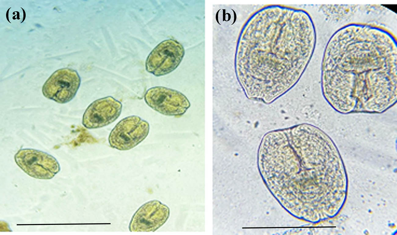
Echinococcus granulosus protoscolices at 10× (a), and 40× (b) magnifications. Scale-bars: 100 µm.

(a) Coenurus cyst attached to a muscle. (b) Multiple scolices appeared after cyst opening. (c) Isolated protoscolex large and small rostellar hooks: blue arrow and 4 suckers: red arrows. (d) Isolated rostellar hooks. Scale bars: 100 µm.
3.2 PCR, sequencing, and phylogenetic analysis
Mitochondrial DNA was used to amplify two separate gene loci, COX1 and NAD1, for both E. granulosus and T. multiceps cysts to yield amplicons of 446 bp and 520 bp, respectively. The present results report the success of PCR amplification for both selected portions (Figures 3 and 4). However, NAD1 amplification shows other lower nonspecific bands.
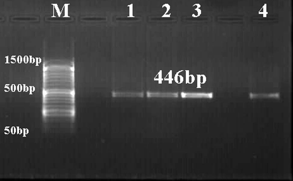
Representative agarose gel stained with ethidium bromide (1.5%) showing PCR product (446 bp) of COX1 of E. granulosus (lanes 1 and 2) and T. multiceps (lanes 3 and 4) cysts. Lane M: low-molecular-weight marker (50–1,500 bp).
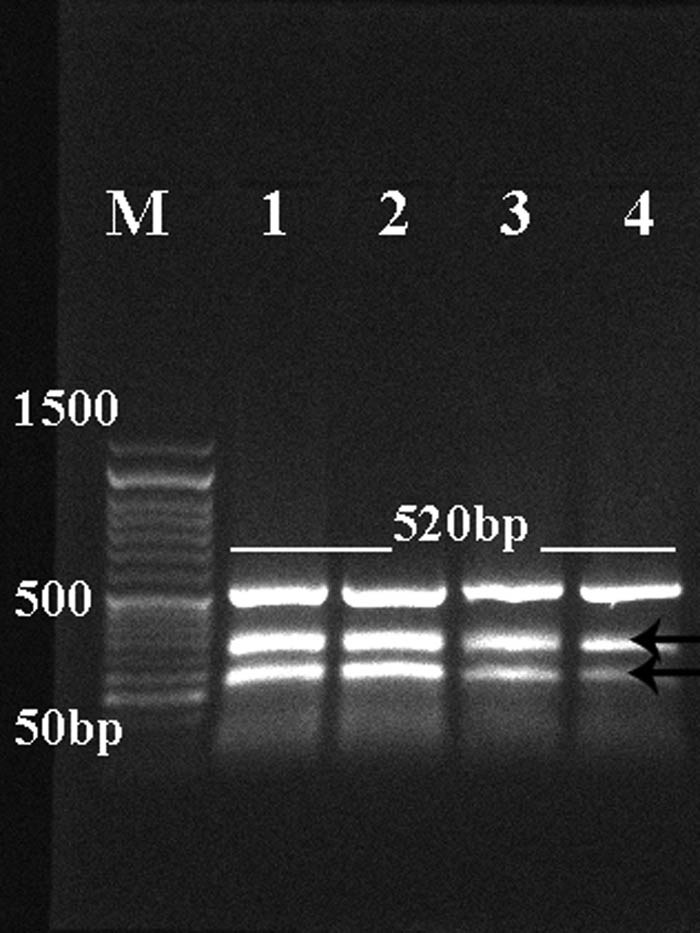
Representative 1.5% agarose gel showing PCR product (main amplicon size 520 bp) of NAD1 of E. granulosus (lanes 1 and 2) and T. multiceps (lanes 3 and 4) cysts. Low bands are nonspecific bands. Lane M: low-molecular-weight marker (50–1,500 bp).
Randomly selected PCR products from the two loci were sequenced by using their forward/reverse primers. Sequences of COX1 were deposited in Genbank and have been assigned different accession numbers: E. granulosus (COX1) MZ345697.1 and MZ350810.1; and T. multiceps coenurus cyst (COX1) MZ346598.1 and MZ348363.1, respectively. MZ345697.1, MZ350810.1, MZ346598.1, and MZ348363.1 were blasted with other related sequences in different countries. Phylogenetic trees were constructed from those sequences according to higher percentage identity and query coverage range, as shown in Figures 5 and 6. Sequences of NAD1 could not be blasted with other sequences nor submitted in Genbank due to the presence of nonspecific bands in PCR products that lead to the high noisy background in FASTA sequence (data not shown).
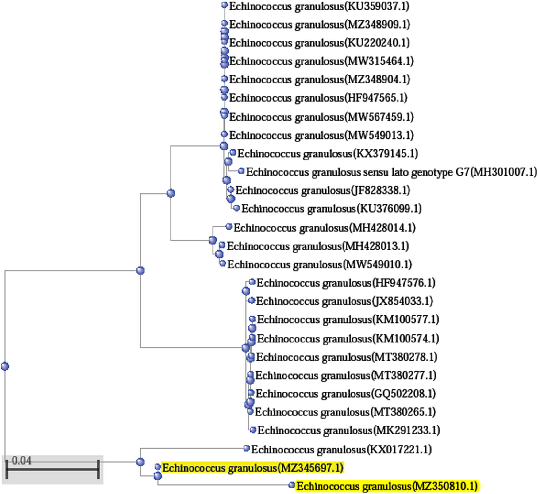
Phylogenetic relationships between Taif_Sheep E. granulosus (COX1) MZ345697.1 and MZ350810.1 (yellow highlights) with other reference sequences of E. granulosus from NCBI GenBank. GenBank sequences were shown by their name and accession numbers. Bar scale represents 0.05 nucleotide substitution per site.
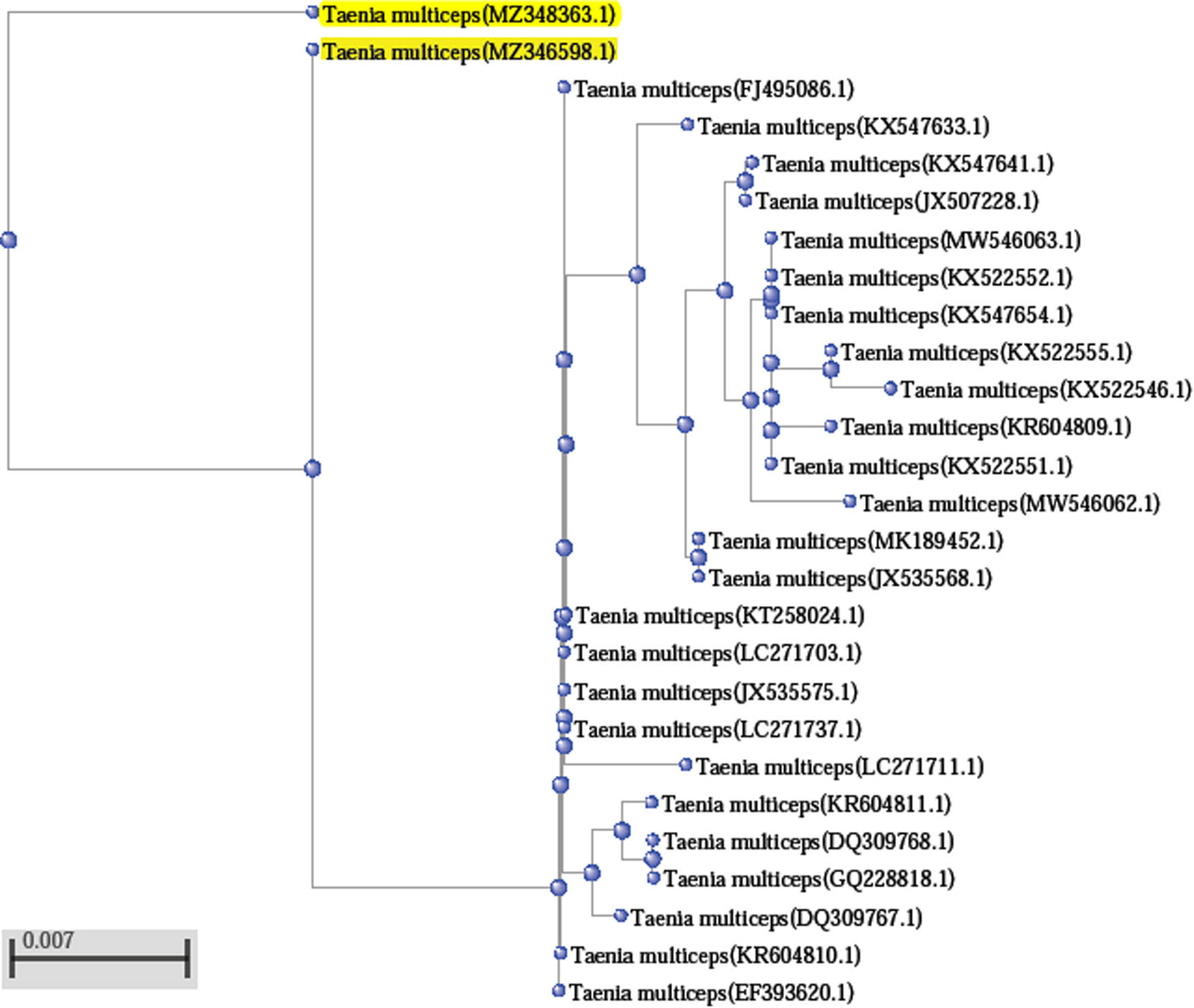
Phylogenetic relationships between Taif_Sheep coenurus cyst of T. multiceps (COX1) MZ346598.1 and MZ348363.1 (yellow highlights) with other reference sequences of T. multiceps from NCBI GenBank. GenBank sequences were shown by their name and accession numbers. Bar scale represents 0.007 nucleotide substitution per site.
For E. granulosus (COX1): The BLAST analysis showed that Taif sheep isolate of COX1 sequence exhibited percentage identity 84–95.67% with Query coverage ranging from 94 to 100% of E. granulosus in GenBank isolates that were collected from other different countries. Alignment of MZ345697.1 with the other isolate MZ350810.1 showed identity with 94.95%. MZ345697.1 showed a higher alignment identity (95.67%) with E. granulosus KX017221.1 from dog stool in Palestine. MZ345697.1 showed 85% homology with Iran isolates KU359037.1, MW549010.1, MW567459.1, MW315464.1, MW549013.1, KU220240.1, and KU376099.1 from different hosts such as camel, sheep, and goat. MZ345697.1 showed 85% identity with E. granulosus isolates from Egypt MZ348909.1 and MZ348904.1 in camel and Homo sapiens, respectively. In addition, the COX1 isolate of Taif sheep showed the same identity percentage with other isolates from buffalos (MH428014.1 and MH428013.1) in India, cattle (HF947565.1) in Portugal, goat (KX379145.1) in Italy, pig (JF828338.1) in Brazil, and sheep (GQ502208.1) in Chile (Figure 5). MZ345697.1 showed lower homology (84%) with isolates from cattle in Turkey (KM100577.1, KM100574.1, MT380278.1, MT380277.1, and MT380265.1), Portugal (HF947576.1), Homo sapiens in India (JX854033.1), Iran (MK291233.1), and Homo sapiens in Poland (MH301007.1).
For larval stage (coenurus) of T. multiceps (COX1): The BLAST analysis showed that COX1 sequence (Taif-sheep isolate) exhibited a higher percentage identity 99–98% (Query coverage 100–97%) with that of T. multiceps COX1 in GenBank isolates that were collected from other different countries. Taif isolate 1, MZ346598.1, showed 97% homology with our second isolate, MZ348363.1, in the present study. MZ346598.1 showed a higher alignment identity (99%) with coenurus cyst isolates found in brains of sheep in Egypt (LC271737.1 and LC271703.1), isolates from sheep and humans in China (JX535575.1, FJ495086.1, and KT258024.1), Greece (KR604810.1), and Turkey (EF393620.1). In addition, MZ346598.1 showed 98% identity with coenurus (larval stage of T. multiceps) isolates from different hosts of China (KX547633.1, KX547641.1, MW546063.1, KX522551.1, KX547654.1, KX522552.1, JX535568.1, GQ228818.1, and JX507228.1), from goat brain, dog intestine, goat muscle, and brain of domestic yak. Moreover, MZ346598.1 showed 98% homology with coenurus COX1 isolates from Italy (DQ309767.1 and DQ309768.1), Greece (KR604811.1), and Egypt (LC271711.1). Lower identity percent was found in MZ346598.1 alignment (97%) with isolates from China (KX522555.1, MW546062.1, and KX522546.1) and Greece (KR604809.1).
4 Discussion
Cystic echinococcosis and coenurosis are zoonotic diseases that affect humans and livestock due to infection with the larval stages of Echinococcus granulosus and of Taenia multiceps cysts, leading to economic loss that increases annually [4,9,11]. Dogs, especially stray unvaccinated dogs, are definitive hosts for different cestodes of Taeniidae family, including E. granulosus and T. multiceps. Dogs were commonly associated with extensive livestock herding to protect domestic animals against predators that could lead to their infection [20,21]. For the first time, the present study reports the genetic characterization of two different parasite cysts, hydatid (the larval stage of E. granulosus) and coenurus (the larval stage of T. multiceps), in Taif governorate, Makkah Province, Saudi Arabia.
Sheep, goats, cattle, and camels represent the primary livestock species producing red meat in Saudi Arabia, with an estimated total population of 13,444,435 heads [22]. Sheep represent the significant population (72%) of the livestock, and they import large numbers of sheep to satisfy the needs of Saudi citizens. Awassi, Harri, and Najdi are the most available sheep species in Saudi Arabia [23]. Any endemic infection for livestock, such as hydatidosis and coenurosis, could lead to economic setbacks; therefore, there is a need to highlight their study, especially in KSA.
Hydatidosis and coenurosis due to E. granulosus and T. multiceps larval infection, respectively, cause significant damage in livestock that leads to production losses, especially in endemic areas [2,24]. Moreover, these parasites were distributed worldwide in different intermediate hosts, especially in sheep and goats, accidentally humans, reflecting their medical and veterinary importance [25].
The present study aims to identify two Taeniids parasites found in slaughtered Harri sheep meat based on COX1 and NAD1 partial sequencing and phylogenetic relationship. It was reported that the development of molecular techniques rather than traditional morphological criteria has provided improved tools for taeniid species identification and investigating relationships among them. Sequencing of mitochondrial DNA, especially COX1 and NAD1 loci, has been successfully used for molecular characterization and identification of taeniids tapeworm [26,27].
Our choice to use mitochondrial DNA (mtDNA) sequence, especially COX1 and NAD1, for molecular characterization of E. granulosus and T. multiceps larva was based on the previous assessment. mtDNA is widely used in molecular characterization and phylogenetic evaluation studies. COX1 gene represents the most common gene of mtDNA for phylogenetic analysis and evolutionary biology of helminth parasites to determine interspecific and/or intraspecific variation [8,28,29,30]. The mitochondrial COX1 gene is very suitable for genetic diversity detection and haplotype analysis. Its evolutionary change rate is slow enough for the same species but fast enough for differentiation between different species. Therefore, the mitochondrial COX1 gene has been selected for use in the creation of DNA barcodes and species differentiation of different helminths as our present study [31,32].
Worldwide, there have been well-documented research studies about the genetic variability among many species found within the Taeniidae, especially for the genus Echinococcus. Still, there are a few related species of Taenia, especially for T. multiceps [33]. However, there is a scarcity of studies related to the genetic characterization of E. granulosus and T. multiceps larval stages in KSA and their phylogenetic relationship.
In the current study, a genetically characterized sample based on COX1 isolated from a hydatid cyst of MZ345697.1 showed identity about 94.95% with the other isolate MZ350810.1. For coenurus cyst, isolate1 MZ346598.1 showed 97% homology with the second isolate MZ348363.1. In addition, comparing partial sequences of Taif COX1 isolates from both cysts showed different identities with other isolates from organs present in various hosts found in other countries. The present results are inconsistent with previous studies, in which Al-Hizab et al. [34] and AL-Mutairi et al. [35] have reported E. granulosus species variation based on molecular characterization in infected sheep and camels in the Arabian Peninsula and KSA. AL-Mutairi et al. [35] have reported the phenotypic and genetic characterization of hydatid cysts isolated from sheep and camel meat in Al-Madinah, KSA. Genetic characterization of E. granulosus was based on random amplified polymorphic DNA polymerase chain reaction (RAPD-PCR) for the whole genome, PCR amplification of COX1 and 12S rRNA genes followed by single-stranded conformation polymorphism (SSCP), and then sequencing. They have investigated that cyst isolates slightly vary from each other and other isolates found in Genbank. They have concluded that there is an intraspecific variation in E. granulosus found in camels and local sheep.
In addition, Christodoulopoulos et al. [36] have investigated COX1 nucleotide diversity of coenurus cyst isolates from sheep and goats in Pakistan, and their phylogenetic analysis shows high related homology with China isolates but highly different from other isolates from different countries. They have reported that coenurus cerebralis commonly found in the brain (cerebral form) could also be found in other non-cerebral tissue such as intramuscular and subcutaneous tissues.
Diversity in the nucleotide sequences of the presently studied isolates was recorded before within the same region, such as in Iran [5,13], China [26], and Italy [37,38]. Most of their phylogenetic analysis depends on one or more mtDNA sequences such as nad1, cox1, and 12S rRNA. They have concluded that there are no monophyletic groups based on the intermediate host species, the organ from which the parasite was isolated, and geographical origin [33,36]. However, reporting parasite characterization and genetic identification will be crucial for controlling and preventing parasitic infections [8].
The limitation of this study is the use of the limited sample size, and only partial sequencing was done for the COX1 gene that could have implications on data interpretation. Thus, we recommend future studies considering a larger sample size, complete sequencing COX1, and other characterization-related genes in a massive epidemiological survey for further evaluation.
5 Conclusion
The present study reports the molecular characterization of hydatid (the larval stage of E. granulosus) and coenurus (the larval stage of T. multiceps) cysts present in slaughtered sheep meat in Taif, KSA. We have determined sequences differences within isolates of the same species that we submitted in GenBank and other isolates found in Genbank from different host organs in different countries. For a more precise identification and characterization of Taeniids in KSA, additional isolates from other hosts, other geographic areas, different molecular protocols, long-sequenced genes in the mitochondrial and nuclear genome, and further information concerning biological characteristics may be necessary to increase our understanding of the epidemiological distribution in KSA.
Acknowledgments
This work was supported by Taif University Researchers Supporting Project number (TURSP-2020/299), Taif University, Taif, Saudi Arabia. The authors would like to thank the Ministry of Environment, Water and Agriculture in Taif governorate (KSA) for facilitating data and sample collections.
-
Funding information: Taif University Researchers Supporting Project number (TURSP-2020/299).
-
Author contributions: J.M., and N.H. participated in the design of the study. J.M. collected samples and performed parasitological assay. J.M. and N.H. carried out the molecular evaluation. N.H. performed data analysis and wrote the article draft. J.M., and N.H. read and approved the final manuscript.
-
Conflict of interest: The authors state no conflict of interest.
-
Ethics approval: Data and samples were collected from veterinarians as per their periodic examination according to the ministerial recommendation. We have a permission letter from the Ministry of Environment, Water, and Agriculture (KSA) to collect cattle samples from different locations in Taif for our research study with the number 106082/1074/1442, dated 21-02-1442H. Sample collection and all experimental procedures were performed in accordance with a national ethical requirement. National Committee of Bioethics (NCBE) at King Abdulaziz City for Science and Technology (KACST) with number: 10023117, valid till October 1, 2023.
-
Data availability statement: The data sets generated during and/or analyzed during the current study are available from the corresponding author on reasonable request.
References
[1] Eckert J, Schantz PM, Gasser RB, Torgerson PR, Bessonov AS, Movsessian SO, et al. Geographic distribution and prevalence. In: Eckert J, Gemmell MA, Meslin FX, Pawlowski ZS, editors. WHO/OIE manual on Echinococcosis in humans and animals: a public health problem of global concern. Geneva: World Health Organisation; 2001. p. 100–42.Suche in Google Scholar
[2] Hoberg EP. Taenia tapeworms: their biology, evolution and socioeconomic significance. Microbes Infect. 2002;4(8):859–66. 10.1016/s1286-4579(02)01606-4.Suche in Google Scholar
[3] Grosso G, Gruttadauria S, Biondi A, Marventano S, Mistretta A. Worldwide epidemiology of liver hydatidosis including the Mediterranean area. World J Gastroenterol. 2012;18(13):1425–37. 10.3748/wjg.v18.i13.1425.Suche in Google Scholar
[4] Dalimi A, Motamedi G, Hosseini M, Mohammadian B, Malaki H, Ghamari Z, et al. Echinococcosis/hydatidosis in western Iran. Vet Parasitol. 2002;105:161–71. 10.1016/S0304-4017(02)00005-5.Suche in Google Scholar
[5] Harandi MF, Hobbs RP, Adams PJ, Mobedi I, Morgan-Ryan UM, Thompson RC. Molecular and morphological characterization of Echinococcus granulosus of human and animal origin in Iran. Parasitology. 2002;125:367–73. 10.1017/S0031182002002172.Suche in Google Scholar
[6] Rokni M. Echinococcosis/hydatidosis in Iran. Iran J Parasitol. 2009;4(2):1–16.Suche in Google Scholar
[7] Basinger SC, Khan A, Ahmed H, Afzal MS, Simsek S, Budke CM. Estimation of the monetary burden of treated human cystic echinococcosis in Pakistan. Acta tropica. 2021;222:106026. 10.1016/j.actatropica.2021.106026.Suche in Google Scholar PubMed
[8] Harandi MF, Budke CM, Rostami S. The monetary burden of cystic echinococcosis in Iran. PLoS Negl Trop Dis. 2012;6:e1915. 10.1371/journal.pntd.0001915.Suche in Google Scholar PubMed PubMed Central
[9] Khan A, Ahmed H, Khan H, Saleem S, Simsek S, Brunetti E, et al. Cystic Echinococcosis in Pakistan: a review of reported cases, diagnosis, and management. Acta tropica. 2020;212:105709. 10.1016/j.actatropica.2020.105709.Suche in Google Scholar PubMed
[10] Amrabadi O, Oryan A, Moazeni M, Shari-Fiyazdi H, Akbari M. Histopathological and molecular evaluation of the experimentally infected goats by the larval forms of Taenia multiceps. Iran J Parasitol. 2019;14:95–105. 10.18502/ijpa.v14i1.722.Suche in Google Scholar
[11] Sharma DK, Chauhan PPS. Coenurosis status in Afro-Asian region: a review. Small Ruminant Res. 2006;64:197–202. 10.1016/j.smallrumres.2005.05.021.Suche in Google Scholar
[12] Christodoulopoulos G, Theodoropoulos G, Petrakos G. Epidemiological survey of cestode-larva disease in Greek sheep flocks. Vet Parasitol. 2008;153:368–73. 10.1016/j.vetpar.2008.02.002.Suche in Google Scholar PubMed
[13] Oryan A, Nazifi S, Sharifiyazdi H, Ahmadnia S. Pathological, molecular, and biochemical characterization of Coenurus gaigeri in Iranian native goats. J Parasitol. 2010;96:961–7. 10.1645/GE-2399.1.Suche in Google Scholar PubMed
[14] Schuster RK, Sivakumar S, Wieckowsky T. Non-cerebral coenurosis in goats. Parasitol Res. 2010;107:721–6. 10.1007/s00436-010-1919-6.Suche in Google Scholar PubMed
[15] Giadinis ND, Psychas V, Polizopoulou Z, Papadopoulos E, Papaioannou N, Komnenou AT, et al. Acute coenurosis of dairy sheep from 11 flocks in Greece. N Z Vet J. 2012;60:247–53. 10.1080/00480169.2012.665343.Suche in Google Scholar PubMed
[16] Akbari M, Moazeni M, Oryan A, Sharifiyazdi H, Amrabadi O. Experimental cerebral and non-cerebral coenurosis in goats: a comparative study on the morphological and molecular characteristics of the parasite. Vet Parasitol. 2015;211:201–7. 10.1016/j.vetpar.2015.06.013.Suche in Google Scholar PubMed
[17] Barazesh A, Sarkari B, Ebrahimi S, Hami M. DNA extraction from hydatid cyst protoscolices: Comparison of five different methods. Vet. World. 2018;11(2):231–4.10.14202/vetworld.2018.231-234Suche in Google Scholar PubMed PubMed Central
[18] Sambrook J, Fritsch EF, Maniatis T. In: Sambrook J, Fritsch EF, Maniatis T, editors. Molecular cloning: a laboratory manual. 2nd edn. New York, NY, USA: CSH Cold Spring Harbor Press; 1989.Suche in Google Scholar
[19] Kuznetsov A, Bollin CJ. NCBI genome workbench: desktop software for comparative genomics, visualization, and GenBank data submission. Methods Mol Biol. 2021;2231:261–95.10.1007/978-1-0716-1036-7_16Suche in Google Scholar PubMed
[20] Abdi J, Asadolahi KH, Maleki MH, Ashrafi-Hafez A. Prevalence of helminthes infection of stray dogs in Ilam province. J Paramed Sci. 2013;4(2):58–61.Suche in Google Scholar
[21] Khan A, Ahmed H, Simsek S, Afzal MS, Cao J. Spread of cystic echinococcosis in pakistan due to stray dogs and livestock slaughtering habits: research priorities and public health importance. Public Health Front. 2020;7:412. 10.3389/fpubh.2019.00412.Suche in Google Scholar PubMed PubMed Central
[22] GASTAT. Agriculture, water, and environment. In: General authority for statistics. Statistical Yearbook of 2018. Available online at: https://www.stats.gov.sa/en/1011 (accessed March 3, 2021). 2018.Suche in Google Scholar
[23] Suliman GM, Al-Owaimer AN, El-Waziry AM, Hussein E, Abuelfatah K, Swelum A. A comparative study of sheep breeds: fattening performance, carcass characteristics, meat chemical composition and quality attributes. Front. Vet. Sci. 2021;8:647192. 10.3389/fvets.2021.647192.Suche in Google Scholar PubMed PubMed Central
[24] Raissi V, Etemadi S, Sohrabi N, Raiesi O, Shahraki M, Salimi-Khorashad A, et al. Molecular characterization and phylogeny of Taenia hydatigena and Echinococcus granulosus from Iranian sheep and cattle based on COX1 gene. Curr. Microbiol. 2021;78(4):1202–7. 10.1007/s00284-021-02377-0.Suche in Google Scholar PubMed
[25] Craig P, Pawlowski Z, editors. Cestode zoonoses: echinococcosis and cysticercosis an emergent and global problem. Amsterdam: Ios Press; 2002.Suche in Google Scholar
[26] Zhang Y, Zhao W, Yang D, Tian Y, Zhang W, Liu A. Genetic characterization of three mitochondrial gene sequences of goat/sheep-derived Coenurus cerebralis and Cysticercus tenuicollis isolates in Inner Mongolia, China. Parasite. 2018;25(3–4):1. 10.1051/parasite/2018002.Suche in Google Scholar PubMed PubMed Central
[27] Heidari Z, Sharbatkhori M, Mobedi I, Mirhendi SH, Nikmanesh B, Sharifdini M,et al. Echinococcus multilocularis and Echinococcus granulosus in canines in North-Khorasan Province, northeastern Iran, identified using morphology and genetic characterization of mitochondrial DNA. Parasit Vectors. 2019;606(12):1–13. 10.1186/s13071-019-3859-z.Suche in Google Scholar PubMed PubMed Central
[28] Mohaghegh MA, Yousofi-Darani H, Jafarian AH, Mirbadie SR, Fasihi-Harandi M, Ghavimi R, et al. Isolated human and Livestock Echinococcus granulosus genotypes using real-time PCR of cox1 gene in Northeast Iran. Acta Parasitol. 2019;64(3):679–85. 10.2478/s11686-019-00117-w.Suche in Google Scholar PubMed
[29] Paoletti B, Della Salda L, Di Cesare A, Iorio R, Vergara A, Fava C, et al. Epidemiological survey on cystic echinococcosis in wild boar from Central Italy. Parasitol. Res. 2019;118(1):43–6. 10.1007/s00436-018-6112-3.Suche in Google Scholar PubMed
[30] Mirbadie SR, Nasab AN, Mohaghegh MA, Norouzi P, Mirzaii M, Spotin A. Molecular phylodiagnosis of Echinococcus granulosus sensu lato and Taenia hydatigena determined by mitochondrial Cox1 and SSU-rDNA markers in Iranian dogs: indicating the first record of pig strain (G7) in definitive host in the Middle East. Comp Immunol Microbiol Infect Dis. 2019;65:88–95. 10.1016/j.cimid.2019.05.005.Suche in Google Scholar PubMed
[31] Rach J, Bergmann T, Paknia O, DeSalle R, Schierwater B, Hadrys H. The marker choice: Unexpected resolving power of an unexplored CO1 region for layered DNA barcoding approaches. PLoS One. 2017;12(4):e0174842. 10.1371/journal.pone.0174842.Suche in Google Scholar PubMed PubMed Central
[32] Gunyakti Kilinc S, Celik F, Kesik HK, Simsek S. In silico analysis of the biodiversity and conservation status of mitochondrial cytochrome C oxidase subunit 1 (CO1) gene of Taenia multiceps. Acta Parasitologica. 2020;65(4):852–8. 10.1007/s11686-020-00236-9.Suche in Google Scholar PubMed
[33] Alvi MA, Ohiolei JA, Saqib M, Tayyab MH, Zafar Khan MU, Li L, et al. First report on molecular characterization of taenia multiceps isolates from sheep and goats in faisalabad, Pakistan. Front. Vet. Sci. 2020;7:594599. 10.3389/fvets.2020.594599. Suche in Google Scholar
[34] Al-Hizab FA, Mohamed NS, Wassermann M, Hamouda MA, Ibrahim AM, El-Ghareeb WR, et al. Three species of Echinococcus granulosus sensu lato infect camels on the Arabian Peninsula. Parasitol Res. 2021;120:2077–86. 10.1007/s00436-021-07156-1.Suche in Google Scholar PubMed PubMed Central
[35] AL-Mutairi NM, Taha HA, Nigm AH. Molecular characterization of Echinococcus granulosus in livestock of Al-Madinah (Saudi Arabia). J. Helminthol. 2020;94(e157):1–12. 10.1017/S0022149X20000395. Suche in Google Scholar
[36] Christodoulopoulos G, Dinkel A, Romig T, Ebi D, Mackenstedt U, Loos-Frank B. Cerebral and non-cerebral coenurosis: on the genotypic and phenotypic diversity of Taenia multiceps. Parasitol Res. 2016 Dec;115:4543–58. 10.1007/s00436-016-5246-4.Suche in Google Scholar PubMed
[37] Varcasia A, Lightowlers MW, Cattoli G, Cancedda GM, Canu S, Garippa G, et al. Genetic variation within Taenia multiceps in Sardinia, Western Mediterranean (Italy). Parasitol Res. 2006;99(5):622–6. 10.1007/s00436-006-0179-y.Suche in Google Scholar PubMed
[38] Varcasia A, Pipia AP, Dessì G, Zidda A, Tamponi C, Pau M, et al. Morphology and genetic variability within Taenia multiceps in ruminants from Italy. Vet. Parasitol. 2016;223:181–5. 10.1016/j.vetpar.2016.04.039.Suche in Google Scholar PubMed
© 2021 Jamila S. Al Malki and Nahed Ahmed Hussien, published by De Gruyter
This work is licensed under the Creative Commons Attribution 4.0 International License.
Artikel in diesem Heft
- Biomedical Sciences
- Research progress on the mechanism of orexin in pain regulation in different brain regions
- Adriamycin-resistant cells are significantly less fit than adriamycin-sensitive cells in cervical cancer
- Exogenous spermidine affects polyamine metabolism in the mouse hypothalamus
- Iris metastasis of diffuse large B-cell lymphoma misdiagnosed as primary angle-closure glaucoma: A case report and review of the literature
- LncRNA PVT1 promotes cervical cancer progression by sponging miR-503 to upregulate ARL2 expression
- Two new inflammatory markers related to the CURB-65 score for disease severity in patients with community-acquired pneumonia: The hypersensitive C-reactive protein to albumin ratio and fibrinogen to albumin ratio
- Circ_0091579 enhances the malignancy of hepatocellular carcinoma via miR-1287/PDK2 axis
- Silencing XIST mitigated lipopolysaccharide (LPS)-induced inflammatory injury in human lung fibroblast WI-38 cells through modulating miR-30b-5p/CCL16 axis and TLR4/NF-κB signaling pathway
- Protocatechuic acid attenuates cerebral aneurysm formation and progression by inhibiting TNF-alpha/Nrf-2/NF-kB-mediated inflammatory mechanisms in experimental rats
- ABCB1 polymorphism in clopidogrel-treated Montenegrin patients
- Metabolic profiling of fatty acids in Tripterygium wilfordii multiglucoside- and triptolide-induced liver-injured rats
- miR-338-3p inhibits cell growth, invasion, and EMT process in neuroblastoma through targeting MMP-2
- Verification of neuroprotective effects of alpha-lipoic acid on chronic neuropathic pain in a chronic constriction injury rat model
- Circ_WWC3 overexpression decelerates the progression of osteosarcoma by regulating miR-421/PDE7B axis
- Knockdown of TUG1 rescues cardiomyocyte hypertrophy through targeting the miR-497/MEF2C axis
- MiR-146b-3p protects against AR42J cell injury in cerulein-induced acute pancreatitis model through targeting Anxa2
- miR-299-3p suppresses cell progression and induces apoptosis by downregulating PAX3 in gastric cancer
- Diabetes and COVID-19
- Discovery of novel potential KIT inhibitors for the treatment of gastrointestinal stromal tumor
- TEAD4 is a novel independent predictor of prognosis in LGG patients with IDH mutation
- circTLK1 facilitates the proliferation and metastasis of renal cell carcinoma by regulating miR-495-3p/CBL axis
- microRNA-9-5p protects liver sinusoidal endothelial cell against oxygen glucose deprivation/reperfusion injury
- Long noncoding RNA TUG1 regulates degradation of chondrocyte extracellular matrix via miR-320c/MMP-13 axis in osteoarthritis
- Duodenal adenocarcinoma with skin metastasis as initial manifestation: A case report
- Effects of Loofah cylindrica extract on learning and memory ability, brain tissue morphology, and immune function of aging mice
- Recombinant Bacteroides fragilis enterotoxin-1 (rBFT-1) promotes proliferation of colorectal cancer via CCL3-related molecular pathways
- Blocking circ_UBR4 suppressed proliferation, migration, and cell cycle progression of human vascular smooth muscle cells in atherosclerosis
- Gene therapy in PIDs, hemoglobin, ocular, neurodegenerative, and hemophilia B disorders
- Downregulation of circ_0037655 impedes glioma formation and metastasis via the regulation of miR-1229-3p/ITGB8 axis
- Vitamin D deficiency and cardiovascular risk in type 2 diabetes population
- Circ_0013359 facilitates the tumorigenicity of melanoma by regulating miR-136-5p/RAB9A axis
- Mechanisms of circular RNA circ_0066147 on pancreatic cancer progression
- lncRNA myocardial infarction-associated transcript (MIAT) knockdown alleviates LPS-induced chondrocytes inflammatory injury via regulating miR-488-3p/sex determining region Y-related HMG-box 11 (SOX11) axis
- Identification of circRNA circ-CSPP1 as a potent driver of colorectal cancer by directly targeting the miR-431/LASP1 axis
- Hyperhomocysteinemia exacerbates ischemia-reperfusion injury-induced acute kidney injury by mediating oxidative stress, DNA damage, JNK pathway, and apoptosis
- Potential prognostic markers and significant lncRNA–mRNA co-expression pairs in laryngeal squamous cell carcinoma
- Gamma irradiation-mediated inactivation of enveloped viruses with conservation of genome integrity: Potential application for SARS-CoV-2 inactivated vaccine development
- ADHFE1 is a correlative factor of patient survival in cancer
- The association of transcription factor Prox1 with the proliferation, migration, and invasion of lung cancer
- Is there a relationship between the prevalence of autoimmune thyroid disease and diabetic kidney disease?
- Immunoregulatory function of Dictyophora echinovolvata spore polysaccharides in immunocompromised mice induced by cyclophosphamide
- T cell epitopes of SARS-CoV-2 spike protein and conserved surface protein of Plasmodium malariae share sequence homology
- Anti-obesity effect and mechanism of mesenchymal stem cells influence on obese mice
- Long noncoding RNA HULC contributes to paclitaxel resistance in ovarian cancer via miR-137/ITGB8 axis
- Glucocorticoids protect HEI-OC1 cells from tunicamycin-induced cell damage via inhibiting endoplasmic reticulum stress
- Prognostic value of the neutrophil-to-lymphocyte ratio in acute organophosphorus pesticide poisoning
- Gastroprotective effects of diosgenin against HCl/ethanol-induced gastric mucosal injury through suppression of NF-κβ and myeloperoxidase activities
- Silencing of LINC00707 suppresses cell proliferation, migration, and invasion of osteosarcoma cells by modulating miR-338-3p/AHSA1 axis
- Successful extracorporeal membrane oxygenation resuscitation of patient with cardiogenic shock induced by phaeochromocytoma crisis mimicking hyperthyroidism: A case report
- Effects of miR-185-5p on replication of hepatitis C virus
- Lidocaine has antitumor effect on hepatocellular carcinoma via the circ_DYNC1H1/miR-520a-3p/USP14 axis
- Primary localized cutaneous nodular amyloidosis presenting as lymphatic malformation: A case report
- Multimodal magnetic resonance imaging analysis in the characteristics of Wilson’s disease: A case report and literature review
- Therapeutic potential of anticoagulant therapy in association with cytokine storm inhibition in severe cases of COVID-19: A case report
- Neoadjuvant immunotherapy combined with chemotherapy for locally advanced squamous cell lung carcinoma: A case report and literature review
- Rufinamide (RUF) suppresses inflammation and maintains the integrity of the blood–brain barrier during kainic acid-induced brain damage
- Inhibition of ADAM10 ameliorates doxorubicin-induced cardiac remodeling by suppressing N-cadherin cleavage
- Invasive ductal carcinoma and small lymphocytic lymphoma/chronic lymphocytic leukemia manifesting as a collision breast tumor: A case report and literature review
- Clonal diversity of the B cell receptor repertoire in patients with coronary in-stent restenosis and type 2 diabetes
- CTLA-4 promotes lymphoma progression through tumor stem cell enrichment and immunosuppression
- WDR74 promotes proliferation and metastasis in colorectal cancer cells through regulating the Wnt/β-catenin signaling pathway
- Down-regulation of IGHG1 enhances Protoporphyrin IX accumulation and inhibits hemin biosynthesis in colorectal cancer by suppressing the MEK-FECH axis
- Curcumin suppresses the progression of gastric cancer by regulating circ_0056618/miR-194-5p axis
- Scutellarin-induced A549 cell apoptosis depends on activation of the transforming growth factor-β1/smad2/ROS/caspase-3 pathway
- lncRNA NEAT1 regulates CYP1A2 and influences steroid-induced necrosis
- A two-microRNA signature predicts the progression of male thyroid cancer
- Isolation of microglia from retinas of chronic ocular hypertensive rats
- Changes of immune cells in patients with hepatocellular carcinoma treated by radiofrequency ablation and hepatectomy, a pilot study
- Calcineurin Aβ gene knockdown inhibits transient outward potassium current ion channel remodeling in hypertrophic ventricular myocyte
- Aberrant expression of PI3K/AKT signaling is involved in apoptosis resistance of hepatocellular carcinoma
- Clinical significance of activated Wnt/β-catenin signaling in apoptosis inhibition of oral cancer
- circ_CHFR regulates ox-LDL-mediated cell proliferation, apoptosis, and EndoMT by miR-15a-5p/EGFR axis in human brain microvessel endothelial cells
- Resveratrol pretreatment mitigates LPS-induced acute lung injury by regulating conventional dendritic cells’ maturation and function
- Ubiquitin-conjugating enzyme E2T promotes tumor stem cell characteristics and migration of cervical cancer cells by regulating the GRP78/FAK pathway
- Carriage of HLA-DRB1*11 and 1*12 alleles and risk factors in patients with breast cancer in Burkina Faso
- Protective effect of Lactobacillus-containing probiotics on intestinal mucosa of rats experiencing traumatic hemorrhagic shock
- Glucocorticoids induce osteonecrosis of the femoral head through the Hippo signaling pathway
- Endothelial cell-derived SSAO can increase MLC20 phosphorylation in VSMCs
- Downregulation of STOX1 is a novel prognostic biomarker for glioma patients
- miR-378a-3p regulates glioma cell chemosensitivity to cisplatin through IGF1R
- The molecular mechanisms underlying arecoline-induced cardiac fibrosis in rats
- TGF-β1-overexpressing mesenchymal stem cells reciprocally regulate Th17/Treg cells by regulating the expression of IFN-γ
- The influence of MTHFR genetic polymorphisms on methotrexate therapy in pediatric acute lymphoblastic leukemia
- Red blood cell distribution width-standard deviation but not red blood cell distribution width-coefficient of variation as a potential index for the diagnosis of iron-deficiency anemia in mid-pregnancy women
- Small cell neuroendocrine carcinoma expressing alpha fetoprotein in the endometrium
- Superoxide dismutase and the sigma1 receptor as key elements of the antioxidant system in human gastrointestinal tract cancers
- Molecular characterization and phylogenetic studies of Echinococcus granulosus and Taenia multiceps coenurus cysts in slaughtered sheep in Saudi Arabia
- ITGB5 mutation discovered in a Chinese family with blepharophimosis-ptosis-epicanthus inversus syndrome
- ACTB and GAPDH appear at multiple SDS-PAGE positions, thus not suitable as reference genes for determining protein loading in techniques like Western blotting
- Facilitation of mouse skin-derived precursor growth and yield by optimizing plating density
- 3,4-Dihydroxyphenylethanol ameliorates lipopolysaccharide-induced septic cardiac injury in a murine model
- Downregulation of PITX2 inhibits the proliferation and migration of liver cancer cells and induces cell apoptosis
- Expression of CDK9 in endometrial cancer tissues and its effect on the proliferation of HEC-1B
- Novel predictor of the occurrence of DKA in T1DM patients without infection: A combination of neutrophil/lymphocyte ratio and white blood cells
- Investigation of molecular regulation mechanism under the pathophysiology of subarachnoid hemorrhage
- miR-25-3p protects renal tubular epithelial cells from apoptosis induced by renal IRI by targeting DKK3
- Bioengineering and Biotechnology
- Green fabrication of Co and Co3O4 nanoparticles and their biomedical applications: A review
- Agriculture
- Effects of inorganic and organic selenium sources on the growth performance of broilers in China: A meta-analysis
- Crop-livestock integration practices, knowledge, and attitudes among smallholder farmers: Hedging against climate change-induced shocks in semi-arid Zimbabwe
- Food Science and Nutrition
- Effect of food processing on the antioxidant activity of flavones from Polygonatum odoratum (Mill.) Druce
- Vitamin D and iodine status was associated with the risk and complication of type 2 diabetes mellitus in China
- Diversity of microbiota in Slovak summer ewes’ cheese “Bryndza”
- Comparison between voltammetric detection methods for abalone-flavoring liquid
- Composition of low-molecular-weight glutenin subunits in common wheat (Triticum aestivum L.) and their effects on the rheological properties of dough
- Application of culture, PCR, and PacBio sequencing for determination of microbial composition of milk from subclinical mastitis dairy cows of smallholder farms
- Investigating microplastics and potentially toxic elements contamination in canned Tuna, Salmon, and Sardine fishes from Taif markets, KSA
- From bench to bar side: Evaluating the red wine storage lesion
- Establishment of an iodine model for prevention of iodine-excess-induced thyroid dysfunction in pregnant women
- Plant Sciences
- Characterization of GMPP from Dendrobium huoshanense yielding GDP-D-mannose
- Comparative analysis of the SPL gene family in five Rosaceae species: Fragaria vesca, Malus domestica, Prunus persica, Rubus occidentalis, and Pyrus pyrifolia
- Identification of leaf rust resistance genes Lr34 and Lr46 in common wheat (Triticum aestivum L. ssp. aestivum) lines of different origin using multiplex PCR
- Investigation of bioactivities of Taxus chinensis, Taxus cuspidata, and Taxus × media by gas chromatography-mass spectrometry
- Morphological structures and histochemistry of roots and shoots in Myricaria laxiflora (Tamaricaceae)
- Transcriptome analysis of resistance mechanism to potato wart disease
- In silico analysis of glycosyltransferase 2 family genes in duckweed (Spirodela polyrhiza) and its role in salt stress tolerance
- Comparative study on growth traits and ions regulation of zoysiagrasses under varied salinity treatments
- Role of MS1 homolog Ntms1 gene of tobacco infertility
- Biological characteristics and fungicide sensitivity of Pyricularia variabilis
- In silico/computational analysis of mevalonate pyrophosphate decarboxylase gene families in Campanulids
- Identification of novel drought-responsive miRNA regulatory network of drought stress response in common vetch (Vicia sativa)
- How photoautotrophy, photomixotrophy, and ventilation affect the stomata and fluorescence emission of pistachios rootstock?
- Apoplastic histochemical features of plant root walls that may facilitate ion uptake and retention
- Ecology and Environmental Sciences
- The impact of sewage sludge on the fungal communities in the rhizosphere and roots of barley and on barley yield
- Domestication of wild animals may provide a springboard for rapid variation of coronavirus
- Response of benthic invertebrate assemblages to seasonal and habitat condition in the Wewe River, Ashanti region (Ghana)
- Molecular record for the first authentication of Isaria cicadae from Vietnam
- Twig biomass allocation of Betula platyphylla in different habitats in Wudalianchi Volcano, northeast China
- Animal Sciences
- Supplementation of probiotics in water beneficial growth performance, carcass traits, immune function, and antioxidant capacity in broiler chickens
- Predators of the giant pine scale, Marchalina hellenica (Gennadius 1883; Hemiptera: Marchalinidae), out of its natural range in Turkey
- Honey in wound healing: An updated review
- NONMMUT140591.1 may serve as a ceRNA to regulate Gata5 in UT-B knockout-induced cardiac conduction block
- Radiotherapy for the treatment of pulmonary hydatidosis in sheep
- Retraction
- Retraction of “Long non-coding RNA TUG1 knockdown hinders the tumorigenesis of multiple myeloma by regulating microRNA-34a-5p/NOTCH1 signaling pathway”
- Special Issue on Reuse of Agro-Industrial By-Products
- An effect of positional isomerism of benzoic acid derivatives on antibacterial activity against Escherichia coli
- Special Issue on Computing and Artificial Techniques for Life Science Applications - Part II
- Relationship of Gensini score with retinal vessel diameter and arteriovenous ratio in senile CHD
- Effects of different enantiomers of amlodipine on lipid profiles and vasomotor factors in atherosclerotic rabbits
- Establishment of the New Zealand white rabbit animal model of fatty keratopathy associated with corneal neovascularization
- lncRNA MALAT1/miR-143 axis is a potential biomarker for in-stent restenosis and is involved in the multiplication of vascular smooth muscle cells