Small cell neuroendocrine carcinoma expressing alpha fetoprotein in the endometrium
-
Weiwei Hou


Abstract
Rare small cell neuroendocrine carcinoma (SCNEC) cases showed alpha fetoprotein (AFP) expression in the endometrium. In this study, we reported a case of uterine SCNEC expressing AFP. In addition, a literature review was performed to investigate the potential mechanism and the clinicopathological features of SCNEC to provide clinical guidance. A 65-year-old female was referred to our hospital due to vaginal bleeding for 1 month in November 2020. The clinical features were summarized. After total hysterectomy and removal of bilateral appendages, the histological examination and immunohistochemistry examination were performed. Histological findings showed that the cancer cells were arranged in a nest-like pattern distributed in a lamellar manner. The smooth muscles of the uterus were invaded by cancer cells. Cancer cells were relatively consistent in size. Small glandular duct-like and rosettes-like structures were distinguished, together with necrotic tissues. The deep staining showed that the amount of cytoplasm was lower in the nucleus. Partial cancer cells had small nucleolus with an irregular profile. There were some mitotic figures. Immunohistochemistry examination indicated that there was a diffuse expression of CK, Syn, CgA, CD56, CK8/18, P16, AFP, HepPar-1, Glypican-3, and Ki67 (90%). In this case, we reported a SCNEC patient expressing AFP, Glypican-3, and HepPar-1.
1 Introduction
Small cell neuroendocrine carcinoma (SCNEC) is a malignance usually occurring in the pulmonary tissues [1]. Also, it may occur in the gastrointestinal tract, female reproductive system, head and neck, as well as mammary gland [2]. For the patients with female reproductive system involvement, SCNEC commonly involves the cervix, while rare cases show endometrial involvement with a prevalence of merely 0.8% among all endometrial carcinomas [3].
SCNEC is featured by a high-grade malignancy, strong invasion, distal metastasis, as well as a poor prognosis [4,5]. Rare SCNEC cases showed expression of alpha fetoprotein (AFP) after a literature review [6]. In this study, we reported a case of uterine SCNEC expressing AFP. In addition, a literature review was performed to investigate the potential mechanism and the clinicopathological features of SCNEC in order to provide clinical guidance. We present the following article in accordance with the STROBE reporting checklist.
2 Materials and methods
2.1 Clinical data
A 65-year-old female was referred to our hospital due to vaginal bleeding for 1 month. She showed no abdominal pain and distension. She underwent a hysteroscopy in a local hospital. Diagnostic curettage-based pathology indicated malignance combined with necrosis. On this basis, the patient was highly suspected of poorly differentiated carcinoma or endometrial stromal sarcoma. On physical examination after admitting to our hospital, there were dilation and hydrops in the uterine cavity after the abdominal plain scan. Additionally, irregular lumps with equal density were noticed in the left posterior uterine wall, together with prominence (3.7 cm × 2.7 cm) in the urine cavity, presenting cauliflower-like changes on the surface and heterogeneous enhancement. A CT scan demonstrated occupying lesions in the uterine cavity, and then endometrium malignancy was considered. No abnormality was seen in other organs. Laboratory examination findings were as follows: AFP, 975.30 ng/mL (normal range: 0–10); C-reactive protein (CRP), 165.89 mg/L (normal range: 0.00–9.00); and D-dimer, 2.23 mg/L DDU (normal range: 0–1.00). The patient underwent a total hysterectomy and removal of bilateral appendages. A pathological examination was performed after the surgery.
-
Informed consent: Informed consent has been obtained from all individuals included in this study.
-
Ethical approval: The research related to human use complied with all relevant national regulations, institutional policies and was in accordance with the tenets of the Helsinki Declaration, and has been approved by the Ethics Committee of Tai’an Central Hospital (No.: WD-0019).
2.2 Methods
All the samples were fixed in 10% neutral formalin, followed by embedding in paraffin. Sections (4 μm) were stained by hematoxylin and eosin. The antibodies used were as follows: PAN-cytokeratin (CK, Catalog number: RAB-0050; 1:2,000), synaptophysin (Syn, Catalog number: MAB-0742; 1:1,000), chromogranin A (CgA, Catalog number: MAB-0707; 1:1,000), neural cell adhesion molecule (CD56, Catalog number: MAB-0743; 1:2,000), Glypican-3 (Catalog number: MAB-0617; 1:2,000), cytokeratin 8/18 (CK8/18, Catalog number: MAB-0650; 1:2,000), CD10 (Catalog number: MAB-0668; 1:1,000), estrogen receptor (ER, Catalog number: MAB-0062; 1:1,000), progesterone receptor (PR, Catalog number: MAB-0675; 1:2,000), P53 (Catalog number: MAB-0674; 1:2,000), cyclin dependent kinase-4 (P16, Catalog number: MAB-0673; 1:2,000), cytokeratin-7 (CK7, Catalog number: MAB-0828; 1:1,000), as well as Ki-67 (Catalog number: RMA-0542; 1:1,000). All reagents were purchased from Maixin Biotech (Fuzhou, China). The pathological images were analyzed using CaseViewer software.
3 Results
3.1 General conditions
The uterus with a size of 9 cm × 8 cm × 2 cm had been dissected along the anterior wall. The cervical canal showed a length of 2.5 cm, with a diameter of about 2.5 cm. The mucous membrane at the external orifice was smooth, and the uterine depth was 7 cm. The thickness of the muscular layer was in a range of 1.5–2.0 cm. There was a tumor mass (4 cm × 3.5 cm × 2.5 cm) near the left horn of the uterus. This incisal surface was in a grayish-white color mixed with red color in a hard texture. The depth of invasion was less than half of the myometrium. The bilateral appendix was normal in structure (Figure 1).

A mass in the left horn of the uterus with a size of 4 cm.
3.2 Histological findings
Under a magnification of 40×, the cancer cells were arranged in a nest-like pattern, distributed in a lamellar manner. The smooth muscles of the uterus were invaded by cancer cells (Figure 2a). Cancer cells were relatively consistent in size under a magnification of 200×. Small glandular duct-like and rosette-like structures were distinguished with necrotic tissues (Figure 2b and c). The cytoplasm was less in amount with deep staining in the nucleus under a magnification of 400×. Partial cancer cells showed small nucleolus with an irregular profile. There were some mitotic figures (15 per high power, Figure 2d and e).
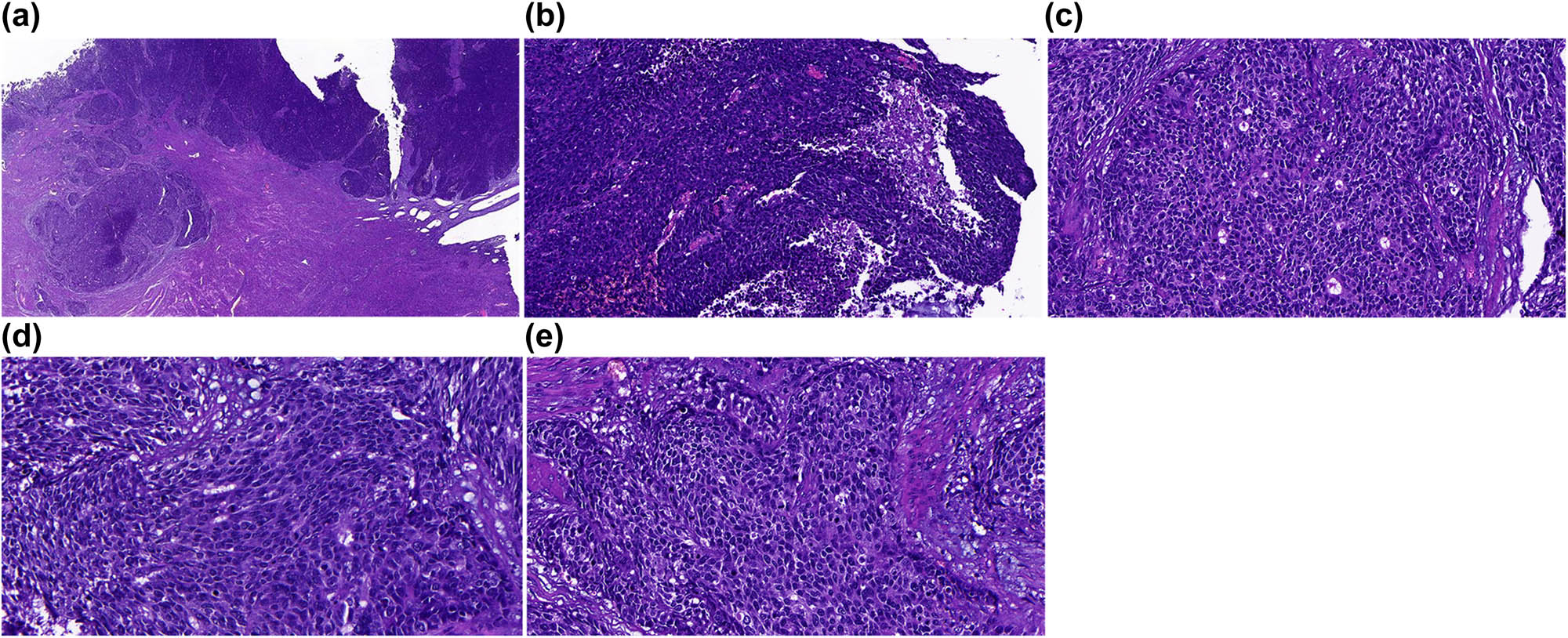
HE staining of the samples. (a) Cancer cells were arranged in a nest or lamellar pattern. Infiltrative growth was noticed in the myometrium. (b) Necrosis in the cancer tissues. (c) Small glandular duct-like and rosettes-like structures. (d) Multiple mitotic phases. (e) Cancer cells with less cytoplasm, and deep staining in nucleus. Part of the cancer cells showed a small nucleus. Magnifications of the images were as follows: (a) 40×; (b and c) 200×; (d and e) 400×.
3.3 Immunophenotypes
There was positivity for the immunophenotypes of cancer cells for CK, AFP, Syn, CgA, CD56, HepPar-1, Glypican-3, CK8/18, P16, and P53 (Figure 3a–d). The expression of CK7, ER, PR, Vimentin, and CD10 was negative. The ki-67 index was approximately 90%.

Immunohistochemistry findings. (a) The cytoplasm in cancer cells was sparse, with the nucleus stained in a deep color. There was a small nucleus in partial cancer cells. (b–d) Expression of Syn, CgA, Glypican-3, and AFP in cancer cells. The images were observed under a magnification of 400×.
3.4 Final diagnosis
Based on these findings mentioned above, the patient was finally diagnosed with SCNEC expressing AFP in the endometrium.
3.5 Follow-up
The patient received no treatment previously. She was followed up for 2 months after surgery. The serum AFP showed a decline after treatment, which was in the normal range.
4 Discussion
SCNEC is a rare malignancy featured by a high possibility of metastasis and a poor prognosis. To date, specific therapeutic options are still lacking for it [7]. Nowadays, there are some hypotheses for the histological origin of SCNEC. The origins were supposed to evolve from endometrial neuroendocrine cells, multifunctional and multiple differentiation potential stem cells in the endometrium, differentiated neuroendocrine cells of endometrial adenocarcinoma, differentiated tumor cells of endometrial adenocarcinoma in the presence of living conditions and endocrine factors, as well as the multidirectional differentiation of the Mullerian ducts such as differentiating into neuroendocrine cells. To the best of our knowledge, rare SCNEC cases expressed AFP [8]. In this study, we reported a SCNEC case expressing AFP and summarized the clinical and pathological features as well as the treatment and outcome.
SCNEC is commonly seen in females of perimenopausal or post-menopause periods. The common symptoms include irregular vaginal bleeding. As previously described [9], patients with SCNEC were mainly manifested as a dystopia, membranous glomerulonephritis, and Cushing syndrome. Partial patients showed a slight elevation in CA125 and NSE. The cancer cells showed a strong invasion capacity, with rapid progression. The lesions were highly malignant, with a high possibility of metastasis to lung, bones, and brain tissues [10].
The lesions of SCNEC were featured by cauliflower-like or polypoid-like mass in the uterine cavity, together with invasive growth in the myometrium. The incisal section was white, with a hard texture. Part of the lesions showed fish-meat-like changes, frequently combined with bleeding and necrosis. The cancer cells were distributed in a nest-like, lamellar and rosette-like pattern. The cellular size was not large, which was relatively consistent in size. The cytoplasm was not much, and the nucleus was stained strongly. Partial cancer cells showed a small nucleus with an irregular profile. There were some mitotic figures combined with endometrial adenocarcinoma or squamous cell carcinoma. In a previous study, Van Hoeven et al. proposed the diagnostic standards for SCNEC as follows: (i) with definite evidence for primary lesions in endometrium; (ii) consisted of cancer cells of similar sizes, in a dense or lamellar growth pattern, with other cancer components; and (iii) expressing at least one neuroendocrine markers in the immunohistochemistry. To date, the commonly utilized immunohistochemistry markers included NSE, CgA, Syn, CD56, P53, P16, and Ki67 in a range of 60–90% [11]. In a previous study, there was a high CgA, Syn, and NSE expression in SCNEC patients [12]. Therefore, the diagnosis of SCNEC relied on the combination of histopathological analysis and immunohistochemistry. In this case, there was expression of Syn, CgA, and CD56, together with AFP, HepPar-1, Glypican-3, CK8/18, and Ki67 (90%). Finally, the patient was diagnosed with SCNEC expressing AFP.
In clinical practice, SCNEC patients with endometrial AFP expression should be differentially diagnosed from the other types of cancers. For example, as in this case, the immunohistochemistry findings were positive for AFP, Glypican-3, and HepPar-1, and it should be distinguished from hepatoid adenocarcinoma of the endometrium. In histology and morphology, the hepatoid adenocarcinoma of the endometrium was similar to hepatocellular liver cancer. The cancer tissues could specifically express AFP, Glypican-3, and HepPar-1; however, they did not express NSE, Syn, and CD56. In addition, it should be distinguished from small cell squamous cell carcinoma, which showed a small and round profile in cancer cells, together with eosin staining in the cytoplasm and deep staining in the nucleus. Occasionally, there might be single-cell hornification. The immunohistochemistry for P63 was positive, while the neuroendocrine markers were all negative. Moreover, it should be distinguished from the poorly differentiated endometrioid carcinoma. The majority (90%) of these patients showed vaginal bleeding, and the cancer cells showed solid and lamellar growth, with none or less an adenoid structure. The cellular differentiation was poor, and there were multiple mitotic phases. There was a strong expression of ER and PR, and the neuroendocrine markers were all negative. Furthermore, SCNEC should be further distinguished from endometrial stromal sarcoma in which the cells were arranged in a lamellar or cluster pattern. The cancer cells were in a short and spindle shape or an oval shape, which distributed along the vessels. In addition, there was collagenization and branch-like vessels. The immunohistochemistry for CD10 was positive, and the neuroendocrine markers were negative. Finally, attention should be paid to the differential diagnosis of metastatic small cell carcinoma. The small cell carcinoma usually occurs in the lungs, while in the female population, it usually involves the cervix and/or ovary gland. Furthermore, SCNEC should be differentially diagnosed with carcinosarcoma, designated as a Mullerian mixed tumor that usually occurs in the aged female. The epithelial component is usually high-grade carcinoma, mainly featured in serous carcinoma and endometrioid carcinoma. The interstitial component consists of homologous and heterogenous types, such as the high-grade endometrial stromal sarcoma and the rhabdomyosarcoma. The immunohistochemistry contributed to the confirmation of the high-grade carcinoma and interstitial components in the carcinosarcoma. The malignant epithelial cells usually expressed p53, p16, and PTEN, while the malignant interstitial cells expressed CD10 and Vimentin rather than Syn, CgA, and CD56. In this case, there were no lesions in other sites after the whole body imaging scan except endometrium, and there were no aberrant changes in the cervix and ovary gland.
At the early stage, there might be local infiltration and lymphatic metastasis. Most cases showed survival of less than 1 year. To date, there is still a lack of SCNEC cases, and its treatment efficiency is still not clear due to a lack of clinical data and treatment standards. To date, the treatment of SCNEC is highly dependent on surgery, chemotherapy, and radiotherapy, and lactate dehydrogenase plays an important role in the evaluation of its prognosis [13]. The surgery scale follows that of the endometrioid carcinoma, including total hysterectomy, bilateral adnexectomy, intrapelvic lymph node dissection, para-abdominal aorta lymph node dissection, and greater omentum resection [14]. In addition, the regimens for chemotherapy and radiotherapy are in accordance with the small cell lung cancer, involving the combination of VP-16 and platinum-based chemotherapy [7,15,16,17].
Indeed, there are some limitations to this study. We could not find out the potential relationship between AFP expression and SCNEC in this study as this is a case report. In the future, animal models should be utilized to investigate the potential relationship.
In summary, SCNEC is rare cancer involving the reproductive system in the female population, with a strong invasion and a high possibility of metastasis. In this case, we reported a SCNEC patient expressing AFP, Glypican-3, and HepPar-1. In addition, the AFP was higher than the normal range before treatment. However, it recovered to the normal range about 2 months after follow-up.
-
Funding information: The authors state no funding involved.
-
Author contributions: Conception and design: L.N.; provision of study materials or patients: H.W.W.; collection and assembly of data: Z.B. and H.G.; data analysis and interpretation: H.W.W. and P.Y.; administrative support: S.J.; manuscript writing: all authors; final approval of manuscript: all authors. The authors applied the SDC approach for the sequence of authors.
-
Conflict of interest: The authors state no conflict of interest.
-
Data availability statement: The datasets generated during and/or analyzed during the current study are available from the corresponding author on reasonable request.
References
[1] Petrović M, Bukumirić Z, Zdravković V, Mitrović S, Atkinson HD, Jurišić V. The prognostic significance of the circulating neuroendocrine markers chromogranin A, pro-gastrin-releasing peptide, and neuron-specific enolase in patients with small-cell lung cancer. Med Oncol. 2014;31(2):823.10.1007/s12032-013-0823-1Search in Google Scholar PubMed
[2] Howard S, O’Regan K, Jagannathan J, Krajewski K, Giardino A, Ramaiya N. Extrapulmonary small cell carcinoma: a pictorial review. AJR Am J Roentgenol. 2011;197(3):W392–8.10.2214/AJR.10.5757Search in Google Scholar PubMed
[3] Kurtay G, Taşkin S, Kadan E, Sertçelik A. Primary endometrial small cell carcinoma. J Obstet Gynaecol. 2012;32(1):104–6.10.3109/01443615.2011.606935Search in Google Scholar PubMed
[4] Ting YY, John A, Maddern G, Kuan LL. Small cell neuroendocrine carcinoma of the gallbladder. ANZ J Surg. 2021;91(5):E357–9.10.1111/ans.16366Search in Google Scholar PubMed
[5] Faisal M, Haider I, Adeel M, Waqas O, Hussain R, Jamshed A. Small cell neuroendocrine carcinoma of nose and paranasal sinuses: The Shaukat Khanum Memorial Cancer Hospital experience and review of literature. J Pak Med Assoc. 2018;68(1):133–6.Search in Google Scholar
[6] Yashi M, Terauchi F, Nukui A, Ochi M, Yuzawa M, Hara Y, et al. Small-cell neuroendocrine carcinoma as a variant form of prostate cancer recurrence: a case report and short literature review. Urol Oncol. 2006;24(4):313–7.10.1016/j.urolonc.2005.08.022Search in Google Scholar PubMed
[7] Pocrnich CE, Ramalingam P, Euscher ED, Malpica A. Neuroendocrine carcinoma of the endometrium: a clinicopathologic study of 25 cases. Am J Surg Pathol. 2016;40(5):577–86.10.1097/PAS.0000000000000633Search in Google Scholar PubMed PubMed Central
[8] Nikulina D, Terentyev A, Galimzyanov K, Jurišić V. Fifty years of discovery of alpha-fetoprotein as the first tumor marker. Srp Arh Celok Lek. 2015;143(1–2):100–4.10.2298/SARH1502100NSearch in Google Scholar
[9] Viau M, Baragar I, Altman AD. Long-term survival in a stage IV small cell carcinoma of the endometrium. Gynecol Oncol Rep. 2020;32:100580.10.1016/j.gore.2020.100580Search in Google Scholar PubMed PubMed Central
[10] DeMarinis A, Malik F, Matin T, Rahmany Z, Putnam T, Nfonoyim J. A rare case of metastatic small cell neuroendocrine carcinoma of the lung presenting as isolated thrombocytopenia. J Commun Hosp Intern Med Perspect. 2019;9(4):327–9.10.1080/20009666.2019.1644916Search in Google Scholar PubMed PubMed Central
[11] Brudie LA, Khan F, Radi MJ, Ahmad S. Serous carcinoma of endometrium in combination with neuroendocrine small-cell: a case report and literature review. Gynecol Oncol Rep. 2016;17:79–82.10.1016/j.gore.2016.07.004Search in Google Scholar PubMed PubMed Central
[12] Katahira A, Akahira J, Niikura H, Ito K, Moriya T, Matsuzawa S, et al. Small cell carcinoma of the endometrium: report of three cases and literature review. Int J Gynecol Cancer. 2004;14(5):1018–23.10.1136/ijgc-00009577-200409000-00041Search in Google Scholar
[13] Jurisic V, Radenkovic S, Konjevic G. The actual role of LDH as tumor marker, biochemical and clinical aspects. Adv Exp Med Biol. 2015;867:115–24.10.1007/978-94-017-7215-0_8Search in Google Scholar PubMed
[14] Koo YJ, Kim DY, Kim KR, Kim JH, Kim YM, Kim YT, et al. Small cell neuroendocrine carcinoma of the endometrium: a clinicopathologic study of six cases. Taiwan J Obstet Gynecol. 2014;53(3):355–9.10.1016/j.tjog.2013.05.006Search in Google Scholar PubMed
[15] Gardner GJ, Reidy-Lagunes D, Gehrig PA. Neuroendocrine tumors of the gynecologic tract: a society of gynecologic oncology (SGO) clinical document. Gynecol Oncol. 2011;122(1):190–8.10.1016/j.ygyno.2011.04.011Search in Google Scholar PubMed
[16] Atienza-Amores M, Guerini-Rocco E, Soslow RA, Park KJ, Weigelt B. Small cell carcinoma of the gynecologic tract: a multifaceted spectrum of lesions. Gynecol Oncol. 2014;134(2):410–8.10.1016/j.ygyno.2014.05.017Search in Google Scholar PubMed
[17] Chuan X, Chenchen W, Minghong S, Lv J. Primary endometrial small cell neuroendocrine carcinoma: a case report and literature review. J Clin Pathol. 2018;38(8):228–33.Search in Google Scholar
© 2021 Weiwei Hou et al., published by De Gruyter
This work is licensed under the Creative Commons Attribution 4.0 International License.
Articles in the same Issue
- Biomedical Sciences
- Research progress on the mechanism of orexin in pain regulation in different brain regions
- Adriamycin-resistant cells are significantly less fit than adriamycin-sensitive cells in cervical cancer
- Exogenous spermidine affects polyamine metabolism in the mouse hypothalamus
- Iris metastasis of diffuse large B-cell lymphoma misdiagnosed as primary angle-closure glaucoma: A case report and review of the literature
- LncRNA PVT1 promotes cervical cancer progression by sponging miR-503 to upregulate ARL2 expression
- Two new inflammatory markers related to the CURB-65 score for disease severity in patients with community-acquired pneumonia: The hypersensitive C-reactive protein to albumin ratio and fibrinogen to albumin ratio
- Circ_0091579 enhances the malignancy of hepatocellular carcinoma via miR-1287/PDK2 axis
- Silencing XIST mitigated lipopolysaccharide (LPS)-induced inflammatory injury in human lung fibroblast WI-38 cells through modulating miR-30b-5p/CCL16 axis and TLR4/NF-κB signaling pathway
- Protocatechuic acid attenuates cerebral aneurysm formation and progression by inhibiting TNF-alpha/Nrf-2/NF-kB-mediated inflammatory mechanisms in experimental rats
- ABCB1 polymorphism in clopidogrel-treated Montenegrin patients
- Metabolic profiling of fatty acids in Tripterygium wilfordii multiglucoside- and triptolide-induced liver-injured rats
- miR-338-3p inhibits cell growth, invasion, and EMT process in neuroblastoma through targeting MMP-2
- Verification of neuroprotective effects of alpha-lipoic acid on chronic neuropathic pain in a chronic constriction injury rat model
- Circ_WWC3 overexpression decelerates the progression of osteosarcoma by regulating miR-421/PDE7B axis
- Knockdown of TUG1 rescues cardiomyocyte hypertrophy through targeting the miR-497/MEF2C axis
- MiR-146b-3p protects against AR42J cell injury in cerulein-induced acute pancreatitis model through targeting Anxa2
- miR-299-3p suppresses cell progression and induces apoptosis by downregulating PAX3 in gastric cancer
- Diabetes and COVID-19
- Discovery of novel potential KIT inhibitors for the treatment of gastrointestinal stromal tumor
- TEAD4 is a novel independent predictor of prognosis in LGG patients with IDH mutation
- circTLK1 facilitates the proliferation and metastasis of renal cell carcinoma by regulating miR-495-3p/CBL axis
- microRNA-9-5p protects liver sinusoidal endothelial cell against oxygen glucose deprivation/reperfusion injury
- Long noncoding RNA TUG1 regulates degradation of chondrocyte extracellular matrix via miR-320c/MMP-13 axis in osteoarthritis
- Duodenal adenocarcinoma with skin metastasis as initial manifestation: A case report
- Effects of Loofah cylindrica extract on learning and memory ability, brain tissue morphology, and immune function of aging mice
- Recombinant Bacteroides fragilis enterotoxin-1 (rBFT-1) promotes proliferation of colorectal cancer via CCL3-related molecular pathways
- Blocking circ_UBR4 suppressed proliferation, migration, and cell cycle progression of human vascular smooth muscle cells in atherosclerosis
- Gene therapy in PIDs, hemoglobin, ocular, neurodegenerative, and hemophilia B disorders
- Downregulation of circ_0037655 impedes glioma formation and metastasis via the regulation of miR-1229-3p/ITGB8 axis
- Vitamin D deficiency and cardiovascular risk in type 2 diabetes population
- Circ_0013359 facilitates the tumorigenicity of melanoma by regulating miR-136-5p/RAB9A axis
- Mechanisms of circular RNA circ_0066147 on pancreatic cancer progression
- lncRNA myocardial infarction-associated transcript (MIAT) knockdown alleviates LPS-induced chondrocytes inflammatory injury via regulating miR-488-3p/sex determining region Y-related HMG-box 11 (SOX11) axis
- Identification of circRNA circ-CSPP1 as a potent driver of colorectal cancer by directly targeting the miR-431/LASP1 axis
- Hyperhomocysteinemia exacerbates ischemia-reperfusion injury-induced acute kidney injury by mediating oxidative stress, DNA damage, JNK pathway, and apoptosis
- Potential prognostic markers and significant lncRNA–mRNA co-expression pairs in laryngeal squamous cell carcinoma
- Gamma irradiation-mediated inactivation of enveloped viruses with conservation of genome integrity: Potential application for SARS-CoV-2 inactivated vaccine development
- ADHFE1 is a correlative factor of patient survival in cancer
- The association of transcription factor Prox1 with the proliferation, migration, and invasion of lung cancer
- Is there a relationship between the prevalence of autoimmune thyroid disease and diabetic kidney disease?
- Immunoregulatory function of Dictyophora echinovolvata spore polysaccharides in immunocompromised mice induced by cyclophosphamide
- T cell epitopes of SARS-CoV-2 spike protein and conserved surface protein of Plasmodium malariae share sequence homology
- Anti-obesity effect and mechanism of mesenchymal stem cells influence on obese mice
- Long noncoding RNA HULC contributes to paclitaxel resistance in ovarian cancer via miR-137/ITGB8 axis
- Glucocorticoids protect HEI-OC1 cells from tunicamycin-induced cell damage via inhibiting endoplasmic reticulum stress
- Prognostic value of the neutrophil-to-lymphocyte ratio in acute organophosphorus pesticide poisoning
- Gastroprotective effects of diosgenin against HCl/ethanol-induced gastric mucosal injury through suppression of NF-κβ and myeloperoxidase activities
- Silencing of LINC00707 suppresses cell proliferation, migration, and invasion of osteosarcoma cells by modulating miR-338-3p/AHSA1 axis
- Successful extracorporeal membrane oxygenation resuscitation of patient with cardiogenic shock induced by phaeochromocytoma crisis mimicking hyperthyroidism: A case report
- Effects of miR-185-5p on replication of hepatitis C virus
- Lidocaine has antitumor effect on hepatocellular carcinoma via the circ_DYNC1H1/miR-520a-3p/USP14 axis
- Primary localized cutaneous nodular amyloidosis presenting as lymphatic malformation: A case report
- Multimodal magnetic resonance imaging analysis in the characteristics of Wilson’s disease: A case report and literature review
- Therapeutic potential of anticoagulant therapy in association with cytokine storm inhibition in severe cases of COVID-19: A case report
- Neoadjuvant immunotherapy combined with chemotherapy for locally advanced squamous cell lung carcinoma: A case report and literature review
- Rufinamide (RUF) suppresses inflammation and maintains the integrity of the blood–brain barrier during kainic acid-induced brain damage
- Inhibition of ADAM10 ameliorates doxorubicin-induced cardiac remodeling by suppressing N-cadherin cleavage
- Invasive ductal carcinoma and small lymphocytic lymphoma/chronic lymphocytic leukemia manifesting as a collision breast tumor: A case report and literature review
- Clonal diversity of the B cell receptor repertoire in patients with coronary in-stent restenosis and type 2 diabetes
- CTLA-4 promotes lymphoma progression through tumor stem cell enrichment and immunosuppression
- WDR74 promotes proliferation and metastasis in colorectal cancer cells through regulating the Wnt/β-catenin signaling pathway
- Down-regulation of IGHG1 enhances Protoporphyrin IX accumulation and inhibits hemin biosynthesis in colorectal cancer by suppressing the MEK-FECH axis
- Curcumin suppresses the progression of gastric cancer by regulating circ_0056618/miR-194-5p axis
- Scutellarin-induced A549 cell apoptosis depends on activation of the transforming growth factor-β1/smad2/ROS/caspase-3 pathway
- lncRNA NEAT1 regulates CYP1A2 and influences steroid-induced necrosis
- A two-microRNA signature predicts the progression of male thyroid cancer
- Isolation of microglia from retinas of chronic ocular hypertensive rats
- Changes of immune cells in patients with hepatocellular carcinoma treated by radiofrequency ablation and hepatectomy, a pilot study
- Calcineurin Aβ gene knockdown inhibits transient outward potassium current ion channel remodeling in hypertrophic ventricular myocyte
- Aberrant expression of PI3K/AKT signaling is involved in apoptosis resistance of hepatocellular carcinoma
- Clinical significance of activated Wnt/β-catenin signaling in apoptosis inhibition of oral cancer
- circ_CHFR regulates ox-LDL-mediated cell proliferation, apoptosis, and EndoMT by miR-15a-5p/EGFR axis in human brain microvessel endothelial cells
- Resveratrol pretreatment mitigates LPS-induced acute lung injury by regulating conventional dendritic cells’ maturation and function
- Ubiquitin-conjugating enzyme E2T promotes tumor stem cell characteristics and migration of cervical cancer cells by regulating the GRP78/FAK pathway
- Carriage of HLA-DRB1*11 and 1*12 alleles and risk factors in patients with breast cancer in Burkina Faso
- Protective effect of Lactobacillus-containing probiotics on intestinal mucosa of rats experiencing traumatic hemorrhagic shock
- Glucocorticoids induce osteonecrosis of the femoral head through the Hippo signaling pathway
- Endothelial cell-derived SSAO can increase MLC20 phosphorylation in VSMCs
- Downregulation of STOX1 is a novel prognostic biomarker for glioma patients
- miR-378a-3p regulates glioma cell chemosensitivity to cisplatin through IGF1R
- The molecular mechanisms underlying arecoline-induced cardiac fibrosis in rats
- TGF-β1-overexpressing mesenchymal stem cells reciprocally regulate Th17/Treg cells by regulating the expression of IFN-γ
- The influence of MTHFR genetic polymorphisms on methotrexate therapy in pediatric acute lymphoblastic leukemia
- Red blood cell distribution width-standard deviation but not red blood cell distribution width-coefficient of variation as a potential index for the diagnosis of iron-deficiency anemia in mid-pregnancy women
- Small cell neuroendocrine carcinoma expressing alpha fetoprotein in the endometrium
- Superoxide dismutase and the sigma1 receptor as key elements of the antioxidant system in human gastrointestinal tract cancers
- Molecular characterization and phylogenetic studies of Echinococcus granulosus and Taenia multiceps coenurus cysts in slaughtered sheep in Saudi Arabia
- ITGB5 mutation discovered in a Chinese family with blepharophimosis-ptosis-epicanthus inversus syndrome
- ACTB and GAPDH appear at multiple SDS-PAGE positions, thus not suitable as reference genes for determining protein loading in techniques like Western blotting
- Facilitation of mouse skin-derived precursor growth and yield by optimizing plating density
- 3,4-Dihydroxyphenylethanol ameliorates lipopolysaccharide-induced septic cardiac injury in a murine model
- Downregulation of PITX2 inhibits the proliferation and migration of liver cancer cells and induces cell apoptosis
- Expression of CDK9 in endometrial cancer tissues and its effect on the proliferation of HEC-1B
- Novel predictor of the occurrence of DKA in T1DM patients without infection: A combination of neutrophil/lymphocyte ratio and white blood cells
- Investigation of molecular regulation mechanism under the pathophysiology of subarachnoid hemorrhage
- miR-25-3p protects renal tubular epithelial cells from apoptosis induced by renal IRI by targeting DKK3
- Bioengineering and Biotechnology
- Green fabrication of Co and Co3O4 nanoparticles and their biomedical applications: A review
- Agriculture
- Effects of inorganic and organic selenium sources on the growth performance of broilers in China: A meta-analysis
- Crop-livestock integration practices, knowledge, and attitudes among smallholder farmers: Hedging against climate change-induced shocks in semi-arid Zimbabwe
- Food Science and Nutrition
- Effect of food processing on the antioxidant activity of flavones from Polygonatum odoratum (Mill.) Druce
- Vitamin D and iodine status was associated with the risk and complication of type 2 diabetes mellitus in China
- Diversity of microbiota in Slovak summer ewes’ cheese “Bryndza”
- Comparison between voltammetric detection methods for abalone-flavoring liquid
- Composition of low-molecular-weight glutenin subunits in common wheat (Triticum aestivum L.) and their effects on the rheological properties of dough
- Application of culture, PCR, and PacBio sequencing for determination of microbial composition of milk from subclinical mastitis dairy cows of smallholder farms
- Investigating microplastics and potentially toxic elements contamination in canned Tuna, Salmon, and Sardine fishes from Taif markets, KSA
- From bench to bar side: Evaluating the red wine storage lesion
- Establishment of an iodine model for prevention of iodine-excess-induced thyroid dysfunction in pregnant women
- Plant Sciences
- Characterization of GMPP from Dendrobium huoshanense yielding GDP-D-mannose
- Comparative analysis of the SPL gene family in five Rosaceae species: Fragaria vesca, Malus domestica, Prunus persica, Rubus occidentalis, and Pyrus pyrifolia
- Identification of leaf rust resistance genes Lr34 and Lr46 in common wheat (Triticum aestivum L. ssp. aestivum) lines of different origin using multiplex PCR
- Investigation of bioactivities of Taxus chinensis, Taxus cuspidata, and Taxus × media by gas chromatography-mass spectrometry
- Morphological structures and histochemistry of roots and shoots in Myricaria laxiflora (Tamaricaceae)
- Transcriptome analysis of resistance mechanism to potato wart disease
- In silico analysis of glycosyltransferase 2 family genes in duckweed (Spirodela polyrhiza) and its role in salt stress tolerance
- Comparative study on growth traits and ions regulation of zoysiagrasses under varied salinity treatments
- Role of MS1 homolog Ntms1 gene of tobacco infertility
- Biological characteristics and fungicide sensitivity of Pyricularia variabilis
- In silico/computational analysis of mevalonate pyrophosphate decarboxylase gene families in Campanulids
- Identification of novel drought-responsive miRNA regulatory network of drought stress response in common vetch (Vicia sativa)
- How photoautotrophy, photomixotrophy, and ventilation affect the stomata and fluorescence emission of pistachios rootstock?
- Apoplastic histochemical features of plant root walls that may facilitate ion uptake and retention
- Ecology and Environmental Sciences
- The impact of sewage sludge on the fungal communities in the rhizosphere and roots of barley and on barley yield
- Domestication of wild animals may provide a springboard for rapid variation of coronavirus
- Response of benthic invertebrate assemblages to seasonal and habitat condition in the Wewe River, Ashanti region (Ghana)
- Molecular record for the first authentication of Isaria cicadae from Vietnam
- Twig biomass allocation of Betula platyphylla in different habitats in Wudalianchi Volcano, northeast China
- Animal Sciences
- Supplementation of probiotics in water beneficial growth performance, carcass traits, immune function, and antioxidant capacity in broiler chickens
- Predators of the giant pine scale, Marchalina hellenica (Gennadius 1883; Hemiptera: Marchalinidae), out of its natural range in Turkey
- Honey in wound healing: An updated review
- NONMMUT140591.1 may serve as a ceRNA to regulate Gata5 in UT-B knockout-induced cardiac conduction block
- Radiotherapy for the treatment of pulmonary hydatidosis in sheep
- Retraction
- Retraction of “Long non-coding RNA TUG1 knockdown hinders the tumorigenesis of multiple myeloma by regulating microRNA-34a-5p/NOTCH1 signaling pathway”
- Special Issue on Reuse of Agro-Industrial By-Products
- An effect of positional isomerism of benzoic acid derivatives on antibacterial activity against Escherichia coli
- Special Issue on Computing and Artificial Techniques for Life Science Applications - Part II
- Relationship of Gensini score with retinal vessel diameter and arteriovenous ratio in senile CHD
- Effects of different enantiomers of amlodipine on lipid profiles and vasomotor factors in atherosclerotic rabbits
- Establishment of the New Zealand white rabbit animal model of fatty keratopathy associated with corneal neovascularization
- lncRNA MALAT1/miR-143 axis is a potential biomarker for in-stent restenosis and is involved in the multiplication of vascular smooth muscle cells