Glucocorticoids induce osteonecrosis of the femoral head through the Hippo signaling pathway
-
Yugang Li


Abstract
Osteonecrosis of the femoral head (ONFH) induced by glucocorticoids (GCs) has been considered to be associated with the dysfunction of bone marrow mesenchymal stem cells (BMSCs). Studies have reported that GCs can regulate the normal differentiation of BMSCs. However, the exact mechanism of this regulation remains unclear. In this study, we used methylprednisolone (MPS) to induce BMSCs, and then found that the Hippo signaling pathway was upregulated in a dose-dependent manner compared to that in the control group. In addition, the osteogenic ability of BMSCs was decreased, as evaluated by Alizarin Red S staining analysis and alkaline phosphatase activity assays, accompanied by the downregulated expression of Runx2, osteopontin, and osteocalcin. Additionally, the adipogenic capacity of BMSCs under the MPS conditions was increased, as identified by Oil Red O staining with upregulated triglyceride and PPARγ expression. Moreover, suppression by knockdown of MST1 was found to attenuate the Hippo signaling pathway and adipogenic differentiation, while enhancing osteogenic differentiation. In conclusion, our findings revealed that the Hippo signaling pathway was involved in GC-ONFH by affecting the osteogenic and adipogenic differentiation capacities of BMSCs. Our study could provide a basis for further investigation of the specific function of the Hippo pathway in ONFH.
1 Introduction
Osteonecrosis of the femoral head (ONFH), which mostly occurs following overdose glucocorticoid (GC) therapy, remains the most serious side effect of long-term or excessive steroid therapy [1]. ONFH is mainly due to insufficiency or interruption of the blood supply of the femoral head, resulting in apoptosis and necrosis of the cells. Epidemiological investigations in China have found that hormonal osteonecrosis accounts for approximately 24% of cases, especially in lupus erythematosus and rheumatoid arthritis patients requiring hormone therapy [2]. When ONFH is not properly treated, total hip arthroplasty is the only optional treatment. Unfortunately, most patients who suffer ONFH often need multiple operations over their lifetime. Consequently, the development of new targets for OHFH has become a primary current research topic.
A previous study demonstrated that bone marrow mesenchymal stem cells (BMSCs) play vital roles in bone formation [3]. BMSCs have been reported to be involved in balancing osteogenic and adipogenic differentiation, which may lead to osteoporosis [4]. Thus, dysregulation of BMSCs may result in orthopedic disorders [5]. In this study, we hypothesized that BMSCs also play important roles in the pathogenesis of GC-ONFH through abnormal directional differentiation.
The Hippo pathway is a kinase chain composed of protein kinases and transcription factors, which are also involved in regulating the cellular proliferation, differentiation, and maintenance of stem cells [6,7]. A previous study reported that the Hippo signaling pathway is involved in osteogenic differentiation though a transcriptional coactivator with a PDZ-binding motif (TAZ) [8]. Another study also showed that Hippo/TAZ was confirmed to promote alkaline phosphatase (ALP) activity, while the mineralized nodules increased significantly in human periodontal ligament stem cells [9]. In addition, the Hippo signaling pathway can regulate the adipogenic differentiation regulator peroxisome proliferator-activated receptor γ (PPARγ) and inhibit its downstream gene transcription [10]. Dong and Li found that activation of the Hippo signaling pathway could enhance the adipogenic capacity of mouse BMSCs in vitro, in which LATS2 played an important regulatory role [11]. Given that the Hippo pathway is associated with the processes of osteogenic differentiation and adipogenic differentiation, it is necessary to further explore whether the Hippo pathway may be the key mechanism in the regulation of GC-ONFH [12,13].
In this study, we demonstrated the role of the Hippo signaling pathway in regulating the adipo-osteogenic balance in MPS-induced BMSCs. The relationship between the Hippo signaling pathway and BMSCs treated with MPS was assessed, and the expressions of mammalian sterile 20-like kinase 1 (MST1), p-MST1, large tumor suppressor 1 and 2 (LATS1/2), p-LATS1/2, Yes-associated protein 1 (YAP), p-YAP, transcriptional coactivator with PDZ-binding motif (TAZ), and p-TAZ were examined by western blot. The osteogenic differentiation of BMSCs was analyzed by Alizarin Red S staining analysis and ALP activity assay, while the adipogenic differentiation ability was tested by Oil Red O staining and triglyceride (TG) analysis. We also observed the expression of PPARγ, runt-related gene 2 (Runx2), osteopontin (OPN), and osteocalcin (OCN) detected by qRT-PCR after methylprednisolone (MPS) treatment. Moreover, we further explored the mechanism of the Hippo signaling pathway in BMSCs treated with MPS after MST1 knockdown. The results showed that the Hippo signaling pathway plays an important role in the adipo-osteogenesis balance of BMSCs. Collectively, Hippo promoted the adipogenic differentiation of BMSCs and inhibited osteogenic differentiation under MPS conditions. These results also strongly suggest that the Hippo signaling pathway is an effective method to study human GC-ONFH.
2 Materials and methods
2.1 Cell culture
BMSCs were purchased from Guangzhou Cyagen Inc. (OriCell®, Article No: RASMX-01001) and cultured in low-glucose DMEM (Gibco, Germany) with 10% fetal bovine serum (FBS; Gibco, Germany) and 1% penicillin–streptomycin. The BMSCs were passaged upon reaching 80% confluence.
2.2 Flow cytometry
BMSCs were grown in 6-hole plate, digested with 0.25% trypsin, and centrifuged at 4°C (1,000 rpm) for 5 min. Then, the cells were collected and washed three times in PBS and the associated flow cell antibodies (CD44, CD45, and CD90) were added for 0.5 h at 4°C. Ultimately, after washing two times with PBS, the surface markers of the hematopoietic cells CD44, CD45, and CD90 were examined by flow cytometry (BD Company, America) according to the manufacturer’s protocol [11].
2.3 Cell treatment
BMSCs were continuously treated with MPS (Pfizer Pharmaceutical, China) at different concentrations (0, 10−9, 10−7, 10−5 mol/L) after the density of BMSCs reached 80% according to the CCK-8 assay. BMSCs were cultured in osteogenic differentiation medium or adipogenic differentiation medium and were treated with different concentrations of MPS (0, 10−9, 10−7, 10−5 mol/L) for 21 days for differentiation correlation experiments. To investigate the effects of MST1-specific siRNA (MST1-siRNA), BMSCs were treated with 10−7 mol/L MPS in osteogenic differentiation medium or adipogenic differentiation medium for 21 days.
2.4 Cell counting kit-8 (CCK-8) assay
The effect of MPS on the proliferation of BMSCs was determined using a CCK-8 kit (Beyotime, China). Briefly, the treated cells were cultivated in a 96-well plate, with 3 × 10³ cells/well. Ten microliters of CCK-8 reagent were added at different time points (days 0, 1, 2, 3, and 4) and tested at 450 nm with a microplate reader (Thermo Fisher, America).
2.5 Alizarin red S staining analysis (calcium nodule staining)
The cells were washed with PBS 1−2 times, fixed for 10 min with 95% ethanol, and rewashed 1−2 times using PBS. The cells were covered with 0.1% Alizarin Red S solution for 10 min. Finally, the cells were observed under an inverted light microscope (Olympus, Japan).
2.6 ALP activity assay
ALP activity was examined using an ALP activity kit (Beyotime, China) according to the manufacturer’s protocol. Cells were grown in 96-well plates (1 × 10⁴ cell/mL) and cultured after 14 days containing 10% fetal bovine serum for the ALP activity assay. After the operation, the mixture was lysed and incubated in 37°C light avoidance solution for 30 min, and 100 μL of reaction termination liquid was added to each hole to terminate the reaction. The absorbance was determined at 405 nm with enzyme and labeling instrument (Thermo Fisher, America).
2.7 Oil red O staining
The cells were washed twice and fixed with PBS solution and 4% paraformaldehyde for 30 min. Then, the cells were washed twice with PBS solution, stained with Oil Red O, and stained at room temperature for 30 min. After washing the cells twice, they were observed and photographed by ordinary optical microscopy (Olympus, Japan).
2.8 Determination of TG
The cells in the 6-well plate were digested with 0.25% trypsin, the supernatant was discarded, and the cells were precipitated. After washing with PBS twice, 1% TritonX-100 was added for 30 min, the mixture was mixed well at 37°C for 10 min, and the absorbance was measured at 510 nm by enzyme labeling instrument (Thermo Fisher, USA).
2.9 qRT-PCR
TRIzol reagent (Invitrogen, USA) was used to extract total RNA according to the manufacturer’s instructions. Then cDNA was synthesized by an PrimeScript RT Master Mix kit (TaKaRa Biotechnology, China). qRT-PCR was performed using SYBR Green Master Mix (Roche, USA). RNA expressions levels were analyzed using the 2−ΔΔCt-method. The cloned sequences were constructed by RiboBio (Guangzhou, China). The primers used are described in Table 1.
Real-time PCR gene markers
| Gene markers | Forward | Reverse |
|---|---|---|
| Runx | TTCAACGATCTGAGATTTGTGGG | GGATGAGGAATGCGCCCTA |
| OPN | CTGGCAGCTCAGGGAGAAG | TTCTGTGGCGCAAGGAGATT |
| OCN | GGGCTCCAAGTCCATTGTT | ACCCGAATGTTGAGCGAGAG |
| PPAPγ | CCCTTTACCACGGTTGATTTC | CTTCAATCGGATGGTTCTTCG |
| GAPDH | CCCAGAAGACTGTGGATGG | CACATTGGGGGTAGGAACAC |
2.10 Western blot
In brief, the cells were washed with PBS three times and lysed and denatured in RIPA lysate (Beyotime Biotechnology, China), with protease inhibitor for 30 min. The protein samples were separated by SDS-PAGE and transferred to PVDF membranes (Millipore, USA). The membrane was blocked with TBST (T1085-500, Solarbio) containing 5% skimmed milk for 2 h and then incubated with primary antibodies including: anti-MST1 (0.2 µg/mL, ab245826, Abcam), anti-p-MST1 (1/500, ab79199, Abcam), anti-LATS1/2 (1/100, orb193134, Biobyt), anti-p-LATS1/2 (1/100, ab111344, Abcam), anti-YAP (1/5,000, ab52771, Abcam), anti-p-YAP (1/10,000, ab76252, Abcam), anti-TAZ (1 μg/mL, ab84927, Abcam), anti-p-TAZ (1/200, sc17610, Santa Cruz), and anti-GAPDH (1/2,500, ab9485, Abcam) overnight at 4°C. After being washed with PBS twice, HRP-conjugated secondary antibodies (1:5,000, Abcam) were incubated with the membranes at room temperature for 1 h. The diluent for secondary antibodies was TBST buffer. The membranes were detected using an ECL western blot kit (K820500, Biovision Inc., USA) and analyzed by ImageJ.
2.11 Transfection assay
MST1-siRNA was obtained from Thermo Fisher Company (GenePharma, China). The target sequence of MST1-siRNA was 5′-CCAUGACUGAUGGAGCCAATT-3′. MST1-siRNA was then transfected into BMSCs with Lipofectamine®RNAiMAX reagent following the manufacturer’s protocol. Forty-eight hours after transfection, the cells were collected for the following studies.
2.12 Statistical analysis
All data were analyzed by Graphpad 6.0 and are presented as mean ± SD. Statistical analysis was performed with one-way analysis of variance (ANOVA). P < 0.05 was regarded as statistically significant. The experiments were repeated three times.
3 Results
3.1 BMSC surface markers examination
Our study used commercially available BMSCs. After 3 days of culture, the cells were of a typical long-spindle-like appearance (Figure 1a). The results of flow cytometry showed that the expression levels of CD44 and CD90, characteristic markers of bone marrow-derived mesenchymal stem cells, were significantly increased (99.14 and 99.37%, respectively) compared with that of CD45 (0.52%) (Figure 1b).

BMSC surface markers examination. (a) The BMSCs had a typical long-spindle-like appearance. (b) CD44 and CD90 in BMSCs were significantly increased compared with CD45.
3.2 The Hippo signaling pathway in BMSCs was mediated by MPS.
Next, we identified the mechanism of the Hippo signaling pathway in MPS-induced BMSCs. The western blot results showed that MPS significantly increased the expression levels of p-MST1, p-LATS1/2, p-YAP, and p-TAZ in a dose-dependent manner compared to the control, indicating that MPS could activate the Hippo signaling pathway (Figure 2).

MPS inhibited BMSC proliferation and suppressed osteogenesis. (a) The viability of BMSCs under MPS conditions was evaluated by CCK-8. (b) The ALP activity under MPS conditions was detected by ALP activity assay. (c) The mRNA expressions levels of OCN, OPN, and Runx2 in BMSCs treated with MPS were evaluated by qRT-PCR. (d) The calcium nodule form in BMSCs was detected by Alizarin Red S staining analysis. *p < 0.05, **p < 0.01, ***p < 0.001. The experiment was repeated three times.
3.3 MPS inhibited BMSC proliferation and suppressed osteogenesis.
To investigate the function of MPS in BMSCs, we verified the effects of different concentrations of MPS (10−9, 10−7, 10−5 mol/L) on BMSCs in the following experiments. The CCK-8 results showed that the proliferation of BMSCs was blocked upon stimulation with MPS, especially on the fourth day after MPS treatment (Figure 3a). In addition, to investigate the osteogenic effects of different concentrations of MPS, BMSCs were cultured in osteogenic induction medium with MPS treatment. The ALP activity assay results showed that MPS treatment inhibited the activity of ALP, especially at concentrations of 10−9 and 10−7 (Figure 3b). qRT-PCR results showed that MPS decreased the expressions levels of Runx2, OPN, and OCN, especially at concentrations of 10−9 and 10−7 MPS (Figure 3c). The Alizarin Red S staining analysis revealed that calcium in the control group had a darker staining than that in MPS-treated groups; meanwhile, with the increase of MPS concentration, the Alizarin Red S staining gradually decreased, suggesting that MPS could reduce the osteogenic differentiation capacity of BMSCs (Figure 3d).

MPS promoted BMSC adipogenesis. (a) The mRNA expression of PPARγ was evaluated by qRT-PCR. (b) The content of TG was measured in BMSCs. (c) Oil droplets in BMSCs were observed using Oil Red O staining (×100). *p < 0.05, **p < 0.01, ***p < 0.001. The experiment was repeated three times.
3.4 MPS promoted BMSC adipogenesis.
The adipogenic differentiation of BMSCs was then carried out under MPS treatment. After 21 days of adipogenic induction, qRT-PCR results showed that PPARγ expression was decreased with increasing MPS concentration (Figure 4a). The TG content of BMSCs was increased (Figure 4b) and the number of droplets was increased using Oil Red O staining under MPS conditions (Figure 4c).

The Hippo signaling pathway in BMSCs was mediated by MPS. The protein expression levels of MST1, p-MST1, Yap, p-Yap, LATS1/2, p-LATS1/2, TAZ, and p-TAZ were detected by western blot. *p < 0.05, **p < 0.01, ***p < 0.001. The experiment was repeated three times.
3.5 Evaluation of the effects of MST1-siRNA on MPS-treated BMSCs.
To verify our hypothesis, we used MST1-siRNA-treated BMSCs to observe the changes in osteogenic differentiation, adipogenic differentiation abilities, and Hippo signaling. The group treated with 10−7 mol/L MPS was regarded as the control. We repeated the above experiments to verify the effect of MST1-siRNA on BMSC osteogenic and adipogenic abilities and Hippo signaling pathway. The results showed that MST1-siRNA promoted BMSC osteogenic differentiation and increased the mRNA expression levels of Runx2, OPN, and OCN, with increasing ALP activity (Figure 5a–c). Meanwhile, MST1-siRNA significantly reduced the number of droplets, the mRNA expression of PPARγ, and the TG content (Figure 5d–f). In addition, the expression level of protein related to the Hippo signaling pathway was significantly reduced under si-MST1 treatment (Figure 5g).
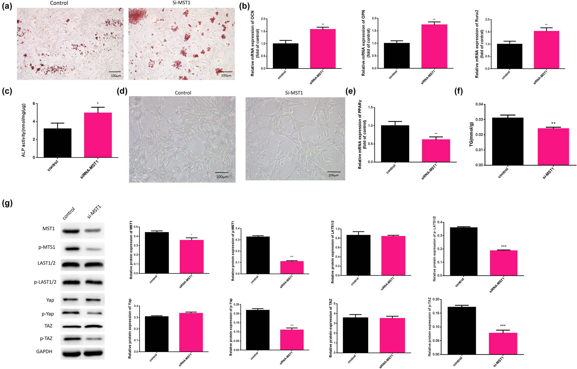
Downregulated MST1 alleviated the effects of osteogenesis and Hippo signaling and enhanced adipogenesis in BMSCs. (a) The calcium nodule form in BMSCs was detected by Alizarin Red S staining analysis. (b) The mRNA expression levels of OCN, OPN, and Runx2 in BMSCs treated with MPS were evaluated by qRT-PCR. (c) The ALP activity in MPS conditions was detected by ALP activity assay. (d) Oil droplets in BMSCs were observed using Oil Red O staining (×100). (e) The mRNA expression of PPARγ was evaluated by qRT-PCR. (b) The content of TG was measured in BMSCs. (f) The content of TG was measured in BMSCs. (g) The protein expression levels of MST1, p-MST1, Yap, p-Yap, LATS1/2, p-LATS1/2, TAZ, and p-TAZ were detected by western blot. *p < 0.05, **p < 0.01, ***p < 0.001. The experiment was repeated three times.
4 Discussion
Necrosis of the femoral head due to pathological changes can result in joint surface collapse and the restriction of joint activity. A number of mechanisms for demonstrating the pathogenesis of steroid-based femoral head necrosis have been identified, including osteogenic differentiation balance disorder [14], fat embolism [15], cycle-blockade [16], cell apoptosis [17], and functional disorder [18]. In this study, we demonstrate that the abnormal control of osteogenesis and adipogenic differentiation in BMSCs plays an important role in the occurrence and development of GC-ONFH. Additionally, the impact of Hippo signaling on GC-ONFH progression was explored, and its potential mechanism was demonstrated.
Although the clinical overdose therapy of GC is related to the occurrence of ONFH, the exact mechanisms are still unclear. In general, bone marrow necrosis was found in GC-ONFH model with increased adipose tissue [19]. Additionally, numerous studies have demonstrated that excessive use of GC can lead to decreased osteogenic differentiation and increased adipogenesis of BMSCs [20,21]. Meanwhile, some reports suggest that BMSCs in GC-ONFH cases show decreased proliferation activity and reduced osteogenic differentiation ability [22]. In brief, these results indicated that GC-ONFH was involved in both BMSC-defective osteogenic and adipogenic differentiation. In our study, we found that BMSCs treated with MPS showed abnormal osteogenic differentiation and adipogenic differentiation, which was consistent with the results of previous studies.
The Hippo signaling pathway is composed of a group of conserved kinases [6]. Previous studies have reported that when Hippo signaling is activated, phosphorylated YAP is replaced in the cytoplasm to promote degradation, while when Hippo signaling is inactivated, nonphosphorylated YAP is transferred to the nucleus to induce the transcriptional activity of genes involved in cell growth [23,24]. DuPont et al. believe that MST1/2 and Lats1/2, as upstream regulatory proteins of TAZ/YAP, balance the phosphorylation of TAZ/YAP during the normal metabolism of Hippo signaling [25]. Runx2 is a transcription factor that stimulates osteogenesis and can bind to the WW domain of TAZ [26]. A prior study suggested that TAZ, a downstream effector molecule of Hippo signaling, could promote the transcription of genes downstream of Runx2 and the expression of osteoblast marker genes ALP, OCN, and OPN [27]. Our results confirmed that the Hippo pathway was activated in BMSCs under MPS treatment and that osteogenesis was increased with Runx2, ALP, OCN, and OPN activation. In recent years, it has been reported that MST1, as the upstream gene of the Hippo signaling pathway, is widely involved in regulating cell differentiation, cell homeostasis, autophagy, and other core proteins regulating Hippo signaling through phosphorylation [28]. Therefore, MST1 is considered to be an important indicator of Hippo signaling pathway activation. As expected, our results also showed that siRNA-MST1 inhibited the activity of Hippo signaling and promoted the osteogenic effects of BMSCs, indicating that osteogenesis in BMSCs might be regulated by Hippo signaling. From an earlier study, BMSCs in GC-ONFH cases showed decreased proliferation activity and reduced osteogenic differentiation ability [21]. In a study by Hong, TAZ was found bind to the adipogenic transcription factor PPARγ and inhibit the transcription of downstream target genes [26]. In our study, we also found that the Hippo pathway was activated in BMSCs under MPS conditions, accompanied by the inhibition of adipogenic differentiation and decreased expression of PPARγ, which can be reversed by siRNA-MST1. Collectively, based on the expression of proteins related to adipogenic and osteogenic differentiation and Hippo signaling-related protein expression, we hypothesized that Hippo signaling is an important signaling pathway involved in the differentiation of adipogenesis and osteogenesis in the pathological process of GC-ONFH.
In conclusion, for the first time, we found that the Hippo signaling pathway increased significantly in BMSCs under MPS conditions, which was consistent with the expression of the adipogenic differentiation-related PPARγ protein and related proteins of osteogenic differentiation, including Runx2, OPN, and OCN. In addition, si-MST1 decreased the adipogenic differentiation of BMSCs and promoted osteogenic differentiation by inhibiting Hippo signaling. Based on the above analysis, we predict that the Hippo signal may act as an important signaling pathway in the regulation of adipogenic and osteogenic differentiation in the presence of GC-ONFH.
Our study had some limitations. First, many signaling pathways regulate BMSCs; however, our present study only investigated the Hippo signaling pathway. We should provide further evidence by analyzing TCGA data to provide more mechanistic evidence as a next step. Second, two or more siRNAs are generally selected to ensure experimental quality, but given the limited experimental time of this study, there was not enough time to supplement the finding with another siRNA experiment. We plan to supplement two or three siRNAs in a subsequent study as a next step. Third, the effects of GC treatment on other Hippo-related signaling pathways downstream of BMSCs were not assessed. In the present study, only osteogenesis and adipogenic differentiation were applied, which were based on data from preliminary studies. Further studies are needed to elucidate the effects of GC treatment.
-
Funding information: The authors state no funding involved.
-
Author contributions: Y.L. wrote the manuscript and explained the data. Y.L. and Z.X. analyzed the data and revised the manuscript, and Z.X. retrieved the literature and conducted experiments. S.C. designed the study. The authors applied the S.D.C. approach for the sequence of authors.
-
Conflict of interest: The authors state no conflict of interest.
-
Data availability statement: The datasets generated during and/or analyzed during the current study are available from the corresponding author on reasonable request.
Appendix
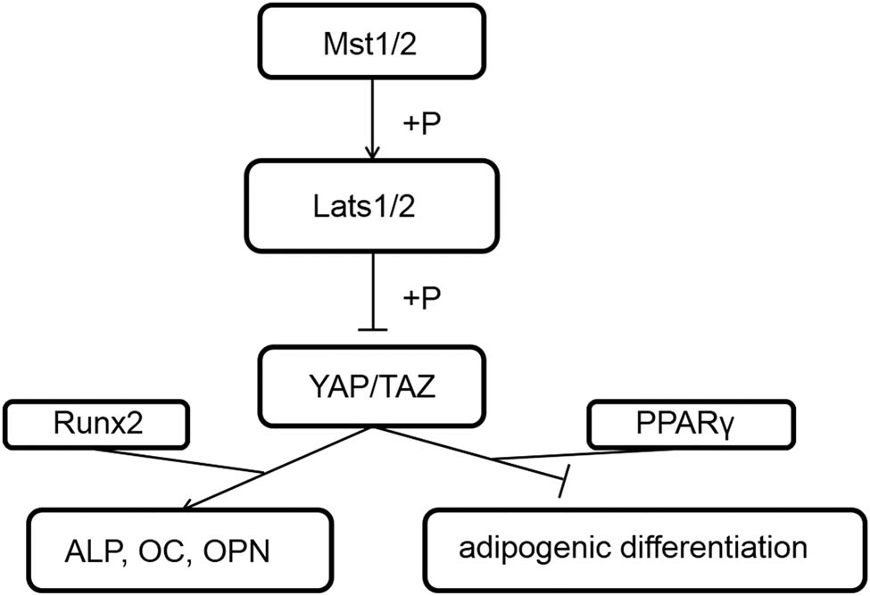
The Hippo signaling pathway schematic diagram.
References
[1] Wang XS, Zhuang QY, Weng XS, Lin J, Qian WW. Etiological and clinical analysis of osteonecrosis of the femoral head in Chinese patients. Chin Med J (Engl). 2013;126(2):290–5.10.3760/cma.j.issn.0366-6999.20120663Search in Google Scholar
[2] Cui L, Zhuang Q, Lin J, Jin J, Zhang K, Cao L, et al. Multicentric epidemiologic study on six thousand three hundred and ninetyfive cases of femoral head osteonecrosis in China. Int Orthop. 2016;40(2):267–76.10.1007/s00264-015-3061-7Search in Google Scholar
[3] Griffin M, Iqbal SA, Bayat A. Exploring the application of mesenchymal stem cells in bone repair and regeneration. J Bone Jt Surg Br. 2011;93(4):427–34.10.1302/0301-620X.93B4.25249Search in Google Scholar
[4] Dragojevič J, Logar DB, Komadina R, Marc J. Osteoblastogenesis and adipogenesis are higher in osteoarthritic than in osteoporotic bone tissue. Arch Med Res. 2011;42(5):392–7.10.1016/j.arcmed.2011.08.005Search in Google Scholar
[5] Compston JE, McClung MR, Leslie WD. Osteoporosis. Lancet. 2019;393(10169):364–76.10.1016/S0140-6736(18)32112-3Search in Google Scholar
[6] Maugeri-Saccà M, De Maria R. The Hippo pathway in normal development and cancer. Pharmacol Ther. 2018;186:60–72.10.1016/j.pharmthera.2017.12.011Search in Google Scholar PubMed
[7] Ou C, Sun Z, He X, Li X, Fan S, Zheng X, et al. Targeting YAP1/LINC00152/FSCN1 signaling axis prevents the progression of colorectal cancer. Adv Sci (Weinh). 2019;7(3):1901380.10.1002/advs.201901380Search in Google Scholar PubMed PubMed Central
[8] Byun MR, Kim AR, Hwang JH, Sung MK, Lee YK, Hwang BS, et al. Phorbaketal A stimulates osteoblast differentiation through TAZ mediated Runx2 activation. FEBS Lett. 2012;586(8):1086–92.10.1016/j.febslet.2012.03.008Search in Google Scholar PubMed
[9] Gu K, Fu X, Tian H, Zhang Y, Li A, Wang Y, et al. TAZ promotes the proliferation and osteogenic differentiation of human periodontal ligament stem cells via the p-SMAD3. J Cell Biochem. 2020;121(2):1101–13.10.1002/jcb.29346Search in Google Scholar PubMed
[10] Hong W, Guan KL. The YAP and TAZ transcription co-activators: key downstream effectors of the mammalian Hippo pathway. Semin Cell Dev Biol. 2012;23(7):785–93.10.1016/j.semcdb.2012.05.004Search in Google Scholar PubMed PubMed Central
[11] Dong L, Li L. Large tumor suppressor gene 2-mediated Hippo signaling pathway reglates the biological behavior of mesenchymal stem cells in vitro. Zhonghua Wei Zhong Bing Ji Jiu Yi Xue. 2019;31(9):1143–8.Search in Google Scholar
[12] Kuang MJ, Zhang KH, Qiu J, Wang AB, Che WW, Li XM, et al. Exosomal miR-365a-5p derived from HUC-MSCs regulates osteogenesis in GIONFH through the Hippo signaling pathway. Mol Ther Nucleic Acids. 2020;23:565–76.10.1016/j.omtn.2020.12.006Search in Google Scholar PubMed PubMed Central
[13] Shahdadfar A, Frønsdal K, Haug T, Reinholt FP, Brinchmann JE. In vitro expansion of human mesenchymal stem cells: choice of serum is a determinant of cell proliferation, differentiation, gene expression, and transcriptome stability. Stem Cell. 2005;23(9):1357–66.10.1634/stemcells.2005-0094Search in Google Scholar PubMed
[14] Li X, Jin L, Cui Q, Wang GJ, Balian G. Steroid effects on osteogenesis through mesenchymal cell gene expression. Osteoporos Int. 2005;16(1):101–8.10.1007/s00198-004-1649-7Search in Google Scholar PubMed
[15] Murata M, Kumagai K, Miyata N, Osaki M, Shindo H. Osteonecrosis in stroke-prone spontaneously hypertensive rats: effect of glucocorticoid. J Orthop Sci. 2007;12(3):289–95.10.1007/s00776-007-1129-ySearch in Google Scholar PubMed
[16] Zou W, Yang S, Zhang T, Sun H, Wang Y, Xue H, et al. Hypoxia enhances glucocorticoid-induced apoptosis and cell cycle arrest via the PI3K/Akt signaling pathway in osteoblastic cells. J Bone Min Metab. 2015;33(6):615–24.10.1007/s00774-014-0627-1Search in Google Scholar PubMed
[17] Zhang WL, Chi CT, Meng XH, Liang SD. miRNA-15a-5p facilitates the bone marrow stem cell apoptosis of femoral head necrosis through the Wnt/β-catenin/PPARγ signaling pathway. Mol Med Rep. 2019;19(6):4779–87.10.3892/mmr.2019.10130Search in Google Scholar PubMed PubMed Central
[18] Nie Z, Chen S, Deng S, Long L, Peng P, Gao M, et al. Gene expression profiling of osteoblasts subjected to dexamethasone-induced apoptosis with/without GSK3β-shRNA. Biochem Biophys Res Commun. 2018;506(1):41–7.10.1016/j.bbrc.2018.10.043Search in Google Scholar PubMed
[19] Wu X, Yang S, Wang H, Meng C, Xu W, Duan D, et al. G-CSF/SCF exert beneficial effects via anti-apoptosis in rabbits with steroid-associated osteonecrosis. Exp Mol Pathol. 2013;94(1):247–54.10.1016/j.yexmp.2012.06.003Search in Google Scholar PubMed
[20] Hao C, Yang S, Xu W, Shen JK, Ye S, Liu X, et al. MiR-708 promotes steroid-induced osteonecrosis of femoral head, suppresses osteogenic differentiation by targeting SMAD3. Sci Rep. 2016;6:22599.10.1038/srep22599Search in Google Scholar PubMed PubMed Central
[21] Pengde K, Fuxing P, Bin S, Jing Y, Jingqiu C. Lovastatin inhibits adipogenesis and prevents osteonecrosis in steroid-treated rabbits. Jt Bone Spine. 2008;75(6):696–701.10.1016/j.jbspin.2007.12.008Search in Google Scholar PubMed
[22] Sun ZB, Wang JW, Xiao H, Zhang QS, Kan WS, Mo FB, et al. Icariin may benefit the mesenchymal stem cells of patients with steroid-associated osteonecrosis by ABCB1-promoter demethylation: a preliminary study. Osteoporos Int. 2015;26(1):187–97.10.1007/s00198-014-2809-zSearch in Google Scholar PubMed
[23] Saucedo LJ, Edgar BA. Filling out the Hippo pathway. Nat Rev Mol Cell Biol. 2007 Aug;8(8):613–21.10.1038/nrm2221Search in Google Scholar PubMed
[24] Sohn BH, Shim JJ, Kim SB, Jang KY, Kim SM, Kim JH, et al. Inactivation of Hippo pathway is significantly associated with poor prognosis in hepatocellular carcinoma. Clin Cancer Res. 2016;22(5):1256–64.10.1158/1078-0432.CCR-15-1447Search in Google Scholar PubMed PubMed Central
[25] Dupont S, Morsut L, Aragona M, Enzo E, Giulitti S, Cordenonsi M, et al. Role of YAP/TAZ in mechanotransduction. Nature. 2011;474(7350):179–83.10.1038/nature10137Search in Google Scholar PubMed
[26] Murakami M, Nakagawa M, Olson EN, Nakagawa O. A WW domain protein TAZ is a critical coactivator for TBX5, a transcription factor implicated in Holt-Oram syndrome. Proc Natl Acad Sci USA. 2005;102(50):18034–9.10.1073/pnas.0509109102Search in Google Scholar PubMed PubMed Central
[27] Hong JH, Hwang ES, McManus MT, Amsterdam A, Tian Y, Kalmukova R, et al. TAZ, a transcriptional modulator of mesenchymal stem cell differentiation. Science. 2005;309(5737):1074–8.10.1126/science.1110955Search in Google Scholar PubMed
[28] Zhao B, Lei QY, Guan KL. The Hippo-YAP pathway: new connections between regulation of organ size and cancer. Curr Opin Cell Biol. 2008;20(6):638–46.10.1016/j.ceb.2008.10.001Search in Google Scholar PubMed PubMed Central
© 2021 Yugang Li et al., published by De Gruyter
This work is licensed under the Creative Commons Attribution 4.0 International License.
Articles in the same Issue
- Biomedical Sciences
- Research progress on the mechanism of orexin in pain regulation in different brain regions
- Adriamycin-resistant cells are significantly less fit than adriamycin-sensitive cells in cervical cancer
- Exogenous spermidine affects polyamine metabolism in the mouse hypothalamus
- Iris metastasis of diffuse large B-cell lymphoma misdiagnosed as primary angle-closure glaucoma: A case report and review of the literature
- LncRNA PVT1 promotes cervical cancer progression by sponging miR-503 to upregulate ARL2 expression
- Two new inflammatory markers related to the CURB-65 score for disease severity in patients with community-acquired pneumonia: The hypersensitive C-reactive protein to albumin ratio and fibrinogen to albumin ratio
- Circ_0091579 enhances the malignancy of hepatocellular carcinoma via miR-1287/PDK2 axis
- Silencing XIST mitigated lipopolysaccharide (LPS)-induced inflammatory injury in human lung fibroblast WI-38 cells through modulating miR-30b-5p/CCL16 axis and TLR4/NF-κB signaling pathway
- Protocatechuic acid attenuates cerebral aneurysm formation and progression by inhibiting TNF-alpha/Nrf-2/NF-kB-mediated inflammatory mechanisms in experimental rats
- ABCB1 polymorphism in clopidogrel-treated Montenegrin patients
- Metabolic profiling of fatty acids in Tripterygium wilfordii multiglucoside- and triptolide-induced liver-injured rats
- miR-338-3p inhibits cell growth, invasion, and EMT process in neuroblastoma through targeting MMP-2
- Verification of neuroprotective effects of alpha-lipoic acid on chronic neuropathic pain in a chronic constriction injury rat model
- Circ_WWC3 overexpression decelerates the progression of osteosarcoma by regulating miR-421/PDE7B axis
- Knockdown of TUG1 rescues cardiomyocyte hypertrophy through targeting the miR-497/MEF2C axis
- MiR-146b-3p protects against AR42J cell injury in cerulein-induced acute pancreatitis model through targeting Anxa2
- miR-299-3p suppresses cell progression and induces apoptosis by downregulating PAX3 in gastric cancer
- Diabetes and COVID-19
- Discovery of novel potential KIT inhibitors for the treatment of gastrointestinal stromal tumor
- TEAD4 is a novel independent predictor of prognosis in LGG patients with IDH mutation
- circTLK1 facilitates the proliferation and metastasis of renal cell carcinoma by regulating miR-495-3p/CBL axis
- microRNA-9-5p protects liver sinusoidal endothelial cell against oxygen glucose deprivation/reperfusion injury
- Long noncoding RNA TUG1 regulates degradation of chondrocyte extracellular matrix via miR-320c/MMP-13 axis in osteoarthritis
- Duodenal adenocarcinoma with skin metastasis as initial manifestation: A case report
- Effects of Loofah cylindrica extract on learning and memory ability, brain tissue morphology, and immune function of aging mice
- Recombinant Bacteroides fragilis enterotoxin-1 (rBFT-1) promotes proliferation of colorectal cancer via CCL3-related molecular pathways
- Blocking circ_UBR4 suppressed proliferation, migration, and cell cycle progression of human vascular smooth muscle cells in atherosclerosis
- Gene therapy in PIDs, hemoglobin, ocular, neurodegenerative, and hemophilia B disorders
- Downregulation of circ_0037655 impedes glioma formation and metastasis via the regulation of miR-1229-3p/ITGB8 axis
- Vitamin D deficiency and cardiovascular risk in type 2 diabetes population
- Circ_0013359 facilitates the tumorigenicity of melanoma by regulating miR-136-5p/RAB9A axis
- Mechanisms of circular RNA circ_0066147 on pancreatic cancer progression
- lncRNA myocardial infarction-associated transcript (MIAT) knockdown alleviates LPS-induced chondrocytes inflammatory injury via regulating miR-488-3p/sex determining region Y-related HMG-box 11 (SOX11) axis
- Identification of circRNA circ-CSPP1 as a potent driver of colorectal cancer by directly targeting the miR-431/LASP1 axis
- Hyperhomocysteinemia exacerbates ischemia-reperfusion injury-induced acute kidney injury by mediating oxidative stress, DNA damage, JNK pathway, and apoptosis
- Potential prognostic markers and significant lncRNA–mRNA co-expression pairs in laryngeal squamous cell carcinoma
- Gamma irradiation-mediated inactivation of enveloped viruses with conservation of genome integrity: Potential application for SARS-CoV-2 inactivated vaccine development
- ADHFE1 is a correlative factor of patient survival in cancer
- The association of transcription factor Prox1 with the proliferation, migration, and invasion of lung cancer
- Is there a relationship between the prevalence of autoimmune thyroid disease and diabetic kidney disease?
- Immunoregulatory function of Dictyophora echinovolvata spore polysaccharides in immunocompromised mice induced by cyclophosphamide
- T cell epitopes of SARS-CoV-2 spike protein and conserved surface protein of Plasmodium malariae share sequence homology
- Anti-obesity effect and mechanism of mesenchymal stem cells influence on obese mice
- Long noncoding RNA HULC contributes to paclitaxel resistance in ovarian cancer via miR-137/ITGB8 axis
- Glucocorticoids protect HEI-OC1 cells from tunicamycin-induced cell damage via inhibiting endoplasmic reticulum stress
- Prognostic value of the neutrophil-to-lymphocyte ratio in acute organophosphorus pesticide poisoning
- Gastroprotective effects of diosgenin against HCl/ethanol-induced gastric mucosal injury through suppression of NF-κβ and myeloperoxidase activities
- Silencing of LINC00707 suppresses cell proliferation, migration, and invasion of osteosarcoma cells by modulating miR-338-3p/AHSA1 axis
- Successful extracorporeal membrane oxygenation resuscitation of patient with cardiogenic shock induced by phaeochromocytoma crisis mimicking hyperthyroidism: A case report
- Effects of miR-185-5p on replication of hepatitis C virus
- Lidocaine has antitumor effect on hepatocellular carcinoma via the circ_DYNC1H1/miR-520a-3p/USP14 axis
- Primary localized cutaneous nodular amyloidosis presenting as lymphatic malformation: A case report
- Multimodal magnetic resonance imaging analysis in the characteristics of Wilson’s disease: A case report and literature review
- Therapeutic potential of anticoagulant therapy in association with cytokine storm inhibition in severe cases of COVID-19: A case report
- Neoadjuvant immunotherapy combined with chemotherapy for locally advanced squamous cell lung carcinoma: A case report and literature review
- Rufinamide (RUF) suppresses inflammation and maintains the integrity of the blood–brain barrier during kainic acid-induced brain damage
- Inhibition of ADAM10 ameliorates doxorubicin-induced cardiac remodeling by suppressing N-cadherin cleavage
- Invasive ductal carcinoma and small lymphocytic lymphoma/chronic lymphocytic leukemia manifesting as a collision breast tumor: A case report and literature review
- Clonal diversity of the B cell receptor repertoire in patients with coronary in-stent restenosis and type 2 diabetes
- CTLA-4 promotes lymphoma progression through tumor stem cell enrichment and immunosuppression
- WDR74 promotes proliferation and metastasis in colorectal cancer cells through regulating the Wnt/β-catenin signaling pathway
- Down-regulation of IGHG1 enhances Protoporphyrin IX accumulation and inhibits hemin biosynthesis in colorectal cancer by suppressing the MEK-FECH axis
- Curcumin suppresses the progression of gastric cancer by regulating circ_0056618/miR-194-5p axis
- Scutellarin-induced A549 cell apoptosis depends on activation of the transforming growth factor-β1/smad2/ROS/caspase-3 pathway
- lncRNA NEAT1 regulates CYP1A2 and influences steroid-induced necrosis
- A two-microRNA signature predicts the progression of male thyroid cancer
- Isolation of microglia from retinas of chronic ocular hypertensive rats
- Changes of immune cells in patients with hepatocellular carcinoma treated by radiofrequency ablation and hepatectomy, a pilot study
- Calcineurin Aβ gene knockdown inhibits transient outward potassium current ion channel remodeling in hypertrophic ventricular myocyte
- Aberrant expression of PI3K/AKT signaling is involved in apoptosis resistance of hepatocellular carcinoma
- Clinical significance of activated Wnt/β-catenin signaling in apoptosis inhibition of oral cancer
- circ_CHFR regulates ox-LDL-mediated cell proliferation, apoptosis, and EndoMT by miR-15a-5p/EGFR axis in human brain microvessel endothelial cells
- Resveratrol pretreatment mitigates LPS-induced acute lung injury by regulating conventional dendritic cells’ maturation and function
- Ubiquitin-conjugating enzyme E2T promotes tumor stem cell characteristics and migration of cervical cancer cells by regulating the GRP78/FAK pathway
- Carriage of HLA-DRB1*11 and 1*12 alleles and risk factors in patients with breast cancer in Burkina Faso
- Protective effect of Lactobacillus-containing probiotics on intestinal mucosa of rats experiencing traumatic hemorrhagic shock
- Glucocorticoids induce osteonecrosis of the femoral head through the Hippo signaling pathway
- Endothelial cell-derived SSAO can increase MLC20 phosphorylation in VSMCs
- Downregulation of STOX1 is a novel prognostic biomarker for glioma patients
- miR-378a-3p regulates glioma cell chemosensitivity to cisplatin through IGF1R
- The molecular mechanisms underlying arecoline-induced cardiac fibrosis in rats
- TGF-β1-overexpressing mesenchymal stem cells reciprocally regulate Th17/Treg cells by regulating the expression of IFN-γ
- The influence of MTHFR genetic polymorphisms on methotrexate therapy in pediatric acute lymphoblastic leukemia
- Red blood cell distribution width-standard deviation but not red blood cell distribution width-coefficient of variation as a potential index for the diagnosis of iron-deficiency anemia in mid-pregnancy women
- Small cell neuroendocrine carcinoma expressing alpha fetoprotein in the endometrium
- Superoxide dismutase and the sigma1 receptor as key elements of the antioxidant system in human gastrointestinal tract cancers
- Molecular characterization and phylogenetic studies of Echinococcus granulosus and Taenia multiceps coenurus cysts in slaughtered sheep in Saudi Arabia
- ITGB5 mutation discovered in a Chinese family with blepharophimosis-ptosis-epicanthus inversus syndrome
- ACTB and GAPDH appear at multiple SDS-PAGE positions, thus not suitable as reference genes for determining protein loading in techniques like Western blotting
- Facilitation of mouse skin-derived precursor growth and yield by optimizing plating density
- 3,4-Dihydroxyphenylethanol ameliorates lipopolysaccharide-induced septic cardiac injury in a murine model
- Downregulation of PITX2 inhibits the proliferation and migration of liver cancer cells and induces cell apoptosis
- Expression of CDK9 in endometrial cancer tissues and its effect on the proliferation of HEC-1B
- Novel predictor of the occurrence of DKA in T1DM patients without infection: A combination of neutrophil/lymphocyte ratio and white blood cells
- Investigation of molecular regulation mechanism under the pathophysiology of subarachnoid hemorrhage
- miR-25-3p protects renal tubular epithelial cells from apoptosis induced by renal IRI by targeting DKK3
- Bioengineering and Biotechnology
- Green fabrication of Co and Co3O4 nanoparticles and their biomedical applications: A review
- Agriculture
- Effects of inorganic and organic selenium sources on the growth performance of broilers in China: A meta-analysis
- Crop-livestock integration practices, knowledge, and attitudes among smallholder farmers: Hedging against climate change-induced shocks in semi-arid Zimbabwe
- Food Science and Nutrition
- Effect of food processing on the antioxidant activity of flavones from Polygonatum odoratum (Mill.) Druce
- Vitamin D and iodine status was associated with the risk and complication of type 2 diabetes mellitus in China
- Diversity of microbiota in Slovak summer ewes’ cheese “Bryndza”
- Comparison between voltammetric detection methods for abalone-flavoring liquid
- Composition of low-molecular-weight glutenin subunits in common wheat (Triticum aestivum L.) and their effects on the rheological properties of dough
- Application of culture, PCR, and PacBio sequencing for determination of microbial composition of milk from subclinical mastitis dairy cows of smallholder farms
- Investigating microplastics and potentially toxic elements contamination in canned Tuna, Salmon, and Sardine fishes from Taif markets, KSA
- From bench to bar side: Evaluating the red wine storage lesion
- Establishment of an iodine model for prevention of iodine-excess-induced thyroid dysfunction in pregnant women
- Plant Sciences
- Characterization of GMPP from Dendrobium huoshanense yielding GDP-D-mannose
- Comparative analysis of the SPL gene family in five Rosaceae species: Fragaria vesca, Malus domestica, Prunus persica, Rubus occidentalis, and Pyrus pyrifolia
- Identification of leaf rust resistance genes Lr34 and Lr46 in common wheat (Triticum aestivum L. ssp. aestivum) lines of different origin using multiplex PCR
- Investigation of bioactivities of Taxus chinensis, Taxus cuspidata, and Taxus × media by gas chromatography-mass spectrometry
- Morphological structures and histochemistry of roots and shoots in Myricaria laxiflora (Tamaricaceae)
- Transcriptome analysis of resistance mechanism to potato wart disease
- In silico analysis of glycosyltransferase 2 family genes in duckweed (Spirodela polyrhiza) and its role in salt stress tolerance
- Comparative study on growth traits and ions regulation of zoysiagrasses under varied salinity treatments
- Role of MS1 homolog Ntms1 gene of tobacco infertility
- Biological characteristics and fungicide sensitivity of Pyricularia variabilis
- In silico/computational analysis of mevalonate pyrophosphate decarboxylase gene families in Campanulids
- Identification of novel drought-responsive miRNA regulatory network of drought stress response in common vetch (Vicia sativa)
- How photoautotrophy, photomixotrophy, and ventilation affect the stomata and fluorescence emission of pistachios rootstock?
- Apoplastic histochemical features of plant root walls that may facilitate ion uptake and retention
- Ecology and Environmental Sciences
- The impact of sewage sludge on the fungal communities in the rhizosphere and roots of barley and on barley yield
- Domestication of wild animals may provide a springboard for rapid variation of coronavirus
- Response of benthic invertebrate assemblages to seasonal and habitat condition in the Wewe River, Ashanti region (Ghana)
- Molecular record for the first authentication of Isaria cicadae from Vietnam
- Twig biomass allocation of Betula platyphylla in different habitats in Wudalianchi Volcano, northeast China
- Animal Sciences
- Supplementation of probiotics in water beneficial growth performance, carcass traits, immune function, and antioxidant capacity in broiler chickens
- Predators of the giant pine scale, Marchalina hellenica (Gennadius 1883; Hemiptera: Marchalinidae), out of its natural range in Turkey
- Honey in wound healing: An updated review
- NONMMUT140591.1 may serve as a ceRNA to regulate Gata5 in UT-B knockout-induced cardiac conduction block
- Radiotherapy for the treatment of pulmonary hydatidosis in sheep
- Retraction
- Retraction of “Long non-coding RNA TUG1 knockdown hinders the tumorigenesis of multiple myeloma by regulating microRNA-34a-5p/NOTCH1 signaling pathway”
- Special Issue on Reuse of Agro-Industrial By-Products
- An effect of positional isomerism of benzoic acid derivatives on antibacterial activity against Escherichia coli
- Special Issue on Computing and Artificial Techniques for Life Science Applications - Part II
- Relationship of Gensini score with retinal vessel diameter and arteriovenous ratio in senile CHD
- Effects of different enantiomers of amlodipine on lipid profiles and vasomotor factors in atherosclerotic rabbits
- Establishment of the New Zealand white rabbit animal model of fatty keratopathy associated with corneal neovascularization
- lncRNA MALAT1/miR-143 axis is a potential biomarker for in-stent restenosis and is involved in the multiplication of vascular smooth muscle cells