Calcineurin Aβ gene knockdown inhibits transient outward potassium current ion channel remodeling in hypertrophic ventricular myocyte
-
Long Yang
 ,
Na Deng
,
Na Deng

Abstract
It has been shown that the activation of calcineurin is involved in regulating ion channel remodeling in hypertrophic cardiomyocytes. But the precise role of calcineurin in the regulation of transient outward potassium current (I to), an ion channel associated with fatal arrhythmia, remains controversial. This study aimed to examine the effects of calcineurin Aβ (CnAβ) gene knockdown on I to channel remodeling and action potential duration (APD) in the hypertrophic ventricular myocytes of neonatal rats. Results showed that phenylephrine stimulation caused hypertrophy of ventricular myocytes, upregulation of CnAβ protein expression, downregulation of Kv4.2 mRNA and protein expression, a decrease in I to current density, and prolongation of APD. CnAβ gene knockdown significantly inhibited the effects of phenylephrine stimulation. Our data indicate that CnAβ gene knockdown can inhibit I to channel remodeling and APD prolongation in hypertrophic neonatal rat ventricular myocytes. This finding suggests that calcineurin may be a potential target for the prevention of malignant ventricular arrhythmia in a hypertrophic heart.
1 Introduction
Ventricular remodeling caused by pathological cardiac hypertrophy is chronic congestive heart failure’s most important pathophysiological mechanism [1,2]. Sudden cardiac death is the dominant reason of death in patients with congestive heart failure [3]. Ion channel remodeling in ventricular myocytes is the main pathophysiological basis leading to changes in the action potential duration (APD) of ventricular myocytes, resulting in malignant ventricular arrhythmia [4,5]. Congenitally, dysregulation of transient outward potassium current (I to) has also been demonstrated to play a pivotal role in the Brugada syndrome [6,7].
I to is a rapidly activated and inactivated outward potassium current, mainly involved in phase 1 of action potentials. The activity of I to channels influences the activation of voltage-gated Ca2+ channels and the balance of inward and outward currents during the plateau, thereby mediating the duration and the amplitude of phase 2. In cultured hypertrophic ventricular myocytes, or the ventricular myocytes from a myocardial infarction heart, downregulated or dysfunctional I to channels of ventricular myocytes lead to delayed repolarization and prolonged APD, which may easily cause fatal arrhythmia [8,9].
Calcineurin, a Ca2+-calmodulin-regulated phosphatase, has been shown to participate in hypertrophic signal transduction. Research have shown that the continuous activation of calcineurin promotes cardiac hypertrophic remodeling, decompensated heart failure, and arrhythmic death [10,11]. Additionally, calcineurin regulates ion channel remodeling in hypertrophic cardiomyocytes [9,12,13,14,15].
In transgenic mice, overexpression of calcineurin results in cardiac hypertrophy and the downregulation of I to, which was reversed by the calcineurin inhibitor cyclosporine [13]. In rats after myocardial infarction, cyclosporine significantly attenuated the decreases in mRNA levels of Kv4.2 and Kv4.3, the components of α subunit in I to channel, and I to density in the left ventricular [14]. In cultured adult canine left ventricular cardiomyocytes, rapid pacing reduced I to density and Kv4.3 mRNA and protein expression, which was markedly prevented by inhibiting calcineurin with cyclosporine [15]. Those results indicated that the activation of calcineurin may lead to I to downregulation. Conversely, there was also evidence in cultured neonatal rat ventricular myocytes that the overexpression of constitutive calcineurin upregulates Kv4.2 expression without affecting Kv4.3 [9]. Thus, the precise role of calcineurin in the regulation of I to remains unclear.
The purpose of this study is to clarify the regulatory effect of calcineurin on I to channel remodeling and APD alterations in the hypertrophic ventricular myocytes of rats by way of knockdown of calcineurin-related genes.
2 Materials and methods
2.1 Materials
The Axopatch 700B patch clamp amplifier and Digidata 1322 data converter were from Axon (USA). The Sutter P-97 microelectrode puller was from Sutter (USA) and the BJ-40 glass microelectrode was from Beijing Zhengtianyi Electronics (China). Trypsin and collagenase (type II) were purchased from Sigma (USA). High-glucose Dulbecco’s modified Eagle’s medium (DMEM), premium fetal bovine serum (FBS), phenylephrine (PE), and 5-bromo-2-deoxyuridine (5-BrdU) were purchased from Gibco-BRL (USA). The following antibodies were used: rabbit anti-calcineurin Aβ (CnAβ) antibody (Merck Millipore, Germany), α striated muscle sarcomere actin (α-SCA) antibody (Sigma), horseradish peroxidase-conjugated anti-rabbit antibody (Santa Cruz, USA), rabbit anti-rat Kv4.2 antibody (Abcam, USA), and mouse-derived anti-rat glyceraldehyde-3-phosphate dehydrogenase (GAPDH) antibody (Shanghai Kangcheng Biotech, China). The Genomic DNA Purification Kit was purchased from Fermentas (Canada). The RNeasy Mini Kit was purchased from Qiagen (China). Ad-CnAβshRNA, the gene mediated by the recombinant adenovirus shRNA interference vector for silencing the A subunit β subtype of CaN, and the empty viral vector (null) were prepared by HanBio (Shanghai, China).
2.2 Isolation and culture of ventricular myocytes from neonatal rats
One-day-old neonatal Sprague-Dawley rats of clean grade were provided by the Animal Center of Peking University Health Science Center (license number: SYXK [Beijing] 2011-0039). The rats were sacrificed under anesthesia and the animals were fully immersed in 75% ethanol. The ventricle was harvested, the ventricular tissue was cut into small pieces, and the cells were isolated by enzymatic hydrolysis (final concentration of trypsin is 0.1%; the final concentration of type II collagenase is 0.03%). Ventricular myocytes were obtained by differential adherence and 5-BrdU affinity purification. The cells were cultured in high-glucose DMEM containing 10% FBS at 37°C in a 5% CO2 incubator. After 48 h, the cells were cultured with serum-free DMEM with high glucose for the next experiment.
-
Ethical approval: The research related to animal use has complied with all the relevant national regulations and institutional policies for the care and use of animals and has been approved by the Ethics Committee of Guizhou Provincial People’s Hospital (Hospital Ethics Review [2012] No. 001).
2.3 Identification of ventricular myocytes
Cells were cultured on fibrin-coated glass slides for 48 h and α-SCA was detected by immunofluorescence staining. Neonatal rat cardiac fibroblasts were used as a negative control (Figure A).
2.4 Ad-CnAβshRNA sequence screening
Primary ventricular myocytes were cultured for 48 h. Ad-CnAβshRNA1 (A1, interference base sequence 5′-3′: CAGAAAGGGTCTATGAAGCTTGTAT), Ad-CnAβshRNA2 (A2, interference base sequence 5′-3′: CCGCCAGTTTAACTGTTCTCCACAT), Ad-CnAβshRNA3 (A3, interference base sequence 5′-3′: GCAAGATGGCAAGAGTCTTCT), and null at a multiplicity of infection (MOI) of 50 were selected to infect cultured ventricular myocytes for 48 h. CnAβ protein expression in the ventricular myocytes of each group was detected by western blotting. The Ad-CnAβshRNA corresponding to the lowest CnAβ protein expression was regarded as the optimal Ad-CnAβshRNA. A1 caused the most obvious decrease in CnAβ protein expression after its infection of ventricular myocytes at 48 h, and thus it was used in subsequent experiments (Figure A2).
2.5 Grouping and interventions
Ventricular myocytes were cultured for 48 h and then cultured in serum-free DMEM. These cells were divided into four groups. (1) In the null group, an adenovirus empty vector at an MOI of 50 was added. After 6 h of infection, the cells were cultured in two volumes of fresh serum-free DMEM for 48 h. (2) In the null + PE group, an adenovirus empty vector at an MOI of 50 was added. After 6 h of infection, the cells were cultured in two volumes of fresh serum-free DMEM with a total of 100 μM of PE (Gibco-BRL, USA) for 48 h. (3) In the A1 group, A1 at an MOI of 50 was added. After 6 h of infection, the cells were cultured in two volumes of fresh serum-free DMEM for 48 h. (4) In the A1 + PE group, A1 at an MOI of 50 was added. After 6 h of infection, the cells were cultured in two volumes of fresh serum-free DMEM with a total of 100 μM of PE for 48 h.
2.6 Determining the effectiveness of intervention on hypertrophy in cultured cells
Cell hypertrophy was identified by the measurement of brain natriuretic peptide (BNP) mRNA expression and cell size after 48 h of intervention. Upon completion of cell grouping and intervention, real-time reverse transcription-polymerase chain reaction (RT-PCR) was conducted to determine BNP mRNA expression in ventricular myocytes. Cells were cultured on glass slides. Crystal violet staining assay was performed after grouping and intervention. Three fields of view were randomly selected in each group. The surface area of the cells was assessed using Image J software.
2.7 RT-qPCR
Total RNA was extracted from ventricular myocytes using the RNeasy Mini Kit according to the manufacturer’s instructions. A total of 1 µg of RNA was reverse transcribed using random hexamers from a first-strand cDNA synthesis kit. The mRNAs expression was measured by the real-time PCR method. The cycling conditions were as follows: 95°C for 10 min, followed by 40 cycles of 15 s at 95°C, and 30 s at 60°C. mRNA levels were normalized to GAPDH and determined using the 2-ΔΔCq method. Primers were designed using the Oligo 6.0 software. Specific primers were used to identify and amplify BNP (sense primer: 5′-GTCTCCAGAACAATCCACGA-3′; antisense primer: 5′-CTAAAACAACCTCAGCCCGT-3′), Kv4.2 (sense primer: 5′- GCTCTTCAGCAAGCAAGTTC-3′; antisense primer: 5′- TCCGACTGAAGTTAGACACG-3′), and GAPDH (sense primer: 5′- TGATGACATCAAGAAGGTGGTGAAG-3′; antisense primer: 5′- TCCTTGGAGGCCATGTAGGCCAT-3′).
2.8 Western blotting
Total protein (40 μg) was loaded and then transferred to a nitrocellulose membrane after electrophoresis. The membrane was blocked with 5% skim milk for 1 h. Rabbit anti-rat GAPDH antibody (1:1,000), rabbit anti-rat rabbit anti-CnAβ antibody (1:500), and rabbit anti-rat rabbit anti-rat Kv4.2 antibody (1:1,000) were added before overnight incubation at 4°C. After thorough rinsing, horseradish peroxidase-labeled secondary antibody was added for incubation at room temperature for 1 h. After thorough rinsing again, color development, photography, and quantitative measurement of the gray scale were performed.
2.9 Whole-cell patch clamp detection
A glass microelectrode formed a high resistance seal with the cells and ruptured the membrane. I to was recorded under the voltage clamp mode. Current density analysis was used (current density [pA/pF] = current intensity/capacitance) to avoid errors caused by cell size. The action potential of the individual cells was recorded under the current clamp mode. The current signal was guided by an Ag/AgCl electrode, amplified by a patch clamp AXON 700B amplifier through an AD/DA converter board, and stored in a computer hard disk. During the experimental procedure, stimulation discharge and signal acquisition were controlled by pCLAMP 10.0 software.
In the I to depolarization step, the clamping voltage was set to −80 mV with an −40 mV to +70 mV pulse stimulation series, with a step voltage of 10 mV, wave width of 300 ms, and frequency of 0.2 Hz. The I to steady-state activation curve stimulation protocol was as follows. The clamping voltage was set to −80 mV with a −40 to + 70 mV pulse stimulation series, with a step voltage of 10 mV and a wave width of 300 ms. The I to current was then recorded. The I to steady-state inactivation curve stimulation protocol was as follows. The clamping voltage was set to −80 mV with a −40 mV to +50 mV pulse stimulation series, with a step voltage of 10 mV and a wave width of 1,000 ms. The residual current was then recorded. The steady-state activation and inactivation curves were created using the normalized current values obtained by the two above-mentioned stimulation schemes as the ordinate and the stimulation pulses under different voltages as the abscissa. The semi-activated voltage (V 1/2,act) and semi-inactivated voltage (V 1/2,inact) were calculated by curve fitting using the Boltzmann equation (I/I max = 1/{1 + exp[(V 1/2 − V m)/k]}).
The action potential recording method was similar to the voltage clamp mode. After membrane sealing, membrane breaking, and compensation, the recording was switched to the current clamp mode. A 1 nA current pulse was then applied, with a wave width of 2.5 ms, which induced the action potential in the ventricular myocytes. The APDs at 20, 50, and 90% repolarization (APD20, APD50, and APD90) were recorded and analyzed.
2.10 Statistical analysis
Statistical analysis was performed using the Graphpad Prism 6 software. All data are expressed as mean ± SD. Differences among groups were compared by one-way analysis of variance, and the q test was used for comparison of the two groups. A P value of <0.05 was considered statistically significant.
3 Results
3.1 Effectiveness of stimulation on BNP mRNA expression and the surface area of ventricular myocyte
In the null + PE group, PE treatment for 48 h significantly upregulated BNP mRNA expression by an average of 2.24 times (P < 0.01), and significantly increased the surface area ((1,360 ± 90) µm2 vs (700 ± 40) µm2, P < 0.01) of ventricular myocytes compared with the null group. Therefore, PE intervention led to hypertrophy of ventricular myocytes. The BNP mRNA expression in the A1 + PE group was markedly attenuated compared with that in the null + PE group (1.15 ± 0.09 vs 2.24 ± 0.48, P < 0.05), as well as the surface area of cells ((680 ± 180) µm2 vs (1,360 ± 90) µm2, P < 0.01), indicating that the cellular hypertrophy induced by PE was significantly inhibited by Ad-CnAβshRNA intervention (Figures 1 and 2, and Table 1).
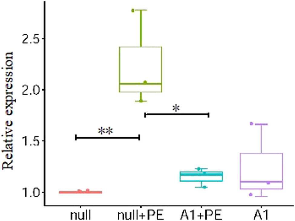
PE stimulation promoted the BNP mRNA expression in ventricular myocytes which was attenuated by pre-treatment with Ad-CnAβshRNA1. Bar chart showed the quantitative analysis results of BNP mRNA by real-time reverse transcription-polymerase chain reaction. *P < 0.05; n = 3. PE: Phenylephrine. A1: Ad-CnAβshRNA.
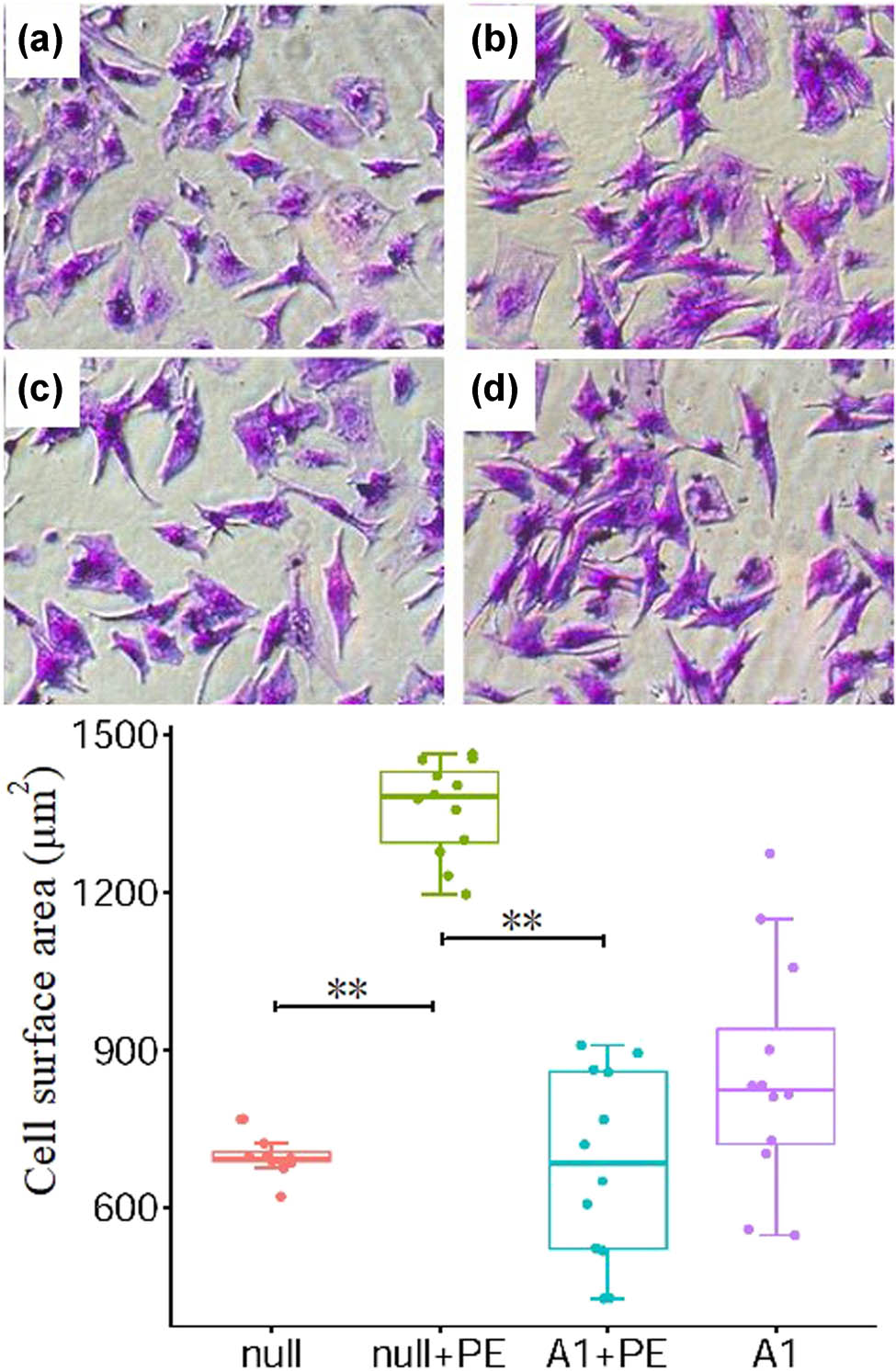
PE stimulation promoted cell hypertrophy which was attenuated by pre-treatment with Ad-CnAβshRNA1. Color picture showed the ventricular cells stained with crystal violet in four groups. a, b, c, and d are the null, null + PE, A1 + PE, and A1 groups, respectively (×100). The histogram showed the comparative results of cell area in groups. *P < 0.05, **P < 0.01. PE: Phenylephrine. A1: Ad-CnAβshRNA.
Comparison of the expression of mRNAs and proteins, and the cell area in each group (͞x ± s)
| n | Null | Null + PE | A1 + PE | A1 | |
|---|---|---|---|---|---|
| BNP mRNA | 3 | 1** | 2.24 ± 0.48 | 1.15 ± 0.09* | 1.24 ± 0.37 |
| Cell area (µm2) | 12 | 700 ± 40** | 1360 ± 90 | 680 ± 180** | 850 ± 220 |
| CnAβ protein | 3 | 1** | 2.29 ± 0.24 | 0.90 ± 0.12** | 1.18 ± 0.07 |
| Kv4.2 mRNA | 5 | 1* | 0.62 ± 0.07 | 1.35 ± 0.18** | 1.2 ± 0.21 |
| Kv4.2 protein | 4 | 1* | 0.58 ± 0.11 | 0.88 ± 0.09* | 1.05 ± 0.2 |
The expression of mRNAs and proteins were exhibited as relative data. Compared with null + PE group, *P < 0.05, **P < 0.01. PE: phenylephrine. A1: Ad-CnAβshRNA. BNP: brain natriuretic peptide. CnAβ: calcineurin Aβ.
3.2 Effect of Ad-CnAβshRNA intervention on PE-induced CnAβ protein expression in ventricular myocytes
In the null + PE group, PE treatment significantly upregulated CnAβ protein expression by an average of 2.29 times (P < 0.01) when compared with the null group. In contrast, CnAβ protein expression was significantly lower in the A1 + PE group than in the null + PE group (0.90 ± 0.12 vs 2.29 ± 0.24, P < 0.01) (Figure 3 and Table 1).
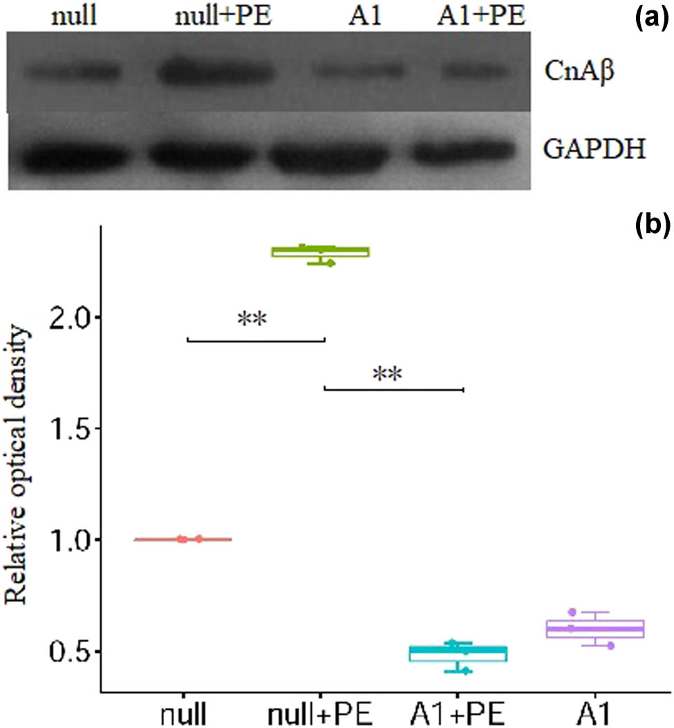
PE stimulation increased CnAβ protein expression which was attenuated by pre-treatment with Ad-CnAβshRNA1. (a) The representative picture of CnAβ protein detected by Western blotting. (b) The semiquantitative analysis results of CnAβ protein in groups. **P < 0.01, n = 3. CnAβ: Calcineurin Aβ. PE: Phenylephrine. A1: Ad-CnAβshRNA.
3.3 Effect of Ad-CnAβshRNA intervention on PE-induced Kv4.2 mRNA and protein expression in ventricular myocytes
In the null + PE group, PE treatment significantly downregulated Kv4.2 mRNA (1 vs 0.62 ± 0.07, P < 0.05) and protein (1 vs 0.58 ± 0.11, P < 0.05) expression when compared to the null group. In contrast, Kv4.2 mRNA (1.35 ± 0.18 vs 0.62 ± 0.07, P < 0.01) and protein (0.88 ± 0.09 vs 0.58 ± 0.11, P < 0.05) expression was significantly higher in the A1 + PE group than in the null + PE group (Figure 4 and Table 1).
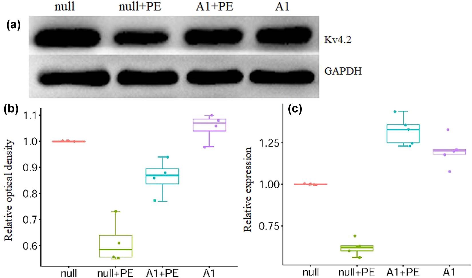
PE intervention inhibited the protein and mRNA expression of Kv4.2 which was attenuated by pre-treatment with Ad-CnAβshRNA1. (a) The representative picture of Kv4.2 protein detected by western blotting. (b and c) The semiquantitative analysis results of Kv4.2 protein (n = 4) and mRNA (n = 5) in groups, respectively. *P < 0.05, **P < 0.01. PE: Phenylephrine. A1: Ad-CnAβshRNA.
3.4 Effect of Ad-CnAβshRNA intervention on I to in ventricular myocytes
At a stimulation voltage of +20 to +70 mV, I to current density in the null + PE group was significantly lower than that in the null group (Table 2), the averaged current–voltage (I–V) curve relations of I to remarkably shifted downward, and the peak current density was decreased by 49% (P < 0.05; Figure 5). The A1 + PE group had significantly higher I to current density than the null + PE group (P < 0.05; Figure 5 and Table 2). The shape and distribution of the activation curves of I to were similar among the different groups (Figure 6a), and the V 1/2,act showed no significant difference among the groups (P > 0.05; Figure 6b and Table 3). The inactivation curve of I to was significantly shifted to the left in the null + PE group when compared with the null group and was significantly shifted to the right in the A1 + PE group compared to the null + PE group (Figure 6c). The V 1/2,inact was significantly lower in the null + PE group than in the null group (P < 0.05) and significantly higher in the A1 + PE group than in the null + PE group (P < 0.05; Figure 6d and Table 3). Therefore, stimulation of ventricular myocytes with PE accelerated the inactivation of I to, whereas Ad-CnAβshRNA1 intervention inhibited such an effect.
Comparison of current densities (pA/pF) at different voltages in each group (͞x ± s)
| n | 20 mV | 30 mV | 40 mV | 50 mV | 60 mV | 70 mV | |
|---|---|---|---|---|---|---|---|
| Null | 14 | 7.83 ± 1.04 | 11.03 ± 1.15 | 14.28 ± 1.22 | 17.48 ± 1.45 | 20.99 ± 1.76 | 24.02 ± 1.86 |
| Null + PE | 12 | 5.04 ± 1.24 | 6.27 ± 1.18 | 7.80 ± 1.21 | 9.10 ± 1.25 | 10.89 ± 1.51 | 12.27 ± 1.88 |
| A1 + PE | 12 | 6.38 ± 0.79 | 8.45 ± 0.79 | 10.46 ± 0.88 | 12.33 ± 1.12 | 14.65 ± 1.24 | 16.85 ± 1.38 |
| A1 | 12 | 7.41 ± 1.10 | 9.86 ± 1.06 | 12.76 ± 0.86 | 15.38 ± 0.51 | 18.51 ± 0.64 | 21.15 ± 0.89 |
At a stimulation voltage of 20–70 mV: null vs null + PE, P < 0.05; A1 + PE vs null + PE, P < 0.05. PE: phenylephrine. A1: Ad-CnAβshRNA.

Densities of transient outward potassium current (I to) in ventricular myocytes. (a) A typical I to recording from isolated ventricular myocytes in groups. (b) Current–voltage (I–V) curve relations. At a stimulation voltage of +20 to +70 mV, PE intervention significantly decreased I to density (P < 0.05), which was markedly attenuated by pre-treatment with Ad-CnAβshRNA1 (P < 0.05; the cell numbers are 14, 12, 12, and 12 in null, null + PE, A1 + PE and A1 groups, respectively). PE: Phenylephrine. A1: Ad-CnAβshRNA.

The activation and inactivation curves of transient outward potassium current (I to). (a) The steady-state activation curve of I to. (b) The semi-activated voltage (V 1/2,act) of I to. There were no significant differences of V 1/2, act among groups (P > 0.05; the cell numbers are 14, 12, 12, and 9 in null, null + PE, A1 + PE, and A1 groups, respectively). (c) The steady-state inactivation curve of I to. (d) The semi-inactivated voltage (V 1/2,inact) of I to. PE stimulation accelerated inactivation of I to, which was inhibited by Ad-CnAβshRNA1 intervention (*P < 0.05; n = 10 in each group). PE: Phenylephrine. A1: Ad-CnAβshRNA.
Comparison of V 1/2 of the activation curves and inactivation curves in each group (͞x ± s)
| n act | V 1/2,act (mV) | n inact | V 1/2,inact (mV) | |
|---|---|---|---|---|
| Null | 14 | 31.57 ± 2.38 | 10 | −16.34 ± 1.07 |
| Null + PE | 12 | 28.21 ± 4.47 | 10 | −28.15 ± 1.00* |
| A1 + PE | 12 | 30.21 ± 2.91 | 10 | −21.01 ± 1.07 |
| A1 | 9 | 30.72 ± 3.51 | 10 | −18.07 ± 1.34 |
Compared with null group or A1 + PE group, *P < 0.05. PE: phenylephrine.
A1: Ad-CnAβshRNA. V 1/2,act: semi-activated voltage. V 1/2, inact: semi-inactivated voltage.
n act and n inact: cell number in each group enrolled testing of V 1/2,act or V 1/2,inact.
3.5 Effect of Ad-CnAβshRNA intervention on APD in ventricular myocytes
APD20, APD50, and APD90 were significantly longer in the null + PE group than in the null group and were significantly shorter in the A1 + PE group than in the null + PE group (Figure 7 and Table 4). Therefore, PE intervention led to significant prolongation of APD in ventricular myocytes, whereas Ad-CnAβshRNA intervention attenuated such an effect.

Action potentials of ventricular myocytes. (a) Representative action potentials of cultured ventricular myocytes. (b–d) The analysis results of the APD20, APD50, and APD90 in groups. PE stimulation prolonged the APD, which was inhibited by Ad-CnAβshRNA1 intervention. **P < 0.001, *P < 0.05; the cell numbers are 12, 12, 12, and 11 in null, null + PE, A1 + PE, and A1 groups, respectively. PE: Phenylephrine. A1: Ad-CnAβshRNA.
Comparison of APD (ms) in each group (͞x ± s)
| n | APD20 | APD50 | APD90 | |
|---|---|---|---|---|
| Null | 12 | 1.31 ± 0.26** | 15.08 ± 2.65** | 126.66 ± 35.55** |
| Null + PE | 12 | 2.30 ± 0.41 | 21.75 ± 3.24 | 216.44 ± 39.43 |
| A1 + PE | 12 | 1.87 ± 0.42* | 17.25 ± 1.19** | 136.41 ± 17.66** |
| A1 | 11 | 1.33 ± 0.35 | 15.30 ± 1.98 | 129.48 ± 27.51 |
Compared with null group or A1 + PE group, *P < 0.05, **P < 0.01. PE: phenylephrine, A1: Ad-CnAβshRNA. APD: action potential duration.
4 Discussion
The reduction of I to slows the repolarization of the action potential in the first phase and reduces the depth of the phase 1 notch, thus affecting the activity of other ion channels. The Kv4.3 channel is expressed in human left ventricular muscle and shows a gradient of protein across the ventricular wall, thus forming electrophysiological transmural heterogeneity [16,17]. In the hypertrophic ventricular myocardium in animal models of cardiac hypertrophy or patients with organic heart disease, especially in patients with co-existing heart failure and myocardial injury, the expression of Kv4.3 and Kv4.2 in ventricular myocytes is downregulated and the activity of I to channels is reduced. This leads to abnormal early repolarization, repolarization delay, and APD prolongation, which may easily cause fatal ventricular arrhythmias [8,9,18,19].
The molecular structure of the I to ion channel includes a pore-forming α subunit and an auxiliary β subunit. The α subunit of I to channel has two functionally distinct components. The fast component, I to,fast (I to,f), for example, recovers from inactivation very rapidly with time constants in the range of 60–100 ms [20,21]. In contrast, the slow component, I to,slow (I to,s), recovers from inactivation slowly with time constants on the order of seconds [22,23]. Usually, I to is referred to the I to,fast. The fast component of α subunit is formed by assembly of Kv4.2 subunits, Kv4.3 subunits, or a combination of the two, which shows the heterogeneity of the species and the different regions of the same heart [23,24,25,26,27].
Selective gene silencing using antisense oligonucleotides (AsODNs) targeted against Kv4.2 and Kv4.3 reduced I to,f in cultured rodent ventricular myocytes [28,29]. In rat atrial myocytes, AsODNs targeted against Kv4.2, but not Kv4.3, attenuated I to,f [30], whereas in human atrial myocytes, I to,f was significantly attenuated by Kv4.3, but not by Kv4.2 and AsODNs [31]. In addition, targeted gene deletion of Kv4.2 in mice eliminates ventricular I to,f, further revealing the critical role of Kv4.2 in the generation of I to,f channels in rodents [32]. So, in this study, Kv4.2 was chosen as the research component of I to channel other than Kv4.3.
Ventricular hypertrophy is an effective compensation for chronic heart overload, but eventually develops into congestive heart failure because of decompensation following ventricular remodeling [1,2]. Remodeling of numerous ion channels in ventricular myocytes during this process is an important basis for malignant ventricular arrhythmias. Many cell signaling factors are involved in regulating ion channel remodeling in hypertrophic cardiomyocytes. Promoting calcineurin mRNA and protein expression and enhancing calcineurin activity can promote hypertrophy of cardiomyocytes and participate in regulating multiple ion channel remodeling in cardiomyocytes [10,12,13,29]. In rat models of myocardial infarction, the calcineurin inhibitor cyclosporin A can significantly inhibit ventricular remodeling and hypertrophy, improve diastolic function, inhibit a decrease in I to current density in ventricular myocytes, and downregulate Kv4.2 and Kv4.3 mRNA and protein expression [14]. In ventricular myocytes of mouse myocardial infarction models, I to current density is decreased, and Kv4.2 and Kv4.3 mRNA and protein expression is downregulated [8]. Furthermore, the β-blocker metoprolol, the calcineurin inhibitor cyclosporin A, and knockout of calcineurin-specific downstream signaling factors (i.e., NFAT4 gene, a member of the nuclear factor of activated T cells [NFAT] family) can significantly inhibit a decrease in I to current density and inhibit changes in Kv4.2 and Kv4.3 mRNA and protein expression. In hypertrophic neonatal rat cardiomyocytes, activation of the calcineurin-NFAT signal upregulates the transcriptional expression of Kv4.2 mRNA and protein and increases I to current density [9]. In canine ventricular myocytes with simulated ventricular tachycardia, Kv4.3 mRNA and protein expression is downregulated, and I to current density is significantly reduced, which manifests as significant inhibition of cyclosporine [15].
In this study, the knockdown of the CnAβ gene, which encoded the main functional unit of calcineurin, completely inhibited CnAβ protein expression, which defected in the substance base that enables calcineurin to function. Therefore, we evaluated the effect of completely suppressed calcineurin activity on I to. When the stimulation voltage was between +20 and +70 mV, intervention with the conventional α1 adrenergic receptor agonist PE significantly reduced I to current density in the ventricular myocytes of neonatal rats. The peak current density decreased by 49%, and the I–V curves of I to remarkably shifted downward. Furthermore, APD20, APD50, and APD90 were significantly prolonged. Knockdown of the CnAβ gene significantly inhibited the effect of PE intervention on I to current density and APD. Moreover, PE stimulation did not affect the activation of I to, but PE reduced I to current density by accelerating its inactivation. Additionally, the knockdown of the CnAβ gene inhibited the effect of PE on I to inactivation.
The results of earlier studies showed that the precise role of calcineurin in the regulation of I to remains unclear [9,13,14,15]. In those studies, calcineurin activity was promoted by the agonist or transgenic method, and was depressed by cyclosporine [9,13,14,15]. As per our knowledge, this study is the first one to assess the role of calcineurin in the regulation of I to by way of calcineurin gene silence. Our results potently showed that calcineurin is a negative regulator of I to activity in ventricular myocytes from neonatal rats.
In summary, our findings indicated that CnAβ gene knockdown can inhibit PE-induced I to channel remodeling and APD prolongation in hypertrophic neonatal rat ventricular myocytes. This finding suggested that calcineurin may be a potential target for the prevention of malignant ventricular arrhythmia in hypertrophic hearts. However, the study had some limitations. Many ion channels are involved in the formation of action potential in ventricular myocytes. However, this study only detected the I to ion channel. Moreover, as one of the coding genes of I to ion channel, the expression of Kv4.3 was not detected.
-
Funding information: This study was supported by the Guizhou Provincial Science and Technology Fund (Grant No. (2019) 1205), and the Clinical Research Center Project of the Department of Science and Technology of Guizhou Province (NO. (2017)5405).
-
Author contributions: Long Yang, Na Deng, and Jionghong He designed the research and carried out the experiment. Na Deng and Jionghong He completed data analysis and manuscript writing. Long yang completed the final revision and finalization of the manuscript. Guiling Xia, Ying Yang, Yidong Zhao, Zhaomei Huo, and Chuxian Guo participated in the experiment in varying degrees.
-
Conflict of interest: The authors state no conflict of interest.
-
Data availability statement: The datasets generated during and/or analyzed during the current study are available from the corresponding author on reasonable request.
Appendix
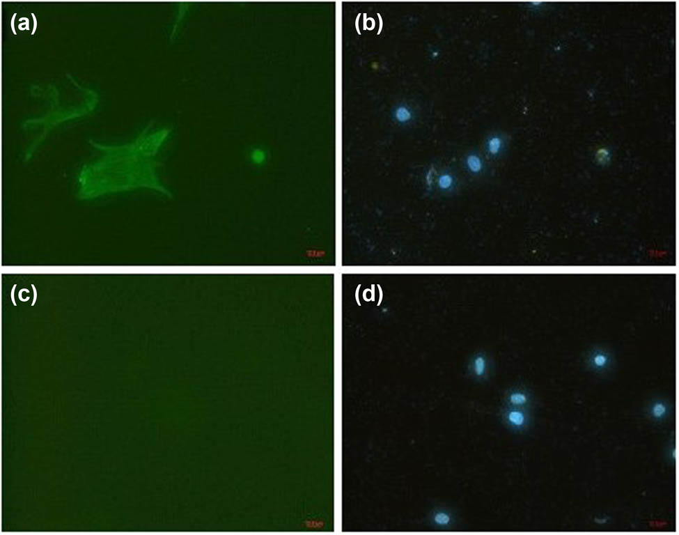
Identification of ventricular myocytes. After 48 h of culture, myocardial cells were demonstrated by immunofluorescent staining with α striated muscle sarcomere actin (α-SCA) antibody that is specialized to striated muscle. (a) Cells appearing green were identified as myocardial cells. (c) Staining for fibroblasts was negative. (b and d) The blue was nuclei stained with diamidine phenylindole. Panels (a) and (b), (c), and (d) are the same view, respectively (×200).
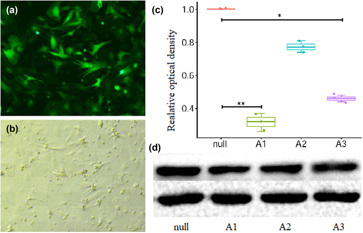
The results of Ad-CnAβshRNA sequence screening. A1 caused the most obvious decrease in the A subunit β subtype of calcineurin (CnAβ) protein expression after its infection of ventricular myocytes for 48 h. (a) Cells appearing green were identified as cells infected by recombinant adenovirus. (b) The photo taken in bright background at the same view of (a). (c) The semiquantitative analysis results of CnAβ protein. *P < 0.05, **P < 0.01; n = 3. (d) The representative picture of CnAβ protein detected by western blotting. A1: Ad-CnAβshRNA1. A2: Ad-CnAβshRNA2. A3: Ad-CnAβshRNA3.
References
[1] Oldfield CJ, Duhamel TA, Dhalla NS. Mechanisms for the transition from physiological to pathological cardiac hypertrophy. Can J Physiol Pharmacol. 2020;98(2):74–84.10.1139/cjpp-2019-0566Suche in Google Scholar
[2] Shimizu I, Minamino T. Physiological and pathological cardiac hypertrophy. J Mol Cell Cardiol. 2016;97:245–62.10.1016/j.yjmcc.2016.06.001Suche in Google Scholar
[3] Huang D, Hua W, Fang Q, Yan J, Su Y, Liu B, et al. POSCD-China Biventricular pacemaker and defibrillator implantation in patients with chronic heart failure in China. ESC Heart Fail. 2021;8(1):546–54.10.1002/ehf2.13114Suche in Google Scholar
[4] Wang Y, Hill JA. Electrophysiological remodeling in heart failure. J Mol Cell Cardiol. 2010;48(4):619–32.10.1016/j.yjmcc.2010.01.009Suche in Google Scholar
[5] Kepenek ES, Ozcinar E, Tuncay E, Akcali KC, Akar AR, Turan B. Differential expression of genes participating in cardiomyocyte electrophysiological remodeling via membrane ionic mechanisms and Ca2+-handling in human heart failure. Mol Cell Biochem. 2020;463(1–2):33–44.10.1007/s11010-019-03626-4Suche in Google Scholar
[6] David JP, Lisewski U, Crump SM, Jepps TA, Bocksteins E, Wilck N, et al. Deletion in mice of X-linked, Brugada syndrome- and atrial fibrillation-associated Kcne5 augments ventricular K V currents and predisposes to ventricular arrhythmia. FASEB J. 2019;33(2):2537–52.10.1096/fj.201800502RSuche in Google Scholar
[7] Portero V, Le Scouarnec S, Es-Salah-Lamoureux Z, Burel S, Gourraud JB, Bonnaud S, et al. Dysfunction of the voltage-gated K+ channel β2 subunit in a familial case of Brugada syndrome. J Am Heart Assoc. 2016;5(6):e003122.10.1161/JAHA.115.003122Suche in Google Scholar
[8] Rossow CF, Minami E, Chase EG, Murry CE, Santana LF. NFATc3-induced reductions in voltage-gated K+ currents after myocardial infarction. Circ Res. 2004;94(10):1340–50.10.1161/01.RES.0000128406.08418.34Suche in Google Scholar
[9] Gong N, Bodi I, Zobel C, Schwartz A, Molkentin JD, Backx PH. Calcineurin increases cardiac transient outward K+ currents via transcriptional up-regulation of Kv4.2 channel subunits. J Biol Chem. 2006;281(50):38498–506.10.1074/jbc.M607774200Suche in Google Scholar
[10] Molkentin JD, Lu JR, Antos CL, Markham B, Richardson J, Robbins J, et al. A calcineurin-dependent transcriptional pathway for cardiac hypertrophy. Cell. 1998;93(2):215–28.10.1016/S0092-8674(00)81573-1Suche in Google Scholar
[11] Gao L, Liu Y, Guo S, Xiao L, Liang C, Wang X. Testin protects against cardiac hypertrophy by targeting a calcineurin-dependent signalling pathway. J Cell Mol Med. 2019;23(1):328–39.10.1111/jcmm.13934Suche in Google Scholar
[12] He J, Xu Y, Yang L, Xia G, Deng N, Yang Y, et al. Regulation of inward rectifier potassium current ionic channel remodeling by AT1-Calcineurin-NFAT signaling pathway in stretch-induced hypertrophic atrial myocytes. Cell Biol Int. 2018;42(9):1149–59.10.1002/cbin.10983Suche in Google Scholar
[13] Dong D, Duan Y, Guo J, Roach DE, Swirp SL, Wang L, et al. Overexpression of calcineurin in mouse causes sudden cardiac death associated with decreased density of K+ channels. Cardiovasc Res. 2003;57(2):320–32.10.1016/S0008-6363(02)00661-2Suche in Google Scholar
[14] Deng L, Huang B, Qin D, Ganguly K, El-Sherif N. Calcineurin inhibition ameliorates structural, contractile, and electrophysiologic consequences of postinfarction remodeling. J Cardiovasc Electrophysiol. 2001;12(9):1055–61.10.1046/j.1540-8167.2001.01055.xSuche in Google Scholar
[15] Xiao L, Coutu P, Villeneuve LR, Tadevosyan A, Maguy A, Le Bouter S, et al. Mechanisms underlying rate-dependent remodeling of transient outward potassium current in canine ventricular myocytes. Circ Res. 2008;103(7):733–42.10.1161/CIRCRESAHA.108.171157Suche in Google Scholar
[16] Niwa N, Nerbonne JM. Molecular determinants of cardiac transient outward potassium current (I(to)) expression and regulation. J Mol Cell Cardiol. 2010;48(1):12–25.10.1016/j.yjmcc.2009.07.013Suche in Google Scholar
[17] Wickenden AD, Kaprielian R, Kassiri Z, Tsoporis JN, Tsushima R, Fishman GI, et al. The role of action potential prolongation and altered intracellular calcium handling in the pathogenesis of heart failure. Cardiovasc Res. 1998;37(2):312–23.10.1016/S0008-6363(97)00256-3Suche in Google Scholar
[18] Guo Y, Zhang C, Ye T, Chen X, Liu X, Chen X, et al. Pinocembrin ameliorates arrhythmias in rats with chronic ischaemic heart failure. Ann Med. 2021;53(1):830–40.10.1080/07853890.2021.1927168Suche in Google Scholar PubMed PubMed Central
[19] Taigen T, De Windt LJ, Lim HW, Molkentin JD. Targeted inhibition of calcineurin prevents agonist-induced cardiomyocyte hypertrophy. Proc Natl Acad Sci USA. 2000;97(3):1196–201.10.1073/pnas.97.3.1196Suche in Google Scholar PubMed PubMed Central
[20] Patel SP, Campbell DL. Transient outward potassium current, ‘Ito’, phenotypes in the mammalian left ventricle: underlying molecular, cellular and biophysical mechanisms. J Physiol. 2005;569(Pt 1):7–39.10.1113/jphysiol.2005.086223Suche in Google Scholar PubMed PubMed Central
[21] Oudit GY, Kassiri Z, Sah R, Ramirez RJ, Zobel C, Backx PH. The molecular physiology of the cardiac transient outward potassium current (Ito) in normal and diseased myocardium. J Mol Cell Cardiol. 2001;33(5):851–72.10.1006/jmcc.2001.1376Suche in Google Scholar PubMed
[22] Xu H, Guo W, Nerbonne JM. Four kinetically distinct depolarization-activated K+ currents in adult mouse ventricular myocytes. J Gen Physiol. 1999;113(5):661–78.10.1085/jgp.113.5.661Suche in Google Scholar PubMed PubMed Central
[23] Brahmajothi MV, Campbell DL, Rasmusson RL, Morales MJ, Trimmer JS, Nerbonne JM, et al. Distinct transient outward potassium current (Ito) phenotypes and distribution of fast inactivating potassium channel α subunits in ferret left ventricular myocytes. J Gen Physiol. 1999;113(4):581–600.10.1085/jgp.113.4.581Suche in Google Scholar PubMed PubMed Central
[24] Alday A, Ahyayauch H, Fernández-López V, Echeazarra L, Urrutia J, Casis O, et al. CaMKIi modulates the cardiac transient outward K+ current through its association with Kv4 channels in non-caveolar membrane rafts. Cell Physiol Biochem. 2020;54(1):27–39.10.33594/000000203Suche in Google Scholar
[25] Li X, Xue YM, Guo HM, Deng CY, Peng DW, Yang H, et al. High hydrostatic pressure induces atrial electrical remodeling through upregulation of inflammatory cytokines. Life Sci. 2020;242:117209.10.1016/j.lfs.2019.117209Suche in Google Scholar PubMed
[26] Gaborit N, Le Bouter S, Szuts V, Varro A, Escande D, Nattel S, et al. Regional and tissue specific transcript signatures of ion channel genes in the non-diseased human heart. J Physiol. 2007;582(Pt 2):675–93.10.1113/jphysiol.2006.126714Suche in Google Scholar PubMed PubMed Central
[27] Dixon JE, McKinnon D. Quantitative analysis of potassium channel mRNA expression in atrial and ventricular muscle of rats. Circ Res. 1994;75(2):252–60.10.1161/01.RES.75.2.252Suche in Google Scholar PubMed
[28] Fiset C, Clark RB, Shimoni Y, Giles WR. Shal-type channels contribute to the Ca2+-independent transient outward K+ current in rat ventricle. J Physiol. 1997;500(Pt 1):51–64.10.1113/jphysiol.1997.sp021998Suche in Google Scholar PubMed PubMed Central
[29] Guo W, Li H, Aimond F, Johns DC, Rhodes KJ, Trimmer JS, et al. Role of heteromultimers in the generation of myocardial transient outward K+ currents. Circ Res. 2002;90(5):586–93.10.1161/01.RES.0000012664.05949.E0Suche in Google Scholar PubMed
[30] Xu H, Li H, Nerbonne JM. Elimination of the transient outward current and action potential prolongation in mouse atrial myocytes expressing a dominant negative Kv4 α subunit. J Physiol. 1999;519(Pt 1):11–21.10.1111/j.1469-7793.1999.0011o.xSuche in Google Scholar PubMed PubMed Central
[31] Wang Z, Feng J, Shi H, Pond A, Nerbonne JM, Nattel S. Potential molecular basis of different physiological properties of the transient outward K+ current in rabbit and human atrial myocytes. Circ Res. 1999;84(5):551–61.10.1161/01.RES.84.5.551Suche in Google Scholar PubMed
[32] Guo W, Jung WE, Marionneau C, Aimond F, Xu H, Yamada KA, et al. Targeted deletion of Kv4.2 eliminates Ito,f and results in electrical and molecular remodeling, with no evidence of ventricular hypertrophy or myocardial dysfunction. Circ Res. 2005;97(12):1342–50.10.1161/01.RES.0000196559.63223.aaSuche in Google Scholar PubMed
© 2021 Long Yang et al., published by De Gruyter
This work is licensed under the Creative Commons Attribution 4.0 International License.
Artikel in diesem Heft
- Biomedical Sciences
- Research progress on the mechanism of orexin in pain regulation in different brain regions
- Adriamycin-resistant cells are significantly less fit than adriamycin-sensitive cells in cervical cancer
- Exogenous spermidine affects polyamine metabolism in the mouse hypothalamus
- Iris metastasis of diffuse large B-cell lymphoma misdiagnosed as primary angle-closure glaucoma: A case report and review of the literature
- LncRNA PVT1 promotes cervical cancer progression by sponging miR-503 to upregulate ARL2 expression
- Two new inflammatory markers related to the CURB-65 score for disease severity in patients with community-acquired pneumonia: The hypersensitive C-reactive protein to albumin ratio and fibrinogen to albumin ratio
- Circ_0091579 enhances the malignancy of hepatocellular carcinoma via miR-1287/PDK2 axis
- Silencing XIST mitigated lipopolysaccharide (LPS)-induced inflammatory injury in human lung fibroblast WI-38 cells through modulating miR-30b-5p/CCL16 axis and TLR4/NF-κB signaling pathway
- Protocatechuic acid attenuates cerebral aneurysm formation and progression by inhibiting TNF-alpha/Nrf-2/NF-kB-mediated inflammatory mechanisms in experimental rats
- ABCB1 polymorphism in clopidogrel-treated Montenegrin patients
- Metabolic profiling of fatty acids in Tripterygium wilfordii multiglucoside- and triptolide-induced liver-injured rats
- miR-338-3p inhibits cell growth, invasion, and EMT process in neuroblastoma through targeting MMP-2
- Verification of neuroprotective effects of alpha-lipoic acid on chronic neuropathic pain in a chronic constriction injury rat model
- Circ_WWC3 overexpression decelerates the progression of osteosarcoma by regulating miR-421/PDE7B axis
- Knockdown of TUG1 rescues cardiomyocyte hypertrophy through targeting the miR-497/MEF2C axis
- MiR-146b-3p protects against AR42J cell injury in cerulein-induced acute pancreatitis model through targeting Anxa2
- miR-299-3p suppresses cell progression and induces apoptosis by downregulating PAX3 in gastric cancer
- Diabetes and COVID-19
- Discovery of novel potential KIT inhibitors for the treatment of gastrointestinal stromal tumor
- TEAD4 is a novel independent predictor of prognosis in LGG patients with IDH mutation
- circTLK1 facilitates the proliferation and metastasis of renal cell carcinoma by regulating miR-495-3p/CBL axis
- microRNA-9-5p protects liver sinusoidal endothelial cell against oxygen glucose deprivation/reperfusion injury
- Long noncoding RNA TUG1 regulates degradation of chondrocyte extracellular matrix via miR-320c/MMP-13 axis in osteoarthritis
- Duodenal adenocarcinoma with skin metastasis as initial manifestation: A case report
- Effects of Loofah cylindrica extract on learning and memory ability, brain tissue morphology, and immune function of aging mice
- Recombinant Bacteroides fragilis enterotoxin-1 (rBFT-1) promotes proliferation of colorectal cancer via CCL3-related molecular pathways
- Blocking circ_UBR4 suppressed proliferation, migration, and cell cycle progression of human vascular smooth muscle cells in atherosclerosis
- Gene therapy in PIDs, hemoglobin, ocular, neurodegenerative, and hemophilia B disorders
- Downregulation of circ_0037655 impedes glioma formation and metastasis via the regulation of miR-1229-3p/ITGB8 axis
- Vitamin D deficiency and cardiovascular risk in type 2 diabetes population
- Circ_0013359 facilitates the tumorigenicity of melanoma by regulating miR-136-5p/RAB9A axis
- Mechanisms of circular RNA circ_0066147 on pancreatic cancer progression
- lncRNA myocardial infarction-associated transcript (MIAT) knockdown alleviates LPS-induced chondrocytes inflammatory injury via regulating miR-488-3p/sex determining region Y-related HMG-box 11 (SOX11) axis
- Identification of circRNA circ-CSPP1 as a potent driver of colorectal cancer by directly targeting the miR-431/LASP1 axis
- Hyperhomocysteinemia exacerbates ischemia-reperfusion injury-induced acute kidney injury by mediating oxidative stress, DNA damage, JNK pathway, and apoptosis
- Potential prognostic markers and significant lncRNA–mRNA co-expression pairs in laryngeal squamous cell carcinoma
- Gamma irradiation-mediated inactivation of enveloped viruses with conservation of genome integrity: Potential application for SARS-CoV-2 inactivated vaccine development
- ADHFE1 is a correlative factor of patient survival in cancer
- The association of transcription factor Prox1 with the proliferation, migration, and invasion of lung cancer
- Is there a relationship between the prevalence of autoimmune thyroid disease and diabetic kidney disease?
- Immunoregulatory function of Dictyophora echinovolvata spore polysaccharides in immunocompromised mice induced by cyclophosphamide
- T cell epitopes of SARS-CoV-2 spike protein and conserved surface protein of Plasmodium malariae share sequence homology
- Anti-obesity effect and mechanism of mesenchymal stem cells influence on obese mice
- Long noncoding RNA HULC contributes to paclitaxel resistance in ovarian cancer via miR-137/ITGB8 axis
- Glucocorticoids protect HEI-OC1 cells from tunicamycin-induced cell damage via inhibiting endoplasmic reticulum stress
- Prognostic value of the neutrophil-to-lymphocyte ratio in acute organophosphorus pesticide poisoning
- Gastroprotective effects of diosgenin against HCl/ethanol-induced gastric mucosal injury through suppression of NF-κβ and myeloperoxidase activities
- Silencing of LINC00707 suppresses cell proliferation, migration, and invasion of osteosarcoma cells by modulating miR-338-3p/AHSA1 axis
- Successful extracorporeal membrane oxygenation resuscitation of patient with cardiogenic shock induced by phaeochromocytoma crisis mimicking hyperthyroidism: A case report
- Effects of miR-185-5p on replication of hepatitis C virus
- Lidocaine has antitumor effect on hepatocellular carcinoma via the circ_DYNC1H1/miR-520a-3p/USP14 axis
- Primary localized cutaneous nodular amyloidosis presenting as lymphatic malformation: A case report
- Multimodal magnetic resonance imaging analysis in the characteristics of Wilson’s disease: A case report and literature review
- Therapeutic potential of anticoagulant therapy in association with cytokine storm inhibition in severe cases of COVID-19: A case report
- Neoadjuvant immunotherapy combined with chemotherapy for locally advanced squamous cell lung carcinoma: A case report and literature review
- Rufinamide (RUF) suppresses inflammation and maintains the integrity of the blood–brain barrier during kainic acid-induced brain damage
- Inhibition of ADAM10 ameliorates doxorubicin-induced cardiac remodeling by suppressing N-cadherin cleavage
- Invasive ductal carcinoma and small lymphocytic lymphoma/chronic lymphocytic leukemia manifesting as a collision breast tumor: A case report and literature review
- Clonal diversity of the B cell receptor repertoire in patients with coronary in-stent restenosis and type 2 diabetes
- CTLA-4 promotes lymphoma progression through tumor stem cell enrichment and immunosuppression
- WDR74 promotes proliferation and metastasis in colorectal cancer cells through regulating the Wnt/β-catenin signaling pathway
- Down-regulation of IGHG1 enhances Protoporphyrin IX accumulation and inhibits hemin biosynthesis in colorectal cancer by suppressing the MEK-FECH axis
- Curcumin suppresses the progression of gastric cancer by regulating circ_0056618/miR-194-5p axis
- Scutellarin-induced A549 cell apoptosis depends on activation of the transforming growth factor-β1/smad2/ROS/caspase-3 pathway
- lncRNA NEAT1 regulates CYP1A2 and influences steroid-induced necrosis
- A two-microRNA signature predicts the progression of male thyroid cancer
- Isolation of microglia from retinas of chronic ocular hypertensive rats
- Changes of immune cells in patients with hepatocellular carcinoma treated by radiofrequency ablation and hepatectomy, a pilot study
- Calcineurin Aβ gene knockdown inhibits transient outward potassium current ion channel remodeling in hypertrophic ventricular myocyte
- Aberrant expression of PI3K/AKT signaling is involved in apoptosis resistance of hepatocellular carcinoma
- Clinical significance of activated Wnt/β-catenin signaling in apoptosis inhibition of oral cancer
- circ_CHFR regulates ox-LDL-mediated cell proliferation, apoptosis, and EndoMT by miR-15a-5p/EGFR axis in human brain microvessel endothelial cells
- Resveratrol pretreatment mitigates LPS-induced acute lung injury by regulating conventional dendritic cells’ maturation and function
- Ubiquitin-conjugating enzyme E2T promotes tumor stem cell characteristics and migration of cervical cancer cells by regulating the GRP78/FAK pathway
- Carriage of HLA-DRB1*11 and 1*12 alleles and risk factors in patients with breast cancer in Burkina Faso
- Protective effect of Lactobacillus-containing probiotics on intestinal mucosa of rats experiencing traumatic hemorrhagic shock
- Glucocorticoids induce osteonecrosis of the femoral head through the Hippo signaling pathway
- Endothelial cell-derived SSAO can increase MLC20 phosphorylation in VSMCs
- Downregulation of STOX1 is a novel prognostic biomarker for glioma patients
- miR-378a-3p regulates glioma cell chemosensitivity to cisplatin through IGF1R
- The molecular mechanisms underlying arecoline-induced cardiac fibrosis in rats
- TGF-β1-overexpressing mesenchymal stem cells reciprocally regulate Th17/Treg cells by regulating the expression of IFN-γ
- The influence of MTHFR genetic polymorphisms on methotrexate therapy in pediatric acute lymphoblastic leukemia
- Red blood cell distribution width-standard deviation but not red blood cell distribution width-coefficient of variation as a potential index for the diagnosis of iron-deficiency anemia in mid-pregnancy women
- Small cell neuroendocrine carcinoma expressing alpha fetoprotein in the endometrium
- Superoxide dismutase and the sigma1 receptor as key elements of the antioxidant system in human gastrointestinal tract cancers
- Molecular characterization and phylogenetic studies of Echinococcus granulosus and Taenia multiceps coenurus cysts in slaughtered sheep in Saudi Arabia
- ITGB5 mutation discovered in a Chinese family with blepharophimosis-ptosis-epicanthus inversus syndrome
- ACTB and GAPDH appear at multiple SDS-PAGE positions, thus not suitable as reference genes for determining protein loading in techniques like Western blotting
- Facilitation of mouse skin-derived precursor growth and yield by optimizing plating density
- 3,4-Dihydroxyphenylethanol ameliorates lipopolysaccharide-induced septic cardiac injury in a murine model
- Downregulation of PITX2 inhibits the proliferation and migration of liver cancer cells and induces cell apoptosis
- Expression of CDK9 in endometrial cancer tissues and its effect on the proliferation of HEC-1B
- Novel predictor of the occurrence of DKA in T1DM patients without infection: A combination of neutrophil/lymphocyte ratio and white blood cells
- Investigation of molecular regulation mechanism under the pathophysiology of subarachnoid hemorrhage
- miR-25-3p protects renal tubular epithelial cells from apoptosis induced by renal IRI by targeting DKK3
- Bioengineering and Biotechnology
- Green fabrication of Co and Co3O4 nanoparticles and their biomedical applications: A review
- Agriculture
- Effects of inorganic and organic selenium sources on the growth performance of broilers in China: A meta-analysis
- Crop-livestock integration practices, knowledge, and attitudes among smallholder farmers: Hedging against climate change-induced shocks in semi-arid Zimbabwe
- Food Science and Nutrition
- Effect of food processing on the antioxidant activity of flavones from Polygonatum odoratum (Mill.) Druce
- Vitamin D and iodine status was associated with the risk and complication of type 2 diabetes mellitus in China
- Diversity of microbiota in Slovak summer ewes’ cheese “Bryndza”
- Comparison between voltammetric detection methods for abalone-flavoring liquid
- Composition of low-molecular-weight glutenin subunits in common wheat (Triticum aestivum L.) and their effects on the rheological properties of dough
- Application of culture, PCR, and PacBio sequencing for determination of microbial composition of milk from subclinical mastitis dairy cows of smallholder farms
- Investigating microplastics and potentially toxic elements contamination in canned Tuna, Salmon, and Sardine fishes from Taif markets, KSA
- From bench to bar side: Evaluating the red wine storage lesion
- Establishment of an iodine model for prevention of iodine-excess-induced thyroid dysfunction in pregnant women
- Plant Sciences
- Characterization of GMPP from Dendrobium huoshanense yielding GDP-D-mannose
- Comparative analysis of the SPL gene family in five Rosaceae species: Fragaria vesca, Malus domestica, Prunus persica, Rubus occidentalis, and Pyrus pyrifolia
- Identification of leaf rust resistance genes Lr34 and Lr46 in common wheat (Triticum aestivum L. ssp. aestivum) lines of different origin using multiplex PCR
- Investigation of bioactivities of Taxus chinensis, Taxus cuspidata, and Taxus × media by gas chromatography-mass spectrometry
- Morphological structures and histochemistry of roots and shoots in Myricaria laxiflora (Tamaricaceae)
- Transcriptome analysis of resistance mechanism to potato wart disease
- In silico analysis of glycosyltransferase 2 family genes in duckweed (Spirodela polyrhiza) and its role in salt stress tolerance
- Comparative study on growth traits and ions regulation of zoysiagrasses under varied salinity treatments
- Role of MS1 homolog Ntms1 gene of tobacco infertility
- Biological characteristics and fungicide sensitivity of Pyricularia variabilis
- In silico/computational analysis of mevalonate pyrophosphate decarboxylase gene families in Campanulids
- Identification of novel drought-responsive miRNA regulatory network of drought stress response in common vetch (Vicia sativa)
- How photoautotrophy, photomixotrophy, and ventilation affect the stomata and fluorescence emission of pistachios rootstock?
- Apoplastic histochemical features of plant root walls that may facilitate ion uptake and retention
- Ecology and Environmental Sciences
- The impact of sewage sludge on the fungal communities in the rhizosphere and roots of barley and on barley yield
- Domestication of wild animals may provide a springboard for rapid variation of coronavirus
- Response of benthic invertebrate assemblages to seasonal and habitat condition in the Wewe River, Ashanti region (Ghana)
- Molecular record for the first authentication of Isaria cicadae from Vietnam
- Twig biomass allocation of Betula platyphylla in different habitats in Wudalianchi Volcano, northeast China
- Animal Sciences
- Supplementation of probiotics in water beneficial growth performance, carcass traits, immune function, and antioxidant capacity in broiler chickens
- Predators of the giant pine scale, Marchalina hellenica (Gennadius 1883; Hemiptera: Marchalinidae), out of its natural range in Turkey
- Honey in wound healing: An updated review
- NONMMUT140591.1 may serve as a ceRNA to regulate Gata5 in UT-B knockout-induced cardiac conduction block
- Radiotherapy for the treatment of pulmonary hydatidosis in sheep
- Retraction
- Retraction of “Long non-coding RNA TUG1 knockdown hinders the tumorigenesis of multiple myeloma by regulating microRNA-34a-5p/NOTCH1 signaling pathway”
- Special Issue on Reuse of Agro-Industrial By-Products
- An effect of positional isomerism of benzoic acid derivatives on antibacterial activity against Escherichia coli
- Special Issue on Computing and Artificial Techniques for Life Science Applications - Part II
- Relationship of Gensini score with retinal vessel diameter and arteriovenous ratio in senile CHD
- Effects of different enantiomers of amlodipine on lipid profiles and vasomotor factors in atherosclerotic rabbits
- Establishment of the New Zealand white rabbit animal model of fatty keratopathy associated with corneal neovascularization
- lncRNA MALAT1/miR-143 axis is a potential biomarker for in-stent restenosis and is involved in the multiplication of vascular smooth muscle cells