Facilitation of mouse skin-derived precursor growth and yield by optimizing plating density
-
Yiming Li


Abstract
Multiple methodologies have been reported to facilitate skin-derived precursor (SKP) growth, but the impact of plating density on SKP growth has not been studied. To determine the optimal plating density, we used six plating densities and two types of flasks for mouse SKP (mSKP) culture. On the 14th day, the number, diameter, and viability of mSKP spheres were compared by morphological assessment and cell counting kit 8, and we found the optimal plating density was 2.5 × 10⁵–5 × 10⁵ cells/mL. In addition, we investigated the correlation between the SKP spheres and the adherent cell colonies in the serum-free culture system. We treated the adherent cell colonies with two culture conditions and characterized the cells generated from two conditions by immunocytochemistry and induced differentiation, respectively. The results elucidated that the adherent cell colonies differentiated into either mSKPs or dermal mesenchymal stem cells under appropriate culture conditions. In conclusion, mSKP spheres differentiated from the adherent cell colonies. The optimal plating density significantly promoted and advanced the proliferation of adherent cell colonies, which optimized mSKP growth and yield. The adherent cell colonies possessed the capacity of differentiating into different types of cells under appropriate culture conditions.
1 Introduction
Persistent within the dermis throughout adulthood, skin-derived precursors (SKPs) are multipotent dermal stem cells that share multiple properties with embryonic neural crest stem cells [1,2]. SKPs are deemed to be promising in regenerative medicine and cell replacement therapy, given their capacity of the neuron [3], Schwann cell- [4], insulin-producing cell- [5], corneal epithelial-like cell- [6], skeletogenic cell- [7], and mesenchymal cell [8]-induced differentiation. A recent study demonstrated that except for the difference in morphology, stem cell antigen expression, and cell cycle distribution, mouse SKPs (mSKPs), and dermal mesenchymal stem cells (DMSCs) possessed distinct transcriptome profiles. Significantly enriched immune or inflammation-related differentially expressed genes and Kyoto encyclopedia of genes and genomes pathways presented in mSKPs [9]. These findings implied mSKPs’ possible application in immune modulation, which was in accordance with the preceding research [10].
Traditionally, SKPs were isolated according to the standard protocol described by Biernaskie et al. [11], but the growth and production remained limited. Multiple methodologies to promote SKP proliferation and sphere formation have been reported, including optimizing growth factor concentration [12], introducing transforming growth factor-β (TGF-β) [13], or fibroblast growth factor binding protein (FGF-BP) [14], employing stirred suspension bioreactors [15], and applying hydrogel scaffolds [16]. Some researchers even achieved induction of SKP-like spheres from established dermal cultures or primary fibroblast cultures [17,18,19].
The efficacy of the methodologies mentioned above varies. For instance, 1 ng/mL TGF-β increased the number of mSKP spheres by 2.6 times and increased the diameter by 1.5 times [13]. The culture medium containing 10 ng/mL FGF-BP and 40 ng/mL bFGF increased the number of mSKP spheres by 1.8 times [14]. The 60 rpm stirred suspension bioreactors raised the density of human SKPs (hSKPs) 4–5 times [15]. One major concern is these methodologies are apparatus-demanding or expensive and may alter SKP’s biological characteristics. Expensive and challenging cell culture and yet unsatisfactory production have hampered further research and clinical application. Cell growth optimization would be vital.
The growth rate of SKPs is cell density dependent, so that the lower the cell density, the slower their growth [11]. To date, no detailed research concerning the impact of plating density has been reported. In addition, no study on the adherent cell colonies in the serum-free SKP culture system was performed. In this study, we monitored the impact of various plating densities on mSKP proliferation without altering other culture conditions. We then explored the biological characteristics of adherent cell colonies. This study optimized mSKP growth and accelerated further research and possible clinical application.
2 Materials and methods
2.1 Animals and ethical approval
Cell suspension was collected from neonatal male Balb/C mice (aged 1–3 days) dermis according to the standard culture protocols [11] and conducted routinely in our lab [9,20,21,22,23].
-
Ethical approval: The research related to animal use has been complied with all the relevant national regulations and institutional policies for the care and use of animals and was approved by the Animal Ethics Committee of West China Hospital, Sichuan University (Approval No. 2017064A).
2.2 Culture medium setup
The SKP culture medium used in the study was commercially available DMEM/F12 (3:1, Invitrogen, USA), containing 0.1% penicillin/streptomycin (Invitrogen, USA), 40 ng/mL bFGF (Millipore, USA), 20 ng/mL EGF (Millipore, USA), and 2% B27 supplement (Gibco, USA). The culture medium for DMSC was low glucose DMEM (Invitrogen, USA), containing 10% FBS (Clarks, Australia) and 1% penicillin/streptomycin. The basal medium for spheres was SKP culture medium containing 5% FBS. The basal medium for adherent cell colonies was the same as the DMSC culture medium.
2.3 mSKP cell isolation
Neonatal male Balb/C mice dorsal skin was dissected and cut into 2–3 mm2 pieces. These dissected pieces were washed 3 times with PBS (Solarbio, Beijing, China) and digested with 0.1% trypsin (Invitrogen, USA) under gentle agitation for 1 h at 37°C. When tissue pieces became pale, they were washed 3 times with PBS. The epidermis was then removed from the dermis. Afterward, dermis pieces were digested by collagenase type XI (Sigma-Aldrich, USA) for 1 h at 37°C, mechanically dissociated with scissors, and subsequently triturated repeatedly in SKP culture medium with a 1,000 µL pipette tip. The supernatant was collected, and the trituration was repeated until tissue pieces became thin. After the dissociated cell suspension was filtered through a 40 µm cell strainer and centrifuged at 1,200 rpm for 7 min, the pellet was suspended in SKP culture medium.
2.4 mSKP culture at six plating densities and in two types of culture flasks
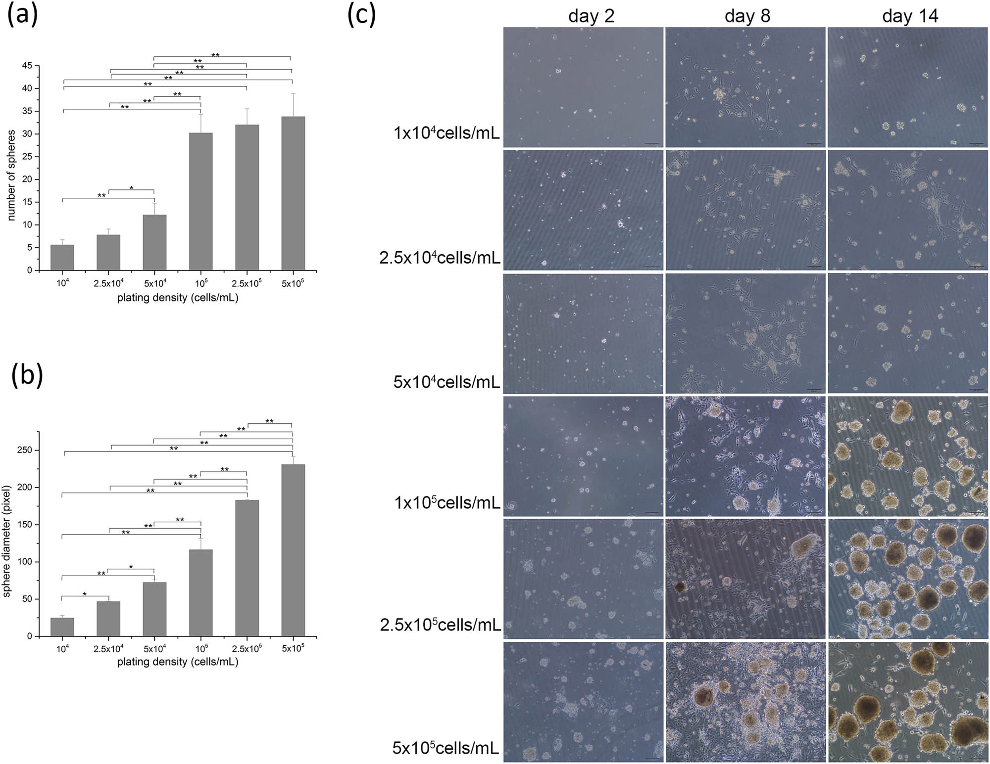
After achieving the cell suspension mentioned in the “mSKP cell isolation” part, we detected the cell density by hemocytometer (Millipore, USA). Six plating densities (104, 2.5 × 10⁴, 5 × 10⁴, 105, 2.5 × 10⁵, 5 × 10⁵ cells/mL) were used. Meanwhile, two types of flasks, including Corning tissue-culture-treated flasks (Corning, NY, USA) and Nest untreated flasks (Nest, Wuxi, China), were used for mSKP culture. The final volume was 5 mL in a 25 cm2 flask. Cultures were fed every 3 days with an addition of 1 mL fresh medium containing all growth factors and supplements (bFGF, EGF, B27) at a concentration that would replenish the entirety of the culture medium. The proliferation of mSKPs at different plating densities in different flasks was monitored and recorded using a light microscope (Olympus, Japan) daily. The experiment design for this section can be found in Figure 1a.

The experiment design for determining the optimal plating density and mSKP growth at multiple plating densities in Corning tissue-culture-treated flasks. (a) The experiment design; (b) mSKP growth at multiple plating densities in Corning tissue-culture-treated flasks. Adherent cell colonies are indicated by arrowheads; (c) diagram representing the number of mSKP spheres on the 14th day; (d) diagram representing the diameter of mSKP spheres on the 14th day. Bar: 100 µm. (Results of three independent experiments, ANOVA for multiple comparisons, *p < 0.05, **p < 0.01.).
2.5 mSKP sphere counting and diameter measurement
On the 14th day, five pictures were taken randomly for every culture condition, mSKP spheres were counted, and their diameters were measured using Image-pro Plus 7.0 (Media Cybernetics, USA). Then the number and diameter of mSKP spheres obtained from every culture condition were compared.
2.6 Cell counting kit 8 (CCK8) viability assay
The mSKP viability under every culture condition was tested using CCK8 (Dojindo, Japan) according to the manufacturer’s instructions. Briefly, CCK8 detected cell viability based upon intracellular WST-8 (C20H14N6NaO11S2) concentration, a compound similar to methyl thiazolyl tetrazolium. WST-8 could be transformed to orange-colored formazan through a reduction reaction catalyzed by dehydrogenases located in mitochondria. The faster the cell proliferate, the darker the orange color would be. The color shade and the number of cells present a linear relation for the same cell line.
2.7 Treat adherent cell colonies with culture condition A or B
mSKPs were cultured in Corning tissue-culture-treated flasks at the plating density of 5 × 10⁵ cells/mL. On the 10th day, both spheres and adherent cell colonies were present. The spheres were then poured out and transferred to the new flasks.
After being washed with PBS 5 times, the original flasks containing the adherent cell colonies were divided into groups A (culture condition A) and B (culture condition B), with 5 flasks in each group. Around 5 mL SKP culture medium was added to group A flasks, and 5 mL DMSC culture medium was added to group B flasks. For group A, cultures were fed every 3 days with an addition of 1 mL fresh medium containing all growth factors and supplements at a concentration that would replenish the entirety of the culture medium. For group B, we changed the fresh DMSC culture medium every 3 days. Cell growth was monitored, and pictures were taken daily. On the 8th day, the cell colonies generated from both groups were collected for characterization by immunocytochemistry and induced differentiation, respectively. The poured-out spheres were cultured with SKP medium in new flasks and fed every 3 days with a fresh medium containing all growth factors and supplements. The experiment design for this section can be found in Figure 4a.
2.8 Immunocytochemistry assay for cell colonies generated from culture condition A or B
Cells were plated on slides, fixed by 4% paraformaldehyde. The fixed cells were blocked with 3% BSA for 30 min and subsequently incubated with primary antibody overnight at 4°C. After being washed with PBS 3 times, cells were incubated with a secondary antibody for 1 h at room temperature. Finally, cells were incubated with DAPI (Dogindo, Japan) for 1 min. Primary antibodies were anti-α-SMA (Abcam, UK, 1:500), anti-Nanog (Abcam, UK, 1:250), anti-Oct4 (Abcam, UK, 1:250), anti-Pan Cytokeratin (Boster, Wuhan, 1:1,000), anti-Ssea 4 (Abcam, UK, 1:250), anti-Versican (Boster, Wuhan, 1:250), anti-Vwf (Boster, Wuhan, 1:500), anti-Fibronectin (Abcam, UK, 1:250), anti-Vimentin (Abcam, UK, 1:200), anti-Nestin (Abcam, UK, 1:500), anti-Sox2 (Boster, Wuhan, 1:250), and anti-Collagen I (Abcam, UK, 1:500). Secondary antibodies were Alexa Fluor® 488 donkey anti-mouse (Abcam, UK, 1:500) and Alexa Fluor® 594 goat anti-rabbit (Abcam, UK, 1:500). The protocol was performed in triplicate for both cell types.
2.9 Osteocyte-, adipocyte-, and chondrocyte-induced differentiation for cell colonies generated from culture condition A or B
Cells were trypsinized, dissociated into single cells, and then resuspended and cultured in basal medium for spheres or adherent cell colonies. The basal medium was replaced by osteocytes or adipocytes differentiation medium (Cyagen, USA) when the confluency reached 60–70%. The following steps were conducted according to the protocol within the kits. At the end of a 28 day induced differentiation, cells were stained with Alizarin Red Solution or Oil Red Solution provided in the kits, respectively.
For chondrocyte-induced differentiation, cells were collected, induced, and differentiated according to the protocol within the chondrogenic differentiation kit (Cyagen, USA). The chondrogenic pellets were harvested after 30 days in the 15 mL tubes culture. According to the protocol, pellets were 10% formalin-fixed, paraffin-embedded, machine-sliced, and then stained with Alcian blue (Leagene biotechnology, Beijing).
2.10 Statistical analysis
All data were expressed as mean value ± SE. Statistical significance was evaluated by analysis of variance (ANOVA) for multiple comparisons. A value of p < 0.05 was considered statistically significant.
3 Results
3.1 mSKP growth in Corning tissue-culture-treated flasks
In Corning tissue-culture-treated flasks, compared with lower plating densities (104–105 cells/mL), the plating densities of 2.5 × 10⁵ and 5 × 10⁵ cells/mL significantly promoted mSKP proliferation (p < 0.01). A large number of adherent cell colonies were present on the 5th or 6th day (Figure 1b, indicated by arrowheads). mSKPs then started to form on the adherent cell colonies on the 7th or 8th day. They were attached to the adherent cell colonies and then detached and migrated out to form floating mSKP spheres as they grew. On the 14th day, the number and the diameter of mSKP spheres generated from different plating densities varied significantly (p < 0.01) (Figure 1b–d). Compared with 104 cells/mL, the number of mSKP spheres increased by 7.7 times, and the diameter of mSKP spheres increased by 8.3 times at the plating density of 5 × 10⁵ cells/mL (Figure 1c and d and Table 1).
Number and diameter of mSKP spheres under multiple plating densities
| Plating density (cells/mL) | Corning tissue-culture-treated flasks (mean ± SE) | Nest untreated flasks (mean ± SE) | ||
|---|---|---|---|---|
| Number of mSKP spheres | Diameter of mSKP spheres (pixel) | Number of mSKP spheres | Diameter of mSKP spheres (pixel) | |
| 104 | 4.2 ± 0.84 | 26.96 ± 0.66 | 5.6 ± 1.14 | 24.58 ± 3.62 |
| 2.5 × 10⁴ | 5.4 ± 1.14 | 41.50 ± 6.91 | 7.8 ± 1.30 | 46.62 ± 0.52 |
| 5 × 10⁴ | 16.8 ± 3.11 | 61.42 ± 0.66 | 12.2 ± 2.59 | 72.43 ± 3.47 |
| 105 | 23.4 ± 5.03 | 142.93 ± 10.50 | 30.2 ± 4.15 | 116.39 ± 15.40 |
| 2.5 × 10⁵ | 30.2 ± 4.49 | 187.37 ± 6.30 | 32.0 ± 3.54 | 182.80 ± 1.10 |
| 5 × 10⁵ | 32.4 ± 2.07 | 222.99 ± 25.20 | 33.8 ± 5.07 | 230.81 ± 10.91 |
3.2 mSKP growth in Nest untreated flasks
A similar phenomenon was present in Nest untreated flasks (Figure 2). On the 14th day, the number and the diameter of mSKP spheres generated from different plating densities varied significantly (p < 0.01) (Figure 2a–c). Compared with 104 cells/mL, the number of mSKP spheres increased by 6 times, and the diameter of mSKP spheres increased by 9.4 times when the plating density was 5 × 10⁵ cells/mL (Figure 2a and b and Table 1).
mSKP growth at multiple plating densities in Nest untreated flasks. (a) Diagram representing the number of mSKP spheres on the 14th day; (b) diagram representing the diameter of mSKP spheres on the 14th day; (c) mSKP growth at multiple plating densities in Nest untreated flasks. Bar: 100 µm. (Results of three independent experiments, ANOVA for multiple comparisons, *p < 0.05, **p < 0.01.).
3.3 mSKP viability
The CCK8 results indicated that compared with lower plating densities (104–105 cells/mL), the plating densities of 2.5 × 10⁵ cells/mL and 5 × 10⁵ cells/mL significantly promoted mSKP viability (p < 0.01) (Figure 3).

CCK8 results: the plating densities of 2.5 × 10⁵ and 5 × 10⁵ cells/mL promoted mSKP viability in both (a) Corning tissue-culture-treated flasks and (b) Nest untreated flasks (Results of three independent experiments, ANOVA for multiple comparisons, *p < 0.05, **p < 0.01.).
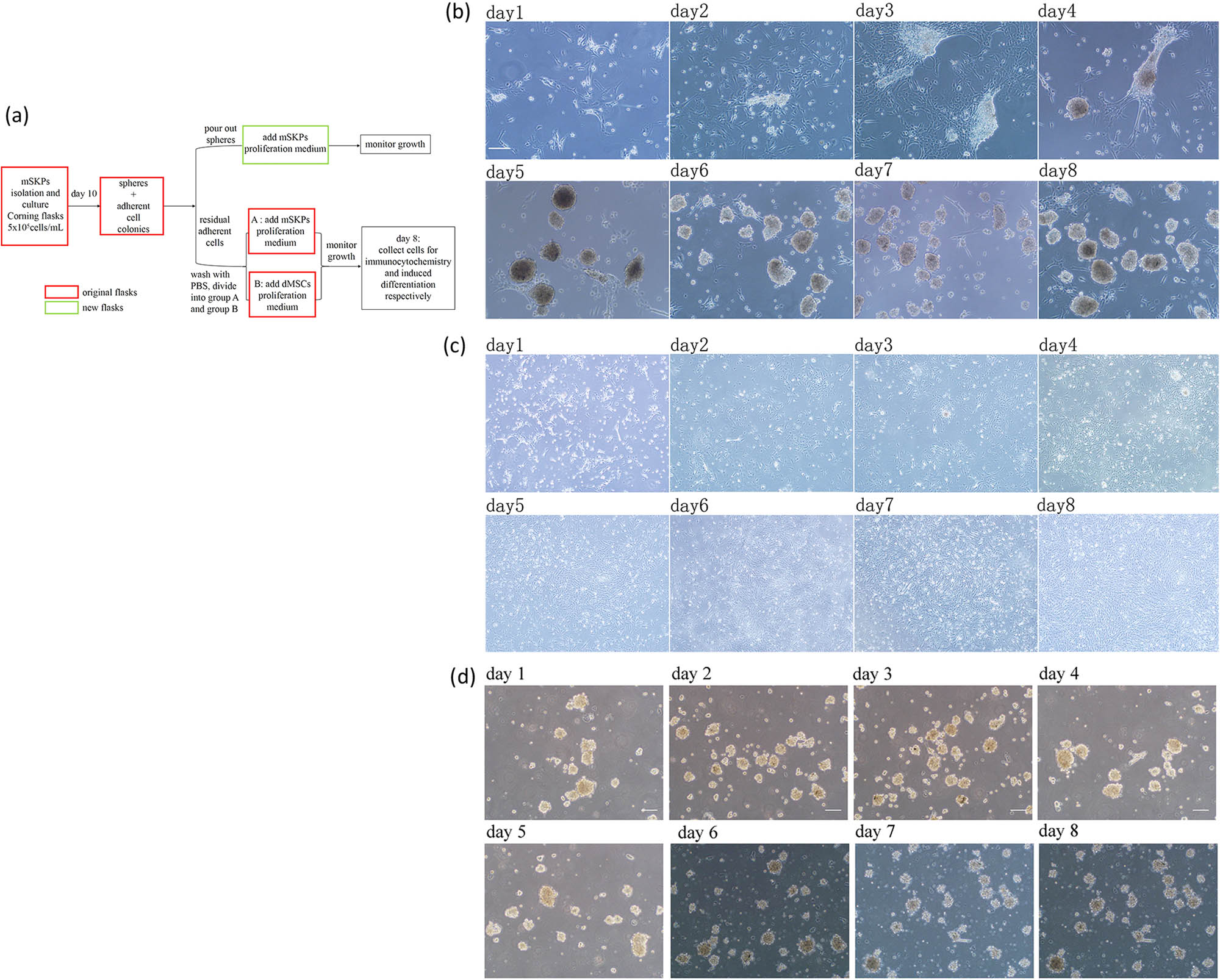
The experiment design and results of treating adherent cell colonies with culture condition A (group A) or B (group B). (a) The experiment design; (b) adherent cell colonies differentiated into SKP-like spheres under the SKP culture condition, in culture condition A, day 1–day 8; (c) adherent cell colonies differentiated into DMSC-like colonies under the DMSC culture condition, in culture condition B, day 1–day 8; (d) the poured-out spheres stopped growing in new flasks. Bar: 100 µm.
3.4 Treating adherent cell colonies with culture condition A or B
3.4.1 Cell colonies in culture condition A or B
Culture condition A: on the 1st day, only a few residual adherent cells were noticed, and then the cells proliferated and formed multiple adherent cell colonies. On the 3rd or 4th day, spheres started to form on the colonies, attached and then detached and migrated from the adherent cell colonies as growing larger. On the 7th–8th day, a large number of spheres were noticed, while the adherent cell colonies reduced considerably (Figure 4b). Culture condition B: the residual adherent cells proliferated to form colonies. The cells demonstrated a flattened and fibroblast-like morphology, with a bright lining (Figure 4c).
3.4.2 The poured-out spheres
Although being cultured in SKP medium and fed every 3 days with an addition of fresh medium containing all growth factors and supplements, the poured-out spheres stopped growing in both number and diameter in the new flasks (Figure 4d).
3.5 Antigen expression
According to the immunocytochemistry results, the spheres obtained from culture condition A expressed α-SMA, Nanog, Nestin, Oct4, Sox2, Ssea4, Versican, and Vimentin (Figure 5a). The adherent cells obtained from culture condition B expressed α-SMA, Collagen I, Nestin, Sox2 (weakly positive), Ssea4, Versican, and Vimentin (Figure 5b).
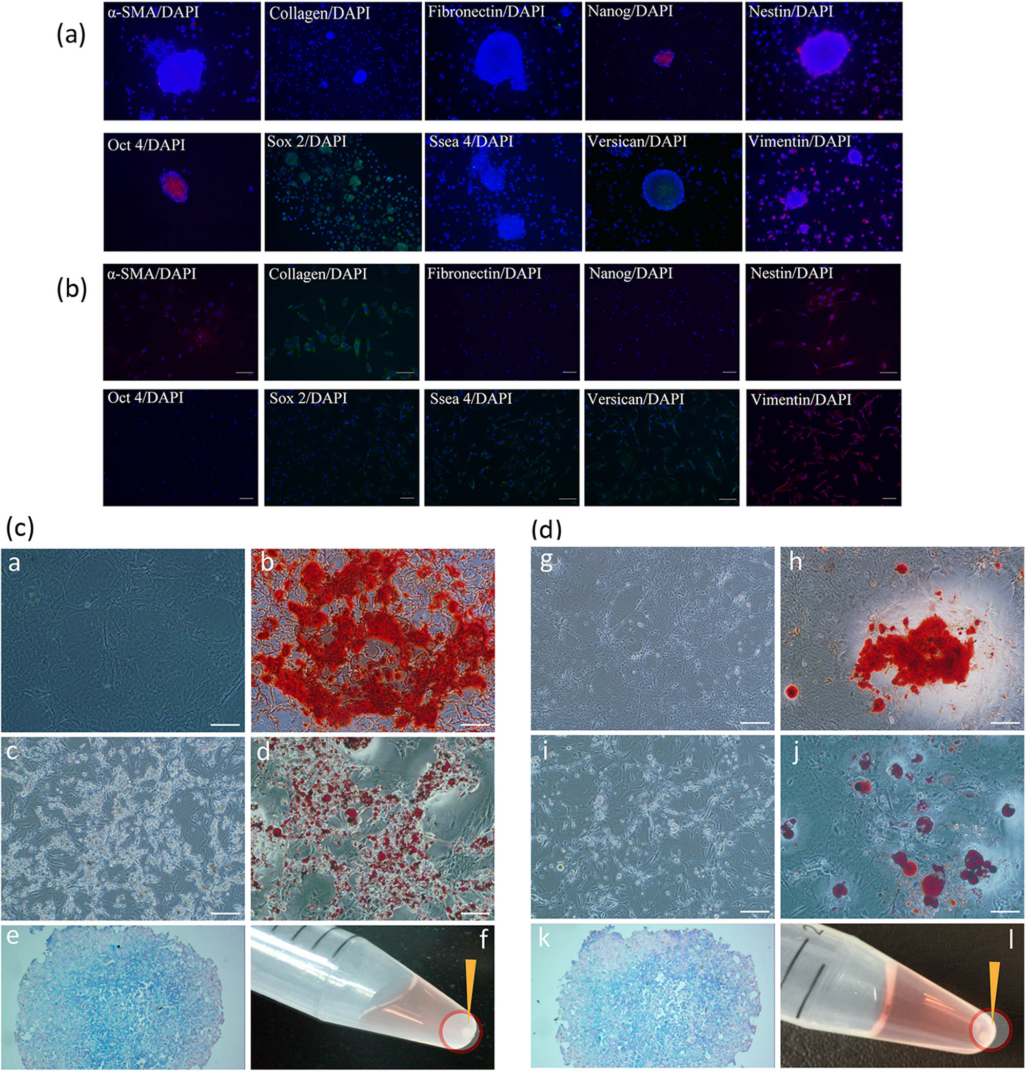
Results of immunocytochemistry and induced differentiation of cells obtained from culture condition A or B. (a) The spheres obtained from culture condition A expressed α-SMA, Nanog, Nestin, Oct4, Sox2, Ssea4, Versican, and Vimentin; (b) the adherent cells obtained from culture condition B expressed α-SMA, Collagen I, Nestin, Sox2 (weak positive), Ssea4, Versican, and Vimentin; (c) osteocyte-, adipocyte-, and chondrocyte-induced differentiation of spheres obtained from culture condition A; (d) osteocyte-, adipocyte-, and chondrocyte-induced differentiation of adherent cell colonies obtained from culture condition B. ((a and g) Osteocyte calcified matrix (osteogenesis, day 28); (b and h) alizarin red stain (day 28); (c and i) adipocyte lipid droplets (adipogenesis, day 28); (d and j) oil red stain (day 28); (e and k) alcian blue stain, pellets consisted of chondrocytes glycosaminoglycan (chondrogenesis, day 30); (f and l) chondrogenic pellets formed at the bottom of the tubes (indicated by arrowheads, day 30)). Bar: 100 µm.
3.6 Induced differentiation
After a 28 day induction, cells obtained from both culture conditions A and B differentiated into osteocyte calcified matrix (osteogenesis) or adipocyte lipid droplets (adipogenesis) (Figure 5c and d). Chondrogenic pellets formed at the bottom of the 15 mL tubes after a 30 day induction slides stained with Alcian blue indicated pellets consisted of chondrocytes glycosaminoglycan (chondrogenesis) (Figure 5c and d).
4 Discussion
Biernaskie et al. [11] believed the growth rate of mSKPs was cell density dependent and recommended the optimal 104–2.5 × 10⁴ cells/mL plating density for mSKPs. They noticed the adherent cell colonies under the serum-free culture condition, and they stated, “it is extremely important to use tissue-culture-treated plastic in the flasks to prevent adherence of cells to the plastic.” Nevertheless, according to our experience, mSKP growth remained very limited even if all the requirements mentioned above were fulfilled.
In this study, two types of flasks, including tissue-culture-treated flasks and untreated flasks, were used. For the tissue-culture-treated flasks, the cell culture surface treatment promotes cell attachment and growth. Untreated flasks are preferred in traditional SKP culture because SKPs are floating spheres, and the attachment of cells to the flask surface is unexpected. Noteworthily, our research revealed that surface treatment did not influence SKP growth. In addition, we explored the impact of plating density on mSKP production, as well as the biological characteristics of the adherent cell colonies. The research demonstrated that both mSKPs and adherent cell colonies increased as the plating density ascended. Inconsistent with Biernaskie’s findings [11], mSKPs formed on the adherent cell colonies firstly and then detached to form floating spheres as the amount and size increased. Lower plating densities (104–105 cells/mL) generated significantly less adherent cell colonies, which resulted in significantly fewer floating mSKP spheres. Taken together, the optimal plating density promoted mSKP proliferation by promoting adherent cell colonies growth. Conventionally adherent cells were avoided in SKP culture (for example, by decreasing cell density), which strictly limited SKP sphere development.
Considering mSKP spheres were 3D and might vary largely in size, we detected the spheres’ number, diameter, and cell viability when evaluating efficiency. All three parameters increased significantly under the optimal density. In addition, we managed to shorten the culture duration by 7 days by obtaining adequate mSKPs on the 12th or 14th day. More importantly, no extra growth factors, cytokines, additional apparatus, or biomedical materials would be needed, hence managed budget control. Optimizing plating density was easy to perform, and the SKP biological pattern would not be interrupted.
Vast chunks of mSKP aggregation were noticed on the 8th or 10th day at 106 cells/mL density. The dark center of aggregation implied insufficient nutrients and unhealthy cells, which implied that the plating density of 106 cells/mL did not prove to be more productive than 5 × 10⁵ cells/mL. A similar situation was also noticed in hSKPs (data on file), and the plating density of 5 × 10⁵ cells/mL proved to be more productive than the lower densities (1 × 10⁵ and 2.5 × 10⁵ cells/mL).
Treating adherent cell colonies with two culture conditions further elucidated the correlation between mSKP spheres and adherent cell colonies. The cells that adhered to both tissue-culture-treated flasks and untreated flasks under a serum-free culture condition seemed to possess the capacity to proliferate and differentiate toward two distinct directions under two different culture conditions. The immunocytochemistry and induced differentiation results confirmed that the spheres achieved from culture condition A were mSKPs. The adherent cell colonies achieved from culture condition B displayed the most important markers and characteristics that a primary culture should possess to be classified as DMSCs [24]. Forni et al. [25] reported simultaneously isolating and characterizing three stem cell populations from the dermis and epidermis of murine skin, namely epidermal stem cells, SKPs, and DMSCs. They believed DMSCs and SKPs could be isolated simultaneously from one skin sample and showed no overt difference with DMSCs and SKPs isolated in a routine way. Nevertheless, we did not consider that multiple types of cells were isolated simultaneously. We believed that mSKPs and DMSCs are derived from the same cell type isolated (i.e., the adherent cell colonies), and different culture conditions generated two types of cells with distinct biological properties. Taken together, the characteristics of cells were shown to be altered by culture conditions.
Notably, the poured-out mSKP spheres failed to grow in new flasks even if cultures were fed with the addition of fresh medium containing all growth factors and supplements, implying mSKP spheres did not proliferate without the presence of adherent cell colonies. This finding confirmed again that mSKP spheres originated from the adherent cell colonies. It also explained the observation by Zong et al. [26], who reported isolating SKPs and DMSCs simultaneously from a single human skin sample. According to them, the suspended cells were collected 4 h later and transferred to another flask, and cultured in SKP proliferation medium. No obvious floating sphere formed even 10 days later, and they mentioned: “the definite reasons were unknown.” Interestingly, they also reported that many cells adhered to the base of the flask 4 h after isolation, which was not observed in our experiment. At the plating density of 5 × 10⁵ cells/mL, the adherent cells did not present until the 2nd or 3rd day.
In summary, mSKPs are derived from the adherent cell colonies in the primary cell culture, regardless of their floating characteristic. Within a certain limit, the plating density played a major role in mSKP development. The optimal plating density (2.5 × 10⁵–5 × 10⁵ cells/mL) accelerated mSKP growth and guaranteed the following stem cell-conditioned medium and exomes research. In addition, the research data on mouse stem cells might provide insights for hSKP culture and promote hSKP basic research and clinical application.
-
Funding information: The work was supported by the following funding: Science & Technology Department of Sichuan Province Project (No. 2020YJ0058); Post-Doctor Research Project, West China Hospital, Sichuan University (No. 2020HXBH050); China Postdoctoral Science Foundation (No. 2021M692292); National Natural Science Foundation of China (No. 82103753); Natural Science Foundation of Zhejiang Province (No. LQ20H110002); Key Science & Technology Department of Sichuan Province Project (No. 2018SZ0196); 1.3.5 project for disciplines of excellence, West China Hospital, Sichuan University.
-
Author contributions: Yiming Li designed the research, carried out the experiments, analyzed and discussed the data, and drafted the manuscript. Lidan Xiong analyzed and discussed the data, and reviewed the manuscript. Jie Tang, Ru Dai, and Shiyi Li analyzed and discussed the data. Li Li designed the research and reviewed the manuscript. All authors have read and approved the final manuscript.
-
Conflict of interest: The authors state no conflict of interest.
-
Data availability statement: The datasets generated during and/or analyzed during the current study are available from the corresponding author on reasonable request.
References
[1] Toma JG, McKenzie IA, Bagli D, Miller FD. Isolation and characterization of multipotent skin-derived precursors from human skin. Stem Cells. 2005;23:727–37.10.1634/stemcells.2004-0134Suche in Google Scholar PubMed
[2] Suflita MT, Pfaltzgraff ER, Mundell NA, Pevny LH, Labosky PA. Ground-state transcriptional requirements for skin-derived precursors. Stem Cells Dev. 2013;22:1779–88.10.1089/scd.2012.0501Suche in Google Scholar PubMed PubMed Central
[3] Gingras M, Champigny MF, Berthod F. Differentiation of human adult skin-derived neuronal precursors into mature neurons. J Cellular Physiol. 2007;210:498–506.10.1002/jcp.20889Suche in Google Scholar PubMed
[4] Kumar R, Sinha S, Hagner A, Stykel M, Raharjo E, Singh KK, et al. Adult skin-derived precursor Schwann cells exhibit superior myelination and regeneration supportive properties compared to chronically denervated nerve-derived Schwann cells. Exp Neurol. 2016;278:127–42.10.1016/j.expneurol.2016.02.006Suche in Google Scholar PubMed
[5] Mehrabi M, Mansouri K, Hosseinkhani S, Yarani R, Yari K, Bakhtiari M, et al. Differentiation of human skin-derived precursor cells into functional islet-like insulin-producing cell clusters. In Vitro Cell Dev Biol-Animal. 2015;51:595–603.10.1007/s11626-015-9866-2Suche in Google Scholar PubMed
[6] Saichanma S, Bunyaratvej A, Sila-Asna M. In vitro transdifferentiation of corneal epithelial-like cells from human skin-derived precursor cells. Int J Ophthalmol. 2012;5(2):158–63.Suche in Google Scholar
[7] Lavoie JF, Biernaskie JA, Chen Y, Bagli D, Alman B, Kaplan DR, et al. Skin-derived precursors differentiate into skeletogenic cell types and contribute to bone repair. Stem Cells Develop. 2009;18:893–906.10.1089/scd.2008.0260Suche in Google Scholar PubMed
[8] Hunt DP, Jahoda C, Chandran S. Multipotent skin-derived precursors: from biology to clinical translation. Curr Opin Biotechnol. 2009;20(5):522–30.10.1016/j.copbio.2009.10.004Suche in Google Scholar PubMed
[9] Li Y, Li X, Xiong L, Tang J, Li L. Comparison of phenotypes and transcriptomes of mouse skin-derived precursors and dermal mesenchymal stem cells. Differentiation. 2018;102:30–9.10.1016/j.diff.2018.07.001Suche in Google Scholar PubMed
[10] De Kock J, Meuleman P, Raicevic G, Rodrigues RM, Branson S, Meganathan K, et al. Human skin-derived precursor cells are poorly immunogenic and modulate the allogeneic immune response. Stem Cells. 2014;32:2215–28.10.1002/stem.1692Suche in Google Scholar PubMed
[11] Biernaskie JA, McKenzie IA, Toma JG, Miller FD. Isolation of skin-derived precursors (SKPs) and differentiation and enrichment of their Schwann cell progeny. Nat Protoc. 2006;1(6):2803–12.10.1038/nprot.2006.422Suche in Google Scholar PubMed
[12] Riekstina U, Muceniece R, Cakstina I, Muiznieks I, Ancans J. Characterization of human skin-derived mesenchymal stem cell proliferation rate in different growth conditions. Cytotechnology. 2008;58:153–62.10.1007/s10616-009-9183-2Suche in Google Scholar PubMed PubMed Central
[13] Kawase Y, Yanagi Y, Takato T, Fujimoto M, Okochi H. Characterization of multipotent adult stem cells from the skin: transforming growth factor-beta (TGF-beta) facilitates cell growth. Exp Cell Res. 2004;295(1):194–203.10.1016/j.yexcr.2003.12.027Suche in Google Scholar PubMed
[14] Huang Y, Qi SH, Shu B, Chen L, Xie JL, Xu YB, et al. Fibroblast growth factor-binding protein facilitates the growth and migration of skin-derived precursors. J Cutan Med Surg. 2011;15(4):201–9.10.2310/7750.2011.10049Suche in Google Scholar PubMed
[15] Surrao DC, Boon K, Borys B, Sinha S, Kumar R, Biernaskie J, et al. Large-scale expansion of human skin-derived precursor cells (hSKPs) in stirred suspension bioreactors. Biotechnol Bioeng. 2016;113(12):2725–38.10.1002/bit.26040Suche in Google Scholar PubMed
[16] Wang X, Liu S, Zhao Q, Li N, Zhang H, Zhang X, et al. Three-dimensional hydrogel scaffolds facilitate in vitro self-renewal of human skin-derived precursors. Acta Biomater. 2014;10(7):3177–87.10.1016/j.actbio.2014.03.018Suche in Google Scholar PubMed
[17] Hill RP, Gledhill K, Gardner A, Higgins CA, Crawford H, Lawrence C, et al. Generation and characterization of multipotent stem cells from established dermal cultures. PLoS One. 2012;7(11):e50742.10.1371/journal.pone.0050742Suche in Google Scholar PubMed PubMed Central
[18] Budel L, Djabali K. Rapid isolation and expansion of skin-derived precursor cells from human primary fibroblast cultures. Biol Open. 2017;6(11):1745–55.10.1242/bio.025130Suche in Google Scholar PubMed PubMed Central
[19] Prescott HM, Manning C, Gardner A, Ritchie WA, Pizzi R, Girling S, et al. Giant panda (Ailuropoda melanoleuca) buccal mucosa tissue as a source of multipotent progenitor cells. PLoS One. 2015;10(9):e0138840.10.1371/journal.pone.0138840Suche in Google Scholar PubMed PubMed Central
[20] Li Y, Xiong L, Tang J, Zhu G, Dai R, Li L. Mouse skin-derived precursors alleviates ultraviolet B irradiation damage via early activation of TGF-β/Smad pathway by thrombospondin1. Cell Cycle. 2020;19(4):492–503.10.1080/15384101.2020.1717042Suche in Google Scholar PubMed PubMed Central
[21] Wang S, Zhong J, Li L. Protective effect of skin-derived precursors on photoaging in nude mice. Australas J Dermatol. 2019;60(1):e20–8.10.1111/ajd.12867Suche in Google Scholar PubMed
[22] Zhong J, Li L. Skin-derived precursors against UVB-induced apoptosis via Bcl-2 and Nrf2 upregulation. Biomed Res Int. 2016;2016:6894743.10.1155/2016/6894743Suche in Google Scholar PubMed PubMed Central
[23] Mao Y, Xiong L, Wang S, Zhong J, Zhou R, Li L. Comparison of the transcriptomes of mouse skin derived precursors (SKPs) and SKP-derived fibroblasts (SFBs) by RNA-Seq. PLoS One. 2015;10(2):e0117739.10.1371/journal.pone.0117739Suche in Google Scholar PubMed PubMed Central
[24] Dominici M, Le Blanc K, Mueller I, Slaper-Cortenbach I, Marini F, Krause D, et al. Minimal criteria for defining multipotent mesenchymal stromal cells. The international society for cellular therapy position statement. Cytotherapy. 2006;8(4):315–7.10.1080/14653240600855905Suche in Google Scholar PubMed
[25] Forni MF, Ramos Maia Lobba A, Pereira Ferreira AH, Sogayar MC. Simultaneous isolation of three different stem cell populations from murine skin. PLoS One. 2015;10(10):e0140143.10.1371/journal.pone.0140143Suche in Google Scholar PubMed PubMed Central
[26] Zong Z, Li N, Ran X, Su Y, Shen Y, Shi CM, et al. Isolation and characterization of two kinds of stem cells from the same human skin back sample with therapeutic potential in spinal cord injury. PLoS One. 2012;7(11):e50222.10.1371/journal.pone.0050222Suche in Google Scholar PubMed PubMed Central
© 2021 Yiming Li et al., published by De Gruyter
This work is licensed under the Creative Commons Attribution 4.0 International License.
Artikel in diesem Heft
- Biomedical Sciences
- Research progress on the mechanism of orexin in pain regulation in different brain regions
- Adriamycin-resistant cells are significantly less fit than adriamycin-sensitive cells in cervical cancer
- Exogenous spermidine affects polyamine metabolism in the mouse hypothalamus
- Iris metastasis of diffuse large B-cell lymphoma misdiagnosed as primary angle-closure glaucoma: A case report and review of the literature
- LncRNA PVT1 promotes cervical cancer progression by sponging miR-503 to upregulate ARL2 expression
- Two new inflammatory markers related to the CURB-65 score for disease severity in patients with community-acquired pneumonia: The hypersensitive C-reactive protein to albumin ratio and fibrinogen to albumin ratio
- Circ_0091579 enhances the malignancy of hepatocellular carcinoma via miR-1287/PDK2 axis
- Silencing XIST mitigated lipopolysaccharide (LPS)-induced inflammatory injury in human lung fibroblast WI-38 cells through modulating miR-30b-5p/CCL16 axis and TLR4/NF-κB signaling pathway
- Protocatechuic acid attenuates cerebral aneurysm formation and progression by inhibiting TNF-alpha/Nrf-2/NF-kB-mediated inflammatory mechanisms in experimental rats
- ABCB1 polymorphism in clopidogrel-treated Montenegrin patients
- Metabolic profiling of fatty acids in Tripterygium wilfordii multiglucoside- and triptolide-induced liver-injured rats
- miR-338-3p inhibits cell growth, invasion, and EMT process in neuroblastoma through targeting MMP-2
- Verification of neuroprotective effects of alpha-lipoic acid on chronic neuropathic pain in a chronic constriction injury rat model
- Circ_WWC3 overexpression decelerates the progression of osteosarcoma by regulating miR-421/PDE7B axis
- Knockdown of TUG1 rescues cardiomyocyte hypertrophy through targeting the miR-497/MEF2C axis
- MiR-146b-3p protects against AR42J cell injury in cerulein-induced acute pancreatitis model through targeting Anxa2
- miR-299-3p suppresses cell progression and induces apoptosis by downregulating PAX3 in gastric cancer
- Diabetes and COVID-19
- Discovery of novel potential KIT inhibitors for the treatment of gastrointestinal stromal tumor
- TEAD4 is a novel independent predictor of prognosis in LGG patients with IDH mutation
- circTLK1 facilitates the proliferation and metastasis of renal cell carcinoma by regulating miR-495-3p/CBL axis
- microRNA-9-5p protects liver sinusoidal endothelial cell against oxygen glucose deprivation/reperfusion injury
- Long noncoding RNA TUG1 regulates degradation of chondrocyte extracellular matrix via miR-320c/MMP-13 axis in osteoarthritis
- Duodenal adenocarcinoma with skin metastasis as initial manifestation: A case report
- Effects of Loofah cylindrica extract on learning and memory ability, brain tissue morphology, and immune function of aging mice
- Recombinant Bacteroides fragilis enterotoxin-1 (rBFT-1) promotes proliferation of colorectal cancer via CCL3-related molecular pathways
- Blocking circ_UBR4 suppressed proliferation, migration, and cell cycle progression of human vascular smooth muscle cells in atherosclerosis
- Gene therapy in PIDs, hemoglobin, ocular, neurodegenerative, and hemophilia B disorders
- Downregulation of circ_0037655 impedes glioma formation and metastasis via the regulation of miR-1229-3p/ITGB8 axis
- Vitamin D deficiency and cardiovascular risk in type 2 diabetes population
- Circ_0013359 facilitates the tumorigenicity of melanoma by regulating miR-136-5p/RAB9A axis
- Mechanisms of circular RNA circ_0066147 on pancreatic cancer progression
- lncRNA myocardial infarction-associated transcript (MIAT) knockdown alleviates LPS-induced chondrocytes inflammatory injury via regulating miR-488-3p/sex determining region Y-related HMG-box 11 (SOX11) axis
- Identification of circRNA circ-CSPP1 as a potent driver of colorectal cancer by directly targeting the miR-431/LASP1 axis
- Hyperhomocysteinemia exacerbates ischemia-reperfusion injury-induced acute kidney injury by mediating oxidative stress, DNA damage, JNK pathway, and apoptosis
- Potential prognostic markers and significant lncRNA–mRNA co-expression pairs in laryngeal squamous cell carcinoma
- Gamma irradiation-mediated inactivation of enveloped viruses with conservation of genome integrity: Potential application for SARS-CoV-2 inactivated vaccine development
- ADHFE1 is a correlative factor of patient survival in cancer
- The association of transcription factor Prox1 with the proliferation, migration, and invasion of lung cancer
- Is there a relationship between the prevalence of autoimmune thyroid disease and diabetic kidney disease?
- Immunoregulatory function of Dictyophora echinovolvata spore polysaccharides in immunocompromised mice induced by cyclophosphamide
- T cell epitopes of SARS-CoV-2 spike protein and conserved surface protein of Plasmodium malariae share sequence homology
- Anti-obesity effect and mechanism of mesenchymal stem cells influence on obese mice
- Long noncoding RNA HULC contributes to paclitaxel resistance in ovarian cancer via miR-137/ITGB8 axis
- Glucocorticoids protect HEI-OC1 cells from tunicamycin-induced cell damage via inhibiting endoplasmic reticulum stress
- Prognostic value of the neutrophil-to-lymphocyte ratio in acute organophosphorus pesticide poisoning
- Gastroprotective effects of diosgenin against HCl/ethanol-induced gastric mucosal injury through suppression of NF-κβ and myeloperoxidase activities
- Silencing of LINC00707 suppresses cell proliferation, migration, and invasion of osteosarcoma cells by modulating miR-338-3p/AHSA1 axis
- Successful extracorporeal membrane oxygenation resuscitation of patient with cardiogenic shock induced by phaeochromocytoma crisis mimicking hyperthyroidism: A case report
- Effects of miR-185-5p on replication of hepatitis C virus
- Lidocaine has antitumor effect on hepatocellular carcinoma via the circ_DYNC1H1/miR-520a-3p/USP14 axis
- Primary localized cutaneous nodular amyloidosis presenting as lymphatic malformation: A case report
- Multimodal magnetic resonance imaging analysis in the characteristics of Wilson’s disease: A case report and literature review
- Therapeutic potential of anticoagulant therapy in association with cytokine storm inhibition in severe cases of COVID-19: A case report
- Neoadjuvant immunotherapy combined with chemotherapy for locally advanced squamous cell lung carcinoma: A case report and literature review
- Rufinamide (RUF) suppresses inflammation and maintains the integrity of the blood–brain barrier during kainic acid-induced brain damage
- Inhibition of ADAM10 ameliorates doxorubicin-induced cardiac remodeling by suppressing N-cadherin cleavage
- Invasive ductal carcinoma and small lymphocytic lymphoma/chronic lymphocytic leukemia manifesting as a collision breast tumor: A case report and literature review
- Clonal diversity of the B cell receptor repertoire in patients with coronary in-stent restenosis and type 2 diabetes
- CTLA-4 promotes lymphoma progression through tumor stem cell enrichment and immunosuppression
- WDR74 promotes proliferation and metastasis in colorectal cancer cells through regulating the Wnt/β-catenin signaling pathway
- Down-regulation of IGHG1 enhances Protoporphyrin IX accumulation and inhibits hemin biosynthesis in colorectal cancer by suppressing the MEK-FECH axis
- Curcumin suppresses the progression of gastric cancer by regulating circ_0056618/miR-194-5p axis
- Scutellarin-induced A549 cell apoptosis depends on activation of the transforming growth factor-β1/smad2/ROS/caspase-3 pathway
- lncRNA NEAT1 regulates CYP1A2 and influences steroid-induced necrosis
- A two-microRNA signature predicts the progression of male thyroid cancer
- Isolation of microglia from retinas of chronic ocular hypertensive rats
- Changes of immune cells in patients with hepatocellular carcinoma treated by radiofrequency ablation and hepatectomy, a pilot study
- Calcineurin Aβ gene knockdown inhibits transient outward potassium current ion channel remodeling in hypertrophic ventricular myocyte
- Aberrant expression of PI3K/AKT signaling is involved in apoptosis resistance of hepatocellular carcinoma
- Clinical significance of activated Wnt/β-catenin signaling in apoptosis inhibition of oral cancer
- circ_CHFR regulates ox-LDL-mediated cell proliferation, apoptosis, and EndoMT by miR-15a-5p/EGFR axis in human brain microvessel endothelial cells
- Resveratrol pretreatment mitigates LPS-induced acute lung injury by regulating conventional dendritic cells’ maturation and function
- Ubiquitin-conjugating enzyme E2T promotes tumor stem cell characteristics and migration of cervical cancer cells by regulating the GRP78/FAK pathway
- Carriage of HLA-DRB1*11 and 1*12 alleles and risk factors in patients with breast cancer in Burkina Faso
- Protective effect of Lactobacillus-containing probiotics on intestinal mucosa of rats experiencing traumatic hemorrhagic shock
- Glucocorticoids induce osteonecrosis of the femoral head through the Hippo signaling pathway
- Endothelial cell-derived SSAO can increase MLC20 phosphorylation in VSMCs
- Downregulation of STOX1 is a novel prognostic biomarker for glioma patients
- miR-378a-3p regulates glioma cell chemosensitivity to cisplatin through IGF1R
- The molecular mechanisms underlying arecoline-induced cardiac fibrosis in rats
- TGF-β1-overexpressing mesenchymal stem cells reciprocally regulate Th17/Treg cells by regulating the expression of IFN-γ
- The influence of MTHFR genetic polymorphisms on methotrexate therapy in pediatric acute lymphoblastic leukemia
- Red blood cell distribution width-standard deviation but not red blood cell distribution width-coefficient of variation as a potential index for the diagnosis of iron-deficiency anemia in mid-pregnancy women
- Small cell neuroendocrine carcinoma expressing alpha fetoprotein in the endometrium
- Superoxide dismutase and the sigma1 receptor as key elements of the antioxidant system in human gastrointestinal tract cancers
- Molecular characterization and phylogenetic studies of Echinococcus granulosus and Taenia multiceps coenurus cysts in slaughtered sheep in Saudi Arabia
- ITGB5 mutation discovered in a Chinese family with blepharophimosis-ptosis-epicanthus inversus syndrome
- ACTB and GAPDH appear at multiple SDS-PAGE positions, thus not suitable as reference genes for determining protein loading in techniques like Western blotting
- Facilitation of mouse skin-derived precursor growth and yield by optimizing plating density
- 3,4-Dihydroxyphenylethanol ameliorates lipopolysaccharide-induced septic cardiac injury in a murine model
- Downregulation of PITX2 inhibits the proliferation and migration of liver cancer cells and induces cell apoptosis
- Expression of CDK9 in endometrial cancer tissues and its effect on the proliferation of HEC-1B
- Novel predictor of the occurrence of DKA in T1DM patients without infection: A combination of neutrophil/lymphocyte ratio and white blood cells
- Investigation of molecular regulation mechanism under the pathophysiology of subarachnoid hemorrhage
- miR-25-3p protects renal tubular epithelial cells from apoptosis induced by renal IRI by targeting DKK3
- Bioengineering and Biotechnology
- Green fabrication of Co and Co3O4 nanoparticles and their biomedical applications: A review
- Agriculture
- Effects of inorganic and organic selenium sources on the growth performance of broilers in China: A meta-analysis
- Crop-livestock integration practices, knowledge, and attitudes among smallholder farmers: Hedging against climate change-induced shocks in semi-arid Zimbabwe
- Food Science and Nutrition
- Effect of food processing on the antioxidant activity of flavones from Polygonatum odoratum (Mill.) Druce
- Vitamin D and iodine status was associated with the risk and complication of type 2 diabetes mellitus in China
- Diversity of microbiota in Slovak summer ewes’ cheese “Bryndza”
- Comparison between voltammetric detection methods for abalone-flavoring liquid
- Composition of low-molecular-weight glutenin subunits in common wheat (Triticum aestivum L.) and their effects on the rheological properties of dough
- Application of culture, PCR, and PacBio sequencing for determination of microbial composition of milk from subclinical mastitis dairy cows of smallholder farms
- Investigating microplastics and potentially toxic elements contamination in canned Tuna, Salmon, and Sardine fishes from Taif markets, KSA
- From bench to bar side: Evaluating the red wine storage lesion
- Establishment of an iodine model for prevention of iodine-excess-induced thyroid dysfunction in pregnant women
- Plant Sciences
- Characterization of GMPP from Dendrobium huoshanense yielding GDP-D-mannose
- Comparative analysis of the SPL gene family in five Rosaceae species: Fragaria vesca, Malus domestica, Prunus persica, Rubus occidentalis, and Pyrus pyrifolia
- Identification of leaf rust resistance genes Lr34 and Lr46 in common wheat (Triticum aestivum L. ssp. aestivum) lines of different origin using multiplex PCR
- Investigation of bioactivities of Taxus chinensis, Taxus cuspidata, and Taxus × media by gas chromatography-mass spectrometry
- Morphological structures and histochemistry of roots and shoots in Myricaria laxiflora (Tamaricaceae)
- Transcriptome analysis of resistance mechanism to potato wart disease
- In silico analysis of glycosyltransferase 2 family genes in duckweed (Spirodela polyrhiza) and its role in salt stress tolerance
- Comparative study on growth traits and ions regulation of zoysiagrasses under varied salinity treatments
- Role of MS1 homolog Ntms1 gene of tobacco infertility
- Biological characteristics and fungicide sensitivity of Pyricularia variabilis
- In silico/computational analysis of mevalonate pyrophosphate decarboxylase gene families in Campanulids
- Identification of novel drought-responsive miRNA regulatory network of drought stress response in common vetch (Vicia sativa)
- How photoautotrophy, photomixotrophy, and ventilation affect the stomata and fluorescence emission of pistachios rootstock?
- Apoplastic histochemical features of plant root walls that may facilitate ion uptake and retention
- Ecology and Environmental Sciences
- The impact of sewage sludge on the fungal communities in the rhizosphere and roots of barley and on barley yield
- Domestication of wild animals may provide a springboard for rapid variation of coronavirus
- Response of benthic invertebrate assemblages to seasonal and habitat condition in the Wewe River, Ashanti region (Ghana)
- Molecular record for the first authentication of Isaria cicadae from Vietnam
- Twig biomass allocation of Betula platyphylla in different habitats in Wudalianchi Volcano, northeast China
- Animal Sciences
- Supplementation of probiotics in water beneficial growth performance, carcass traits, immune function, and antioxidant capacity in broiler chickens
- Predators of the giant pine scale, Marchalina hellenica (Gennadius 1883; Hemiptera: Marchalinidae), out of its natural range in Turkey
- Honey in wound healing: An updated review
- NONMMUT140591.1 may serve as a ceRNA to regulate Gata5 in UT-B knockout-induced cardiac conduction block
- Radiotherapy for the treatment of pulmonary hydatidosis in sheep
- Retraction
- Retraction of “Long non-coding RNA TUG1 knockdown hinders the tumorigenesis of multiple myeloma by regulating microRNA-34a-5p/NOTCH1 signaling pathway”
- Special Issue on Reuse of Agro-Industrial By-Products
- An effect of positional isomerism of benzoic acid derivatives on antibacterial activity against Escherichia coli
- Special Issue on Computing and Artificial Techniques for Life Science Applications - Part II
- Relationship of Gensini score with retinal vessel diameter and arteriovenous ratio in senile CHD
- Effects of different enantiomers of amlodipine on lipid profiles and vasomotor factors in atherosclerotic rabbits
- Establishment of the New Zealand white rabbit animal model of fatty keratopathy associated with corneal neovascularization
- lncRNA MALAT1/miR-143 axis is a potential biomarker for in-stent restenosis and is involved in the multiplication of vascular smooth muscle cells