Clinical significance of activated Wnt/β-catenin signaling in apoptosis inhibition of oral cancer
-
Yufeng Wang
 ,
Fengjia Liu
,
Fengjia Liu

Abstract
Wnt/β‐catenin signaling is an evolutionarily conserved pathway and plays a crucial role in regulating cancer cell proliferation and tumorigenesis. However, the molecular mechanism behind the Wnt/β‐catenin signaling-mediated carcinogenesis and apoptosis resistance in oral squamous cell carcinoma is not well characterized so far. In the present study, we have investigated the effect of β‐catenin depletion of the perversely activated Wnt/β-catenin signaling pathway on apoptosis resistance and tumorigenesis of the human OSCC cell line SCC-55. RT-PCR and western blot analysis demonstrated that the Wnt/β-catenin signaling pathway and its downstream targets such as DKK1 and AXIN2 are aberrantly activated in SCC-55 cells. Furthermore, upon silencing (RNA interference) of β‐catenin in SCC-55, cells became more sensitive toward the chemotherapeutic drugs and thus resulted in apoptotic cell death. Meanwhile, flow cytometry analysis confirmed the enhanced apoptosis and activation of caspases in β‐catenin RNAi cells. Besides ensuing β-catenin–siRNA transfection, the cell proliferation and cancer colony generating efficiencies are significantly impeded compared to the non-transfected cells. Furthermore, the tumorigenicity was inhibited by the downregulation of OCT-4 in β‐catenin-silenced SCC-55 cells. Altogether, Wnt/β‐catenin signaling could potentially target anti-cancer drugs to induce apoptosis and achieve a better clinical outcome.
1 Introduction
Oral squamous cell carcinoma (OSCC) is the most common type of oral cancer that originates in the oral mucosa or dysplasia in oral mucosa or dysplasia, signifying the sixth most common cancer in the world [1,2]. OSCC is associated with an aggressive type of differentiated tumors and extensive metastasis to the lymph node region even at an early stage. In the past few years, upon diagnosis, the average survival rate of OSCC patients (less than 50%) at the metastasis stage is about 5 years [3] and thus indicates oral cancer has significant health implications worldwide. Conventional treatment methods for OSCC patients include surgery, chemotherapy, radiotherapy, and phytotherapy in recent times [3,4]. Oral carcinogenesis is a multifaceted process that comprises both genetic and molecular modifications that result in defected DNA repair mechanisms, modulated signaling pathways, deregulated cell cycle, and limited apoptosis.
The irrevocable genetic and epigenetic modifications in oral cancers lead to distorted protein expression and functions, such as Notch1, EGFR, cyclin, p53, pRB, and Wnt/β-catenin pathway factors [5,6,7,8]. Among them, the Wnt/β-catenin signaling has been proposed as an essential pathway, plays a crucial role in cell survival and tumorigenesis [9,10]. Aberrant activation of Wnt/β-catenin signaling has been reported in different types of human cancers [11,12,13,14], characterized by β-catenin nuclear translocation and overexpression of Wnt ligands. As a result, accelerated cancer growth and tumorigenesis occur by activating downstream targets of Wnt/β-catenin signaling [15].
Previous reports have demonstrated that Wnt/β-catenin signaling pathways are significantly upregulated in oral cancer cells and malignant oral lesions [13,14,15,16]. However, the research data regarding the transcriptional regulation of Wnt/β-catenin signaling components and their molecular mechanism in cell survival, proliferation, apoptosis resistance, and tumorigenic potential are still lacking. Therefore, meticulous research for molecular and signaling pathways involved in oral carcinogenesis is imperative to develop novel anti-cancer drugs with potential targets. Considering this fact, in the present study, we have performed an RNA interference (RNAi) approach for β-catenin to unravel the Wnt/β-catenin signaling targets and its regulation involved in apoptosis resistance, cell proliferation, and tumorigenesis of oral squamous cell carcinoma cell line SCC-55.
2 Materials and methods
2.1 Cell line and cell culture
The specifications of the SCC-55 cell line used in this study are as follows: site – oral squamous cell carcinoma; region – mandibular, recurrence type of tumor; tumor grade – III. As a control, we have used healthy (non-cancerous) oral squamous epithelial cells obtained in the mandibular region, and no lymphatic nodes are affected. Control cells are named HS-129 and, hereafter, throughout this article, the HS-129 cell line is referred to as “control.” Both SC-55 and HS-129 cell lines are maintained in our Huzhou Central Hospital cell culture depository. These cell lines were cultured in DMEM (Dulbecco’s modified Eagle’s medium; Thermofisher Scientific) containing fetal bovine serum (10%) and antibiotics such as penicillin and streptomycin. The cell culturing conditions/parameters, passaging, and cryopreservation were performed as described previously [17].
2.2 siRNA transfection
SCC-55 cells were grown on six well-plates for 24 h in DMEM with 10% FBS at 37°C with a supply of 5% CO2. Study groups were assigned as (a) SCC-55 (non-transfected which acts as control); (b) SCC55 + scramble RNAi; and (c) SCC-55 + β-catenin RNAi. Cells at the logarithmic growth phase were subjected to RNAi transfection, mediated by transfection reagent 6 μL of Lipofectamine® 2000 (Invitrogen). The transfection procedure was performed exactly as described in the manufacturer’s protocol. The targeting siRNA for the β-catenin gene (CATGUGUTGGUAAGCUCUA) and scramble RNAi sequence (AUGCUGATCAGUGUCGATU) were used as described previously [14], and they were purchased from Gene Pharma, Co., Ltd, Shanghai.
2.3 Reverse transcription (RT)-polymerase chain reaction (PCR)
Complementary DNA was prepared from the extracted total RNA using the Fermentas Reverse Transcriptase kit. Subsequently, 1 μL of reverse transcription reaction was subjected to qPCR (Bio-Rad-iCycler) with a total volume of 30 μL reaction with Biorad IQ supermix (Syber Green). The primer sequences for AXIN2 and DKK1 used in this study were as described previously [18,19]. The primers sequences for β-catenin, OCT-4, BMI-1, and GAPDH were described previously [20,21]. OCT-4: F- GCAATTTGCCAAGCTCCTGAA and R-GCAGATGGTCGTTTGGCTGA; GAPDH: F-ATGTCGTGGAGTCTACTGGC and R-TGACCTTGCCCACAGCCTTG; β-catenin: F-GCTACTCAAGCTGATTTGATGGA and R-GGTAGTGGCACCAGAATGGATT. PCR parameters include 35 cycles 95°C for 30 s (denaturation), 58°C for 40 s (annealing), 72°C for 30 s (extension) and 72°C for 7 min (final extension). 1% agarose gel electrophoresis with ethidium bromide (EtBr) staining was used to visualize the amplicons. The mRNA band intensity was measured using ImageJ software, and the relative mRNA expression level was adjusted with the GAPDH gene. The change in the expression level (in the fold) was also measured by using the 2-ΔΔcT method.
2.4 Luciferase assay
Cells were grown for 24 h, subjected to transfection with a reaction mix containing TOPFLASH/FOPFLASH of 75 ng and pCMV-RL (20 ng), mediated by Lipofectamine® 2000. Cell lysates were prepared from 24 h post-transfected cells, and the luciferase activity was determined according to the manufacturer’s protocol (Promega Dual-Luciferase Reporter Assay System).
2.5 Western blot
Total cellular proteins were separated by 10% sodium dodecyl sulfate (SDS)-polyacrylamide gel electrophoresis (PAGE). Subsequently, proteins were transferred to nitrocellulose membranes and blocked with a 5% skim milk solution in TBST. Then, they were incubated with the primary antibodies such as cytochrome c (Cell Signaling, 1:500); caspase 3 (Cell Signaling, 1:1,000); caspases 9 (1:1,000); BCL-2 (1:2,000) and GAPDH (1:5,000; Cell signaling); Axin2 (1:500); DKK1 (1:2,000) and β-catenin (1:1,000) from Thermofishers. The HRP conjugated secondary antibody, either mouse or rabbit conjugated, was purchased from cell signaling (1:5,000). The protein signal was determined with the ECL chemiluminescence kit (Biorad ECL).
2.6 Flow cytometry analysis
Propidium iodide staining (Sigma-Aldrich Kit) was performed in 48 h post-transfected siRNA cells. After overnight incubation at 37°C in dark conditions, cells were fixed in 70% ice-cold methanol and the apoptosis rate was evaluated by flow cytometry. The apoptosis percentage was measured by the mean intensity of the fluorescence signal emitted by dead cells. The mean values represented in the quantification graph were obtained from three individual experiments.
2.7 In vitro cell proliferation assay
Cells (106) were seeded in 96 well plates, cultured, and the proliferation rate at OD 450 nm was measured from day 1 to day 7. Before each measurement, CCK-8 solution (10 μL) was added to the culture and incubated for 3 h. Thus, the growth curve represents the average OD values of three independent experiments.
2.8 Chemoresistance assay
Cells were seeded in 96 well plates and grown for 24 h. Then they were treated with 5-fluorouracil (5-FU;10 μg/mL) and cisplatin (20 μmol/L). After 48 h, 10 μL of CCK-8 solution was added to wells, incubated for 3 h, and then chemoresistance assay was performed. The resistance was evaluated precisely using the formula as described previously [22]: rate of cell resistance (%) = (experimental group OD450 value/control group OD450 value) × 100.
2.9 Soft agar assay
The six-well plates were made of two types of agar layers: bottom layer – 6% agar in DMEM with 10% FBS; top layer – 0.3% agar in DMEM with 10% FBS. Upon solidification, approximately 1 × 10⁴ cells were seeded in the top layer and incubated for 2–3 weeks at 37°C. Subsequently, crystal violet staining was performed to visualize and count the colonies.
2.10 Statistical analysis
The statistical software package was used to perform statistical analysis. The values represented in the error bars are ± standard deviation (SD). In addition, for statistical comparisons between two groups, a one-way analysis of variance (ANOVA) was performed.
3 Results
3.1 Aberrant activation of Wnt/β-catenin signaling in oral squamous cell carcinoma
To investigate whether Wnt/β-catenin signaling is activated in oral squamous cell carcinoma (OSCC) SCC-55 cells, we have examined the expression pattern of Wnt/β-catenin genes and its downstream targets. As a result, by western blot analysis, the protein expression level of β-catenin and the downstream targets such as DKK1 and Axin2 are highly upregulated in oral cancer SCC-55 cells compared to control cells (Figure 1a). Analogously, our RT-PCR analysis revealed that the transcriptional upregulation of genes such as β-catenin (>7-fold), DKK1 (>5-fold), and AXIN2 (>7-fold) than control cells (Figure 1b). Conclusively, we have performed another confirmatory assay, such as the TOP FLASH luciferase reporter assay. In this assay, the activation of Wnt/β-catenin is directly correlated to the activation of the TOPFLASH reporter gene. Moreover, the FOPFLASH reporter gene acts as a negative control as it contains mutated β-catenin. Furthermore, luciferase reporter assay data displayed significantly enhanced transcriptional activation of Wnt/β-catenin activity in SCC-55 cells than control cells (Figure 1c). Therefore, these data suggest that Wnt/β-catenin activity is aberrantly upregulated in SCC-55 cells.
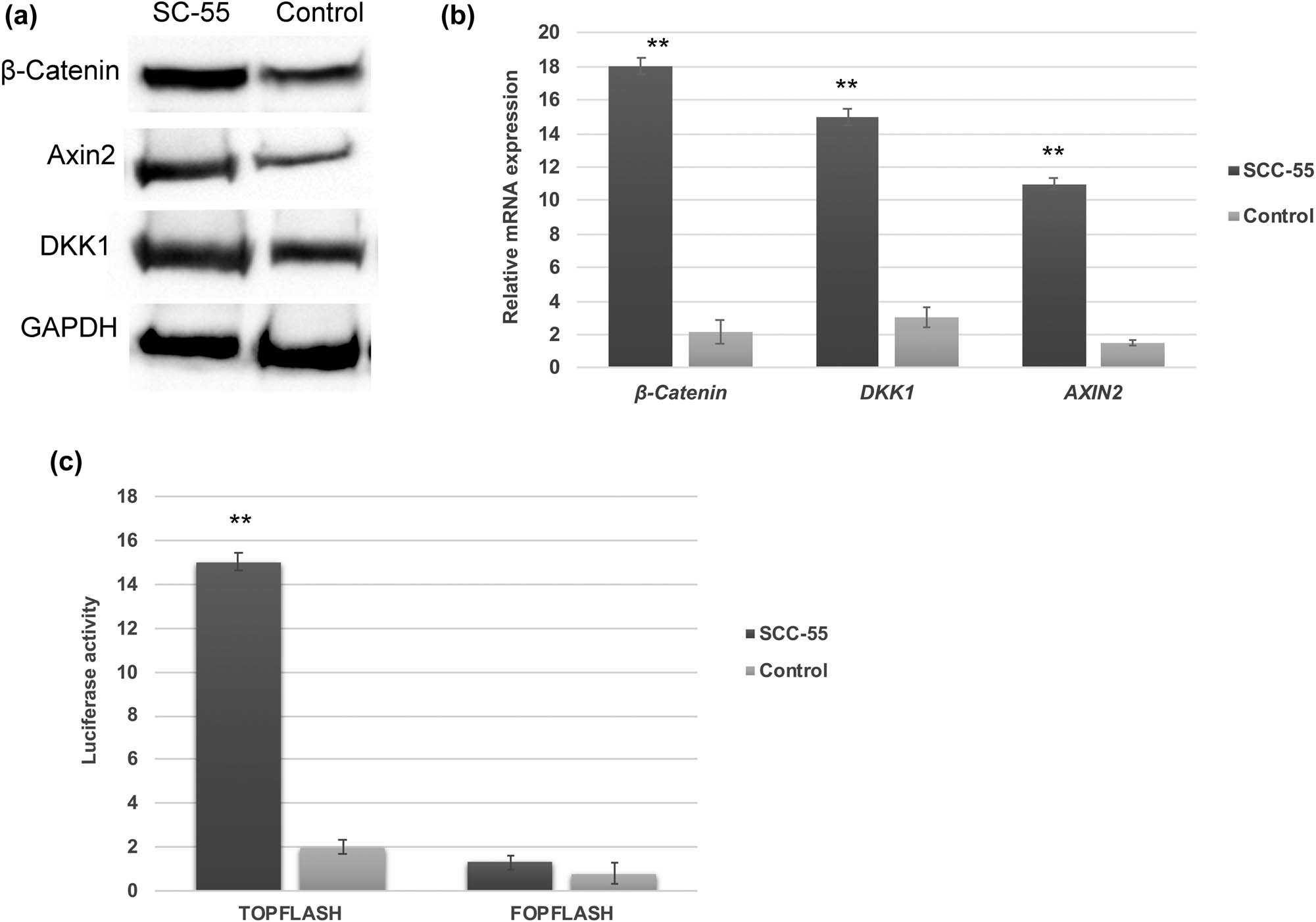
Aberrant activation of Wnt/β-catenin signaling in OSCC. Western blot (a) and RT-PCR analysis (b) showing that Wnt/β-catenin and its downstream targets are activated and upregulated in OSCC SCC-55 cells. (c) TOPFLASH luciferase activity showing the elevated transcriptional level of Wnt/β-catenin in SCC-55 cells. The values in the error bar are± standard deviation (SD); ** p < 0.01.
3.2 β-catenin depletion influences apoptosis in SCC-55 cells
As Wnt/β-catenin signaling is highly activated in SCC-55 cells, we attempted to knock out β-catenin by RNA interference approach. As a result, our western blot analysis demonstrated that β-catenin expression was dramatically reduced upon β-catenin–siRNA transfection in SCC-55 cells when compared to scramble RNAi and non-transfected (control) SCC-55 cells (Figure 2a). Cancer cells are highly resistant to apoptosis, and therefore, they developed resistance to chemotherapy. Hence, the β-catenin depleted cells are subjected to the evaluation of chemotherapy resistance and apoptosis induction. As a result, we found that SCC-55-β-catenin RNAi cells became more sensitive to DNA-binding drugs such as 5-FU and cisplatin. As a result, the survival rate of β-catenin RNAi cells significantly declined and thus indicated apoptosis (Figure 2b). In addition, the evaluation of apoptosis by flow cytometry revealed that the enhanced rate of apoptosis in β-catenin-depleted SCC-55 rather than scramble RNAi and non-transfected SC-55 cells (Figure 2c). Collectively, these findings suggest that the downregulation of β-catenin was able to induce apoptosis effectively in oral cancer cells, and therefore, cancer cell survival was compromised.

β-catenin knockdown induces apoptosis in SCC-55 cells. (a) Western blot data showing decreased β-catenin expression after β-catenin siRNA transfection in SCC-55 cells. (b) Chemoresistance assay showing that β-catenin RNAi cells viability are significantly reduced. (c) Flow cytometry assessment confirms the enhanced rate of apoptosis in β-catenin RNAi SCC-55 cells when compared to controls. The values in the error bar are ± standard deviation (SD); **, p < 0.01.
3.3 β-catenin downregulation impedes cell proliferation and tumorigenesis
Next, we have examined whether the β-catenin downregulation has a deleterious effect on the tumorigenic potential of oral cancer cells. In vitro proliferation assay demonstrated that the proliferation rate of β-catenin-depleted SCC-55 cells was significantly deteriorated from day 1 to day 7 than the non-transfected and negative control ones (Figure 3a). Also, the colony formation efficiency of β-catenin RNAi cells was reduced (<3-fold times), as they regenerated significantly fewer colonies on soft-agar assay (Figure 3b). Hence, these data indicate that β-catenin–siRNA was able to limit cell survival and self-renewal properties of oral cancer cells, which may be governed by the interaction of stemness genes.
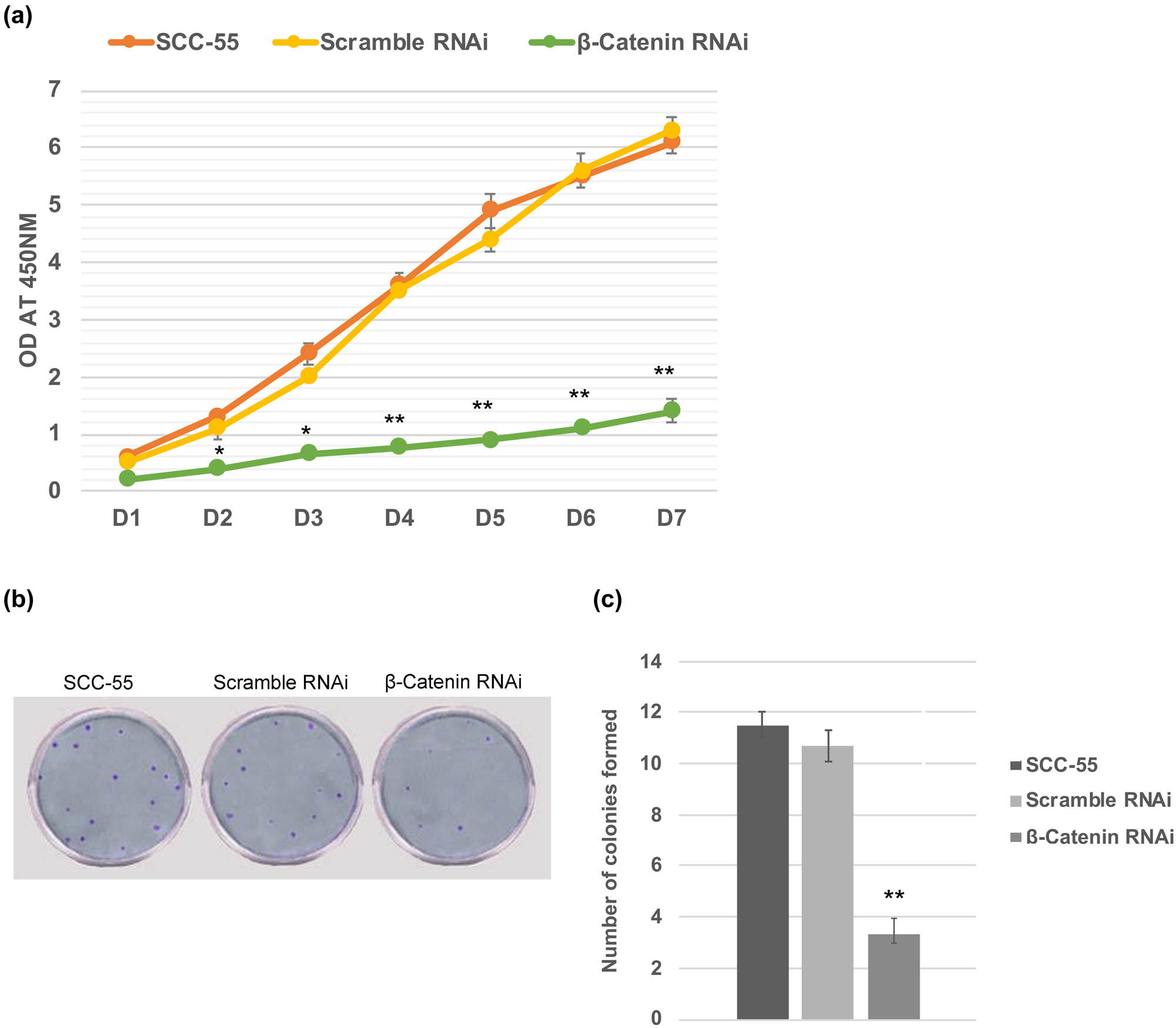
β-catenin knockdown impedes cell proliferation and inhibits tumorigenicity. (a) In vitro cell proliferation assay displaying that the growth rate of SCC-55 cells was compromised upon silencing of β-catenin. (b) The number of colonies formed in the soft agar assay was dramatically less upon siRNA of β-catenin transfection in SCC-55 cells. The values in the error bar are ± standard deviation (SD). * p < 0.05; ** p < 0.01.
3.4 Inactivation of caspases and activation of Oct-4 by Wnt/β-catenin signaling
It has been well demonstrated that cytochrome c release is crucial for the onset of mitochondrial-mediated intrinsic apoptosis through the activation of caspases. Consequently, the protein expression pattern of cytochrome c, caspases 3 and 9 were evaluated by western blot analysis, and they were all found to be highly upregulated in β-catenin–siRNA cells (Figure 4a). Concurrently, the relative mRNA expression of the OCT-4 gene was accelerated in β-catenin–siRNA cells (Figure 4b), compared to non-transfected SCC-55 cells. Altogether, these data suggest that β-catenin downregulation might promote caspases activation, leading to apoptosis in β-catenin–siRNA cells. On the other hand, β-catenin–siRNA caused decreased tumorigenic potential, which could be due to transcriptional downregulation of OCT-4.
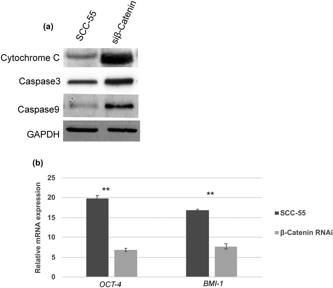
Activation of intrinsic apoptotic pathway and downregulation of OCT-4 in β-catenin RNAi cells. (a) Western blot analysis showing activation and enhanced expression of cytochrome c, caspase 3, and caspase 9 in β-catenin RNAi SCC-55 cells. (b) RT-PCR data showing relative mRNA expression of OCT-4 was significantly downregulated in β-catenin RNAi cells compared to the controls. The values in the error bar are ± standard deviation (SD); ** p < 0.01.
4 Discussion
In recent times, tumor initiation, progression, and invasion have become complex mechanisms that involved many cell adhesion molecules and intracellular signaling pathways for cell migration and invasion [23,24]. Mainly, Wnt/β-catenin signaling has gained more attention as it plays a crucial role in regulating cell proliferation, self-renewal, and differentiation of cancer cells and embryonic stem cells [25]. According to pathological studies, human neck and squamous cell carcinoma (HNSCC) has been associated with hyperactivation of Wnt/β-catenin signaling, contributing to HNSCC invasion [26,27,28]. In the present study, we have found abnormal activation of Wnt/β-catenin signaling in OSCC SCC-55 cells, whose protein and relative mRNA expression are significantly upregulated in SCC-55 cells.
β-catenin is an essential stimulation center for Wnt signaling pathways; cytoplasmic multifunctional protein can interact with E-cadherin adhesion molecule in a calcium-dependent manner [29,31]. Overexpression of β-catenin has been reported in different cancers, including breast, skin, blood, head, and neck [11,12,13], and they are associated with high-risk tumorigenesis and invasion [14]. Thus, it has been suggested that two different roles might be performed by the aberrantly activated Wnt/β-catenin signaling in cancer cells: (a) promote tumorigenesis and invasion and (b) suppress cell detachment-mediated apoptosis [28]. Keeping this in mind, we investigated whether β-catenin depletion influences apoptosis and tumorigenic potential of SCC-55 cells.
In the present article, we have used siRNA that could specifically target β-catenin and suppress its expression. Our RT-PCR and western blot data confirmed the significantly downregulated levels of β-catenin, and thus, the siRNA used could effectively inhibit the Wnt signaling pathway. Interestingly, our flow cytometry analysis demonstrated that apoptosis was significantly initiated in β-catenin-depleted SCC-55 cells. As a result, these cells became more susceptible when treated with DNA targeting drugs such as 5-FU and cisplatin. The precise molecular mechanism of apoptosis induction in SCC-55 cells upon β-catenin gene depletion is not known. However, we found elevated caspase-3 and caspase-9 in β-catenin RNAi cells, indicating that PI3/AKT signaling might be downregulated, promoting mitochondria-mediated intrinsic apoptotic pathways [30,31,32,33]. Similar to our findings, studies in the colon cancer cell line demonstrated that knocking of β-catenin significantly increased the rate of apoptosis through the activation of caspase-3 and limited the tumor invasion process in SW480 cells [14].
The present study has also demonstrated that the cell proliferation rate slowed down dramatically, and the number of colonies formed in the soft agar assay was significantly compromised when the β-catenin gene expression was downregulated. By considering these data, it can be suggested that the silencing of β-catenin may directly disrupt the expression of genes involved in the regulation of the cell cycle, cell proliferation, and self-renewal of oral cancer cells. Accordingly, our RT-PCR analysis revealed that OCT-4 gene transcription was significantly downregulated in SCC-55 cells upon β-catenin silencing. Elevated expression of OCT-4 has been identified in several cancers, and it acts as a critical regulator of cancer invasion and colony formation [34,35,36]. Furthermore, knockdown of OCT-4 results in decreased tumor invasion and colony formation efficiency in lung cancer [35].
On the other hand, it has been reported that the therapeutic drug resistance of liver cancer cells is mediated by overexpression of OCT-4 [35,36]. Therefore, our results suggest here that Wnt/β-catenin signaling aberrant activation and traits in oral squamous cell carcinoma SCC-55 cells might occur through the activation of OCT-4. However, further research is required for elucidating the β-catenin-mediated anti-apoptotic mechanism, and its downstream effectors would undoubtedly provide more insights for precisely disrupting the cascade events of Wnt signaling pathways to kill the cancer cells.
-
Funding information: This study was supported by the Project of Huzhou Science and Technology Bureau. Funding No: 2020GYB11.
-
Conflict of interest: The authors state no conflict of interest.
-
Data availability statement: The datasets generated during and/or analyzed during the current study are available from the corresponding author on reasonable request.
References
[1] Reyes M, Flores T, Betancur D, Peña-Oyarzún D, Torres VA. Wnt/β-Catenin signaling in oral carcinogenesis. Int J Mol Sci. 2020;21(13):4682.10.3390/ijms21134682Search in Google Scholar PubMed PubMed Central
[2] Leemans CR, Braakhuis BJ, Brakenhoff RH. The molecular biology of head and neck cancer. Nat Rev Cancer. 2011;11:9–22.10.1038/nrc2982Search in Google Scholar PubMed
[3] Lee TY, Tseng YH. The potential of phytochemicals in oral cancer prevention and therapy: a review of the evidence. Biomolecules. 2020;10(8):1150.10.3390/biom10081150Search in Google Scholar PubMed PubMed Central
[4] Polanska H, Raudenska M, Gumulec J, Sztalmachova M, Adam V, Kizek R, et al. Clinical significance of head and neck squamous cell cancer biomarkers. Oral Oncol. 2014;50:168–77.10.1016/j.oraloncology.2013.12.008Search in Google Scholar PubMed
[5] Nasser W, Flechtenmacher C, Holzinger D, Hofele C, Bosch FX. Aberrant expression of p53, p16INK4a and Ki-67 as basic biomarker for malignant progression of oral leukoplakias. J Oral Pathol Med. 2011;40:629–35.10.1111/j.1600-0714.2011.01026.xSearch in Google Scholar PubMed
[6] Ramakrishna A, Shreedhar B, Narayan T, Mohanty L, Shenoy S, Jamadar S. Cyclin D1 an early biomarker in oral carcinogenesis. J Oral Maxillofac Pathol. 2013;17:351–7.10.4103/0973-029X.125189Search in Google Scholar PubMed PubMed Central
[7] Guan G, Bakr MM, Firth N, Love RM. Expression of cyclin D1 correlates with p27KIP1 and regulates the degree of oral dysplasia and squamous cell carcinoma differentiation. Oral Surg Oral Med Oral Pathol Oral Radiol. 2018;126:174–83.10.1016/j.oooo.2018.01.015Search in Google Scholar PubMed
[8] Shah NG, Trivedi TI, Tankshali RA, Goswami JV, Jetly DH, Shukla SN, et al. Prognostic significance of molecular markers in oral squamous cell carcinoma: a multivariate analysis. Head Neck. 2009;31:1544–56.10.1002/hed.21126Search in Google Scholar PubMed
[9] Fodde R, Brabletz T. Wnt/beta-catenin signaling in cancer stemness and malignant behavior. Curr Opin Cell Biol. 2007;19:150–8.10.1016/j.ceb.2007.02.007Search in Google Scholar PubMed
[10] Ring A, Kim YM, Kahn M. Wnt/catenin signaling in adult stem cell physiology and disease. Stem Cell Rev. 2014;10:512–25.10.1007/s12015-014-9515-2Search in Google Scholar PubMed PubMed Central
[11] Clevers H, Nusse R. Wnt/β-catenin signaling and disease. Cell. 2012;149:1192–1205.10.1016/j.cell.2012.05.012Search in Google Scholar PubMed
[12] Schmeel LC, Schmeel FC, Kim Y, Endo T, Lu D, Schmidt-Wolf IG. Targeting the Wnt/beta-catenin pathway in multiple myeloma. Anticancer Res. 2013;33:4719–26.Search in Google Scholar
[13] Yao H, Ashihara E, Maekawa T. Targeting the Wnt/β-catenin signaling pathway in human cancers. Expert Opin Ther Targets. 2011;15:873–87.10.1517/14728222.2011.577418Search in Google Scholar PubMed
[14] Li K, Zhou ZY, Ji PP, Luo HS. Knockdown of β-catenin by siRNA influences proliferation, apoptosis and invasion of the colon cancer cell line. Oncol Lett. 2016;11(6):3896–900.10.3892/ol.2016.4481Search in Google Scholar PubMed PubMed Central
[15] Fan K, Li N, Qi J, Yin P, Zhao C, Wang L, et al. Wnt/β-catenin signaling induces the transcription of cystathionine-γ-lyase, a stimulator of tumor in colon cancer. Cell Signal. 2014;26:2801–8.10.1016/j.cellsig.2014.08.023Search in Google Scholar PubMed
[16] Behrens J. Control of beta-catenin signaling in tumor development. Ann N Y Acad Sci. 2000;910:21–35.10.1111/j.1749-6632.2000.tb06698.xSearch in Google Scholar PubMed
[17] Liu Y, Cui P, Chen J, Li W. Isolation and phenotypic characterization of side population cells in oral squamous cell carcinoma. Mol Med Rep. 2015;11(5):3642–6.10.3892/mmr.2014.3133Search in Google Scholar PubMed
[18] Song J, Chang I, Chen Z, Kang M, Wang CY. Characterization of side populations in HNSCC: highly invasive, chemoresistant and abnormal Wnt signaling. PLoS One. 2010;5(7):e11456.10.1371/journal.pone.0011456Search in Google Scholar PubMed PubMed Central
[19] Wamunyokoli FW, Bonome T, Lee JY, Feltmate CM, Welch WR, Radanovich M, et al. Expression profiling 23 of mucinous tumors of the ovary identifies genes of clinicopathologic importance. Clin Cancer Res. 2006;12:690–700.10.1158/1078-0432.CCR-05-1110Search in Google Scholar PubMed
[20] Song P, Zheng XJ, Liu JZ, Xu J, Wu LY, Liu C, et al. Effect of the Wnt1/β-catenin signalling pathway on human embryonic pulmonary fibroblasts. Mol Med Rep. 2014;10(2):1030–6.10.3892/mmr.2014.2261Search in Google Scholar PubMed
[21] Guan GF, Zhang DJ, Zheng Y, Wen LJ, Yu DJ, Lu YQ, et al. Significance of ATP-binding cassette transporter proteins in multidrug resistance of head and neck squamous cell carcinoma. Oncol Lett. 2015;10(2):631–6.10.3892/ol.2015.3359Search in Google Scholar PubMed PubMed Central
[22] He QZ, Luo XZ, Wang K, Zhou Q, Ao H, Yang Y, et al. Isolation and characterization of cancer stem cells from high-grade serous ovarian carcinomas. Cell Physiol Biochem. 2014;33(1):173–84.10.1159/000356660Search in Google Scholar PubMed
[23] Tan CW, Gardiner BS, Hirokawa Y, Layton MJ, Smith DW, Burgess AW. Wnt signalling pathway parameters for mammalian cells. PLoS One. 2012;7:e31882.10.1371/journal.pone.0031882Search in Google Scholar PubMed PubMed Central
[24] Paul S, Dey A. Wnt signaling and cancer development: Therapeutic implication. Neoplasma. 2008;55:165–76.Search in Google Scholar
[25] Lee SH, Koo BS, Kim JM, Huang S, Rho YS, Bae WJ, et al. Wnt/β‐catenin signalling maintains self‐renewal and tumourigenicity of head and neck squamous cell carcinoma stem‐like cells by activating Oct4. J Pathol. 2014;234(1):99–107.10.1002/path.4383Search in Google Scholar PubMed
[26] Rhee CS, Sen M, Lu D, Wu C, Leoni L, Rubin J, et al. Wnt and frizzled receptors as potential targets for immunotherapy in head and neck squamous cell carcinomas. Oncogene. 2002;21(43):6598–605.10.1038/sj.onc.1205920Search in Google Scholar PubMed
[27] Uraguchi M, Morikawa M, Shirakawa M, Sanada K, Imai K. Activation of WNT family expression and signaling in squamous cell carcinomas of the oral cavity. J Dent Res. 2004;83(4):327–32.10.1177/154405910408300411Search in Google Scholar PubMed
[28] Yang F, Zeng Q, Yu G, Li S, Wang CY. Wnt/β-catenin signaling inhibits death receptor-mediated apoptosis and promotes invasive growth of HNSCC. Cell Signal. 2006;18(5):679–87.10.1016/j.cellsig.2005.06.015Search in Google Scholar PubMed
[29] Lai T, Su C, Kuo W, Yeh Y, Kuo W, Tsai F, et al. β-catenin plays a key role in metastasis of human hepatocellular carcinoma. Oncol Rep. 2011;26:415–22.Search in Google Scholar
[30] Voronkov A, Krauss S. Wnt/beta-catenin signaling and small molecule inhibitors. Curr Pharm Des. 2013;19:634–64.10.2174/138161213804581837Search in Google Scholar PubMed PubMed Central
[31] McIlwain DR, Berger T, Mak TW. Caspase functions in cell death and disease. Cold Spring Harb Perspect Biol. 2013;5(4):a008656.10.1101/cshperspect.a008656Search in Google Scholar PubMed PubMed Central
[32] Elena-Real CA, Díaz-Quintana A, González-Arzola K, Velázquez-Campoy A, et al. Cytochrome c speeds up caspase cascade activation by blocking 14-3-3ε-dependent Apaf-1 inhibition. Cell Death Dis. 2018;9(3):365.10.1038/s41419-018-0408-1Search in Google Scholar PubMed PubMed Central
[33] Parsons MJ, Rehm M, Bouchier-Hayes L. Imaging-based methods for assessing caspase activity in single cells. Cold Spring Harb Protoc. 2015;2015(1):PDB.top070342.10.1101/pdb.top070342Search in Google Scholar PubMed
[34] Atlas Y, Mowla SJ, Ziaee SA, Bahrami AR. OCT-4, an embryonic stem cell marker, is highly expressed in bladder cancer. Int J Cancer. 2007;120:1598–602.10.1002/ijc.22508Search in Google Scholar PubMed
[35] Chen YC, Hsu HS, Chen YW, Tsai TH, How CK, Wang CY, et al. Oct-4 expression maintained cancer stem-like properties in lung cancer-derived CD133-positive cells. PLoS One. 2008;3:e2637.10.1371/journal.pone.0002637Search in Google Scholar PubMed PubMed Central
[36] Wang XQ, Ongkeko WM, Chen L, Yang ZF, Lu P, Chen KK, et al. Octamer 4 (Oct4) mediates chemotherapeutic drug resistance in liver cancer cells through a potential Oct4–AKT–ATP-binding cassette G2 pathway. Hepatology. 2010;52:528–39.10.1002/hep.23692Search in Google Scholar PubMed
© 2021 Yufeng Wang et al., published by De Gruyter
This work is licensed under the Creative Commons Attribution 4.0 International License.
Articles in the same Issue
- Biomedical Sciences
- Research progress on the mechanism of orexin in pain regulation in different brain regions
- Adriamycin-resistant cells are significantly less fit than adriamycin-sensitive cells in cervical cancer
- Exogenous spermidine affects polyamine metabolism in the mouse hypothalamus
- Iris metastasis of diffuse large B-cell lymphoma misdiagnosed as primary angle-closure glaucoma: A case report and review of the literature
- LncRNA PVT1 promotes cervical cancer progression by sponging miR-503 to upregulate ARL2 expression
- Two new inflammatory markers related to the CURB-65 score for disease severity in patients with community-acquired pneumonia: The hypersensitive C-reactive protein to albumin ratio and fibrinogen to albumin ratio
- Circ_0091579 enhances the malignancy of hepatocellular carcinoma via miR-1287/PDK2 axis
- Silencing XIST mitigated lipopolysaccharide (LPS)-induced inflammatory injury in human lung fibroblast WI-38 cells through modulating miR-30b-5p/CCL16 axis and TLR4/NF-κB signaling pathway
- Protocatechuic acid attenuates cerebral aneurysm formation and progression by inhibiting TNF-alpha/Nrf-2/NF-kB-mediated inflammatory mechanisms in experimental rats
- ABCB1 polymorphism in clopidogrel-treated Montenegrin patients
- Metabolic profiling of fatty acids in Tripterygium wilfordii multiglucoside- and triptolide-induced liver-injured rats
- miR-338-3p inhibits cell growth, invasion, and EMT process in neuroblastoma through targeting MMP-2
- Verification of neuroprotective effects of alpha-lipoic acid on chronic neuropathic pain in a chronic constriction injury rat model
- Circ_WWC3 overexpression decelerates the progression of osteosarcoma by regulating miR-421/PDE7B axis
- Knockdown of TUG1 rescues cardiomyocyte hypertrophy through targeting the miR-497/MEF2C axis
- MiR-146b-3p protects against AR42J cell injury in cerulein-induced acute pancreatitis model through targeting Anxa2
- miR-299-3p suppresses cell progression and induces apoptosis by downregulating PAX3 in gastric cancer
- Diabetes and COVID-19
- Discovery of novel potential KIT inhibitors for the treatment of gastrointestinal stromal tumor
- TEAD4 is a novel independent predictor of prognosis in LGG patients with IDH mutation
- circTLK1 facilitates the proliferation and metastasis of renal cell carcinoma by regulating miR-495-3p/CBL axis
- microRNA-9-5p protects liver sinusoidal endothelial cell against oxygen glucose deprivation/reperfusion injury
- Long noncoding RNA TUG1 regulates degradation of chondrocyte extracellular matrix via miR-320c/MMP-13 axis in osteoarthritis
- Duodenal adenocarcinoma with skin metastasis as initial manifestation: A case report
- Effects of Loofah cylindrica extract on learning and memory ability, brain tissue morphology, and immune function of aging mice
- Recombinant Bacteroides fragilis enterotoxin-1 (rBFT-1) promotes proliferation of colorectal cancer via CCL3-related molecular pathways
- Blocking circ_UBR4 suppressed proliferation, migration, and cell cycle progression of human vascular smooth muscle cells in atherosclerosis
- Gene therapy in PIDs, hemoglobin, ocular, neurodegenerative, and hemophilia B disorders
- Downregulation of circ_0037655 impedes glioma formation and metastasis via the regulation of miR-1229-3p/ITGB8 axis
- Vitamin D deficiency and cardiovascular risk in type 2 diabetes population
- Circ_0013359 facilitates the tumorigenicity of melanoma by regulating miR-136-5p/RAB9A axis
- Mechanisms of circular RNA circ_0066147 on pancreatic cancer progression
- lncRNA myocardial infarction-associated transcript (MIAT) knockdown alleviates LPS-induced chondrocytes inflammatory injury via regulating miR-488-3p/sex determining region Y-related HMG-box 11 (SOX11) axis
- Identification of circRNA circ-CSPP1 as a potent driver of colorectal cancer by directly targeting the miR-431/LASP1 axis
- Hyperhomocysteinemia exacerbates ischemia-reperfusion injury-induced acute kidney injury by mediating oxidative stress, DNA damage, JNK pathway, and apoptosis
- Potential prognostic markers and significant lncRNA–mRNA co-expression pairs in laryngeal squamous cell carcinoma
- Gamma irradiation-mediated inactivation of enveloped viruses with conservation of genome integrity: Potential application for SARS-CoV-2 inactivated vaccine development
- ADHFE1 is a correlative factor of patient survival in cancer
- The association of transcription factor Prox1 with the proliferation, migration, and invasion of lung cancer
- Is there a relationship between the prevalence of autoimmune thyroid disease and diabetic kidney disease?
- Immunoregulatory function of Dictyophora echinovolvata spore polysaccharides in immunocompromised mice induced by cyclophosphamide
- T cell epitopes of SARS-CoV-2 spike protein and conserved surface protein of Plasmodium malariae share sequence homology
- Anti-obesity effect and mechanism of mesenchymal stem cells influence on obese mice
- Long noncoding RNA HULC contributes to paclitaxel resistance in ovarian cancer via miR-137/ITGB8 axis
- Glucocorticoids protect HEI-OC1 cells from tunicamycin-induced cell damage via inhibiting endoplasmic reticulum stress
- Prognostic value of the neutrophil-to-lymphocyte ratio in acute organophosphorus pesticide poisoning
- Gastroprotective effects of diosgenin against HCl/ethanol-induced gastric mucosal injury through suppression of NF-κβ and myeloperoxidase activities
- Silencing of LINC00707 suppresses cell proliferation, migration, and invasion of osteosarcoma cells by modulating miR-338-3p/AHSA1 axis
- Successful extracorporeal membrane oxygenation resuscitation of patient with cardiogenic shock induced by phaeochromocytoma crisis mimicking hyperthyroidism: A case report
- Effects of miR-185-5p on replication of hepatitis C virus
- Lidocaine has antitumor effect on hepatocellular carcinoma via the circ_DYNC1H1/miR-520a-3p/USP14 axis
- Primary localized cutaneous nodular amyloidosis presenting as lymphatic malformation: A case report
- Multimodal magnetic resonance imaging analysis in the characteristics of Wilson’s disease: A case report and literature review
- Therapeutic potential of anticoagulant therapy in association with cytokine storm inhibition in severe cases of COVID-19: A case report
- Neoadjuvant immunotherapy combined with chemotherapy for locally advanced squamous cell lung carcinoma: A case report and literature review
- Rufinamide (RUF) suppresses inflammation and maintains the integrity of the blood–brain barrier during kainic acid-induced brain damage
- Inhibition of ADAM10 ameliorates doxorubicin-induced cardiac remodeling by suppressing N-cadherin cleavage
- Invasive ductal carcinoma and small lymphocytic lymphoma/chronic lymphocytic leukemia manifesting as a collision breast tumor: A case report and literature review
- Clonal diversity of the B cell receptor repertoire in patients with coronary in-stent restenosis and type 2 diabetes
- CTLA-4 promotes lymphoma progression through tumor stem cell enrichment and immunosuppression
- WDR74 promotes proliferation and metastasis in colorectal cancer cells through regulating the Wnt/β-catenin signaling pathway
- Down-regulation of IGHG1 enhances Protoporphyrin IX accumulation and inhibits hemin biosynthesis in colorectal cancer by suppressing the MEK-FECH axis
- Curcumin suppresses the progression of gastric cancer by regulating circ_0056618/miR-194-5p axis
- Scutellarin-induced A549 cell apoptosis depends on activation of the transforming growth factor-β1/smad2/ROS/caspase-3 pathway
- lncRNA NEAT1 regulates CYP1A2 and influences steroid-induced necrosis
- A two-microRNA signature predicts the progression of male thyroid cancer
- Isolation of microglia from retinas of chronic ocular hypertensive rats
- Changes of immune cells in patients with hepatocellular carcinoma treated by radiofrequency ablation and hepatectomy, a pilot study
- Calcineurin Aβ gene knockdown inhibits transient outward potassium current ion channel remodeling in hypertrophic ventricular myocyte
- Aberrant expression of PI3K/AKT signaling is involved in apoptosis resistance of hepatocellular carcinoma
- Clinical significance of activated Wnt/β-catenin signaling in apoptosis inhibition of oral cancer
- circ_CHFR regulates ox-LDL-mediated cell proliferation, apoptosis, and EndoMT by miR-15a-5p/EGFR axis in human brain microvessel endothelial cells
- Resveratrol pretreatment mitigates LPS-induced acute lung injury by regulating conventional dendritic cells’ maturation and function
- Ubiquitin-conjugating enzyme E2T promotes tumor stem cell characteristics and migration of cervical cancer cells by regulating the GRP78/FAK pathway
- Carriage of HLA-DRB1*11 and 1*12 alleles and risk factors in patients with breast cancer in Burkina Faso
- Protective effect of Lactobacillus-containing probiotics on intestinal mucosa of rats experiencing traumatic hemorrhagic shock
- Glucocorticoids induce osteonecrosis of the femoral head through the Hippo signaling pathway
- Endothelial cell-derived SSAO can increase MLC20 phosphorylation in VSMCs
- Downregulation of STOX1 is a novel prognostic biomarker for glioma patients
- miR-378a-3p regulates glioma cell chemosensitivity to cisplatin through IGF1R
- The molecular mechanisms underlying arecoline-induced cardiac fibrosis in rats
- TGF-β1-overexpressing mesenchymal stem cells reciprocally regulate Th17/Treg cells by regulating the expression of IFN-γ
- The influence of MTHFR genetic polymorphisms on methotrexate therapy in pediatric acute lymphoblastic leukemia
- Red blood cell distribution width-standard deviation but not red blood cell distribution width-coefficient of variation as a potential index for the diagnosis of iron-deficiency anemia in mid-pregnancy women
- Small cell neuroendocrine carcinoma expressing alpha fetoprotein in the endometrium
- Superoxide dismutase and the sigma1 receptor as key elements of the antioxidant system in human gastrointestinal tract cancers
- Molecular characterization and phylogenetic studies of Echinococcus granulosus and Taenia multiceps coenurus cysts in slaughtered sheep in Saudi Arabia
- ITGB5 mutation discovered in a Chinese family with blepharophimosis-ptosis-epicanthus inversus syndrome
- ACTB and GAPDH appear at multiple SDS-PAGE positions, thus not suitable as reference genes for determining protein loading in techniques like Western blotting
- Facilitation of mouse skin-derived precursor growth and yield by optimizing plating density
- 3,4-Dihydroxyphenylethanol ameliorates lipopolysaccharide-induced septic cardiac injury in a murine model
- Downregulation of PITX2 inhibits the proliferation and migration of liver cancer cells and induces cell apoptosis
- Expression of CDK9 in endometrial cancer tissues and its effect on the proliferation of HEC-1B
- Novel predictor of the occurrence of DKA in T1DM patients without infection: A combination of neutrophil/lymphocyte ratio and white blood cells
- Investigation of molecular regulation mechanism under the pathophysiology of subarachnoid hemorrhage
- miR-25-3p protects renal tubular epithelial cells from apoptosis induced by renal IRI by targeting DKK3
- Bioengineering and Biotechnology
- Green fabrication of Co and Co3O4 nanoparticles and their biomedical applications: A review
- Agriculture
- Effects of inorganic and organic selenium sources on the growth performance of broilers in China: A meta-analysis
- Crop-livestock integration practices, knowledge, and attitudes among smallholder farmers: Hedging against climate change-induced shocks in semi-arid Zimbabwe
- Food Science and Nutrition
- Effect of food processing on the antioxidant activity of flavones from Polygonatum odoratum (Mill.) Druce
- Vitamin D and iodine status was associated with the risk and complication of type 2 diabetes mellitus in China
- Diversity of microbiota in Slovak summer ewes’ cheese “Bryndza”
- Comparison between voltammetric detection methods for abalone-flavoring liquid
- Composition of low-molecular-weight glutenin subunits in common wheat (Triticum aestivum L.) and their effects on the rheological properties of dough
- Application of culture, PCR, and PacBio sequencing for determination of microbial composition of milk from subclinical mastitis dairy cows of smallholder farms
- Investigating microplastics and potentially toxic elements contamination in canned Tuna, Salmon, and Sardine fishes from Taif markets, KSA
- From bench to bar side: Evaluating the red wine storage lesion
- Establishment of an iodine model for prevention of iodine-excess-induced thyroid dysfunction in pregnant women
- Plant Sciences
- Characterization of GMPP from Dendrobium huoshanense yielding GDP-D-mannose
- Comparative analysis of the SPL gene family in five Rosaceae species: Fragaria vesca, Malus domestica, Prunus persica, Rubus occidentalis, and Pyrus pyrifolia
- Identification of leaf rust resistance genes Lr34 and Lr46 in common wheat (Triticum aestivum L. ssp. aestivum) lines of different origin using multiplex PCR
- Investigation of bioactivities of Taxus chinensis, Taxus cuspidata, and Taxus × media by gas chromatography-mass spectrometry
- Morphological structures and histochemistry of roots and shoots in Myricaria laxiflora (Tamaricaceae)
- Transcriptome analysis of resistance mechanism to potato wart disease
- In silico analysis of glycosyltransferase 2 family genes in duckweed (Spirodela polyrhiza) and its role in salt stress tolerance
- Comparative study on growth traits and ions regulation of zoysiagrasses under varied salinity treatments
- Role of MS1 homolog Ntms1 gene of tobacco infertility
- Biological characteristics and fungicide sensitivity of Pyricularia variabilis
- In silico/computational analysis of mevalonate pyrophosphate decarboxylase gene families in Campanulids
- Identification of novel drought-responsive miRNA regulatory network of drought stress response in common vetch (Vicia sativa)
- How photoautotrophy, photomixotrophy, and ventilation affect the stomata and fluorescence emission of pistachios rootstock?
- Apoplastic histochemical features of plant root walls that may facilitate ion uptake and retention
- Ecology and Environmental Sciences
- The impact of sewage sludge on the fungal communities in the rhizosphere and roots of barley and on barley yield
- Domestication of wild animals may provide a springboard for rapid variation of coronavirus
- Response of benthic invertebrate assemblages to seasonal and habitat condition in the Wewe River, Ashanti region (Ghana)
- Molecular record for the first authentication of Isaria cicadae from Vietnam
- Twig biomass allocation of Betula platyphylla in different habitats in Wudalianchi Volcano, northeast China
- Animal Sciences
- Supplementation of probiotics in water beneficial growth performance, carcass traits, immune function, and antioxidant capacity in broiler chickens
- Predators of the giant pine scale, Marchalina hellenica (Gennadius 1883; Hemiptera: Marchalinidae), out of its natural range in Turkey
- Honey in wound healing: An updated review
- NONMMUT140591.1 may serve as a ceRNA to regulate Gata5 in UT-B knockout-induced cardiac conduction block
- Radiotherapy for the treatment of pulmonary hydatidosis in sheep
- Retraction
- Retraction of “Long non-coding RNA TUG1 knockdown hinders the tumorigenesis of multiple myeloma by regulating microRNA-34a-5p/NOTCH1 signaling pathway”
- Special Issue on Reuse of Agro-Industrial By-Products
- An effect of positional isomerism of benzoic acid derivatives on antibacterial activity against Escherichia coli
- Special Issue on Computing and Artificial Techniques for Life Science Applications - Part II
- Relationship of Gensini score with retinal vessel diameter and arteriovenous ratio in senile CHD
- Effects of different enantiomers of amlodipine on lipid profiles and vasomotor factors in atherosclerotic rabbits
- Establishment of the New Zealand white rabbit animal model of fatty keratopathy associated with corneal neovascularization
- lncRNA MALAT1/miR-143 axis is a potential biomarker for in-stent restenosis and is involved in the multiplication of vascular smooth muscle cells