
Correlation between peripheral blood lymphocyte subpopulations and primary systemic lupus erythematosus
-
Yan Feng
 ,
Zhijun Li
,
Zhijun Li

Abstract
This study explored the correlation between peripheral blood CD3+, CD3+/CD4+, CD3+/CD8+, CD4+/CD8+, CD3−/CD16+ CD56+, and CD3−CD19+ and disease activity of different subtypes of systemic lupus erythematosus (SLE). The percentages of CD3+, CD3+/CD4+, CD3+/CD8+, CD4+/CD8+, CD3−/CD16+ CD56+, and CD3−CD19+ in the peripheral blood of patients (n = 80) classified into lupus nephritis, blood involvement, and joint involvement and SLE in different active stages were detected by flow cytometry. Their correlations with baseline clinical experimental indicators of SLE patients’ SLE disease activity index score (SLEDAI) and complement C3 were analyzed. The results showed that CD3+, CD3+/CD4+, and CD3+/CD8+ at baseline level were negatively correlated with SLEDAI scores. These were positively correlated with C3. In conclusion, T-lymphocyte subpopulations are closely related to SLE activity and can be used as reference indicators to evaluate the SLE activity.
1 Introduction
Systemic lupus erythematosus (SLE) is an autoimmune disease characterized by excessive B cells and an imbalance of T and natural killer (NK) lymphocytes. In addition to T, B, and NK lymphocytes in SLE, there are disorders and mediators of cytokine communication that can be used as prognostic markers of the disease [1,2]. It involves multiple organs and systems, such as the skin, joints, blood, kidneys, and respiratory system, and its pathogenesis is not yet fully understood [3]. Dysregulated lymphocyte subpopulation ratios and abnormal immune cytokine expression are associated with the paroxysm of SLE and might be major factors in the pathogenesis of SLE [4,5]. This study examined the correlations between the expression percentage of T, NK, and B lymphocytes in the peripheral blood and the disease activity of 80 patients with different subtypes of SLE. In addition, the correlation between the differences in the expression of lymphocyte subpopulations in various clinical subtypes of SLE and the clinical laboratory indexes, such as SLE disease activity index (SLEDAI) score, complement C3, complement C4, IgG, IgA, IgM, erythrocyte sedimentation rate (ESR), and C-reactive protein (CRP), were analyzed. The present study aimed to identify the close correlation between lymphocyte subpopulations and SLE disease activity and provide a basis for the clinical diagnosis and treatment of the disease.
2 Materials and methods
2.1 General materials
The study collected clinical data from 80 in-patients with SLE from January 2019 to early June 2021 at our hospital. Inclusion criteria were as follows: (i) diagnosis in line with the 2019 EULAR/ACR SLE classification criteria [6]; (ii) SLEDAI score ≥5; and (iii) no steroid hormone or immunosuppressive therapy before inclusion. Exclusion criteria were as follows: (i) comorbidities such as other rheumatic immune diseases, tumors, and severe infections; (ii) SLEDAI score <5; and (iii) patients with SLE currently under standard treatment.
-
Informed consent: Informed consent has been obtained from all individuals included in this study.
-
Ethical approval: The research related to human use has been complied with all the relevant national regulations and institutional policies and in accordance with the tenets of the Declaration of Helsinki and has been approved by the First Affiliated Hospital of Anhui University of Science and Technology (2019074X).
2.2 SLEDAI score
Disease activity was assessed according to the SLEDAI 2K scoring system [7], with 0–4 being stable, 5–9 being mildly active, 10–14 being moderately active, and ≥15 being severely active.
2.3 Laboratory
On the second day after admission, 2 ml of fasting venous blood was taken in the morning and anticoagulated with EDTA. Two TruCount tubes were numbered A and B in sequence, and 50 µl of fully mixed anticoagulated whole blood was added into the tubes. Then, 10 µl of the CD3FITC/CD8PE/CD45PercP/CD4APC antibody was added into test tube A, and 10 µl of the CD3FITC/CD16 + 56PE/CD45 PercP/CD19APC antibody was added into test tube B. The tubes were vortexed, mixed evenly, and placed in a dark environment at room temperature for 15 min. Then, 450 µl of red blood cell lysate was added, fully mixed, and kept away from light at room temperature for 15 min. The cells were washed twice with PBS buffer and placed into a flow cytometry system (FC 500 MCL, Beckman Coulter, Brea, CA, USA) [8]. The flow cytometry laser was a cold sub laser with a wavelength of 488 nm. The light path was adjusted by the flow check. The voltage was adjusted. The lateral angular scattering (SSC) was the abscissa, and the forward angular scattering (FSC) was the ordinate. On the scatter diagram, the gating technique was used to analyze the percentage of total T cells (CD3+), CD4+ cells (CD3+/CD4+), CD8+ cells (CD3+/CD8+), NK cells, and B cells in the lymphocyte population. The ratio of the CD4+/CD8+ cells was calculated. The study collected complements C3 and C4, ESR, CRP, IgA, IgM, and IgG from 80 patients with primary SLE using immunoturbidimetric analysis on a BN-II-specific protein analyzer (Siemens, Erlangen, Germany).
2.4 Statistical analysis
The flow cytometry system software was CXP 2.0, and the statistical software for data analysis was SPSS 25.0. The normally distributed continuous data were expressed as mean ± standard deviation, and the t-test was used to compare groups. Correlations were analyzed using the Spearman test. The graphs were plotted using Spearman and GraphPad Prism software for correlation analysis. P < 0.05 indicated statistical significance.
3 Results
3.1 Basic clinical features of SLE patients
The cohort of 80 SLE patients consisted of four males and 76 females, with a male-to-female ratio of 1:19. The patients were 39.3 ± 13.9 (range: 15–75) years of age. The duration of the disease was 1–36 months. Among them, 55 cases had blood involvement (68.8%), 30 had lupus nephritis (LN) (37.5%), 36 had joint involvement (45%), 38 had skin involvement (47.5%), 6 had alopecia (7.5%), 16 had stomatitis (20%), 4 had pleural effusion (5%), 2 had pericardial effusion (2.5%), 19 had a fever (23.8%), and 1 had gastrointestinal vasculitis (1.3%).
3.2 Comparison between the LN and non-LN groups
The expression percentages of peripheral blood T cells (CD3+/CD4+ and CD4+/CD8+) and NK lymphocytes (CD3−/CD16+ CD56+) in the LN group were lower than in the non-LN group. On the other hand, the peripheral blood T cells (CD3+/CD8+) were higher than those in the non-LN group (P < 0.05). Moreover, the percentages of CD3+ and CD3−/CD19+ did not differ significantly in the two groups (P > 0.05; Table 1).
Results of peripheral blood lymphocyte subpopulation testing in the LN and non-LN Groups of SLE (mean ± standard deviation)
| Group | N | CD3+ (%) | CD3+/CD4+ (%) | CD3+/CD8+ (%) | CD4+/CD8+ | CD3−/CD16+CD56+ (%) | CD3−/CD19+ (%) |
|---|---|---|---|---|---|---|---|
| LN | 30 | 72.17 ± 7.13 | 25.27 ± 3.99 | 46.57 ± 6.3 | 0.55 ± 0.11 | 6.13 ± 2.05 | 6.17 ± 2.07 |
| Non-LN | 50 | 69.56 ± 6.36 | 28.64 ± 5.11 | 40.52 ± 3.97 | 0.71 ± 0.17 | 9.08 ± 1.96 | 6.4 ± 1.87 |
| t | 1.646 | −3.286 | 4.727 | −4.917 | −6.337 | −0.506 | |
| P | 0.105 | 0.002 | 0.001 | 0.001 | 0.001 | 0.615 |
3.3 Comparison between the blood system involvement and no blood system involvement groups
Peripheral blood T cells (CD3+/CD4+ and CD4+/CD8+) in the blood system involvement group were lower than in the no blood system involvement group. The peripheral blood T cells CD3+/CD8+ were higher than in the no blood system involvement group (P < 0.05). The percentages of CD3+, CD3−/CD16+ CD56+, and CD3−/CD19+ did not differ significantly between the two groups (P > 0.05; Table 2).
Results of peripheral blood lymphocyte subpopulations in the blood and non-blood groups of SLE (mean ± standard deviation)
| Group | n | CD3+ (%) | CD3+/CD4+ (%) | CD3+/CD8+ (%) | CD4+/CD8+ | CD3−/CD16+CD56+ (%) | CD3−/CD19+ (%) |
|---|---|---|---|---|---|---|---|
| Blood | 55 | 70.93 ± 5.73 | 26.42 ± 4.43 | 44.51 ± 4.98 | 0.60 ± 0.16 | 7.95 ± 2.63 | 6.35 ± 1.83 |
| Non-blood | 25 | 69.88 ± 8.23 | 29.40 ± 5.68 | 39.36 ± 5.28 | 0.75 ± 0.12 | 8.00 ± 2.04 | 6.36 ± 2.06 |
| t | 0.576 | −2.32 | 4.11 | −4.693 | −0.1 | −0.03 | |
| P | 0.568 | 0.026 | 0.001 | 0.001 | 0.92 | 0.976 |
3.4 Comparison between joint and non-joint groups
Peripheral blood T cells (CD3+, CD3+/CD4+) in the joint group were significantly lower than those in the non-joint group (P < 0.05; Table 3).
Results of peripheral blood lymphocyte subpopulations in the joint and non-joint groups of SLE (mean ± standard deviation)
| Group | n | CD3+ (%) | CD3+/CD4+ (%) | CD3+/CD8+ (%) | CD4+/CD8+ | CD3−/CD16+CD56+ (%) | CD3−/CD19+ (%) |
|---|---|---|---|---|---|---|---|
| Joint | 36 | 68.64 ± 5.27 | 26.69 ± 3.30 | 42.47 ± 5.90 | 0.64 ± 0.14 | 8.19 ± 1.86 | 6.17 |
| Non-joint | 44 | 71.5 ± 6.52 | 28.34 ± 5.13 | 42.75 ± 4.62 | 0.68 ± 0.17 | 7.91 ± 2.02 | 6.43 |
| t | −2.17 | −1.73 | −0.23 | −0.55 | −0.49 | −0.6 | |
| P | 0.033 | 0.087 | 0.82 | 0.58 | 0.62 | 0.55 |
3.5 Comparison between mild, moderate, and severe activity groups of SLE
Peripheral blood T cells (CD3+/CD4+ and CD4+/CD8+) in the mild activity group were higher than in the moderate and severe activity groups. The peripheral blood T cells CD3+/CD8+ were lower than in the moderate and severe groups (P < 0.05). NK lymphocytes (CD3−/CD16+ CD56+) in the severe activity group were lower than in the mild and moderate activity groups of SLE (P < 0.05). Moreover, the differences in the total T lymphocytes (CD3+) and B lymphocytes (CD3−/CD19+) of the three groups were not significant (P > 0.05; Table 4).
Results of peripheral blood lymphocyte subpopulations in mild, moderate, and severe active groups of SLE (mean ± standard deviation)
| Group | n | CD3+ (%) | CD3+/CD4+ (%) | CD3+/CD8+ (%) | CD4+/CD8+ | CD3−/CD16+CD56+ (%) | CD3−/CD19+ (%) |
|---|---|---|---|---|---|---|---|
| Mild active | 28 | 70.97 ± 6.57 | 30.18 ± 4.54 | 40 ± 4.71 | 0.76 ± 0.13 | 8.75 ± 1.94 | 6.48 ± 2.01 |
| Moderate active | 31 | 70.54 ± 6.73 | 26.23 ± 4.67 | 44.16 ± 5.16 | 0.59 ± 0.17 | 8.61 ± 2.42 | 6.22 ± 1.99 |
| Severe active | 21 | 70.17 ± 6.45 | 25.33 ± 4.45 | 44.57 ± 6.54 | 0.58 ± 0.12 | 6.00 ± 2.05 | 6.10 ± 1.85 |
3.6 Correlation analysis of peripheral blood lymphocyte subpopulations of SLE with clinical testing indicators
The expression rates of total T lymphocytes (CD3+), helper T lymphocytes (CD3+/CD4+), and suppressor T lymphocytes (CD3+/CD8+) at the baseline level were negatively correlated with SLEDAI scores (rs = −0.313, P = 0.005; rs = −0.435, P = 0.001; rs = −0.442, P = 0.003, respectively). The expression percentages of total T lymphocytes (CD3+), helper T lymphocytes (CD3+/CD4+), and suppressor T lymphocytes (CD3+/CD8+) were positively correlated with C3 (rs = 0.394, P = 0.003; rs = 0.45, P = 0.002; rs = 0.406, P = 0.007, respectively) (Table 5 and Figure 1).
Correlation analysis between peripheral blood lymphocyte subpopulations of SLE and clinical testing indicators
| Name | CD3+ | CD3+/CD4+ | CD3+/CD8+ | CD4+/CD8+ | CD3−/CD16+CD56+ | CD3−/CD19+ | ||||||
|---|---|---|---|---|---|---|---|---|---|---|---|---|
| r | P | r | P | r | P | r | P | r | P | r | P | |
| SLEDAI | −0.313 | 0.005 | −0.435 | 0.001 | −0.442 | 0.003 | 0.1 | 0.934 | 0.07 | 0.856 | 0.09 | 0.812 |
| C3 | 0.394 | 0.003 | 0.45 | 0.002 | 0.406 | 0.007 | 0.163 | 0.215 | 0.149 | 0.186 | 0.177 | 0.283 |
| C4 | 0.218 | 0.091 | 0.195 | 0.227 | 0.171 | 0.353 | 0.146 | 0.281 | 0.206 | 0.115 | 0.101 | 0.420 |
| IgG | 0.089 | 0.674 | 0.234 | 0.155 | 0.056 | 0.740 | 0.188 | 0.132 | 0.092 | 0.464 | 0.083 | 0.352 |
| IgA | 0.099 | 0.522 | 0.165 | 0.213 | −0.147 | 0.257 | 0.203 | 0.071 | 0.167 | 0.322 | 0.134 | 0.264 |
| IgM | 0.141 | 0.381 | 0.066 | 0.582 | −0.101 | 0.447 | 0.135 | 0.172 | 0.071 | 0.728 | 0.138 | 0.205 |
| ESR | −0.078 | 0.634 | −0.095 | 0.825 | 0.033 | 0.542 | −0.055 | 0.796 | −0.014 | 0.561 | 0.088 | 0.432 |
| CRP | −0.056 | 0.417 | −0.087 | 0.365 | 0.052 | 0.772 | −0.044 | 0.625 | 0.095 | 0.518 | 0.069 | 0.481 |
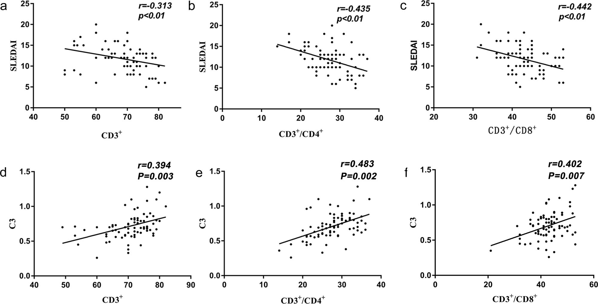
(a) Correlation analysis of peripheral blood CD3+ of SLE with SLEDAI; (b) Correlation analysis of peripheral blood CD3+/CD4+ of SLE with SLEDAI; (c) Correlation analysis of peripheral blood CD3+/CD8+ of SLE with SLEDAI; (d) Correlation analysis of peripheral blood CD3+ of SLE with C3; (e) Correlation analysis of peripheral blood CD3+/CD4+ of SLE with C3; (f) Correlation analysis of peripheral blood CD3+/CD8+ of SLE with C3.
4 Discussion
SLE is heterogeneous; however, the pathogenesis is unclear, and the immune system plays a critical role in SLE [1,2]. The proliferation of T and B lymphocytes inducing abnormal immune tolerance and overactivation is the main cause, and T cells play a crucial role in the overall pathogenesis of SLE [9,10]. CD4+ T cells mediate the activation of other immune cells in adaptive immunity [11], while CD8+ T cells produce toxic granules that induce apoptosis [12]. CD4+ T cells prompt B lymphocytes to produce immunoglobulin G antibodies that bind to self-antigens found in the glomerular basement membrane, the liver, the central nervous system, and the small blood vessels [9,10]. T cells, mainly CD4+ T cells, infiltrate damaged glomeruli and renal interstitium [13]. The number of infiltrating cells is correlated with renal injury, promoting apoptosis, and initiating and perpetuating the inflammatory process in LN [14]. Moreover, CD4+ T cells are present in the urine of patients with active LN [15]. The antigen-reactive T cells expressed by renal CD4+ T cells might lead to renal damage, thereby indicating the degree of inflammation of LN [16]. Gao et al. [17] hypothesized that a decrease in Treg cells decreased the number of CD4+ T cells. Although the proportion of CD4+ T cells is reduced, humoral immunity secretes a variety of cytokines to regulate cellular functions in normal immune defense [9,10]. Immune regulation is diminished, and NK cells lead to B-cell activation [1,2]. In the present study, the percentages of CD8+ T cells, CD4+/CD8+, and NK cells were positively correlated with C3, and it was speculated that CD8+ T cells and NK cells might be involved in the complement pathway.
A decrease in T cells is a risk factor for respiratory and CNS infections in SLE [18,19]. Chen and Chen [20] compared some indicators in the LN group infected with Cryptococcal meningitis with the uninfected group and found that the infected group had significantly decreased CD4+ T cells while SLEDAI was significantly higher. The low-percentage CD4+ T cells and high-percentage SLEDAI 2K scores are independent risk factors for SLE pneumonia [21]. Previous studies reported a significant decrease in peripheral blood CD3+, CD4+, and CD4+/CD8+ in the infected SLE group [22,23]. Abnormal peripheral blood lymphocyte immune response might lead to various infections in SLE patients [24].
The most common changes in SLE are a decrease in lymphocyte subpopulation CD4+ T cells and an imbalance ratio of CD4+/CD8+ [25]. Activated and senescent CD4+ cells in SLE blood cell subpopulations are significantly higher, and resting CD4+ levels are significantly lower than in healthy individuals [26]. Men et al. [27] reported a reduced ratio of CD4+/CD8+ cells in the SLE group and speculated that changes in the CD4+/CD8+ ratio interfere with and disrupt cellular immunity and lead to SLE. As confirmed previously, the absolute values of CD4+ or CD8+ T cells affect SLE progression [28,29]. Maeda et al. [30] showed that SLE CD4+/CD8+ decreased, and human leukocytes expressed the significantly associated antigen HLA-DR through CD8+ T cells, suggesting that the CD4+/CD8+ ratio may be an indicator to assess the treatment efficacy in some SLE patients. This study confirmed that the expression percentage of CD3+/CD4+ and CD4+/CD8+ in the LN group was lower than in the non-LN group and that of CD3+/CD8+ was higher than that in the non-LN group. The proportion of CD3+/CD4+ and CD4+/CD8+ in the blood system involvement group was lower and that of CD3+/CD8+ was higher than that in the no blood system involvement group. CD3+ and CD3+/CD4+ in the joint group were lower than in the non-joint group. These results are supported by Lu et al. [31], who reported that different clusters of T-lymphocyte subsets could help differentiate the different types of SLE.
Kalim et al. [32,33] proposed that the mechanism of cognitive impairment in SLE was accelerated immune senescence, and one of the markers of immune senescence was the immune risk profile (CD4+/CD8+) hypothesis. CD4+ T cells and CD4+/CD8+ levels were decreased, and memory function with visuospatial domains was negatively correlated with memory T cells (CD4+ CD45RO+ T cells and CD8+ CD28− T cells), confirming that accelerated immune senescence in SLE patients led to cognitive dysfunction, especially in attention deficit, recall, and visuospatial domains. Robinson et al. [34] demonstrated that SLE CD8+ T cells increased with disease activity in the baseline adolescent group, with a significantly higher proportion of LN, thereby supporting the role of CD8+ T cells in the pathogenesis of adolescent SLE. The decreased CD4+ T cells, B cells, and altered immune cell subpopulations suggested that adolescent-onset SLE adapted to the dysregulated immune system. In the future, we could study the correlation between the memory function of CD4+ T cells, CD4+/CD8+, and visual space domain and memory T cells (CD4+ CD45RO+ T cells, CD8+ CD28− T cells) to explore the mechanism of accelerated immune aging and cognitive impairment in patients with SLE, especially in attention disorder, memory, and visual space domain.
A close correlation was established between CD4+, CD8+, SLEDAI score, and clinical features, with the remission of SLE, ESR, SLEDAI score, and proteinuria showing a downtrend while C3 showed an uptrend and C4 showed a stable pattern. Moreover, CD4+ was negatively correlated with SLEDAI. CD4+ is a sensitive, specific, reliable, and valid indicator to reflect SLE activity and could be applied to track disease activity [28]. Zhao et al. [35] reported that CD3+/CD4+ and CD4+/CD8+ were negatively correlated with SLEDAI and positively correlated with C3 and C4, suggesting that lymphocyte subpopulations may reflect the severity of SLE activity. The comparison between SLE and healthy control groups indicated that CD3+/CD4+ and CD4+/CD8+ were negatively correlated with SLEDAI scores [23].
NK cells are rapid producers of IFN-γ and influence the adaptive immunity involved in the pathogenesis of systemic SLE [36]. Flow cytometry assays for the NK cell phenotype IFN-γ in SLE and healthy individuals resulted in a significantly lower percentage of peripheral blood NK cells in SLE patients than that in healthy individuals. A study reported that the total number of NK cells in SLE was reduced, but the number of suppressed NK cells was decreased, and the number of activated NK cells was increased, resulting in enhanced NK cell-mediated killing of normal cells [37]. The reduced number and percentage of peripheral blood NK cells in SLE, impaired cytotoxic function, differentiation, altered phenotype, changed cytokines, and NK cells involved in the pathogenesis of SLE were consistent with the results of this study. The number of NK cells was significantly increased in the SLE group and was inversely proportional to disease activity.
The present study has limitations, such as single-center, small sample size, and selection bias. Therefore, additional studies are required to substantiate the conclusion.
The immune expression abnormalities and dysregulation of T-cell subpopulations, B cells, and NK cells play a significant role in the pathogenesis of SLE. T-lymphocyte subpopulations are closely related to SLE activity, and with the popularization of the clinical application of flow cytometry, the analysis of the subpopulation could be one of the comprehensive indicators for assessing SLE activity. In addition, monitoring the changes in peripheral blood lymphocyte subpopulations is conducive to understanding the immune status of SLE patients, which is crucial for accurate diagnosis, the selection of the treatment plan, and judgment of the efficacy and safety of drug therapy.
-
Funding information: This study was funded by Anhui University Natural Science Research Project (KJ2019A0319).
-
Author contributions: Yan Feng and Zhijun Li carried out the studies, participated in collecting data, and drafted the manuscript. Yan Feng and Changhao Xie performed the statistical analysis and participated in its design. Yan Feng and Fanglin Lu participated in the acquisition, analysis, or interpretation of data and drafted the manuscript. All authors read and approved the final article.
-
Conflict of interest: Authors state no conflict of interest.
-
Data availability statement: The datasets generated during and/or analyzed during the current study are available from the corresponding author on reasonable request.
References
[1] Dzopalic T, Bozic-Nedeljkovic B, Jurisic V. Function of innate lymphoid cells in the immune-related disorders. Hum Cell. 2019;32(3):231–9.10.1007/s13577-019-00257-1Suche in Google Scholar PubMed
[2] Živković V, Cvetković T, Mitić B, Stamenković B, Stojanović S, Radovanović-Dinić B, et al. Monocyte chemoattractant protein-1 as a marker of systemic lupus erythematosus: an observational study. Rheumatol Int. 2018;38(6):1003–8.10.1007/s00296-017-3888-xSuche in Google Scholar PubMed
[3] Davidson A. What is damaging the kidney in lupus nephritis? Nat Rev Rheumatol. 2016;12(3):143–53.10.1038/nrrheum.2015.159Suche in Google Scholar PubMed PubMed Central
[4] Moulton VR, Tsokos GC. T cell signaling abnormalities contribute to aberrant immune cell function and autoimmunity. J Clin Invest. 2015;125(6):2220–7.10.1172/JCI78087Suche in Google Scholar PubMed PubMed Central
[5] Luo Q, Ye J, Zeng L, Li X, Fang L, Ju B, et al. Elevated expression of TIGIT on CD3(+)CD4(+) T cells correlates with disease activity in systemic lupus erythematosus. Allergy Asthma Clin Immunol. 2017;13:15–27.10.1186/s13223-017-0188-7Suche in Google Scholar PubMed PubMed Central
[6] Aringer M, Costenbader K, Daikh D, Brinks R, Mosca M, Ramsey-Goldman R, et al. European league against rheumatism/american college of rheumatology classification criteria for systemic lupus erythematosus. Arthritis Rheumatol (Hoboken, NJ). 2019;2019(78):1151–9.Suche in Google Scholar
[7] Gladman DD, Ibañez D, Urowitz MB. Systemic lupus erythematosus disease activity index 2000. J Rheumatol. 2002;29(2):288–91.Suche in Google Scholar
[8] Konjevic G, Mirjacic MK, Vuletic A, Jurisic V, Spuzic I. Distribution of several activating and inhibitory receptors on CD3-CD16+ NK cells and their correlation with NK cell function in healthy individuals. J Membr Biol. 2009;230(3):113–23.10.1007/s00232-009-9191-3Suche in Google Scholar PubMed
[9] Rother N, van der Vlag J. Disturbed t cell signaling and altered th17 and regulatory T cell subsets in the pathogenesis of systemic lupus erythematosus. Front Immunol. 2015;6:610.10.3389/fimmu.2015.00610Suche in Google Scholar PubMed PubMed Central
[10] Moulton VR, Suarez-Fueyo A, Meidan E, Li H, Mizui M, Tsokos GC. Pathogenesis of human systemic lupus erythematosus: a cellular perspective. Trends Mol Med. 2017;23(7):615–35.10.1016/j.molmed.2017.05.006Suche in Google Scholar PubMed PubMed Central
[11] Zhu J, Paul WE. CD4 T cells: fates, functions, and faults. Blood. 2008;112(5):1557–69.10.1182/blood-2008-05-078154Suche in Google Scholar PubMed PubMed Central
[12] Kim JS, Cho BA, Sim JH, Shah K, Woo CM, Lee EB, et al. IL-7Rαlow memory CD8+ T cells are significantly elevated in patients with systemic lupus erythematosus. Rheumatology (Oxford, England). 2012;51(9):1587–94.10.1093/rheumatology/kes100Suche in Google Scholar PubMed PubMed Central
[13] Abdirama D, Tesch S, Grießbach AS, von Spee-Mayer C, Humrich JY, Stervbo U, et al. Nuclear antigen-reactive CD4(+) T cells expand in active systemic lupus erythematosus, produce effector cytokines, and invade the kidneys. Kidney Int. 2021;99(1):238–46.10.1016/j.kint.2020.05.051Suche in Google Scholar PubMed
[14] Park MH, D’Agati V, Appel GB, Pirani CL. Tubulointerstitial disease in lupus nephritis: relationship to immune deposits, interstitial inflammation, glomerular changes, renal function, and prognosis. Nephron. 1986;44(4):309–19.10.1159/000184012Suche in Google Scholar PubMed
[15] Enghard P, Rieder C, Kopetschke K, Klocke JR, Undeutsch R, Biesen R, et al. Urinary CD4 T cells identify SLE patients with proliferative lupus nephritis and can be used to monitor treatment response. Ann Rheumatic Diseases. 2014;73(1):277–83.10.1136/annrheumdis-2012-202784Suche in Google Scholar PubMed
[16] Tesch S, Abdirama D, Grießbach AS, Brand HA, Goerlich N, Humrich JY, et al. Identification and characterization of antigen-specific CD4(+) T cells targeting renally expressed antigens in human lupus nephritis with two independent methods. Sci Rep. 2020;10(1):21312.10.1038/s41598-020-78223-3Suche in Google Scholar PubMed PubMed Central
[17] Gao H, Ma J, Wang X, Lv T, Liu J, Ren Y, et al. Preliminary study on the changes of ovarian reserve, menstruation, and lymphocyte subpopulation in systemic lupus erythematosus (SLE) patients of childbearing age. Lupus. 2018;27(3):445–53.10.1177/0961203317726378Suche in Google Scholar PubMed
[18] Lao M, Zhan Z, Su F, Li H, Yang Z, Chen H, et al. Invasive mycoses in patients with connective tissue disease from southern China: clinical features and associated factors. Arthritis Res Therapy. 2019;21(1):71.10.1186/s13075-019-1851-9Suche in Google Scholar PubMed PubMed Central
[19] Lao M, Chen D, Wu X, Chen H, Qiu Q, Yang X, et al. Active tuberculosis in patients with systemic lupus erythematosus from Southern China: a retrospective study. Clinical Rheumatol. 2019;38(2):535–43.10.1007/s10067-018-4303-zSuche in Google Scholar PubMed
[20] Chen J, Chen P. Cryptococcal meningitis in patients with lupus nephritis. Clinical Rheumatol. 2020;39(2):407–12.10.1007/s10067-019-04844-3Suche in Google Scholar PubMed
[21] Peng L, Wang Y, Zhao L, Chen T, Huang A. Severe pneumonia in chinese patients with systemic lupus erythematosus. Lupus. 2020;29(7):735–42.10.1177/0961203320922609Suche in Google Scholar PubMed
[22] Lu Z, Li J, Ji J, Gu Z, Da Z. Altered peripheral lymphocyte subsets in untreated systemic lupus erythematosus patients with infections. Braz J Med Biol Res. 2019;52(4):e8131.10.1590/1414-431x20198131Suche in Google Scholar
[23] Tian B, Yang C, Wang J, Hou X, Zhao S, Li Y, et al. Peripheral blood brain-derived neurotrophic factor level and tyrosine kinase B expression on T lymphocytes in systemic lupus erythematosus: implications for systemic involvement. Cytokine. 2019;123:154764.10.1016/j.cyto.2019.154764Suche in Google Scholar PubMed
[24] Boomer JS, To K, Chang KC, Takasu O, Osborne DF, Walton AH, et al. Immunosuppression in patients who die of sepsis and multiple organ failure. Jama. 2011;306(23):2594–605.10.1001/jama.2011.1829Suche in Google Scholar PubMed PubMed Central
[25] Xue C, Lan-Lan W, Bei C, Jie C, Wei-Hua F. Abnormal Fas/FasL and caspase-3-mediated apoptotic signaling pathways of T lymphocyte subset in patients with systemic lupus erythematosus. Cellular Immunol. 2006;239(2):121–8.10.1016/j.cellimm.2006.05.003Suche in Google Scholar PubMed
[26] López P, Rodríguez-Carrio J, Caminal-Montero L, Suárez A. Relationship between T-cell exosomes and cellular subsets in SLE according to type I IFN-signaling. Front Med. 2020;7:1–10.10.3389/fmed.2020.604098Suche in Google Scholar PubMed PubMed Central
[27] Men K, Chen Y, Zhang J, Wei D. The evaluation of cellular immune function in elderly patients with systemic lupus erythematosus. Korean J Internal Med. 2019;34(4):932–7.10.3904/kjim.2017.224Suche in Google Scholar PubMed PubMed Central
[28] Sonawale A, Bohara V, Bichile LS. Evaluation of the association between CD4, CD8 and CD25 cell counts and SLE in active disease and in remission. J Assoc Phys India. 2017;65(4):37–42.Suche in Google Scholar
[29] Goropevšek A, Gorenjak M, Gradišnik S, Dai K, Holc I, Hojs R, et al. STAT5 phosphorylation in CD4 T cells from patients with SLE is related to changes in their subsets and follow-up disease severity. J Leukoc Biol. 2017;101(6):1405–18.10.1189/jlb.5A0416-194RSuche in Google Scholar PubMed
[30] Maeda N, Sekigawa I, Iida N, Matsumoto M, Hashimoto H, Hirose S. Relationship between CD4+/CD8+ T cell ratio and T cell activation in systemic lupus erythematosus. Scand J Rheumatol. 1999;28(3):166–70.10.1080/03009749950154248Suche in Google Scholar PubMed
[31] Lu Z, Li W, Tang Y, Da Z, Li X. Lymphocyte subset clustering analysis in treatment-naive patients with systemic lupus erythematosus. Clinical Rheumatol. 2021;40(5):1835–42.10.1007/s10067-020-05480-ySuche in Google Scholar PubMed
[32] Kalim H, Pratama MZ, Mahardini E, Winoto ES, Krisna PA, Handono K. Accelerated immune aging was correlated with lupus-associated brain fog in reproductive-age systemic lupus erythematosus patients. Int J Rheumatic Diseases. 2020;23(5):620–6.10.1111/1756-185X.13816Suche in Google Scholar PubMed
[33] Kalim H, Wahono CS, Permana BPO, Pratama MZ, Handono K. Association between senescence of T cells and disease activity in patients with systemic lupus erythematosus. Reumatologia. 2021;59(5):292–301.10.5114/reum.2021.110318Suche in Google Scholar PubMed PubMed Central
[34] Robinson GA, Peng J, Dönnes P, Coelewij L, Naja M, Radziszewska A, et al. Disease-associated and patient-specific immune cell signatures in juvenile-onset systemic lupus erythematosus: patient stratification using a machine-learning approach. Lancet Rheumatol. 2020;2(8):e485–e96.10.1016/S2665-9913(20)30168-5Suche in Google Scholar
[35] Zhao L, Jiang Z, Jiang Y, Ma N, Wang K, Zhang Y. Changes in immune cell frequencies after cyclophosphamide or mycophenolate mofetil treatments in patients with systemic lupus erythematosus. Clinical Rheumatol. 2012;31(6):951–9.10.1007/s10067-012-1958-8Suche in Google Scholar PubMed
[36] Campbell KS, Hasegawa J. Natural killer cell biology: an update and future directions. J Allergy Clin Immunol. 2013;132(3):536–44.10.1016/j.jaci.2013.07.006Suche in Google Scholar PubMed PubMed Central
[37] Fogel LA, Yokoyama WM, French AR. Natural killer cells in human autoimmune disorders. Arthritis Res Therapy. 2013;15(4):216.10.1186/ar4232Suche in Google Scholar PubMed PubMed Central
© 2022 Yan Feng et al., published by De Gruyter
This work is licensed under the Creative Commons Attribution 4.0 International License.
Artikel in diesem Heft
- Biomedical Sciences
- Effects of direct oral anticoagulants dabigatran and rivaroxaban on the blood coagulation function in rabbits
- The mother of all battles: Viruses vs humans. Can humans avoid extinction in 50–100 years?
- Knockdown of G1P3 inhibits cell proliferation and enhances the cytotoxicity of dexamethasone in acute lymphoblastic leukemia
- LINC00665 regulates hepatocellular carcinoma by modulating mRNA via the m6A enzyme
- Association study of CLDN14 variations in patients with kidney stones
- Concanavalin A-induced autoimmune hepatitis model in mice: Mechanisms and future outlook
- Regulation of miR-30b in cancer development, apoptosis, and drug resistance
- Informatic analysis of the pulmonary microecology in non-cystic fibrosis bronchiectasis at three different stages
- Swimming attenuates tumor growth in CT-26 tumor-bearing mice and suppresses angiogenesis by mediating the HIF-1α/VEGFA pathway
- Characterization of intestinal microbiota and serum metabolites in patients with mild hepatic encephalopathy
- Functional conservation and divergence in plant-specific GRF gene family revealed by sequences and expression analysis
- Application of the FLP/LoxP-FRT recombination system to switch the eGFP expression in a model prokaryote
- Biomedical evaluation of antioxidant properties of lamb meat enriched with iodine and selenium
- Intravenous infusion of the exosomes derived from human umbilical cord mesenchymal stem cells enhance neurological recovery after traumatic brain injury via suppressing the NF-κB pathway
- Effect of dietary pattern on pregnant women with gestational diabetes mellitus and its clinical significance
- Potential regulatory mechanism of TNF-α/TNFR1/ANXA1 in glioma cells and its role in glioma cell proliferation
- Effect of the genetic mutant G71R in uridine diphosphate-glucuronosyltransferase 1A1 on the conjugation of bilirubin
- Quercetin inhibits cytotoxicity of PC12 cells induced by amyloid-beta 25–35 via stimulating estrogen receptor α, activating ERK1/2, and inhibiting apoptosis
- Nutrition intervention in the management of novel coronavirus pneumonia patients
- circ-CFH promotes the development of HCC by regulating cell proliferation, apoptosis, migration, invasion, and glycolysis through the miR-377-3p/RNF38 axis
- Bmi-1 directly upregulates glucose transporter 1 in human gastric adenocarcinoma
- Lacunar infarction aggravates the cognitive deficit in the elderly with white matter lesion
- Hydroxysafflor yellow A improved retinopathy via Nrf2/HO-1 pathway in rats
- Comparison of axon extension: PTFE versus PLA formed by a 3D printer
- Elevated IL-35 level and iTr35 subset increase the bacterial burden and lung lesions in Mycobacterium tuberculosis-infected mice
- A case report of CAT gene and HNF1β gene variations in a patient with early-onset diabetes
- Study on the mechanism of inhibiting patulin production by fengycin
- SOX4 promotes high-glucose-induced inflammation and angiogenesis of retinal endothelial cells by activating NF-κB signaling pathway
- Relationship between blood clots and COVID-19 vaccines: A literature review
- Analysis of genetic characteristics of 436 children with dysplasia and detailed analysis of rare karyotype
- Bioinformatics network analyses of growth differentiation factor 11
- NR4A1 inhibits the epithelial–mesenchymal transition of hepatic stellate cells: Involvement of TGF-β–Smad2/3/4–ZEB signaling
- Expression of Zeb1 in the differentiation of mouse embryonic stem cell
- Study on the genetic damage caused by cadmium sulfide quantum dots in human lymphocytes
- Association between single-nucleotide polymorphisms of NKX2.5 and congenital heart disease in Chinese population: A meta-analysis
- Assessment of the anesthetic effect of modified pentothal sodium solution on Sprague-Dawley rats
- Genetic susceptibility to high myopia in Han Chinese population
- Potential biomarkers and molecular mechanisms in preeclampsia progression
- Silencing circular RNA-friend leukemia virus integration 1 restrained malignancy of CC cells and oxaliplatin resistance by disturbing dyskeratosis congenita 1
- Endostar plus pembrolizumab combined with a platinum-based dual chemotherapy regime for advanced pulmonary large-cell neuroendocrine carcinoma as a first-line treatment: A case report
- The significance of PAK4 in signaling and clinicopathology: A review
- Sorafenib inhibits ovarian cancer cell proliferation and mobility and induces radiosensitivity by targeting the tumor cell epithelial–mesenchymal transition
- Characterization of rabbit polyclonal antibody against camel recombinant nanobodies
- Active legumain promotes invasion and migration of neuroblastoma by regulating epithelial-mesenchymal transition
- Effect of cell receptors in the pathogenesis of osteoarthritis: Current insights
- MT-12 inhibits the proliferation of bladder cells in vitro and in vivo by enhancing autophagy through mitochondrial dysfunction
- Study of hsa_circRNA_000121 and hsa_circRNA_004183 in papillary thyroid microcarcinoma
- BuyangHuanwu Decoction attenuates cerebral vasospasm caused by subarachnoid hemorrhage in rats via PI3K/AKT/eNOS axis
- Effects of the interaction of Notch and TLR4 pathways on inflammation and heart function in septic heart
- Monosodium iodoacetate-induced subchondral bone microstructure and inflammatory changes in an animal model of osteoarthritis
- A rare presentation of type II Abernethy malformation and nephrotic syndrome: Case report and review
- Rapid death due to pulmonary epithelioid haemangioendothelioma in several weeks: A case report
- Hepatoprotective role of peroxisome proliferator-activated receptor-α in non-cancerous hepatic tissues following transcatheter arterial embolization
- Correlation between peripheral blood lymphocyte subpopulations and primary systemic lupus erythematosus
- A novel SLC8A1-ALK fusion in lung adenocarcinoma confers sensitivity to alectinib: A case report
- β-Hydroxybutyrate upregulates FGF21 expression through inhibition of histone deacetylases in hepatocytes
- Identification of metabolic genes for the prediction of prognosis and tumor microenvironment infiltration in early-stage non-small cell lung cancer
- BTBD10 inhibits glioma tumorigenesis by downregulating cyclin D1 and p-Akt
- Mucormycosis co-infection in COVID-19 patients: An update
- Metagenomic next-generation sequencing in diagnosing Pneumocystis jirovecii pneumonia: A case report
- Long non-coding RNA HOXB-AS1 is a prognostic marker and promotes hepatocellular carcinoma cells’ proliferation and invasion
- Preparation and evaluation of LA-PEG-SPION, a targeted MRI contrast agent for liver cancer
- Proteomic analysis of the liver regulating lipid metabolism in Chaohu ducks using two-dimensional electrophoresis
- Nasopharyngeal tuberculosis: A case report
- Characterization and evaluation of anti-Salmonella enteritidis activity of indigenous probiotic lactobacilli in mice
- Aberrant pulmonary immune response of obese mice to periodontal infection
- Bacteriospermia – A formidable player in male subfertility
- In silico and in vivo analysis of TIPE1 expression in diffuse large B cell lymphoma
- Effects of KCa channels on biological behavior of trophoblasts
- Interleukin-17A influences the vulnerability rather than the size of established atherosclerotic plaques in apolipoprotein E-deficient mice
- Multiple organ failure and death caused by Staphylococcus aureus hip infection: A case report
- Prognostic signature related to the immune environment of oral squamous cell carcinoma
- Primary and metastatic squamous cell carcinoma of the thyroid gland: Two case reports
- Neuroprotective effects of crocin and crocin-loaded niosomes against the paraquat-induced oxidative brain damage in rats
- Role of MMP-2 and CD147 in kidney fibrosis
- Geometric basis of action potential of skeletal muscle cells and neurons
- Babesia microti-induced fulminant sepsis in an immunocompromised host: A case report and the case-specific literature review
- Role of cerebellar cortex in associative learning and memory in guinea pigs
- Application of metagenomic next-generation sequencing technique for diagnosing a specific case of necrotizing meningoencephalitis caused by human herpesvirus 2
- Case report: Quadruple primary malignant neoplasms including esophageal, ureteral, and lung in an elderly male
- Long non-coding RNA NEAT1 promotes angiogenesis in hepatoma carcinoma via the miR-125a-5p/VEGF pathway
- Osteogenic differentiation of periodontal membrane stem cells in inflammatory environments
- Knockdown of SHMT2 enhances the sensitivity of gastric cancer cells to radiotherapy through the Wnt/β-catenin pathway
- Continuous renal replacement therapy combined with double filtration plasmapheresis in the treatment of severe lupus complicated by serious bacterial infections in children: A case report
- Simultaneous triple primary malignancies, including bladder cancer, lymphoma, and lung cancer, in an elderly male: A case report
- Preclinical immunogenicity assessment of a cell-based inactivated whole-virion H5N1 influenza vaccine
- One case of iodine-125 therapy – A new minimally invasive treatment of intrahepatic cholangiocarcinoma
- S1P promotes corneal trigeminal neuron differentiation and corneal nerve repair via upregulating nerve growth factor expression in a mouse model
- Early cancer detection by a targeted methylation assay of circulating tumor DNA in plasma
- Calcifying nanoparticles initiate the calcification process of mesenchymal stem cells in vitro through the activation of the TGF-β1/Smad signaling pathway and promote the decay of echinococcosis
- Evaluation of prognostic markers in patients infected with SARS-CoV-2
- N6-Methyladenosine-related alternative splicing events play a role in bladder cancer
- Characterization of the structural, oxidative, and immunological features of testis tissue from Zucker diabetic fatty rats
- Effects of glucose and osmotic pressure on the proliferation and cell cycle of human chorionic trophoblast cells
- Investigation of genotype diversity of 7,804 norovirus sequences in humans and animals of China
- Characteristics and karyotype analysis of a patient with turner syndrome complicated with multiple-site tumors: A case report
- Aggravated renal fibrosis is positively associated with the activation of HMGB1-TLR2/4 signaling in STZ-induced diabetic mice
- Distribution characteristics of SARS-CoV-2 IgM/IgG in false-positive results detected by chemiluminescent immunoassay
- SRPX2 attenuated oxygen–glucose deprivation and reperfusion-induced injury in cardiomyocytes via alleviating endoplasmic reticulum stress-induced apoptosis through targeting PI3K/Akt/mTOR axis
- Aquaporin-8 overexpression is involved in vascular structure and function changes in placentas of gestational diabetes mellitus patients
- Relationship between CRP gene polymorphisms and ischemic stroke risk: A systematic review and meta-analysis
- Effects of growth hormone on lipid metabolism and sexual development in pubertal obese male rats
- Cloning and identification of the CTLA-4IgV gene and functional application of vaccine in Xinjiang sheep
- Antitumor activity of RUNX3: Upregulation of E-cadherin and downregulation of the epithelial–mesenchymal transition in clear-cell renal cell carcinoma
- PHF8 promotes osteogenic differentiation of BMSCs in old rat with osteoporosis by regulating Wnt/β-catenin pathway
- A review of the current state of the computer-aided diagnosis (CAD) systems for breast cancer diagnosis
- Bilateral dacryoadenitis in adult-onset Still’s disease: A case report
- A novel association between Bmi-1 protein expression and the SUVmax obtained by 18F-FDG PET/CT in patients with gastric adenocarcinoma
- The role of erythrocytes and erythroid progenitor cells in tumors
- Relationship between platelet activation markers and spontaneous abortion: A meta-analysis
- Abnormal methylation caused by folic acid deficiency in neural tube defects
- Silencing TLR4 using an ultrasound-targeted microbubble destruction-based shRNA system reduces ischemia-induced seizures in hyperglycemic rats
- Plant Sciences
- Seasonal succession of bacterial communities in cultured Caulerpa lentillifera detected by high-throughput sequencing
- Cloning and prokaryotic expression of WRKY48 from Caragana intermedia
- Novel Brassica hybrids with different resistance to Leptosphaeria maculans reveal unbalanced rDNA signal patterns
- Application of exogenous auxin and gibberellin regulates the bolting of lettuce (Lactuca sativa L.)
- Phytoremediation of pollutants from wastewater: A concise review
- Genome-wide identification and characterization of NBS-encoding genes in the sweet potato wild ancestor Ipomoea trifida (H.B.K.)
- Alleviative effects of magnetic Fe3O4 nanoparticles on the physiological toxicity of 3-nitrophenol to rice (Oryza sativa L.) seedlings
- Selection and functional identification of Dof genes expressed in response to nitrogen in Populus simonii × Populus nigra
- Study on pecan seed germination influenced by seed endocarp
- Identification of active compounds in Ophiopogonis Radix from different geographical origins by UPLC-Q/TOF-MS combined with GC-MS approaches
- The entire chloroplast genome sequence of Asparagus cochinchinensis and genetic comparison to Asparagus species
- Genome-wide identification of MAPK family genes and their response to abiotic stresses in tea plant (Camellia sinensis)
- Selection and validation of reference genes for RT-qPCR analysis of different organs at various development stages in Caragana intermedia
- Cloning and expression analysis of SERK1 gene in Diospyros lotus
- Integrated metabolomic and transcriptomic profiling revealed coping mechanisms of the edible and medicinal homologous plant Plantago asiatica L. cadmium resistance
- A missense variant in NCF1 is associated with susceptibility to unexplained recurrent spontaneous abortion
- Assessment of drought tolerance indices in faba bean genotypes under different irrigation regimes
- The entire chloroplast genome sequence of Asparagus setaceus (Kunth) Jessop: Genome structure, gene composition, and phylogenetic analysis in Asparagaceae
- Food Science
- Dietary food additive monosodium glutamate with or without high-lipid diet induces spleen anomaly: A mechanistic approach on rat model
- Binge eating disorder during COVID-19
- Potential of honey against the onset of autoimmune diabetes and its associated nephropathy, pancreatitis, and retinopathy in type 1 diabetic animal model
- FTO gene expression in diet-induced obesity is downregulated by Solanum fruit supplementation
- Physical activity enhances fecal lactobacilli in rats chronically drinking sweetened cola beverage
- Supercritical CO2 extraction, chemical composition, and antioxidant effects of Coreopsis tinctoria Nutt. oleoresin
- Functional constituents of plant-based foods boost immunity against acute and chronic disorders
- Effect of selenium and methods of protein extraction on the proteomic profile of Saccharomyces yeast
- Microbial diversity of milk ghee in southern Gansu and its effect on the formation of ghee flavor compounds
- Ecology and Environmental Sciences
- Effects of heavy metals on bacterial community surrounding Bijiashan mining area located in northwest China
- Microorganism community composition analysis coupling with 15N tracer experiments reveals the nitrification rate and N2O emissions in low pH soils in Southern China
- Genetic diversity and population structure of Cinnamomum balansae Lecomte inferred by microsatellites
- Preliminary screening of microplastic contamination in different marine fish species of Taif market, Saudi Arabia
- Plant volatile organic compounds attractive to Lygus pratensis
- Effects of organic materials on soil bacterial community structure in long-term continuous cropping of tomato in greenhouse
- Effects of soil treated fungicide fluopimomide on tomato (Solanum lycopersicum L.) disease control and plant growth
- Prevalence of Yersinia pestis among rodents captured in a semi-arid tropical ecosystem of south-western Zimbabwe
- Effects of irrigation and nitrogen fertilization on mitigating salt-induced Na+ toxicity and sustaining sea rice growth
- Bioengineering and Biotechnology
- Poly-l-lysine-caused cell adhesion induces pyroptosis in THP-1 monocytes
- Development of alkaline phosphatase-scFv and its use for one-step enzyme-linked immunosorbent assay for His-tagged protein detection
- Development and validation of a predictive model for immune-related genes in patients with tongue squamous cell carcinoma
- Agriculture
- Effects of chemical-based fertilizer replacement with biochar-based fertilizer on albic soil nutrient content and maize yield
- Genome-wide identification and expression analysis of CPP-like gene family in Triticum aestivum L. under different hormone and stress conditions
- Agronomic and economic performance of mung bean (Vigna radiata L.) varieties in response to rates of blended NPS fertilizer in Kindo Koysha district, Southern Ethiopia
- Influence of furrow irrigation regime on the yield and water consumption indicators of winter wheat based on a multi-level fuzzy comprehensive evaluation
- Discovery of exercise-related genes and pathway analysis based on comparative genomes of Mongolian originated Abaga and Wushen horse
- Lessons from integrated seasonal forecast-crop modelling in Africa: A systematic review
- Evolution trend of soil fertility in tobacco-planting area of Chenzhou, Hunan Province, China
- Animal Sciences
- Morphological and molecular characterization of Tatera indica Hardwicke 1807 (Rodentia: Muridae) from Pothwar, Pakistan
- Research on meat quality of Qianhua Mutton Merino sheep and Small-tail Han sheep
- SI: A Scientific Memoir
- Suggestions on leading an academic research laboratory group
- My scientific genealogy and the Toronto ACDC Laboratory, 1988–2022
- Erratum
- Erratum to “Changes of immune cells in patients with hepatocellular carcinoma treated by radiofrequency ablation and hepatectomy, a pilot study”
- Erratum to “A two-microRNA signature predicts the progression of male thyroid cancer”
- Retraction
- Retraction of “Lidocaine has antitumor effect on hepatocellular carcinoma via the circ_DYNC1H1/miR-520a-3p/USP14 axis”