Study on the genetic damage caused by cadmium sulfide quantum dots in human lymphocytes
-
Haiping Liu
 and
Qingzhao Li
and
Qingzhao Li

Abstract
Cadmium sulfide quantum dots (CdS QDs) are being developed for sensors, fluorescent probes, and other platforms and are attracting increasing attention. Given the growing demand for QDs, it is clear that there is a need to understand their potential toxicity to organisms. However, little is known regarding the genotoxicity of CdS QDs to humans. Therefore, this study used CdS QDs as the research object, cultured human peripheral blood lymphocytes, and randomly divided them into a control group, CdS I group (CdS QDs), and CdS II group (CdS QDs coated with thioglycolic acid). After cultivation, we measured the olive tail distance, tail length, tail DNA%, lymphocyte micronucleus rate, and aneuploid rate. The comet test results indicated that the indices of the QD group were significantly larger than those of the control group (P < 0.05). The results of the micronucleus and chromosome aberration tests showed that the lymphocyte micronucleus rate and chromosome aneuploid rate in the QD group were significantly increased (P < 0.05) compared with those in the control group. In conclusion, CdS QDs have certain genotoxicity to human peripheral blood lymphocytes, and the DNA damage caused by CdS QDs encapsulated with thioglycolic acid is less severe than that caused by nonencapsulated CdS QDs.
1 Introduction
Nanoparticles (NPs) generally refer to particles with at least one dimension, sized between 1 and 100 nm. They have some novel properties, such as a small size effect, surface interface effect, and quantum size effect, which enable their use in unique applications. Many artificially synthesized NPs have become important tools for scientific research. Among them, the application of semiconductor nanocrystals, called quantum dots (QDs), has attracted increasing attention [1]. QDs are mainly used in solar cells, optoelectronic devices, chemical catalysis, fluorescent probes, and sensors [2,3,4], and they have potential diagnostic and therapeutic effects in the field of biomedicine [5,6,7]. For example, QDs coupled with antibodies have been used to distinguish normal cells from cancer cells [8]. However, the potential threat of QDs to human health hinders their widespread application in life science [9].
Studies have shown that QDs are released into the environment as waste from QD synthesis and leakage during processing and transportation. Additionally, QDs in single electronic devices and various optoelectronic devices can be released into the environment through device use or waste treatment. QDs in the air have a strong adsorption capacity and easily adsorb gas or other particles and react with them. There are three main ways for QDs to enter the human body: the respiratory tract, digestive tract, and skin [10,11,12]. QDs entering the body can avoid phagocytosis of the immune system and accumulate in some target organs. They can also cross different biological barriers and be transported to other tissues and organs of the body, resulting in systematic health effects. Therefore, it is necessary to consider the negative effects of nanomaterials while paying attention to their beneficial biological effects.
Studies have shown that cadmium sulfide quantum dots (CdS QDs) have potential toxicity to organisms, can be distributed in various systems in organisms, and mainly gather in the lung and spleen [13,14,15]. The toxicity mechanism of CdS QDs is mainly related to the induction of reactive oxygen species (ROS) and other free radicals; the inhibition of antioxidant enzyme activity; the imbalance of oxidation and antioxidant systems; and the destruction of the integrity of proteins, nucleic acids, and membranes [16]. However, there are few studies on the genotoxicity of CdS QDs to humans. Therefore, it is of great significance to study the genotoxicity of CdS QDs in humans. Studies have shown that coating the surface of QDs can protect the core of QDs, increase the stability of QDs, and slow their toxic effects on the body, which may remove obstacles to the application of QDs in various fields. CdS QDs are hydrophobic materials and are not easily excreted in the urine. Thioglycolic acid is water-soluble and can be directly excreted in urine through kidney metabolism. Therefore, we chose thioglycolic acid to wrap CdS QDs, which can increase the stability of QDs and reduce their accumulation in various organs to reduce their toxicity.
This study investigated the genotoxicity of CdS QDs in human peripheral blood lymphocytes. The DNA damage caused by CdS QDs and thioglycolic acid-encapsulated cadmium sulfide QDs in human peripheral blood lymphocytes was analyzed through comet, micronucleus, and chromosome aberration tests to provide laboratory data and a scientific basis for the study of the reproductive and genetic toxicity of CdS QDs.
2 Materials and methods
2.1 Main instruments and reagents
HEPA carbon dioxide incubator steri-cycle CO2 (United States THERMO), a JA2003 electronic balance (Shanghai Jingke), a BX-41 fluorescent camera system (Japan OLYMPUS), steady flow programmable electrophoresis (Beijing Liuyi Instrument Factory), a DK-8D digital display constant-temperature water bath (Jintan Medical Instrument Factory), and a KDC-1044 low-speed centrifuge (Keida Innovation Co., Ltd., Zhongjia Branch) were used.
Low-melting-point agarose (SIGMA), normal-melting-point agarose (SIGMA), heparin, ethidium bromide, RPMI 1640 (GIBCOL), 1640 medium (GIBCOL), fetal bovine serum (GIBCOL), heparin, and plant blood coagulation prime were used.
2.2 Synthesis of CdS QDs
CdS QDs were synthesized by the emulsion liquid membrane method (Figure 1). The method is simple and easy to implement, does not require high temperature or high pressure, and can control the monomer particle size. At the same time, the raw material has low toxicity and is relatively stable at room temperature. Thus, it is an efficient “green” synthesis method. The specific method of synthesis was as follows: 50 mL of (0.2 mol/L) Na2S solution, 40 mL of kerosene, 5 mL of surfactant Span80 and 5 mL of tributyl phosphate were mixed in a beaker and stirred at 3,000 rpm for 8 min to form a stable emulsion liquid membrane system. Then, 50 mL of the prepared emulsion liquid membrane was added to 100 mL (0.1 mol/L) of CdCl2 and stirred at 300 rpm for 10 min, and stirring was then stopped. After delamination, the aqueous solution was discarded, and the remaining emulsion was centrifuged. The emulsified liquid membrane was demulsified by a centrifuge, the precipitate was washed 2–3 times with petroleum ether and absolute ethanol, and the precipitate was transferred to a bottle. The particle size of the CdS QD monomer obtained by the emulsion liquid membrane method was 6–8 nm, and the shape was granular. CdS QDs encapsulated by thioglycolic acid were provided by the School of Environmental Science and Engineering, Nankai University. The particle size of the monomer was 6–8 nm, and the shape was granular. Analytical Na2S and CdCl2 were purchased from Beijing Hengye Zhongyuan Chemical Co., Ltd. (Beijing, China); tributyl phosphate was purchased from Tianjin Damao Chemical Reagent Factory (Tianjin, China); Span80 was purchased from Jiangsu Nantong Chenrun Chemical Co., Ltd.; petroleum ether was purchased from Tiande Fine Chemical Co., Ltd. (Zibo, China); and anhydrous ethyl alcohol was purchased from Tianjin Windboat Chemical Technology Co., Ltd. (Tianjin, China).

Synthesis of CdS QDs.
2.3 Preparation of CdS QD suspension
CdS QDs and CdS QD powder coated with thioglycolic acid were autoclaved and then prepared into a suspension in 1640 medium containing 20% fetal bovine serum. After 48 h of ultrasonic vibration dissolution, the mother liquor with uniform dispersion was obtained. Then, it was stored at 4°C until used.
2.4 Cell culture and processing
Twenty milliliters of venous blood from a healthy adult male (without a smoking history and who signed an informed consent form) were obtained. A 0.4 mL blood sample was added to 4.6 mL of RPMI 1640 (containing 20% calf serum, 100 U/mL penicillin, and 2% PHA) culture medium and then randomly divided into a control group, CdS I group (CdS QD group), and CdS II group (CdS QD group wrapped in thioglycolic acid). The CdS I group was supplemented with uniformly dispersed CdS QD mother liquor, and the CdS II group was supplemented with uniformly dispersed thioglycolic acid-coated CdS QD mother liquor. The final concentration of the sample suspension of the two groups was 2 µg/mL, the control group was treated with the same volume of normal saline, and the three groups were cultured in a 37°C, 5% CO2 incubator.
-
Informed consent: Informed consent has been obtained from all individuals included in this study.
-
Ethical approval: The research related to human use has been complied with all the relevant national regulations, institutional policies, and in accordance with the tenets of the Helsinki Declaration, and was approved by the ethics committee of North China University of Science and Technology (Ethics approval number: 2021024; Ethics approval date: 20210412).
2.5 Experimental methods
2.5.1 Comet test
In this study, a comet test was used to assess the extent of DNA damage caused by CdS QDs [17]. The principle is that the damaged DNA breaks and its superhelix structure is damaged because the molecular weight of the DNA fragment is very small; it can leave nuclear DNA in the electrophoretic field and move to the anode in the gel molecular sieve, forming a comet image. The more seriously the DNA is damaged, the more broken strands and variable fragments will be produced, and the smaller the broken strands will be. Under the same electrophoresis conditions, more DNA will migrate to the tail, and the longer the migration distance will be, which manifests as an increase in tail DNA% and tail length. Olive tail distance is a composite index of tail DNA% and migration distance, which can accurately and sensitively respond to DNA damage. Therefore, the severity of DNA damage caused by QDs can be comprehensively judged by three indices: tail length, tail DNA%, and olive tail distance. The comet test was performed according to Singh et al.’s study [18,19].
The lymphocytes were collected immediately after 6 h of incubation and processed as follows: first, a clean frosted glass slide was placed on the horizontal operating table and preheated to 40°C, 0.6% agarose was melted and preheated to 45°C, and 100 μL of 0.6% normal melting point agarose was added to the slide. Then, the cover slide was immediately added, and it was cooled at 4°C for 10 min to solidify the gel. The cover glass was removed, 80 μL of 1% normal melting point agarose containing 20 μL of cells (mixed with a 20 μL cell suspension and 80 μL low-melting-point agarose) was added, the cover glass was immediately added, and it was cooled for 10 min at 4°C to solidify the gel. Then, the cover glass was removed, 80 μL of 1% low-melting-point agarose was added, the cover slide was added immediately, and cooled at 4°C for 10 min to make the glue solidify cover glass removed. Afterward, the slide was placed horizontally into a freshly prepared 4°C lysis buffer for 30 min. After the sliding glass was removed, the lysate was sucked dry with absorbent paper and placed on the anode end of the horizontal electrophoresis tank and precooled freshly configured electrophoresis buffer was added to the electrophoresis tank. Electrophoresis was conducted for 40 min (25 V, 300 mA). The voltage and current can be adjusted by changing the height of the electrophoresis solution. After electrophoresis, the slide was placed on a small porcelain square plate, a neutralizing solution was slowly added, and the slide was then submerged and allowed to stand for 15 min. A 50 mL syringe was used to absorb the liquid in the pan, and absorbent paper was then used to absorb the liquid on the back of the glass slide. Finally, 50 μL of ethidium bromide was added to each slide, and a cover glass was added for staining for 20 min.
A fluorescence microscope was used to randomly select 50 cells from each slide (excluding the edge of the slide) at a magnification of 200×. DNA damage was explained by analyzing the olive tail distance, tail length, and tail DNA percentage (tail DNA%) on the IMI image analysis system. All steps were performed in the dark to prevent additional DNA damage, and the slides were analyzed within 4 h.
2.5.2 Micronucleus test
The micronucleus test is used for the rapid detection of chromosome abnormalities. In this test, the chromosomes of interphase cells are damaged to cause chromatids or chromosomes to break and form fragments or rings without centromeres to form one or more micronuclei outside the main nucleus in the cytoplasm at the end of the division. Among them, binuclear is one of the micronuclei. Therefore, according to the micronucleus rate under a light microscope, the severity of DNA damage caused by toxic substances can be determined. The greater the micronucleus rate, the more serious the DNA damage caused by toxic substances. The micronucleus test was performed according to Fenech et al.’s study [20]. Lymphocytes were harvested immediately after 72 h of culture and treated as follows.
The culture medium was transferred to a 10 mL centrifuge tube and centrifuged at 1,000 rpm for 7 min. The supernatant was discarded, and 0.075 mmol/L KCl (5 mL) preheated at 37°C was added and mixed well. The liquid was placed in a water bath at 37°C for 30 min, fixed with newly prepared methanol and glacial acetic acid (3:1), and a stationary solution (0.5 mL), and centrifuged at 1,000 rpm for 7 min. The supernatants were then discarded, approximately 1 mL of the hypotonic solution was left to mix with the cells, and the solution was drawn into an eyedropper. The new, fixed, 5 mL solution was added to the centrifuge tube, and the cell suspension in the eyedropper was gently injected into the fixed solution, fully mixed, and fixed three times (the first time for 30 min; the second time, it was placed in the refrigerator at 4°C overnight; and after the third centrifugation, the supernatant was removed, 0.2 mL of new stationary solution was added, and then was dropped tablets by ice water method). The 2–4% Giemsa staining solution was prepared with a buffer of pH 7.0. It was dyed for 15–20 min, rinsed with steamed water, and observed under a light microscope after drying. Fifty lymphocytes were observed in each film, and the percentages of micronuclei and binuclear cells were calculated.
2.5.3 Chromosome aberration test
Chromosome aberrations represent significant genetic damage. Generally, lymphocytes in the peripheral blood will not divide under normal conditions. However, by adding an appropriate amount of PHA to the culture, the cells can enter the proliferation cycle, and a large number of mitotic cells can be obtained at this time. After the cells were treated with colchicine, the metaphase chromosome division phase was obtained. When chemically harmful substances act on cells, they can cause damage to the spindle. As a result, cell division cannot be completed, abnormal aneuploid cells are formed, and the aneuploid rate of cells has a good linear relationship with dose (concentration). When chemically harmful substances act on cells, the spindle can be damaged, resulting in the formation of abnormal aneuploid cells by the failure of cell division, and there is a good linear relationship between the aneuploid rate and the dose (concentration). Therefore, the higher the cell aneuploid rate, the more serious the chromosome damage caused by harmful substances.
The cells were harvested immediately after culturing for 72 h. Approximately 2–4 h before the cells were harvested, colchicine was added to the culture medium at a final concentration of 0.3 μg/mL. After harvest, the cells were washed twice with phosphate buffer. An appropriate amount of pre-warmed 0.075 M KCl solution was added, and hypotonic treatment was performed. The fixative (methanol–glacial acetic acid 3:1) was added for fixation, and the tablet was dropped to prepare the specimen. The slides were stained with Giemsa and dried naturally, and well-dispersed cells in metaphase were selected for observation under an oil microscope at a magnification of 1,000×. For each dose group, 50 metaphase cells were analyzed. The percentage of aneuploid peripheral blood lymphocytes was calculated.
2.6 Statistical analysis
The database was established in Excel 2003, and statistical analysis was performed using the SPSS 25.0 software package. The data are expressed as mean ± SE. Differences between the mean of data were assessed using one-way ANOVA and were considered significant at P < 0.05.
3 Results
3.1 Comet test
For the comet test, the results after 6 h of exposure are shown in Figure 2. There was no tailing phenomenon in the peripheral blood lymphocytes of the control group (Figure 2a), indicating that there was no DNA damage. The peripheral blood lymphocytes of the CdS I and CdS II groups exhibited a tailing phenomenon (Figure 2b and c), indicating DNA damage. The data (Figures 3 and 4) showed that the micronucleus and dual-nucleus rates of human peripheral blood lymphocytes in the CdS I and CdS II groups were significantly different from those in the control group (P < 0.05). The olive tail distance, tail length, and tail DNA% of the CdS I and CdS II groups were larger than those of the control group. The olive tail distance of the CdS I group was compared with that of the CdS II group, and the difference was not significant (P > 0.05). The tail length and tail DNA% of the CdS I group were significantly larger than those of the CdS II group (P < 0.05).

Comet test image of human peripheral blood lymphocytes treated with CdS QDs ((a) control group; (b) CdSⅠ group; (c) CdSⅡ group, 1× 200).

DNA damage (average ± SEM) expressed as olive tail moment and tail length. Capital letters represent statistical differences between treatments (P < 0.05). Note: ●: compared with the control group, P < 0.05; Note: ▲: compared with the con P < 0.05.

DNA damage (average ± SEM) expressed as tail DNA%. Capital letters represent statistical differences between treatments (P < 0.05). Note: ●: compared with the control group, P < 0.05; ▲: compared with the low group, P < 0.05.
3.2 Micronucleus test
In the micronucleus test, after 72 h of exposure, the results of each group of cells are shown in Figure 5. The peripheral blood lymphocytes of the control group were normal (Figure 5a). Micronuclei (Figure 5b and e) and dual nuclei (Figure 5c, d and f) were found in both the CdS I and CdS II groups. The data (Figure 6) showed that the micronucleus rate and dual-nucleus rate of human peripheral blood lymphocytes in the CdS I and CdS II groups were significantly different from those of the control group (P < 0.05). The micronucleus and dual-nucleus rates of the CdS I and CdS II groups were higher than those of the control group. The micronucleus and dual-nucleus rates of human peripheral blood lymphocytes in the CdS I group were significantly higher than those in the CdS II group (P < 0.05).
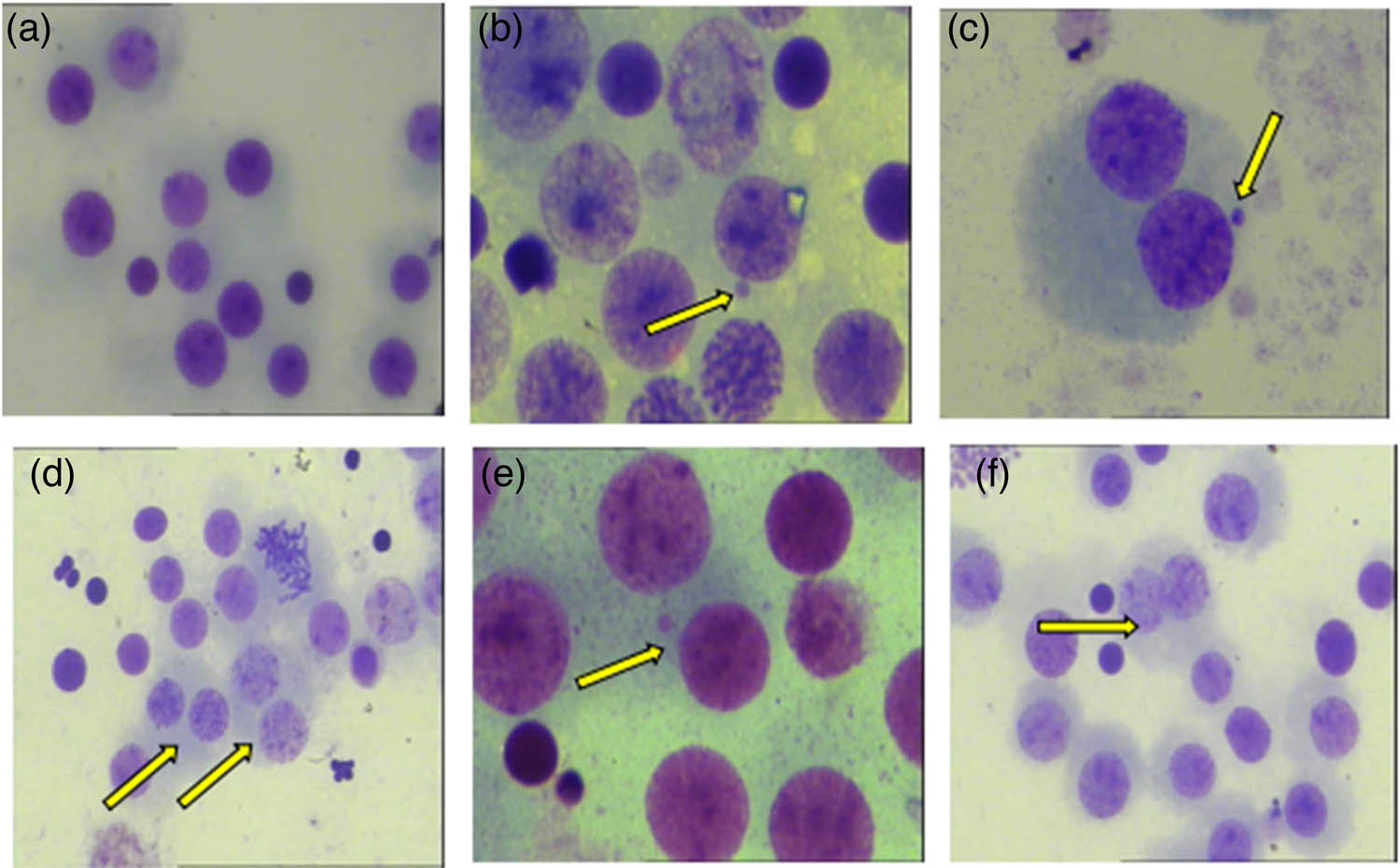
Image of micronucleus test of human peripheral blood lymphocytes treated with CdS QDs ((a): control group, ×400; (b) CdSI group, micronucleus in cells, ×1,000; (c) CdSI group, micronucleus in binuclear cells, ×1,000; (d) CdSI group, 2 binuclear cells, ×400; (e) CdS; group, micronucleus appeared in the cell, ×1,000; (f) CdS0 group, dual nucleus appeared in the cell, ×400).

DNA damage (average ± SEM) expressed as micronucleus rate and binuclear rate. Capital letters represent statistical differences between treatments (P < 0.05). Note: ●: compared with the control group, P < 0.05; ▲: compared with the low group, P < 0.05.
3.3 Chromosome aberration test
For the chromosome aberration test, the results after 72 h of exposure are shown in Figure 7. No aneuploids appeared in the peripheral blood lymphocytes of the control group (Figure 7a), and aneuploid and dicentric chromosomes appeared in the peripheral blood lymphocytes of the CdS I group (Figure 7b and c). Figure 8 shows that the chromosome aneuploid rate of human peripheral blood lymphocytes in the CdS I and CdS II groups was significantly higher than that in the control group (P < 0.05). The chromosome aneuploid rate in the CdS I group was significantly different from that of the CdS II group (P < 0.05), and the chromosome aneuploid rate in the CdS I group was higher than that in the CdS II group.

Image of chromosome aberration test of human peripheral blood lymphocytes treated with CdS QDs ((a) control group, ×1,000; (b) CdS I group, aneuploid in cells, ×1,000; (c) CdS I group, dicentric chromosomes appear in the cell).

DNA damage (average ± SEM) expressed as aneuploid rate. Capital letters represent statistical differences between treatments (P < 0.05). Note: ●: compared with the control group, P < 0.05; ▲: compared with the low group, P < 0.05.
4 Discussion
CdS QDs, as nanomaterials, have attracted increasing attention as platforms, such as sensors and fluorescent probes [1]. Given the growing demand and use for CdS QDs, it is necessary to understand their potential toxicity to organisms and the environment [9]. However, there have been few reports on the genetic toxicity of CdS QDs. Therefore, it is of great importance to study the genetic toxicity of CdS QDs. In this study, CdS QDs were used as the research object. Through comet, micronucleus, and chromosome aberration tests, the damaging effect of CdS QDs on the DNA of human peripheral blood lymphocytes was comprehensively evaluated from the three aspects of single-cell DNA breakage and chromosome integrity and abnormal chromosome separation.
The DNA damage resulting from CdS QDs was evaluated by the comet assay. The results showed that the olive tail distance, tail length, and tail DNA% of the CdS I and CdS II groups were significantly larger than those of the control group, and the tail length and tail DNA% of the CdS I group were significantly larger than those of the CdS II group. This result showed that CdS QDs have a certain damaging effect on the genetic material of human peripheral blood lymphocytes, and the damaging effect after wrapping is lighter than that without wrapping. Katubi et al. [21] studied the effect of exposure dose and time of CdTe QDs on the cytotoxicity and genotoxicity of human liver cancer cells and found that with an increase in the exposure dose of CdTe QDs and prolongation of exposure time, olive tail moment and tail DNA% both significantly increased, indicating that CdTe QDs can induce DNA strand breaks. The related mechanism may be that when QDs infect lymphocytes and are absorbed by lymphocytes through endocytosis, they interact with cells to directly destroy the cellular antioxidant system, increase ROS, cause cellular oxidative stress, produce too many free radicals, attack polyunsaturated fatty acids (PUFAs) in the cell membrane, cause lipid peroxidation, and, finally, cause DNA strand breakage [22]. In addition, free radicals caused by QDs can damage DNA integrity through double-stranded nicks [23].
The ability of CdS QDs to induce chromosome breakage was evaluated by the micronucleus test. The results showed that the olive tail distance, tail length, and tail DNA% of the CdS I and CdS II groups were significantly larger than those of the control group, and the tail length and tail DNA% of the CdS I group were significantly larger than those of the CdS II group. This result shows that CdS QDs have a certain damaging effect on the genetic material of human peripheral blood lymphocytes, and the damaging effect after wrapping is less than that without wrapping. Manshian [24] showed in a study of the genotoxic capacity of CdSe/ZnS QDs with different surface chemistries that Cd/Se QDs can significantly increase the cell micronucleus rate. Demir et al. [25] used in vitro tests to evaluate the cytotoxicity and genotoxicity of cadmium oxide NPs. The results showed that CdO NPs could induce chromosome and DNA single- or double-strand breaks and mutations. These studies have shown that QDs can cause the formation of micronuclei. The mechanism of micronuclei formation might be that QDs damage cell chromosomes by inhibiting, slowing down, or even terminating the normal division of lymphocytes. The mechanism may also be that QDs can induce apoptosis by inducing caspase family expression, downregulating p53 and Bcl-2 gene expression, enabling the apoptosis factor Fas system, and intervening in the mitochondria-mediated apoptosis signaling pathway [26,27]. The important characteristics of apoptosis are chromatin fragmentation and chromatin pyknosis, resulting in a micronucleus formation.
The ability of CdS QDs to induce genetic damage was evaluated by a chromosome aberration test. The results showed that CdS QDs could significantly increase the aneuploid rate of cells, and the aneuploid rate of CdS QDs encapsulated by thioglycolic acid was significantly lower than that of the nonencapsulated group. George [28] studied the genotoxicity and interference of gold NPs in common in vitro mutagenicity and genotoxicity tests and found that gold NPs can cause chromosome aberrations. The mechanism of chromosome aberration formation may be that QDs directly destroy the antioxidant system to increase oxygen-free radicals. Excessive oxygen-free radicals can damage the spindle in the metaphase of cell division such that chromosomes cannot be effectively pulled to the two cell poles and finally induce the formation of polyploidy [29,30].
All three tests showed that the thioglycolic acid-coated CdS QDs caused significantly less damage to genetic material than the uncoated group. Other studies have shown that compared with bare QDs, QDs with a coating as a protective factor are beneficial for reducing cytotoxicity [31]. The reason may be that the coating layer can control dissolution and cellular uptake, increase the stability of QDs, and slow down their oxidation process in cells [24,32].
In summary, CdS QDs can enter human peripheral blood lymphocytes through endocytosis and exert genotoxicity. The damage to the genetic material of CdS QDs encapsulated by thioglycolic acid is significantly reduced. Therefore, QD surface modification may be an effective method to delay the harmful effects of QDs; however, the subcellular localization of QDs, low pH environment, oxidation caused by ultraviolet light penetration of skin and/or an inflammatory reaction may degrade the coating in vivo, and the stability and intrinsic toxicity of nanomaterials together represent the key determinants of genotoxicity induction [24]. Therefore, further research is needed to fully understand the mechanism of toxicity of CdS QDs and estimate their long-term impact on human health. Because there were few toxicity indices selected in this study, there was still a lack of molecular-level results, thus warranting further research in future studies.
Acknowledgments
We would like to thank the National Basic Research Program of China, the National Natural Science Foundation, and the Shanghai Pujiang Program for providing financial support.
-
Funding information: This work was financially supported by the National Basic Research Program of China (973 Program 2013CB932800), the National Natural Science Foundation (21103219), and the Shanghai Pujiang Program (11PJ1412000).
-
Author contributions: Yanhua Cao, Qingzhao Li, and Haiping Liu contributed equally to this work. Yanhua Cao, Qingzhao Li, and Haiping Liu performed the experiments. Huajie Liu and Haiyan Liu designed the experiments. Yanhua Cao and Haiping Liu analyzed the data. Haiping Liu wrote the paper. Duo Zhang, Qian Wang, and Shuang Li revised and edited the manuscript.
-
Conflict of interest: Authors state no conflict of interest.
-
Data availability statement: The datasets generated during and/or analyzed during the current study are available from the corresponding author on reasonable request.
References
[1] Matea CT, Mocan T, Tabaran F, Pop T, Mosteanu O, Puia C, et al. Quantum dots in imaging, drug delivery and sensor applications. Int J Nanomed. 2017;12:5421–31. 10.2147/IJN.S138624.Search in Google Scholar PubMed PubMed Central
[2] Amouzadeh TM, Nazari L, Acedo P. A photo-electrochemical aptasensor for the determination of severe acute respiratory syndrome coronavirus 2 receptor-binding domain by using graphitic carbon nitride-cadmium sulfide quantum dots nanocomposite. Sens Actuators B Chem. 2021;345:130377. 10.1016/j.snb.2021.130377.Search in Google Scholar PubMed PubMed Central
[3] Zhao R, Wang Z, Tian X, Shu H, Yang Y, Xiao X, et al. Excellent fluorescence detection of Cu2+ in water system using N-acetyl-l-cysteines modified CdS quantum dots as fluorescence probe. Nanotechnology. 2021;32(40):405707 (14pp). 10.1088/1361-6528/ac1016.Search in Google Scholar PubMed
[4] Reshma VG, Mohanan PV. Quantum dots: Applications and safety consequences. J Lumin. 2018;205:205–98. 10.1016/j.jlumin.2018.09.015.Search in Google Scholar
[5] Saka C, Tta B, Chen X, Ms A, Hlayja B. Plasmon-enhanced quantum dots electrochemiluminescence aptasensor for selective and sensitive detection of cardiac troponin i. Talanta. 2020;221:121674. 10.1016/j.talanta.2020.121674.Search in Google Scholar PubMed
[6] Liu J, Wang B, Huang H, Jian D, Lu Y, Shan Y, et al. Quantitative ciprofloxacin on-site rapid detections using quantum dot microsphere based immunochromatographic test strips. Food Chem. 2021;335:127596. 10.1016/j.foodchem.2020.127596.Search in Google Scholar PubMed
[7] Shu Y, Gao J, Chen J, Yan J, Sun J, Jin D, et al. A near-infrared fluorescent sensor based on the architecture of low-toxic Ag2S quantum dot and MnO2 nanosheet for sensing glutathione in human serum sample. Talanta. 2021;221:121475. 10.1016/j.talanta.2020.121475.Search in Google Scholar PubMed
[8] Wang Y, Tang M. Review of in vitro toxicological research of quantum dot and potentially involved mechanisms. Sci Total Env. 2018;625:940–62. 10.1016/j.scitotenv.2017.12.334.Search in Google Scholar PubMed
[9] Mo D, Hu L, Zeng G, Chen G, Wan J, Yu Z, et al. Cadmium-containing quantum dots: properties, applications, and toxicity. Appl Microbiol Biotechnol. 2017;101(7):2713–33. 10.1007/s00253-017-8140-9.Search in Google Scholar PubMed
[10] Tan B, Liao L, Yang Z, Long X, Li J. Toward the toxicology of some nitro-compounds. Mod Org Chem Res. 2018;3(1):11–21. 10.22606/mocr.2018.31002.Search in Google Scholar
[11] Cumming AS, Johnson MS. Energetic materials and munitions: life cycle management, environmental impact and demilitarization. Wiley-VCH Verlag GmbH & Co. KGaA, School of Chemistry, University of Edinburgh; US Army Public Health Center; 2019. 10.1002/9783527816651.Search in Google Scholar
[12] Tiwari J, Tarale P, Sivanesan S, Bafana A. Environmental persistence, hazard, and mitigation challenges of nitroaromatic compounds. Env Sci Pollut Res Int. 2019;26(28):28650–67. 10.1007/s11356-019-06043-8.Search in Google Scholar
[13] Jigyasu AK, Siddiqui S, Lohani M, Khan IA, Arshad M. Chemically synthesized CdSe quantum dots inhibit growth of human lung carcinoma cells via ROS generation. EXCLI J. 2016;15:54–63. 10.17179/excli2015-705.Search in Google Scholar
[14] Paesano L, Perotti A, Buschini A, Carubbi C, Marmiroli M, Maestri E, et al. Markers for toxicity to HepG2 exposed to cadmium sulphide quantum dots; damage to mitochondria. Toxicology. 2016;374:18–28. 10.1016/j.tox.2016.11.012.Search in Google Scholar
[15] Brunetti V, Chibli H, Fiammengo R, Galeone A, Malvindi MA, Vecchio G, et al. InP/ZnS as a safer alternative to CdSe/ZnS core/shell quantum dots: in vitro and in vivo toxicity assessment. Nanoscale. 2013;5(1):307–17. 10.1039/c2nr33024e.Search in Google Scholar
[16] He K, Liang X, Wei T, Liu N, Wang Y, Zou L, et al. DNA damage in BV-2 cells: An important supplement to the neurotoxicity of CdTe quantum dots. J Appl Toxicol. 2019;39(3):525–39. 10.1002/jat.3745.Search in Google Scholar
[17] Koppen G, Azqueta A, Pourrut B, Brunborg G, Collins AR, Langie SAS. The next three decades of the comet assay: a report of the 11th International Comet Assay Workshop. Mutagenesis. 2017;32(3):397–408. 10.1093/mutage/gex002.Search in Google Scholar
[18] Singh NP, McCoy MT, Tice RR, Schneider EL. A simple technique for quantitation of low levels of DNA damage in individual cells. Exp Cell Res. 1988;175(1):184–91. 10.1016/0014-4827(88)90265-0.Search in Google Scholar
[19] Ataseven N, Yüzbaşıoğlu D, Keskin AÇ, Ünal F. Genotoxicity of monosodium glutamate. Food Chem Toxicol. 2016;91:8–18. 10.1016/j.fct.2016.02.021.Search in Google Scholar
[20] Fenech M. The in vitro micronucleus technique. Mutat Res. 2000;455(1–2):81–95. 10.1016/s0027-5107(00)00065-8.Search in Google Scholar
[21] Katubi KM, Alzahrani FM, Ali D, Alarifi S. Dose- and duration-dependent cytotoxicity and genotoxicity in human hepato carcinoma cells due to CdTe QDs exposure. Hum Exp Toxicol. 2019;38(8):914–26. 10.1177/0960327119843578.Search in Google Scholar PubMed
[22] De Matteis V. Exposure to inorganic nanoparticles: routes of entry, immune response, biodistribution and in vitro/in vivo toxicity evaluation. Toxics. 2017;5(4):29. 10.3390/toxics5040029.Search in Google Scholar PubMed PubMed Central
[23] Choi YJ, Kim YJ, Lee JW, Lee Y, Lim YB, Chung HW. Cyto-/genotoxic effect of CdSe/ZnS quantum dots in human lung adenocarcinoma cells for potential photodynamic UV therapy applications. J Nanosci Nanotechnol. 2012;12(3):2160–8. 10.1166/jnn.2012.5781.Search in Google Scholar PubMed
[24] Manshian BB, Soenen SJ, Brown A, Hondow N, Wills J, Jenkins GJ, et al. Genotoxic capacity of CdSe/ZnS semiconductor quantum dots with differing surface chemistries. Mutagenesis. 2016;31(1):97–106. 10.1093/mutage/gev061.Search in Google Scholar PubMed PubMed Central
[25] Demir E, Qin T, Li Y, Zhang Y, Guo X, Ingle T, et al. Cytotoxicity and genotoxicity of cadmium oxide nanoparticles evaluated using in vitro assays. Mutat Res Genet Toxicol Env Mutagen. 2020;850–851:503149. 10.1016/j.mrgentox.Search in Google Scholar
[26] Nguyen KC, Willmore WG, Tayabali AF. Cadmium telluride quantum dots cause oxidative stress leading to extrinsic and intrinsic apoptosis in hepatocellular carcinoma HepG2 cells. Toxicology. 2013;306:114–23. 10.1016/j.tox.2013.02.010.Search in Google Scholar PubMed
[27] Singh BR, Singh BN, Khan W, Singh HB, Naqvi AH. ROS-mediated apoptotic cell death in prostate cancer LNCaP cells induced by biosurfactant stabilized CdS quantum dots. Biomaterials. 2012;33:5753–67. 10.1016/j.biomaterials.2012.04.045.Search in Google Scholar PubMed
[28] George JM, Magogotya M, Vetten MA, Buys AV, Gulumian M. From the cover: an investigation of the genotoxicity and interference of gold nanoparticles in commonly used in vitro mutagenicity and genotoxicity assays. Toxicol Sci. 2017;156(1):149–66. 10.1093/toxsci/kfw247.Search in Google Scholar PubMed
[29] Li Y, Zhao J, Shang E, Xia X, Niu J, Crittenden J. Effects of chloride ions on dissolution, ros generation, and toxicity of silver nanoparticles under uv irradiation. Environ Sci Technol. 2018;52(8):4842–9. 10.1021/acs.est.7b04547.Search in Google Scholar PubMed
[30] Shang E, Li Y, Niu J, Zhou Y, Wang T, Crittenden JC. Relative importance of humic and fulvic acid on ros generation, dissolution, and toxicity of sulfide nanoparticles. Water Res. 2017;595–604. 10.1016/j.watres.2017.08.001.Search in Google Scholar PubMed
[31] Peng L, He M, Chen B, Qiao Y, Hu B. Metallomics study of CdSe/ZnS quantum dots in HepG2 cells. ACS Nano. 2015;9(10):10324–34. 10.1021/acsnano.5b04365.Search in Google Scholar PubMed
[32] Geißler D, Wegmann M, Jochum T, Somma V, Sowa M, Scholz J, et al. An automatable platform for genotoxicity testing of nanomaterials based on the fluorometric γ-H2AX assay reveals no genotoxicity of properly surface-shielded cadmium-based quantum dots. Nanoscale. 2019;11(28):13458–68. 10.1039/c9nr01021a.Search in Google Scholar PubMed
© 2022 Haiping Liu et al., published by De Gruyter
This work is licensed under the Creative Commons Attribution 4.0 International License.
Articles in the same Issue
- Biomedical Sciences
- Effects of direct oral anticoagulants dabigatran and rivaroxaban on the blood coagulation function in rabbits
- The mother of all battles: Viruses vs humans. Can humans avoid extinction in 50–100 years?
- Knockdown of G1P3 inhibits cell proliferation and enhances the cytotoxicity of dexamethasone in acute lymphoblastic leukemia
- LINC00665 regulates hepatocellular carcinoma by modulating mRNA via the m6A enzyme
- Association study of CLDN14 variations in patients with kidney stones
- Concanavalin A-induced autoimmune hepatitis model in mice: Mechanisms and future outlook
- Regulation of miR-30b in cancer development, apoptosis, and drug resistance
- Informatic analysis of the pulmonary microecology in non-cystic fibrosis bronchiectasis at three different stages
- Swimming attenuates tumor growth in CT-26 tumor-bearing mice and suppresses angiogenesis by mediating the HIF-1α/VEGFA pathway
- Characterization of intestinal microbiota and serum metabolites in patients with mild hepatic encephalopathy
- Functional conservation and divergence in plant-specific GRF gene family revealed by sequences and expression analysis
- Application of the FLP/LoxP-FRT recombination system to switch the eGFP expression in a model prokaryote
- Biomedical evaluation of antioxidant properties of lamb meat enriched with iodine and selenium
- Intravenous infusion of the exosomes derived from human umbilical cord mesenchymal stem cells enhance neurological recovery after traumatic brain injury via suppressing the NF-κB pathway
- Effect of dietary pattern on pregnant women with gestational diabetes mellitus and its clinical significance
- Potential regulatory mechanism of TNF-α/TNFR1/ANXA1 in glioma cells and its role in glioma cell proliferation
- Effect of the genetic mutant G71R in uridine diphosphate-glucuronosyltransferase 1A1 on the conjugation of bilirubin
- Quercetin inhibits cytotoxicity of PC12 cells induced by amyloid-beta 25–35 via stimulating estrogen receptor α, activating ERK1/2, and inhibiting apoptosis
- Nutrition intervention in the management of novel coronavirus pneumonia patients
- circ-CFH promotes the development of HCC by regulating cell proliferation, apoptosis, migration, invasion, and glycolysis through the miR-377-3p/RNF38 axis
- Bmi-1 directly upregulates glucose transporter 1 in human gastric adenocarcinoma
- Lacunar infarction aggravates the cognitive deficit in the elderly with white matter lesion
- Hydroxysafflor yellow A improved retinopathy via Nrf2/HO-1 pathway in rats
- Comparison of axon extension: PTFE versus PLA formed by a 3D printer
- Elevated IL-35 level and iTr35 subset increase the bacterial burden and lung lesions in Mycobacterium tuberculosis-infected mice
- A case report of CAT gene and HNF1β gene variations in a patient with early-onset diabetes
- Study on the mechanism of inhibiting patulin production by fengycin
- SOX4 promotes high-glucose-induced inflammation and angiogenesis of retinal endothelial cells by activating NF-κB signaling pathway
- Relationship between blood clots and COVID-19 vaccines: A literature review
- Analysis of genetic characteristics of 436 children with dysplasia and detailed analysis of rare karyotype
- Bioinformatics network analyses of growth differentiation factor 11
- NR4A1 inhibits the epithelial–mesenchymal transition of hepatic stellate cells: Involvement of TGF-β–Smad2/3/4–ZEB signaling
- Expression of Zeb1 in the differentiation of mouse embryonic stem cell
- Study on the genetic damage caused by cadmium sulfide quantum dots in human lymphocytes
- Association between single-nucleotide polymorphisms of NKX2.5 and congenital heart disease in Chinese population: A meta-analysis
- Assessment of the anesthetic effect of modified pentothal sodium solution on Sprague-Dawley rats
- Genetic susceptibility to high myopia in Han Chinese population
- Potential biomarkers and molecular mechanisms in preeclampsia progression
- Silencing circular RNA-friend leukemia virus integration 1 restrained malignancy of CC cells and oxaliplatin resistance by disturbing dyskeratosis congenita 1
- Endostar plus pembrolizumab combined with a platinum-based dual chemotherapy regime for advanced pulmonary large-cell neuroendocrine carcinoma as a first-line treatment: A case report
- The significance of PAK4 in signaling and clinicopathology: A review
- Sorafenib inhibits ovarian cancer cell proliferation and mobility and induces radiosensitivity by targeting the tumor cell epithelial–mesenchymal transition
- Characterization of rabbit polyclonal antibody against camel recombinant nanobodies
- Active legumain promotes invasion and migration of neuroblastoma by regulating epithelial-mesenchymal transition
- Effect of cell receptors in the pathogenesis of osteoarthritis: Current insights
- MT-12 inhibits the proliferation of bladder cells in vitro and in vivo by enhancing autophagy through mitochondrial dysfunction
- Study of hsa_circRNA_000121 and hsa_circRNA_004183 in papillary thyroid microcarcinoma
- BuyangHuanwu Decoction attenuates cerebral vasospasm caused by subarachnoid hemorrhage in rats via PI3K/AKT/eNOS axis
- Effects of the interaction of Notch and TLR4 pathways on inflammation and heart function in septic heart
- Monosodium iodoacetate-induced subchondral bone microstructure and inflammatory changes in an animal model of osteoarthritis
- A rare presentation of type II Abernethy malformation and nephrotic syndrome: Case report and review
- Rapid death due to pulmonary epithelioid haemangioendothelioma in several weeks: A case report
- Hepatoprotective role of peroxisome proliferator-activated receptor-α in non-cancerous hepatic tissues following transcatheter arterial embolization
- Correlation between peripheral blood lymphocyte subpopulations and primary systemic lupus erythematosus
- A novel SLC8A1-ALK fusion in lung adenocarcinoma confers sensitivity to alectinib: A case report
- β-Hydroxybutyrate upregulates FGF21 expression through inhibition of histone deacetylases in hepatocytes
- Identification of metabolic genes for the prediction of prognosis and tumor microenvironment infiltration in early-stage non-small cell lung cancer
- BTBD10 inhibits glioma tumorigenesis by downregulating cyclin D1 and p-Akt
- Mucormycosis co-infection in COVID-19 patients: An update
- Metagenomic next-generation sequencing in diagnosing Pneumocystis jirovecii pneumonia: A case report
- Long non-coding RNA HOXB-AS1 is a prognostic marker and promotes hepatocellular carcinoma cells’ proliferation and invasion
- Preparation and evaluation of LA-PEG-SPION, a targeted MRI contrast agent for liver cancer
- Proteomic analysis of the liver regulating lipid metabolism in Chaohu ducks using two-dimensional electrophoresis
- Nasopharyngeal tuberculosis: A case report
- Characterization and evaluation of anti-Salmonella enteritidis activity of indigenous probiotic lactobacilli in mice
- Aberrant pulmonary immune response of obese mice to periodontal infection
- Bacteriospermia – A formidable player in male subfertility
- In silico and in vivo analysis of TIPE1 expression in diffuse large B cell lymphoma
- Effects of KCa channels on biological behavior of trophoblasts
- Interleukin-17A influences the vulnerability rather than the size of established atherosclerotic plaques in apolipoprotein E-deficient mice
- Multiple organ failure and death caused by Staphylococcus aureus hip infection: A case report
- Prognostic signature related to the immune environment of oral squamous cell carcinoma
- Primary and metastatic squamous cell carcinoma of the thyroid gland: Two case reports
- Neuroprotective effects of crocin and crocin-loaded niosomes against the paraquat-induced oxidative brain damage in rats
- Role of MMP-2 and CD147 in kidney fibrosis
- Geometric basis of action potential of skeletal muscle cells and neurons
- Babesia microti-induced fulminant sepsis in an immunocompromised host: A case report and the case-specific literature review
- Role of cerebellar cortex in associative learning and memory in guinea pigs
- Application of metagenomic next-generation sequencing technique for diagnosing a specific case of necrotizing meningoencephalitis caused by human herpesvirus 2
- Case report: Quadruple primary malignant neoplasms including esophageal, ureteral, and lung in an elderly male
- Long non-coding RNA NEAT1 promotes angiogenesis in hepatoma carcinoma via the miR-125a-5p/VEGF pathway
- Osteogenic differentiation of periodontal membrane stem cells in inflammatory environments
- Knockdown of SHMT2 enhances the sensitivity of gastric cancer cells to radiotherapy through the Wnt/β-catenin pathway
- Continuous renal replacement therapy combined with double filtration plasmapheresis in the treatment of severe lupus complicated by serious bacterial infections in children: A case report
- Simultaneous triple primary malignancies, including bladder cancer, lymphoma, and lung cancer, in an elderly male: A case report
- Preclinical immunogenicity assessment of a cell-based inactivated whole-virion H5N1 influenza vaccine
- One case of iodine-125 therapy – A new minimally invasive treatment of intrahepatic cholangiocarcinoma
- S1P promotes corneal trigeminal neuron differentiation and corneal nerve repair via upregulating nerve growth factor expression in a mouse model
- Early cancer detection by a targeted methylation assay of circulating tumor DNA in plasma
- Calcifying nanoparticles initiate the calcification process of mesenchymal stem cells in vitro through the activation of the TGF-β1/Smad signaling pathway and promote the decay of echinococcosis
- Evaluation of prognostic markers in patients infected with SARS-CoV-2
- N6-Methyladenosine-related alternative splicing events play a role in bladder cancer
- Characterization of the structural, oxidative, and immunological features of testis tissue from Zucker diabetic fatty rats
- Effects of glucose and osmotic pressure on the proliferation and cell cycle of human chorionic trophoblast cells
- Investigation of genotype diversity of 7,804 norovirus sequences in humans and animals of China
- Characteristics and karyotype analysis of a patient with turner syndrome complicated with multiple-site tumors: A case report
- Aggravated renal fibrosis is positively associated with the activation of HMGB1-TLR2/4 signaling in STZ-induced diabetic mice
- Distribution characteristics of SARS-CoV-2 IgM/IgG in false-positive results detected by chemiluminescent immunoassay
- SRPX2 attenuated oxygen–glucose deprivation and reperfusion-induced injury in cardiomyocytes via alleviating endoplasmic reticulum stress-induced apoptosis through targeting PI3K/Akt/mTOR axis
- Aquaporin-8 overexpression is involved in vascular structure and function changes in placentas of gestational diabetes mellitus patients
- Relationship between CRP gene polymorphisms and ischemic stroke risk: A systematic review and meta-analysis
- Effects of growth hormone on lipid metabolism and sexual development in pubertal obese male rats
- Cloning and identification of the CTLA-4IgV gene and functional application of vaccine in Xinjiang sheep
- Antitumor activity of RUNX3: Upregulation of E-cadherin and downregulation of the epithelial–mesenchymal transition in clear-cell renal cell carcinoma
- PHF8 promotes osteogenic differentiation of BMSCs in old rat with osteoporosis by regulating Wnt/β-catenin pathway
- A review of the current state of the computer-aided diagnosis (CAD) systems for breast cancer diagnosis
- Bilateral dacryoadenitis in adult-onset Still’s disease: A case report
- A novel association between Bmi-1 protein expression and the SUVmax obtained by 18F-FDG PET/CT in patients with gastric adenocarcinoma
- The role of erythrocytes and erythroid progenitor cells in tumors
- Relationship between platelet activation markers and spontaneous abortion: A meta-analysis
- Abnormal methylation caused by folic acid deficiency in neural tube defects
- Silencing TLR4 using an ultrasound-targeted microbubble destruction-based shRNA system reduces ischemia-induced seizures in hyperglycemic rats
- Plant Sciences
- Seasonal succession of bacterial communities in cultured Caulerpa lentillifera detected by high-throughput sequencing
- Cloning and prokaryotic expression of WRKY48 from Caragana intermedia
- Novel Brassica hybrids with different resistance to Leptosphaeria maculans reveal unbalanced rDNA signal patterns
- Application of exogenous auxin and gibberellin regulates the bolting of lettuce (Lactuca sativa L.)
- Phytoremediation of pollutants from wastewater: A concise review
- Genome-wide identification and characterization of NBS-encoding genes in the sweet potato wild ancestor Ipomoea trifida (H.B.K.)
- Alleviative effects of magnetic Fe3O4 nanoparticles on the physiological toxicity of 3-nitrophenol to rice (Oryza sativa L.) seedlings
- Selection and functional identification of Dof genes expressed in response to nitrogen in Populus simonii × Populus nigra
- Study on pecan seed germination influenced by seed endocarp
- Identification of active compounds in Ophiopogonis Radix from different geographical origins by UPLC-Q/TOF-MS combined with GC-MS approaches
- The entire chloroplast genome sequence of Asparagus cochinchinensis and genetic comparison to Asparagus species
- Genome-wide identification of MAPK family genes and their response to abiotic stresses in tea plant (Camellia sinensis)
- Selection and validation of reference genes for RT-qPCR analysis of different organs at various development stages in Caragana intermedia
- Cloning and expression analysis of SERK1 gene in Diospyros lotus
- Integrated metabolomic and transcriptomic profiling revealed coping mechanisms of the edible and medicinal homologous plant Plantago asiatica L. cadmium resistance
- A missense variant in NCF1 is associated with susceptibility to unexplained recurrent spontaneous abortion
- Assessment of drought tolerance indices in faba bean genotypes under different irrigation regimes
- The entire chloroplast genome sequence of Asparagus setaceus (Kunth) Jessop: Genome structure, gene composition, and phylogenetic analysis in Asparagaceae
- Food Science
- Dietary food additive monosodium glutamate with or without high-lipid diet induces spleen anomaly: A mechanistic approach on rat model
- Binge eating disorder during COVID-19
- Potential of honey against the onset of autoimmune diabetes and its associated nephropathy, pancreatitis, and retinopathy in type 1 diabetic animal model
- FTO gene expression in diet-induced obesity is downregulated by Solanum fruit supplementation
- Physical activity enhances fecal lactobacilli in rats chronically drinking sweetened cola beverage
- Supercritical CO2 extraction, chemical composition, and antioxidant effects of Coreopsis tinctoria Nutt. oleoresin
- Functional constituents of plant-based foods boost immunity against acute and chronic disorders
- Effect of selenium and methods of protein extraction on the proteomic profile of Saccharomyces yeast
- Microbial diversity of milk ghee in southern Gansu and its effect on the formation of ghee flavor compounds
- Ecology and Environmental Sciences
- Effects of heavy metals on bacterial community surrounding Bijiashan mining area located in northwest China
- Microorganism community composition analysis coupling with 15N tracer experiments reveals the nitrification rate and N2O emissions in low pH soils in Southern China
- Genetic diversity and population structure of Cinnamomum balansae Lecomte inferred by microsatellites
- Preliminary screening of microplastic contamination in different marine fish species of Taif market, Saudi Arabia
- Plant volatile organic compounds attractive to Lygus pratensis
- Effects of organic materials on soil bacterial community structure in long-term continuous cropping of tomato in greenhouse
- Effects of soil treated fungicide fluopimomide on tomato (Solanum lycopersicum L.) disease control and plant growth
- Prevalence of Yersinia pestis among rodents captured in a semi-arid tropical ecosystem of south-western Zimbabwe
- Effects of irrigation and nitrogen fertilization on mitigating salt-induced Na+ toxicity and sustaining sea rice growth
- Bioengineering and Biotechnology
- Poly-l-lysine-caused cell adhesion induces pyroptosis in THP-1 monocytes
- Development of alkaline phosphatase-scFv and its use for one-step enzyme-linked immunosorbent assay for His-tagged protein detection
- Development and validation of a predictive model for immune-related genes in patients with tongue squamous cell carcinoma
- Agriculture
- Effects of chemical-based fertilizer replacement with biochar-based fertilizer on albic soil nutrient content and maize yield
- Genome-wide identification and expression analysis of CPP-like gene family in Triticum aestivum L. under different hormone and stress conditions
- Agronomic and economic performance of mung bean (Vigna radiata L.) varieties in response to rates of blended NPS fertilizer in Kindo Koysha district, Southern Ethiopia
- Influence of furrow irrigation regime on the yield and water consumption indicators of winter wheat based on a multi-level fuzzy comprehensive evaluation
- Discovery of exercise-related genes and pathway analysis based on comparative genomes of Mongolian originated Abaga and Wushen horse
- Lessons from integrated seasonal forecast-crop modelling in Africa: A systematic review
- Evolution trend of soil fertility in tobacco-planting area of Chenzhou, Hunan Province, China
- Animal Sciences
- Morphological and molecular characterization of Tatera indica Hardwicke 1807 (Rodentia: Muridae) from Pothwar, Pakistan
- Research on meat quality of Qianhua Mutton Merino sheep and Small-tail Han sheep
- SI: A Scientific Memoir
- Suggestions on leading an academic research laboratory group
- My scientific genealogy and the Toronto ACDC Laboratory, 1988–2022
- Erratum
- Erratum to “Changes of immune cells in patients with hepatocellular carcinoma treated by radiofrequency ablation and hepatectomy, a pilot study”
- Erratum to “A two-microRNA signature predicts the progression of male thyroid cancer”
- Retraction
- Retraction of “Lidocaine has antitumor effect on hepatocellular carcinoma via the circ_DYNC1H1/miR-520a-3p/USP14 axis”