Punica granatum peel extracts mediated the green synthesis of gold nanoparticles and their detailed in vivo biological activities
-
Sami Bawazeer
 ,
Taufiq Nawaz
,
Taufiq Nawaz

Abstract
Requirements for developing new methodologies to biosynthesize nanoparticles are increasing day by day. The typical chemical synthesis of nanoparticles has raised concerns regarding environmental safety and adverse impact on human health. Therefore, there is an urgent need to develop green synthesized nanoparticles that are considered to be safe, ecofriendly, and cost-effective as compared to chemical approaches. Hence, in this study, we synthesized and characterized pomegranate peel extract-based gold nanoparticles (PP-AuNPs) through UV-visible spectroscopy, FT-IR, and AFM microscopy. Furthermore, the biological activities like analgesic, muscle relaxant, and sedative properties of synthesized PP-AuNPs were also determined. The change of color to dark ruby indicates the formation of AuNPs. The surface plasma resonance (SPR) peak in the absorption spectra was shown at 525 nm by using (UV-Vis) spectroscopy. A single distinctive peak implied the shape of nanoparticles to be spherical. AFM images revealed that the biosynthesized nanoparticles were spherical in shape. Furthermore, the images confirm the uniform distribution of PP-AuNPs with particle sizes ranging from 4 to 16 nm. Different classes of phytochemicals were preliminarily identified in extracts. The analgesic effect of extracts (70.04%) and PP-AuNPs (81.98%) demonstrated a significant (p < 0.001) percent reduction in writhing at a dose of 100 and 15 mg·kg−1, respectively. A mild muscle relaxant effect was noted against both the tested samples while a significant sedative effect was observed for both samples; however, PP-AuNPs weres more sedative compared to the extract. Pomegranate peel extracts and synthesized PP-AuNPs were found to possess significant analgesic, muscle relaxant, and sedative properties.
Graphical abstract
1 Introduction
Pomegranate (Punica granatum L.), indigenously known as “Anar,” belongs to the family Punicaceae. It is abundantly available in Pakistan, India, Iran, Afghanistan, Arizona, and California [6]. In folk medicine, P. granatum has been used for the treatment of several diseases like diarrhea, male infertility, fever, dysentery, and bleeding [1]. Evidently, various parts of pomegranate including peel, arils, flower, leaves, roots, and seeds possess phenolics (ellagic acid, punicalagin, ellagitannins, and punicic acid) that are responsible for its health-promoting benefits against diabetes, obesity, cancer, arthritis, Alzheimer, and cardiovascular diseases [2,3,4,5]. Among all these parts, pomegranate peel is an abundant source of ellagitannins, punicalagin, and punicalin. The peel of this fruit contains a cache of polyphenols and has been utilized in different food, cosmetic, and medicinal formulations [6]. Phytochemical profiling of pomegranate peel has revealed an array of bioactive compounds responsible for its antioxidant and antimicrobial properties [7]. Inorganic compounds (silver and gold) are used for the synthesis of NPs to enhance the drug delivery model, antimicrobial, and antioxidant properties [8].
Nano-biotechnology helps significantly in developing new methodologies that may provide a better understanding regarding interactions among nanomaterials and intracellular structures, the process, and the environment. The synthesis of nanoparticles is known to be a key aim in nano-biotechnology. In this context, research studies carried out nowadays are focused on the bio-fabrication of nanoparticles. The synthesis of NPs is preferred using natural resources to have green and eco-friendly end products as compared to traditionally fabricated NPs from physicochemical synthetic routes [9]. Green nano-technological methodologies are characterized by employing natural sources like biomolecules, plants, fungi, and bacteria for the effective synthesis of nanoparticles [10]. Numerous factors such as pH, temperature, reaction time, and content of plant extract have been reported to influence the synthesis of nanoparticles [11]. Amongst all the materials used for the synthesis of nanoparticles, gold (Au) has attained a significant spot with special reference to its biomedical applications. Hence, the production of gold nanoparticles (AuNPs) is a keen area of interest for different disease therapeutics, specifically cancer, owing to their diversified applications, i.e., target drug delivery, cellular bioimaging, and gene therapy [10,12,13,14].
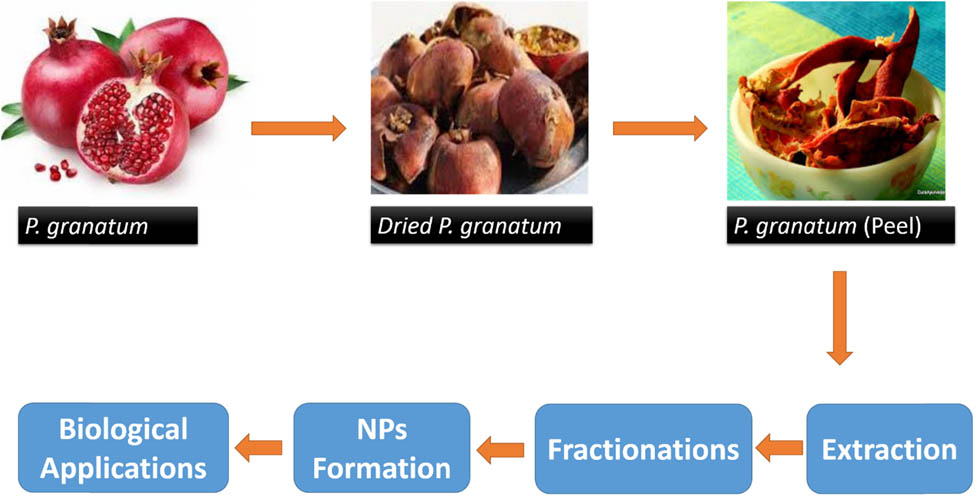
As pomegranate peel is an agro-industrial byproduct, therefore, in this study, valorization was achieved via green production of gold nanoparticles to enhance their biomedical applications. Keeping in view the significance of nanoparticles and the pomegranate peel extract, the current study focused on the synthesis of pomegranate peel-based gold nanoparticles (PP-AuNPs). These biosynthesized PP-AuNPs were characterized using AFM analysis, FTIR (Fourier transform infrared spectroscopy), and UV-visible (UV-Vis) spectroscopy. The conversion of the traditional extract into nanoparticles is the demand of the modern era when scientists are in the search of targeted therapies. Therefore, such chemically modified shapes of extracts might be more fruitful for the treatment of various ailments. In the current study, pomegranate peel extracts and nanoparticles were subjected to various in vivo biological and phytochemical studies.
2 Materials and methods
2.1 Procurement of raw materials and chemicals
Pomegranate peels were procured from the local market of Peshawar, Pakistan. P. granatum peels were dried under shade followed by grinding to collect the respective powder samples [15]. Chemicals and reagents like sodium chloride, methanol, deionized water, and hydrogen tetrachloroaurate trihydrate [HAuCl4·3H2O] used in this study were of analytical grade and were purchased from Merck.
2.2 Extract preparation
Powdered pomegranate peels were soaked in distilled water and methanol for 7 days followed by filtration through Whatman No. 1 filter paper. After filtration, the crude extract was concentrated using a rotary evaporator (45–50°C) [3].
2.3 Preparation of fractions
For the preparation of fractions, the pomegranate peel extract was dissolved in distilled water (100 mL) along with the addition of ethyl acetate (200 mL). Afterwards, the separating funnel was shaken at continuous intervals and was given stay time for the formation of two layers. Ethyl acetate was later evaporated by using a water bath in order to obtain the fractions. A similar procedure was adopted for the collection of chloroform and butanol fractions [16].
2.4 Synthesis of pomegranate peel-based gold nanoparticles (PP-AuNPs)
The pomegranate peel extract (5 mL) was mixed with HAuCl4 (1 mM) in a titration flask along with continuous stirring at room temperature for 4 h. The formation of PP-AuNPs was confirmed from the color (dark ruby) of the solution [17].
2.5 Characterization of the prepared PP-AuNPs
UV-visible spectrophotometry was used to monitor the synthesis of AuNPs at wavelengths ranging from 300 to 700 nm. AFM (atomic force microscopy) was carried out to characterize the particle size of AuNPs, while FTIR analysis was performed to identify probable functional groups responsible for bioreduction.
2.6 Kinetic studies and stability of PP-AuNPs
Kinetic studies were performed to find the time-dependent synthesis of PP-AuNPs. The prepared samples were taken out from the reaction mixtures at specific time intervals followed by UV analysis. For assessing the stability of the prepared PP-AuNPs under various pH conditions, the samples were subjected to different pH intervals. For adjustment of pH, either 1 M HCl or NaOH was added dropwise to adjust the pH value between 2 and 14 of the prepared PP-AuNPs. After each treatment, the UV-Vis spectrum was obtained.
2.7 Phytochemical screening
For determination of phytochemical screening (secondary metabolites) of the pomegranate peel extract and their subfractions, standard protocols were adopted [18,19,20,21,22,23,24].
2.7.1 Alkaloids
For this test, crude extracts and their subfractions (0.5 g) were mixed with H2SO4 (2%) and warmed for 2 min. Afterwards, this mixture was cooled and filtered followed by the addition of Dragengroff’s solution. The appearance of precipitates (orange red) indicates the presence of alkaloids.
2.7.2 Tannins
For the determination of tannins, crude extracts and their subfractions (0.2 g) were mixed with water and heated in a water bath. Moreover, the mixture was filtered followed by dropwise addition of Ferric chloride in each filtrate. Conclusively, the appearance of dark green color precipitates indicated the presence of tannins.
2.7.3 Anthraquinones
For this purpose, each extract and its subfraction (0.5 g) was heated with hydrochloric acid (10%) for a few minutes in a water bath. The resultant mixtures were cooled under tap water and filtered. The same volume of CHCl3 along with 10% NH3 (a few drops) was mixed in each filtrate. The presence of anthraquinones was indicated by the production of rose-pink color.
2.7.4 Glycosides
To assess the presence of glycosides, crude extracts/subfractions (0.3 g) were hydrolyzed with HCl followed by neutralization in the presence of NaOH. Afterwards, in each mixture, Fehling’s solution A and then B were mixed (a few drops). The formation of precipitates (red color) demonstrates the occurrence of glycosides in respective crude extracts/subfractions.
2.7.5 Reducing sugars
For this purpose, crude extracts/subfractions (0.3 g) were thoroughly mixed in distilled water. After filtration, a few drops of Fehling’s solution A and B were added to each filtrate and resultant mixtures were subjected to a few minutes of boiling. The formation of precipitates (orange-red) reveals the presence of reducing sugars.
2.7.6 Saponins
Initially, in this test, 0.5 g of each crude extract/subfractions were mixed thoroughly with distilled water. Later, the resultant reaction mixtures were boiled. Froth formation confirms the presence of saponins.
2.7.7 Flavonoids
For the determination of flavonoids, crude extracts/subfractions (0.2 g) were mixed with NaOH (diluted) followed by the addition of HCl (a few drops). On the addition of HCl, the color change from yellowish to colorless indicated the presence of flavonoids.
2.7.8 Phlobatannins
In order to detect phlobatannins, crude extracts/subfractions (0.5 g) were mixed thoroughly with distilled water followed by filtration. Afterwards, the mixture was boiled in the presence of hydrochloride (2%) solution. The appearance of precipitates (red) revealed the presence of phlobatannins.
2.7.9 Steroids
For this purpose, crude extracts/subfractions (0.3 g) in a test tube were added along with acetic acid (a few drops). Later, the mixture was heated on a burner followed by cooling, and then H2SO4 was added dropwise. The color change of the mixture to green indicated the occurrence of steroids.
2.7.10 Terpenoids
For this test, crude extracts/subfractions (0.3 g) were mixed with CHCl3 (2 mL) followed by filtration. Further, the obtained filtrates were again mixed with H2SO4 (3 mL), shaken, and allowed to stand for a few minutes. The occurrence of golden-yellow color showed the presence of terpenoids.
2.7.11 Caumarine
About 1 mL of aqueous crude extracts/subfractions were treated with 10% NaOH (3 mL). The appearance of yellow color indicated the presence of caumarine.
2.7.12 Emodin
About 0.5 g of crude extracts/subfractions was treated with NH4OH (2 mL) followed by the addition of benzene (3 mL). The formation of red color indicated the presence of emodine.
2.7.13 Anthocyanin and betacyanin
Crude extracts/subfractions (0.2 g) were mixed with 2 N NaOH (1 mL) and heated for 5 min at 100°C using a flame burner. The appearance of bluish-green color revealed the presence of anthocyanin, whereas yellow color indicated the presence of betacyanin.
2.7.14 Carbohydrates
For determination of the presence of carbohydrates, crude extracts/subfractions (0.5 g) were mixed in distilled water followed by the addition of a few drops of Molisch’s reagent. Afterwards, conc. H2SO4 (1 mL) was added to it and after 2 min stay time, distilled water (1 mL) was again added. The production of red/dull violet color in the interphase of two layers was noted as a positive test.
2.8 Biological applications of synthesized PP-AuNPs
2.8.1 Analgesic activity
The acetic acid-induced writhing in vivo paradigm was used for the assessment of the analgesic potential of extract and PP-AuNPs. Animals were classified into different groups (n = 8). Distilled water was used for treating the negative control group (10 mL·kg−1, I.P.), the positive control group received Diclofenac sodium (10 mL·kg−1, I.P.), and the tested groups were treated with extracts (10, 25, 50, and 100 mg·kg−1, PO), and PP-AuNPs (2.5, 5, 10, and 15 mg·kg−1, PO). All the animals were injected with 1% acetic acid solution (IP) after 30 min of the above treatments. After 10 min of the acetic acid injection, the number of abdominal contractions (writhings) was counted (for 10 min) for each group (n = 8) of animals [25].
2.8.2 Muscle relaxant activity
2.8.2.1 Inclined plant test
For the evaluation of fixed oil for muscle co-ordination effect, a plane of two wood was used in such a way that an angle of 65° resulted from the connection. Distilled water was used for treating the negative control group (10 mL·kg−1, I.P.), the positive group was injected with diazepam (1 mg·kg−1), and the tested groups were administered the extract (10, 25, 50, and 100 mg·kg−1, PO) and PP-AuNPs (2.5, 5, 10, and 15 mg·kg−1, PO). After 30, 60, and 90 min of the above treatment, animals were tested for the muscle coordination effect as it was placed on the upper part of the inclined plane for 30 s to hang of fall. This method is the modified form of our published method [26].
2.8.2.2 Traction test
In this in vivo paradigm, the animal was classified and treated as an in the inclined plane model. But, in this case, a metal wire coated with rubber was used, both ends of which were rigidly supported with a stand about 60 cm above the laboratory bench. The animals were exposed to the traction test after 30, 60, and 90 min of treatment. Each animal was hung by its hind legs from the wire and the time of hanging was recorded for 5 s. The failure to hang for less than 5 s was considered as the presence of muscle relaxant activity and vice versa [26].
2.8.3 Sedative activity
For the evaluation of the sedative effect of the extract and PP-AuNPs, a special box was used. The floor of the box was covered with a white sheet (150 cm diameter) and was divided into 20 squares by black lines. This open field box was placed in a soundproof experimental room. Distilled water was used for treating the negative control group (10 mL·kg−1), the positive group was injected with diazepam (1 mg·kg−1), and the tested groups were administered the extract (10, 25, 50, and 100 mg·kg−1, PO) and PP-AuNPs (2.5, 5.0, 10.0, and 15.0 mg·kg−1, PO). After 30 min of the above administration, each animal was tested for the sedative effect by placing them in the centre of the box, and the number of lines crossed by each animal was counted. The smaller number of lines crossed indicated the sedative effect [27].
2.9 Statistical analysis
All results of the biological screening are shown as the mean ± standard error of the mean (SEM), and the obtained results were evaluated by one-way analysis of variance (ANOVA). Statistical analysis was performed with the assistance of Dunnett’s multiple assessment screening using GraphPad prsim 5; the difference was significant at p ≤ 0.05.
3 Results and discussion
3.1 Selection of Au NPs and the extract ratio
Figure 1 shows the UV-Vis spectrum of various experimented ratios of the gold and extract solution (Au:E). The graph almost showed similar peaks in the range of 540–550 nm. By varying the ratio of gold and the extract, the peak intensity changed. The sharpness of the peaks shows the uniformity of Au NPs. Based on the peak sharpness and intensity, the best ratio of 10:1 was selected due to its uniform peak at 545 nm.

UV-Vis spectra of the synthesized gold nanoparticles of P. granatum at various experimental ratios of gold and the extract (Au:E).
3.2 Kinetic study of PP-AuNPs
In order to analyze the time-dependent synthesis of PP-AuNPs (Au:E = 10:1), a kinetic study was conducted. For this purpose, the prepared samples were taken out from the reaction mixtures at specific time intervals followed by UV analysis. Figure 2 reveals that the uniformity and production of nanoparticles increased with the passage of time. Figure 3 reveals the color change for the prepared PP-AuNPs.
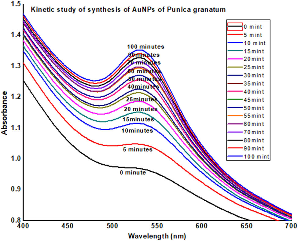
UV-Vis data for the kinetic study of PP-AuNPs.

Change in color of the prepared PP-AuNPs.
3.3 Stability towards pH
For studying the impact of pH on the stability of PP-AuNPs, the pH of the prepared nanoparticles solution was adjusted (1–14). For this purpose, the PP-AuNP solution was incubated at room temperature for 24 h. Afterwards, data of UV-Vis spectra were analyzed. The results of this trait revealed the stability of PP-AuNPs at pH ranging between 3 and 12, whereas lesser stability was noticed in the pH ranging from 1 to 2 and 13 to 14. The instability of PP-AuNPs observed in highly acidic and basic pH was subjected to the removal of stabilizers (plant extract) from the gold surface resulting in destabilization of nanoparticles. Additionally, very low pH resulted in re-oxidation of neutral PP-AuNPs. Furthermore, the change in pH also altered the color of AuNPs, which is due to the removal of the stabilizer (Figure 4). However, enhanced stability was observed at alkaline pH, i.e., 8–10. Moderate stability of the Au NPs at other pH values, i.e., 3–4, where the nanoparticles showed redshift and peak broadening.
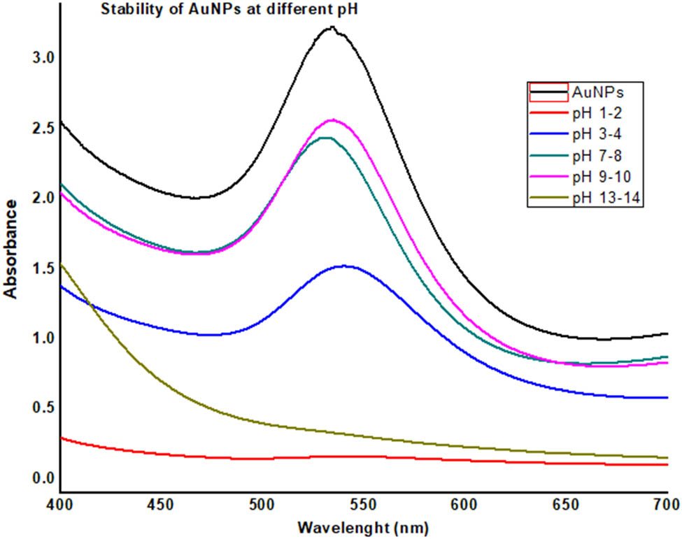
UV data showing the stability of PP-AuNPs (pomegranate peel extract-based gold nanoparticles) at different pH values.
3.4 Characterization of PP-AuNPs
3.4.1 UV-Vis spectroscopic analysis
UV-Vis spectroscopic analysis of PP-AuNPs revealed the surface plasma resonance (SPR) in the wavelength range of 400–600 nm, while a maximum characteristic peak was noticed at 545 nm, as shown in Figure 1.
3.4.2 FT-IR analysis
The crude extract of P. granatum showed a broad peak in the range 3,200–3,400 cm−1, which shows the presence of alcoholic and phenolic groups, and a sharp peak at 3,000 cm−1 corresponds to the C–H stretching frequency. A characteristic peak at 1,000 cm−1 shows the presence of C–F stretching frequency. In the case of Au nanoparticles, the OH peak intensity further increased, while the peak at 3,000 cm−1 completely disappeared, which showed that O–H and C–H olefinic are involved in the formation of nanoparticles. Also, a broadband at 500 cm−1 shows the presence of nanoparticles. The FTIR spectra are shown in the inset of Figure 5.

FTIR spectra of the crude extract (a) and AuNPs (b) of the P. granatum peel extract.
3.4.3 AFM analysis
Atomic force microscopic analysis was conducted to assess the shape of PP-AuNPs. AFM images reveal that biosynthesized nanoparticles are spherical in shape (Figure 6). Furthermore, the images confirm a uniform distribution of PP-AuNPs with particle sizes ranging from 4 to 16 nm.
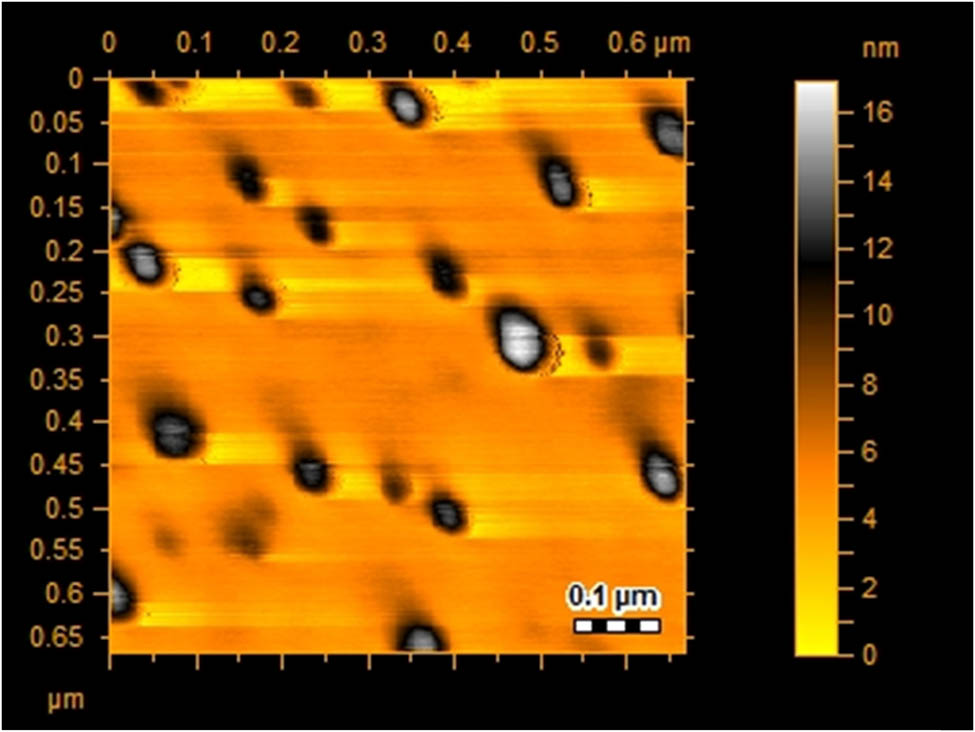
AFM image of AuNPs of P. granatum.
3.5 Phytochemical screening
The phytochemical screening test of ethanolic and aqueous extract of P. granatum is presented in Table 1. The ethanolic extract indicated the presence of tannins, flavonoides, emodins, terpenoids, cardiac glycosides, caumarine, and soluble starch. The aqueous extract showed the presence of tannins, flavonoides, terpenoids, emodins, cardiac glycosides, caumarine, carbohydrates, and soluble starch.
Phytochemical assortment of the P. granatum (peel) extract
| Chemical constituents | Hexane | Chloroform | Ethyl acetate | Ethanolic extract | Aqueous extract |
|---|---|---|---|---|---|
| Alkaloids | − | − | − | − | − |
| Tannins | − | − | + | + | + |
| Anthraquinones | − | − | − | − | − |
| Glycosides | − | − | − | − | − |
| Reducing sugars | + | + | + | − | + |
| Saponins | − | − | − | − | — |
| Flavonoids | − | + | + | + | + |
| Phlobatannins | − | − | − | − | − |
| Steroids | − | − | − | − | − |
| Terpenoids | + | + | + | + | + |
| Cardiac glycoside | + | + | + | + | + |
| Caumarine | − | − | + | + | + |
| Emodines | − | − | + | + | + |
| Anthocyanin and betacyanin | − | − | − | − | − |
| Carbohydrates | + | + | + | − | + |
| Monosaccharides | − | − | − | − | − |
| Reducing sugar | − | − | − | − | − |
| Combined reducing sugar | − | + | + | + | − |
| Soluble starch | − | + | + | + | + |
+: present; −: absent.
3.6 Analgesic effect
The analgesic effects of extracts and PP-AuNPs is shown in Table 2. In both of the tested samples, a uniform dose-dependent analgesic effect was observed. In the case of extract, the significant (p < 0.001) attenuation in induced writhing was noted at a higher dose of 100 mg·kg−1 (70.04%), while PP-AuNPs demonstrated a significant (p < 0.001) percent reduction (81.98) at a dose of 15 mg·kg−1.
Analgesic effect of the extract and nanoparticles
| Treatment | Dose | Percent inhibition of writhing |
|---|---|---|
| Normal saline | 10 mL·kg−1 | − |
| Diclofenic sodium | 10 mg·kg−1 | 85.87 ± 0.65*** |
| Extract | 10 mg·kg−1 | 18.43 ± 1.23 |
| 25 mg·kg−1 | 37.98 ± 1.65 | |
| 50 mg·kg−1 | 56.09 ± 1.43** | |
| 100 mg·kg−1 | 70.04 ± 1.09**** | |
| PP-AuNPs | 2.5 mg·kg−1 | 38.20 ± 1.43 |
| 5 mg·kg−1 | 53.54 ± 1.00** | |
| 10 mg·kg−1 | 67.09 ± 1.43*** | |
| 15 mg·kg−1 | 81.98 ± 1.23 |
The data collected are denoted as the mean ± S.D. for all animals; tolerance to thermal stimuli in seconds; the level of significance was identified by ANOVA following the Dunnets screening model; **p < 0.05; ***p < 0.01; ****p < 0.001.
3.7 Muscle relaxant effect
The muscle co-ordination potential of extracts and PP-AuNPs was tested in two muscle co-ordination models as given in Table 3. A mild muscle relaxant effect was noted after 30 min of sample administration and the effect was improved after 90 min but was not statistically significant. So, in both experimental paradigm, a non-significant effect was noted against the extract and PP-AuNPs.
Muscle relaxant activity of the extract and nanoparticles
| Group | Dose | Inclined plant test | Traction test | ||||
|---|---|---|---|---|---|---|---|
| Percent effect | Percent effect | ||||||
| 30 min | 60 min | 90 min | 30 min | 60 min | 90 min | ||
| Distilled water | 10 mL·kg−1 | 0.00 ± 0.00 | 0.00 ± 0.00 | 0.00 ± 0.00 | 0.00 ± 0.00 | 0.00 ± 0.00 | 0.00 ± 0.00 |
| Diazapam | 1 mg·kg−1 | 100 ± 0.00 | 100 ± 0.00 | 100 ± 0.00 | 100 ± 0.00 | 100 ± 0.00 | 100 ± 0.00 |
| Crude extract | 10 mg·kg−1 | 16.22 ± 1.23 | 21.86 ± 1.09 | 25.94 ± 1.17 | 16.34 ± 1.01 | 17.34 ± 1.23 | 16.91 ± 1.33 |
| 25 mg·kg−1 | 21.77 ± 1.65 | 26.98 ± 1.34 | 31.09 ± 1.13 | 21.97 ± 1.03 | 22.77 ± 1.39 | 21.99 ± 1.23 | |
| 50 mg·kg−1 | 26.34 ± 1.09 | 31.90 ± 1.34 | 32.45 ± 1.10 | 26.44 ± 1.06 | 27.34 ± 1.65 | 28.00 ± 1.88 | |
| 100 mg·kg−1 | 31.32 ± 1.54 | 35.34 ± 1.66 | 39.45 ± 1.63 | 31.34 ± 1.23 | 32.98 ± 1.09 | 31.99 ± 1.23 | |
| PP-AuNPs | 2.5 mg·kg−1 | 21.34 ± 1.43 | 26.34 ± 1.42 | 27.34 ± 1.38 | 21.50 ± 1.09 | 22.43 ± 1.23 | 21.90 ± 1.32 |
| 5 mg·kg−1 | 26.09 ± 1.07 | 30.98 ± 1.09 | 31.34 ± 1.99 | 26.98 ± 1.23 | 27.98 ± 1.32 | 26.87 ± 1.23 | |
| 10 mg·kg−1 | 31.09 ± 1.87 | 34.09 ± 1.89 | 39.09 ± 1.90 | 31.01 ± 1.65 | 32.87 ± 1.34 | 31.67 ± 1.35 | |
| 15 mg·kg−1 | 36.21 ± 1.43 | 40.21 ± 1.32 | 42.09 ± 1.11 | 36.22 ± 1.00 | 37.09 ± 1.35 | 36.00 ± 1.67 | |
The data collected are shown as the mean ± S.D. for all animals; tolerance to thermal stimuli in seconds; and the level of significance was identified by ANOVA following the Dunnets screening model.
3.8 Sedative effect
The sedative effect of both tested samples is presented in Table 4. In the case of extract, a significant (p < 0.05) effect was observed at a tested dose of 100 mg·kg−1. The sedative effect of PP-AuNPs was more significant (p < 0.01) at a dose of 15 mg·kg−1. The sedative effect of PP-AuNPs is better than that of the extract.
Sedative effect of the extract and nanoparticles in open field screening (locomotive activity)
| Treatment | Dose | No. of lines crossed in 10 min |
|---|---|---|
| Distilled water | 10 mL·kg−1 | 124.22 ± 1.16 |
| Diazepam | 0.5 mg·kg−1 | 6.34 ± 1.01 |
| Crude extract | 10 mg·kg−1 | 118.43 ± 2.11 |
| 25 mg·kg−1 | 107.32 ± 2.01 | |
| 50 mg·kg−1 | 97.11 ± 2.98 | |
| 100 mg·kg−1 | 86.23 ± 1.87* | |
| PP-AuNPs | 2.5 mg·kg−1 | 99.54 ± 1.66 |
| 5 mg·kg−1 | 88.54 ± 1.40* | |
| 10 mg·kg−1 | 76.76 ± 1.65** | |
| 15 mg·kg−1 | 64.98 ± 1.20** |
The data collected are shown as the mean±S.D. for all animals; tolerance to thermal stimuli in seconds; the level of significance was identified by ANOVA following the Dunnets screening model; *p < 0.05; **p < 0.01.
4 Discussion
In recent times, the significance of nanotechnology in the field of medicine, optics, and electronics has increased rapidly [28]. The role of nanotechnology in medical sciences has attracted the attention of scientists and researchers owing to its beneficial impact on humans, animals, and plant health. Nanoparticles (NPs) are considered a promising system for drug delivery. Nowadays, various biological, physical, and chemical approaches are used for the synthesis of nanoparticles [29]. Among these, biologically synthesized NPs are preferred owing to their effectiveness, safe, and cost-efficient nature as compared to chemically and physically synthesized NPs [8]. The green synthesis of plant extract-based NPs has acquired a key position in the medical field due to their efficient and less toxic nature [30,31].
The characteristic peak of SPR confirms the synthesis of gold nanoparticles [31]. A single distinctive peak implies the shape of nanoparticles to be spherical [32]. Phytochemicals present in the pomegranate peel extract (terpenoids, flavones, etc.) are considered to be responsible for the bioreduction of Au+ (ion) to Auo (metal) [33]. Our results of UV-Vis spectroscopy are in accordance with the findings of Biao et al. [34]. They also observed a distinct peak by noticing the SPR at 530 nm. FT-IR analysis was performed to determine the probable phytoconstituents present in pomegranate peel extracts that resulted in the efficient stabilization of PP-AuNPs. Signal stretching and vibration bending of peaks could be due to the presence of biomolecules like alkaloids, terpenoids, and flavonoids present in pomegranate peel extracts. Stabilization and reduction of AuNPs may also be due to the presence of these phytomolecules. Pomegranate peel extracts were successfully utilized for the synthesis of gold nanoparticles. The pomegranate peel extract acted as a stabilizing and reducing agent for the formation of Au+ to gold nanoparticles. The results of our study were similar to those reported in the literature [33,34]. The results of AFM analysis are in agreement with the earlier findings of Uz-Zaman et al. [35]. The synthesized PP-AuNPs demonstrated a significant (p < 0.001) percent inhibition (81.98%) of writhing at a dose of 15 mg·kg−1 revealing its analgesic properties. Further, PP-AuNPs possessed significant sedative potential as compared to the PP extract. Similar results regarding analgesic and sedative properties of plant (saffron) extract-based gold nanoparticles (SS-AuNPs) have been shown by Alhumaydhi et al. [36]. They revealed that saffron stigma-based gold nanoparticles had the highest analgesic (84.98%) at 10 mg·kg−1, while all the experimented doses of SS-AuNPs showed a sedative effect.
Natural products are the best source of various classes of phytochemicals. The extracts demonstrated the presence of flavonoids, tannins, and potential classes of phytochemicals. These phytochemicals are responsible for various pharmacologic activities. The preliminary biological screening along with the identification of chemical classes is the way to new drug discovery from natural products. Based on the preliminary evidence, the current pharmaceutical market is supplemented with a lot of natural products like silymarin that is a very good hepatoprotective agent [37]. Green nanotechnology significantly reduces the potential risks associated with human health and the environment and minimizes the production cost of nanomaterials. For the preparation of nanomaterials, amongst all, plant-based materials are in limelight nowadays [38]. Various phytomolecules like terpenes, phenolics, alkaloids, and saponins act as capping agents. The isolated plant phytoconstituents aid in enhancing the reproducibility of shape and size-controlled nanoparticles. Plant phytochemical-based nanoparticles are biologically active and therefore possess various pharmaceutical applications [39].
The analgesic potential of extracts and PP-AuNPs of P. granatum peel strongly supports the antipyretic and anti-arthritis folklore of this plant. The algesia is mostly attributed to the production of prostaglandins (PGs) through the cyclo-oxygenase (COX) pathway. Different PGs exhibit various good or bad biological actions. These PGs are the source of pyrexia, algesia, and inflammation. So, it is possible that the chemical constituents of the P. granatum peel might be COX inhibitors. The COX inhibitory potential is meant for the analgesic effect. It is also possible that chemical moieties of the P. granatum peel might block the sensory receptors as a possible analgesic mechanism.
Traditionally, the P. granatum peels are the best constipating agent and locally the powder of P. granatum peel is used in watery diarrhea. In the current study, a mild muscle relaxant effect was observed. This muscle relaxant effect is adjuvant to the analgesic action because, in many painful situations, analgesic and muscle relaxation are required. One other aspect of the muscle relaxant effect is also related to the curing of diarrhea if our tested samples are also smooth muscle (SM) relaxants. If they also relax the SM, it means they are the best curing candidate in the treatment of diarrhea. The antidiarrheal action is also linked with the inhibition of COX. Because the PGs are also responsible for the contraction of SM and this contraction will lead to diarrhea. The sedative effect of the P. granatum peel is also worth mentioning. The sedative action suggests some further studies for the central analgesic effect. This sedative effect is a good adjuvant pharmacological aspect with analgesia, muscle relaxation, and even as antidiarrheal.
5 Conclusion
Nowadays, the importance of plant extract-based nanoparticles is increasing significantly owing to their effective, safe, and environmentally friendly nature. In the present study, the pomegranate peel extract-based gold nanoparticles (PP-AuNPs) were biosynthesized and evaluated for their analgesic, muscle relaxant, and sedative properties. Keeping in mind the above facts, we concluded that PP-AuNPs could be used for analgesic, muscle relaxation, and sedative effects. All these parameters support the folklore of the P. granatum peel.
Acknowledgment
We thank are thankful to National Science, Technology and Innovation Plan (MAARIFAH), the King Abdul-Aziz City for Science and Technology (KACST), Kingdom of Saudi Arabia.
-
Funding information: The work is funded by grant number 14-MED333-10 from the National Science, Technology and Innovation Plan (MAARIFAH), National Science, Technology and Innovation Plan (MAARIFAH), the King Abdul-Aziz City for Science and Technology (KACST), (KACST), Kingdom of Saudi Arabia. We thank the Science and Technology Unit at Umm Al-Qura University for their continued logistic support.
-
Author contributions: Sami Bawazeer and Abdur Rauf: writing – original draft; Taufiq Nawaz and Anees Ahmed Khalil: writing – review and editing; Muhammad Sameem Javed and Naveed Muhammad: visualization, project administration; and Muhammad Ajmal Shah; resources. All authors edited, read the paper, and agreed on the final version of the manuscript.
-
Conflict of interest: The corresponding author of this article (Abdur Rauf) is a member of the Editorial Board of Green Processing and Synthesis.
-
Data availability statement: Available data are presented in the manuscript.
References
[1] Jayaprakash A, Sangeetha R. Phytochemical screening of Punica granatum Linn. peel extracts. J Acad Ind Res. 2015;4(5):160–2.Suche in Google Scholar
[2] Khalil AA, Khan MR, Shabbir MA, Rahman KU. In vitro antioxidant activity and punicalagin content quantification of pomegranate peel obtained as agro-waste after juice extraction. Pak J Agric Sci. 2018;55(1):197–201.10.21162/PAKJAS/18.5663Suche in Google Scholar
[3] Khalil AA, Khan MR, Shabbir MA, Rahman KU. Comparison of antioxidative potential and punicalagin content of pomegranate peels. J Anim Plant Sci. 2018;27(2):522–7.Suche in Google Scholar
[4] Shabbir MA, Khan MR, Saeed M, Pasha I, Khalil AA, Siraj N. Punicic acid: a striking health substance to combat metabolic syndromes in humans. Lipids Health Dis. 2017;16(1):1–9.10.1186/s12944-017-0489-3Suche in Google Scholar PubMed PubMed Central
[5] Kumar M, Dandapat S, Sinha MP. Phytochemical screening and antibacterial activity of aqueous leaf extract of Punica granatum. Balneo Res J. 2015;6(3):168–71.10.12680/balneo.2015.1100Suche in Google Scholar
[6] Çam M, Hışıl Y. Pressurised water extraction of polyphenols from pomegranate peels. Food Chem. 2010;123(3):878–85.10.1016/j.foodchem.2010.05.011Suche in Google Scholar
[7] Santos ÉS, Hoscheid J, da Mata PT. Antibacterial activity of crude ethanolic and fractionated extracts of Punica granatum Linn. fruit peels. Rev de Cienc Farm Basica e Apl. 2015;36(2):219–25.Suche in Google Scholar
[8] Iqbal M, Bawazeer S, Bakht J, Rauf A, Shah MR, Khalil AA, et al. Green synthesis of silver nanoparticles from Valeriana jatamansi shoots extract and its antimicrobial activity. Green Process Synth. 2020;9(1):715–21.10.1515/gps-2020-0066Suche in Google Scholar
[9] Barabadi H. Nanobiotechnology: A promising scope of gold biotechnology. Cell Mol Biol. 2017;63(12):3–4.10.14715/cmb/2017.63.12.2Suche in Google Scholar PubMed
[10] Saravanan M, Vahidi H, Cruz DM, Vernet-Crua A, Mostafavi E, Stelmach R, et al. Emerging antineoplastic biogenic gold nanomaterials for breast cancer therapeutics: a systematic review. Int J Nanomed. 2020;15:3577.10.2147/IJN.S240293Suche in Google Scholar PubMed PubMed Central
[11] Vijayaraghavan K, Ashokkumar T. Plant-mediated biosynthesis of metallic nanoparticles: a review of literature, factors affecting synthesis, characterization techniques and applications. J Environ Chem Eng. 2017;5(5):4866–83.10.1016/j.jece.2017.09.026Suche in Google Scholar
[12] Barabadi H, Webster TJ, Vahidi H, Sabori H, Kamali KD, Shoushtari FJ, et al. Green nanotechnology-based gold nanomaterials for hepatic cancer therapeutics: a systematic review. Iran J Pharm Res. 2020;19(3):3.Suche in Google Scholar
[13] Khatua A, Prasad A, Priyadarshini E, Patel AK, Naik A, Saravanan M, et al. Emerging antineoplastic plant-based gold nanoparticle synthesis: a mechanistic exploration of their anticancer activity toward cervical cancer cells. J Clust Sci. 2020;31(6):1329–40.10.1007/s10876-019-01742-1Suche in Google Scholar
[14] Barabadi H, Vahidi H, Kamali KD, Hosseini O, Mahjoub MA, Rashedi M, et al. Emerging theranostic gold nanomaterials to combat lung cancer: a systematic review. J Clust Sci. 2020;31(2):323–30.10.1007/s10876-019-01650-4Suche in Google Scholar
[15] Kadi H, Nahal Bouderba N, Benyahia A, Meddah B, Moussaoui A. Phytochemical and antibacterial screening of Punica granatum L. bark of South-west Algeria. J Chem Pharm. 2015;7(8):880–4.Suche in Google Scholar
[16] Iqbal S, Arifeen S, Akbar A, Zahoor S, Maher S, Khan N, et al. 76. Phytochemical screening and antibacterial assay of the crude extract and fractions of Ferula oopoda. Pure Appl Biol. 2019;8(1):742–9.10.19045/bspab.2019.80016Suche in Google Scholar
[17] Ahmad N, Sharma S, Rai R. Rapid green synthesis of silver and gold nanoparticles using peels of Punica granatum. Adv Mater Lett. 2012;3(5):376–80.10.5185/amlett.2012.5357Suche in Google Scholar
[18] Philip D, Kaleena PK, Valivittan K, Kumar CG. Phytochemical screening and antimicrobial activity of Sansevieria roxburghiana Schult. and Schult. F. Middle East. J Sci Res. 2011;10(4):512–8.Suche in Google Scholar
[19] Savithramma N, Rao ML, Suhrulatha D. Screening of medicinal plants for secondary metabolites. Middle East. J Sci Res. 2011;8(3):579–84.Suche in Google Scholar
[20] Usman H, Abdulrahman FI, Usman A. Qualitative phytochemical screening and in vitro antimicrobial effects of methanol stem bark extract of Ficus thonningii (Moraceae). Afr J Tradit Complement Altern Med. 2009;6(3):289–95.10.4314/ajtcam.v6i3.57178Suche in Google Scholar PubMed PubMed Central
[21] Uddin G, Rauf A, Arfan M, Ali M, Qaisar M, Saadiq M, et al. Preliminary phytochemical screening and antioxidant activity of Bergenia caliata. Middle-East. J Sci Res. 2012;11(8):1140–2.Suche in Google Scholar
[22] Rauf A, Muhammad N, Khan A, Uddin N, Atif M. Antibacterial and phytotoxic profile of selected Pakistani medicinal plants. World Appl Sci J. 2012;20(4):540–4.Suche in Google Scholar
[23] Seniya C, Trivedia SS, Verma SK. Antibacterial efficacy and phytochemical analysis of organic solvent extracts of Calotropis gigantea. J Chem Pharm Res. 2011;3(6):330–6.Suche in Google Scholar
[24] Raveendran P, Fu J, Wallen SL. Completely “green” synthesis and stabilization of metal nanoparticles. J Am Chem Soc. 2003;125(46):13940–1.10.1021/ja029267jSuche in Google Scholar PubMed
[25] Rauf A, Ali J, Khan H, Mubarak MS, Patel S. Emerging CAM Ziziphus nummularia with in vivo sedative-hypnotic, antipyretic and analgesic attributes. 3 Biotech. 2016;6(1):1–10.10.1007/s13205-015-0322-5Suche in Google Scholar PubMed PubMed Central
[26] Rauf A, Bawazeer S, Uddin G, Siddiqui BS, Khan H, Hadda TB, et al. Muscle relaxant activities of pistagremic acid isolated from Pistacia integerrima. Z Naturforsch C. 2018;27:413–6.10.1515/znc-2017-0179Suche in Google Scholar PubMed
[27] Hussain A, Abdur R, Abu-Izneid T, Ibrahim M, Abrar S, Khan H, et al. Sedative, muscle relaxant-like effects, and molecular docking study of compounds isolated from Salvia leriifolia. Rev Bras Farmacogn. 2020;30:257–60.10.1007/s43450-020-00046-0Suche in Google Scholar
[28] Rajan R, Chandran K, Harper SL, Yun SI, Kalaichelvan PT. Plantextract synthesized silver nanoparticles: an ongoing source ofnovel biocompatible materials. Ind Crop Prod. 2015;70:356–73.10.1016/j.indcrop.2015.03.015Suche in Google Scholar
[29] Gnanavel V, Palanichamy V, Roopan SM. Biosynthesis and characterization of copper oxide nanoparticles and its anticancer activity on human colon cancer cell lines (HCT-116). J Photochem Photobiol B. 2017;171:133–8.10.1016/j.jphotobiol.2017.05.001Suche in Google Scholar PubMed
[30] Abu-Tahon MA, Ghareib M, Abdallah WE. Environmentally benign rapid biosynthesis of extracellular gold nanoparticles using Aspergillus flavus and their cytotoxic and catalytic activities. Process Biochem. 2020;95:1.10.1016/j.procbio.2020.04.015Suche in Google Scholar
[31] Aljabali AA, Akkam Y, Al Zoubi MS, Al-Batayneh KM, Al-Trad B, Abo Alrob O, et al. Synthesis of gold nanoparticles using leaf extract of Ziziphus zizyphus and their antimicrobial activity. Nanomaterials. 2018;8(3):174.10.3390/nano8030174Suche in Google Scholar PubMed PubMed Central
[32] Nagalingam M, Kalpana VN, Panneerselvam A. Biosynthesis, characterization, and evaluation of bioactivities of leaf extract-mediated biocompatible gold nanoparticles from Alternanthera bettzickiana. Biotechnol Rep. 2018;19:e00268.10.1016/j.btre.2018.e00268Suche in Google Scholar PubMed PubMed Central
[33] ElMitwalli OS, Barakat OA, Daoud RM, Akhtar S, Henari FZ. Green synthesis of gold nanoparticles using cinnamon bark extract, characterization, and fluorescence activity in Au/eosin Y assemblies. J Nanopart Res. 2020;22(10):1–9.10.1007/s11051-020-04983-8Suche in Google Scholar
[34] Biao L, Tan S, Meng Q, Gao J, Zhang X, Liu Z, et al. Green synthesis, characterization and application of proanthocyanidins-functionalized gold nanoparticles. Nanomaterials. 2018;8(1):53.10.3390/nano8010053Suche in Google Scholar PubMed PubMed Central
[35] Uz-Zaman K, Bakht J, Malikovna BK, Elsharkawy ER, Khalil AA, Bawazeer S, et al. Trillium govanianum Wall. Ex. Royle rhizomes extract-medicated silver nanoparticles and their antimicrobial activity. Green Process Synth. 2020;9(1):503–14.10.1515/gps-2020-0054Suche in Google Scholar
[36] Alhumaydhi FA, Khan I, Rauf A, Qureshi MN, Aljohani AS, Khan SA, et al. Synthesis, characterization, biological activities, and catalytic applications of alcoholic extract of saffron (Crocus sativus) flower stigma-based gold nanoparticles. Green Process Synth. 2021;10(1):230–45.10.1515/gps-2021-0024Suche in Google Scholar
[37] Freitag AF, Cardia GFE, da Rocha BA, Aguiar RP, Silva-Comar FMDS, Spironello RA, et al. Hepatoprotective effect of silymarin (Silybum marianum) on hepatotoxicity induced by acetaminophen in spontaneously hypertensive rats. Evidence-Based Complementary Alternative Med. 2015;2015:538317. 10.1155/2015/538317.Suche in Google Scholar PubMed PubMed Central
[38] Saravanan M, Barabadi H, Vahidi H. Green nanotechnology: isolation of bioactive molecules and modified approach of biosynthesis. Biogenic Nanoparticles for Cancer Theranostics. 1st ed. Elsevier; 2021. p. 101–22. 10.1016/B978-0-12-821467-1.00005-7.Suche in Google Scholar
[39] Saravanan M, Barabadi H, Ramachandran B, Venkatraman G, Ponmurugan K. Emerging plant-based anti-cancer green nanomaterials in present scenario. Compr Anal Chem. 2019;87:291–31810.1016/bs.coac.2019.09.001Suche in Google Scholar
© 2021 Sami Bawazeer et al., published by De Gruyter
This work is licensed under the Creative Commons Attribution 4.0 International License.
Artikel in diesem Heft
- Research Articles
- MW irradiation and ionic liquids as green tools in hydrolyses and alcoholyses
- Effect of CaO on catalytic combustion of semi-coke
- Studies of Penicillium species associated with blue mold disease of grapes and management through plant essential oils as non-hazardous botanical fungicides
- Development of leftover rice/gelatin interpenetrating polymer network films for food packaging
- Potent antibacterial action of phycosynthesized selenium nanoparticles using Spirulina platensis extract
- Green synthesized silver and copper nanoparticles induced changes in biomass parameters, secondary metabolites production, and antioxidant activity in callus cultures of Artemisia absinthium L.
- Gold nanoparticles from Celastrus hindsii and HAuCl4: Green synthesis, characteristics, and their cytotoxic effects on HeLa cells
- Green synthesis of silver nanoparticles using Tropaeolum majus: Phytochemical screening and antibacterial studies
- One-step preparation of metal-free phthalocyanine with controllable crystal form
- In vitro and in vivo applications of Euphorbia wallichii shoot extract-mediated gold nanospheres
- Fabrication of green ZnO nanoparticles using walnut leaf extract to develop an antibacterial film based on polyethylene–starch–ZnO NPs
- Preparation of Zn-MOFs by microwave-assisted ball milling for removal of tetracycline hydrochloride and Congo red from wastewater
- Feasibility of fly ash as fluxing agent in mid- and low-grade phosphate rock carbothermal reduction and its reaction kinetics
- Three combined pretreatments for reactive gasification feedstock from wet coffee grounds waste
- Biosynthesis and antioxidation of nano-selenium using lemon juice as a reducing agent
- Combustion and gasification characteristics of low-temperature pyrolytic semi-coke prepared through atmosphere rich in CH4 and H2
- Microwave-assisted reactions: Efficient and versatile one-step synthesis of 8-substituted xanthines and substituted pyrimidopteridine-2,4,6,8-tetraones under controlled microwave heating
- New approach in process intensification based on subcritical water, as green solvent, in propolis oil in water nanoemulsion preparation
- Continuous sulfonation of hexadecylbenzene in a microreactor
- Synthesis, characterization, biological activities, and catalytic applications of alcoholic extract of saffron (Crocus sativus) flower stigma-based gold nanoparticles
- Foliar applications of plant-based titanium dioxide nanoparticles to improve agronomic and physiological attributes of wheat (Triticum aestivum L.) plants under salinity stress
- Simultaneous leaching of rare earth elements and phosphorus from a Chinese phosphate ore using H3PO4
- Silica extraction from bauxite reaction residue and synthesis water glass
- Metal–organic framework-derived nanoporous titanium dioxide–heteropoly acid composites and its application in esterification
- Highly Cr(vi)-tolerant Staphylococcus simulans assisting chromate evacuation from tannery effluent
- A green method for the preparation of phoxim based on high-boiling nitrite
- Silver nanoparticles elicited physiological, biochemical, and antioxidant modifications in rice plants to control Aspergillus flavus
- Mixed gel electrolytes: Synthesis, characterization, and gas release on PbSb electrode
- Supported on mesoporous silica nanospheres, molecularly imprinted polymer for selective adsorption of dichlorophen
- Synthesis of zeolite from fly ash and its adsorption of phosphorus in wastewater
- Development of a continuous PET depolymerization process as a basis for a back-to-monomer recycling method
- Green synthesis of ZnS nanoparticles and fabrication of ZnS–chitosan nanocomposites for the removal of Cr(vi) ion from wastewater
- Synthesis, surface modification, and characterization of Fe3O4@SiO2 core@shell nanostructure
- Antioxidant potential of bulk and nanoparticles of naringenin against cadmium-induced oxidative stress in Nile tilapia, Oreochromis niloticus
- Variability and improvement of optical and antimicrobial performances for CQDs/mesoporous SiO2/Ag NPs composites via in situ synthesis
- Green synthesis of silver nanoparticles: Characterization and its potential biomedical applications
- Green synthesis, characterization, and antimicrobial activity of silver nanoparticles prepared using Trigonella foenum-graecum L. leaves grown in Saudi Arabia
- Intensification process in thyme essential oil nanoemulsion preparation based on subcritical water as green solvent and six different emulsifiers
- Synthesis and biological activities of alcohol extract of black cumin seeds (Bunium persicum)-based gold nanoparticles and their catalytic applications
- Digera muricata (L.) Mart. mediated synthesis of antimicrobial and enzymatic inhibitory zinc oxide bionanoparticles
- Aqueous synthesis of Nb-modified SnO2 quantum dots for efficient photocatalytic degradation of polyethylene for in situ agricultural waste treatment
- Study on the effect of microwave roasting pretreatment on nickel extraction from nickel-containing residue using sulfuric acid
- Green nanotechnology synthesized silver nanoparticles: Characterization and testing its antibacterial activity
- Phyto-fabrication of selenium nanorods using extract of pomegranate rind wastes and their potentialities for inhibiting fish-borne pathogens
- Hydrophilic modification of PVDF membranes by in situ synthesis of nano-Ag with nano-ZrO2
- Paracrine study of adipose tissue-derived mesenchymal stem cells (ADMSCs) in a self-assembling nano-polypeptide hydrogel environment
- Study of the corrosion-inhibiting activity of the green materials of the Posidonia oceanica leaves’ ethanolic extract based on PVP in corrosive media (1 M of HCl)
- Callus-mediated biosynthesis of Ag and ZnO nanoparticles using aqueous callus extract of Cannabis sativa: Their cytotoxic potential and clinical potential against human pathogenic bacteria and fungi
- Ionic liquids as capping agents of silver nanoparticles. Part II: Antimicrobial and cytotoxic study
- CO2 hydrogenation to dimethyl ether over In2O3 catalysts supported on aluminosilicate halloysite nanotubes
- Corylus avellana leaf extract-mediated green synthesis of antifungal silver nanoparticles using microwave irradiation and assessment of their properties
- Novel design and combination strategy of minocycline and OECs-loaded CeO2 nanoparticles with SF for the treatment of spinal cord injury: In vitro and in vivo evaluations
- Fe3+ and Ce3+ modified nano-TiO2 for degradation of exhaust gas in tunnels
- Analysis of enzyme activity and microbial community structure changes in the anaerobic digestion process of cattle manure at sub-mesophilic temperatures
- Synthesis of greener silver nanoparticle-based chitosan nanocomposites and their potential antimicrobial activity against oral pathogens
- Baeyer–Villiger co-oxidation of cyclohexanone with Fe–Sn–O catalysts in an O2/benzaldehyde system
- Increased flexibility to improve the catalytic performance of carbon-based solid acid catalysts
- Study on titanium dioxide nanoparticles as MALDI MS matrix for the determination of lipids in the brain
- Green-synthesized silver nanoparticles with aqueous extract of green algae Chaetomorpha ligustica and its anticancer potential
- Curcumin-removed turmeric oleoresin nano-emulsion as a novel botanical fungicide to control anthracnose (Colletotrichum gloeosporioides) in litchi
- Antibacterial greener silver nanoparticles synthesized using Marsilea quadrifolia extract and their eco-friendly evaluation against Zika virus vector, Aedes aegypti
- Optimization for simultaneous removal of NH3-N and COD from coking wastewater via a three-dimensional electrode system with coal-based electrode materials by RSM method
- Effect of Cu doping on the optical property of green synthesised l-cystein-capped CdSe quantum dots
- Anticandidal potentiality of biosynthesized and decorated nanometals with fucoidan
- Biosynthesis of silver nanoparticles using leaves of Mentha pulegium, their characterization, and antifungal properties
- A study on the coordination of cyclohexanocucurbit[6]uril with copper, zinc, and magnesium ions
- Ultrasound-assisted l-cysteine whole-cell bioconversion by recombinant Escherichia coli with tryptophan synthase
- Green synthesis of silver nanoparticles using aqueous extract of Citrus sinensis peels and evaluation of their antibacterial efficacy
- Preparation and characterization of sodium alginate/acrylic acid composite hydrogels conjugated to silver nanoparticles as an antibiotic delivery system
- Synthesis of tert-amylbenzene for side-chain alkylation of cumene catalyzed by a solid superbase
- Punica granatum peel extracts mediated the green synthesis of gold nanoparticles and their detailed in vivo biological activities
- Simulation and improvement of the separation process of synthesizing vinyl acetate by acetylene gas-phase method
- Review Articles
- Carbon dots: Discovery, structure, fluorescent properties, and applications
- Potential applications of biogenic selenium nanoparticles in alleviating biotic and abiotic stresses in plants: A comprehensive insight on the mechanistic approach and future perspectives
- Review on functionalized magnetic nanoparticles for the pretreatment of organophosphorus pesticides
- Extraction and modification of hemicellulose from lignocellulosic biomass: A review
- Topical Issue: Recent advances in deep eutectic solvents: Fundamentals and applications (Guest Editors: Santiago Aparicio and Mert Atilhan)
- Delignification of unbleached pulp by ternary deep eutectic solvents
- Removal of thiophene from model oil by polyethylene glycol via forming deep eutectic solvents
- Valorization of birch bark using a low transition temperature mixture composed of choline chloride and lactic acid
- Topical Issue: Flow chemistry and microreaction technologies for circular processes (Guest Editor: Gianvito Vilé)
- Stille, Heck, and Sonogashira coupling and hydrogenation catalyzed by porous-silica-gel-supported palladium in batch and flow
- In-flow enantioselective homogeneous organic synthesis